Note: Descriptions are shown in the official language in which they were submitted.
CA 02570933 2006-12-18
PCT/AU2005/000880
- 1 _ Received 31 March 2006
"Ophthalmic Camera and Ophthalmic Camera Adaptor"
Field of the Invention
The present invention relates to an ophthalmic camera and an ophthalmic camera
adaptor. In particular, the invention relates to the optical arrangement that
forms
the basis for the ophthalmic camera and ophthalmic camera adaptor.
Throughout the specification, unless the context requires otherwise, the word
"comprise" or variations such as "comprises" or "comprising", will be
understood to
imply the inclusion of a stated integer or group of integers but not the
exclusion of
any other integer or group of integers.
Background Art
The following discussion of the background to the invention is intended to
facilitate
an understanding of the present invention. However, it should be appreciated
that
the discussion is not an acknowledgement or admission that any of the material
referred to was published, known or part of the common.general knowledge of a
skilled person in any jurisdiction as at the priority date of the application.
Images of the fundus of a patient's eye can degrade due to many factors. Such
factors include:
= reflection of light from the cornea or iris;
= reflection of light from the walls of the ophthalmic lens; and
= use of an incorrect level of illumination for the pupil colour of the
patient's eye.
One method of overcoming some of the above problems is to use low level
illumination devices. However, using such illumination devices, typically,
reduces
the field of view of the image and may not be appropriate for the. fundus
being
examined.
Amended Sheet
IPEA/AU
CA 02570933 2006-12-18
PCT/AU2005/000880
_ 2_ Received 31 March 2006
It is therefore an object of the present invention to provide an optical
arrangement
that reduces the level of reflection by one or more of the cornea, iris, or
walls of
the ophthalmic lens.
Disclosure of the Invention
Broadly, the invention lies in a camera having a lens, at least one
illumination
means; and a second lens; wherein and the centres of the second lens and
camera lens are aligned to form an alignment axis and wherein said one
illumination means is capable of linear movement along a radial axis of the
camera lens and pivotal movement about a pivot point thereof, such that the
circle
of light emitted said one illumination means can be adjusted towards or away
the
alignment axis to remain focused relative to the centre of the second lens.
Preferably, the second lens is an ophthalmic lens and the second lens is equal
to
or smaller than the camera lens.
The illumination angle of the at least one illumination means may be
adjustable.
This may be an inherent characteristic of the at least one illumination means
or
achieved by an external element, such as a collimator. In this manner, the
circle
of light emitted by the at least one illumination means, at the point of
intersection
with the ophthalmic lens, is of a size that, when the light is focused by the
ophthalmic lens onto a fundus, the angle of the focused light provides a wide
field
of view for the size of the fundus being examined.
The field of view of the camera lens may also be adjustable. Ideally, the
field of
view of the camera lens is restricted to the same size as the size of the
fundus
being examined. This may be achieved by means of an iris.
The at least one illumination means may surround the circumference of the
camera lens. Each illumination means is preferably equidistant from its
adjacent
illumination means to provide a homogenous light source. Preferably, the
illumination means are solid state LEDs, however, light bulbs with appropriate
focusing means may also be used.
Ainended Sheet
IPEA/AU
CA 02570933 2006-12-18
PCT/AU2005/000880
Received 31 March 2006
The at least one illumination means may also be of variable intensity. In this
manner, the at least one illumination means can adjust the level of
illumination
provided by the emitted circle of light to more appropriately accord with the
colour
of the fundus being examined. The level of intensity may be a function of a
setting of the ophthalmic camera.
The camera preferably has a high sensitivity to low light. Ideally, the camera
has
a sensitivity level of <0.05 lux and/or a lens of 5 to 8mm in diameter.
The ophthalmic lens is preferably in the range of 20 to 90 dioptres, with 40
being
considered optimal. The ophthalmic lens may have an anti-reflective coating.
The ophthalmic lens may be capable of linear movement along the alignment axis
to allow for focusing. Alternatively, other means of focusing the ophthalmic
camera may be employed.
Ideally, the ophthalmic camera can be set to one or more settings. Each
setting
represents a pupil size. When a setting is changed, the at least one
illumination
means moves linearly along its radial axis to the position specified by the
new
setting and pivots about the radial plane until the circle of light emitted by
the at
least one illumination means is focused on the centre of the ophthalmic lens.
Alternatively, the settings may be omitted and control of the linear and
pivotal
movement of the at least one illumination means may be by means of one or
more manual controls. Both the setting control and the manual control may be
expanded to further control the illumination angle of the at least one
illumination
means and/or the field of view of the camera lens.
Control of one or more of the linear and pivotal movement of the at least one
illumination means, the illumination angle of the at least one illumination
means
and the field of view of the camera lens, may be controlled automatically by a
control means in response to the estimated size of the fundus to be examined
as
determined by an automated measuring means.
The ophthalmic camera may also include magnification lenses. Each
magnification lens may be associated with one or more settings, such that on
choosing a setting, its associated magnification lenses are positioned within
the
Aniended Sheet
IPEA/AU
CA 02570933 2006-12-18
PCT/AU2005/000880
Received 31 March 2006
optical axis of the camera and in-between the ophthalmic lens and the camera
lens.
The camera may be colour or monochromatic, digital or analogue, as required.
Filters may be positioned in front of the camera lens and the at least one
illumination means, the filters being of opposite polarisation to each other.
The invention may also be disclosed in an ophthalmic camera adaptor
incorporating the optics of any of the previous embodiments. The ophthalmic
camera adaptor omits the camera.
The invention may also be disclosed in a method of imaging the fundus of the
eye.
In accordance with a first aspect of the invention there is provided an
ophthalmic
camera for taking an image of the fundus of an eye, comprising a'camera having
a camera lens; at least one illumination means; and a second lens, the centres
of
the second lens and camera lens being aligned to form an alignment axis and
the
illumination means being movable relative to the alignment axis and the second
lens, so that the beam of light emitted by the illumination means is able to
be
focused by the second lens through the pupil onto the fundus.
According to a preferred feature of the invention, the illumination means
comprises a plurality of illumination devices, the illumination devices
disposed to
surround the circumference of the camera lens and be spaced equidistant from
adjacent illumination devices.
According to a preferred feature of the invention, the ophthalmic camera
further
comprises control means, the control means having a plurality of settings such
that, when the setting of the control means is changed, said illumination
means
moves linearly along its radial axis to the position specified by the new
setting and
pivots about the axial plane until the circle of light emitted by said one
illumination
means is focused relative to the centre of the second lens.
Amended Sheet
IPEA/AU
CA 02570933 2006-12-18
PCT/AU2005/000880
Received 31 March 2006
According to a preferred feature of the invention, the ophthalmic camera
includes
automated measuring means, the automated measuring means operable to
analyse a fundus being examined and change the setting of the control means to
the most appropriate setting on the basis of the analysis of the pupil.
According to a preferred feature of the invention, the ophthalmic camera
includes
a first polariser located within the alignment axis and positioned in front of
the
second lens and a second polariser attached to each illumination means such
that
light emitted by the illumination means passes through the second polariser,
the
first polariser being oppositely polarised to the second filter to thereby
filter the
light.
According to a second aspect, the invention resides in an ophthalmic camera
comprising a camera having a camera lens; an illumination means; a second
lens;
a beam splitter; and a light focusing lens; the centres of the second lens,
the
camera lens and the beam splitter being aligned to form an alignment axis, and
the centres of the beam splitter, light focusing lens and the illumination
means
being aligned to form an illumination axis perpendicular to the alignment
axis, the
illumination means being movable relative to the illumination axis and the
light
focusing lens so that the beam of light from the illumination means is focused
by
the light focusing lens towards the beam splitter, and reflected by the beam
splitter along the alignment axis towards and through the pupil, the
illumination
means thereby being movable relative to the alignment axis and the second
lens,
wherein the position of the illumination means is adjustable to focus the beam
of
reflected light so that it is substantially the same size as the pupil to
maximise the
amount of light entering the pupil without impinging upon the iris to thereby
avoid
contraction of the pupil.
According to a preferred embodiment, the beam splitter is a 50/50 beam
splitter.
According to a preferred feature of the invention, the illumination means is
able to
move linearly along the illumination axis such that the light reflected by the
beam
splitter towards the retina is substantially aligned with the centre of a
first surface
of the second lens.
Amended Sheet
IPEA/AU
CA 02570933 2006-12-18
PCT/AU2005/000880
Received 31 March 2006
According to a preferred embodiment, the illumination means is able to pivot
about a pivot point to permit the illumination axis to be moved and adjusted
relative to said alignment axis.
According to a preferred embodiment, the ophthalmic camera includes control
means, the control means having a plurality of settings such that, when the
setting
of the control means is changed, said one illumination means moves linearly
along the illumination axis to a predetermined position associated with the
new
setting.
According to a preferred feature of the invention, the ophthalmic camera
includes
automated measuring means, the automated measuring means operable to
analyse the retina being examined and change the setting of the control means
to
the most appropriate setting on the basis of the analysis of the pupil.
According to a preferred embodiment, the second lens is an ophthalmic lens.
According to a preferred embodiment, the camera has a high sensitivity to low
light.
According to a preferred embodiment, the second lens is in the range of 20 to
90
dioptres.
According to a preferred embodiment, the second lens is substantially 40
dioptres.
According to a preferred embodiment, the ophthalmic camera includes focussing
means for focusing the second lens.
According to a preferred embodiment; the focusing means is means for allowing
linear movement of the second lens along the alignment axis.
According to a preferred feature of the invention, the illumination angle of
the
illumination means is adjustable.
Amended Sheet
IPEA/AU
CA 02570933 2006-12-18
PCT/AU2005/000880
- 7_ Received 31 March 2006
According to a preferred embodiment, the ophthalmic camera includes at least
one collimator, each collimator associated with an illumination means operable
to
adjust the illumination angle of the associated illumination means.
According to a preferred embodiment, the field of view of the camera lens is
adjustable.
According to a preferred feature of the invention, the ophthalmic camera
includes
an iris, the iris operable to adjust the field of view of the camera lens.
According to a preferred embodiment, the intensity of the light generated by
the
illumination means is adjustable.
According to a preferred embodiment, the illumination means is a solid state
light
emitting diode.
According to a preferred embodiment, the illumination means is a light bulb
with
associated appropriate focusing means.
According to a preferred embodiment, at least one surface of at least one lens
lens has an anti-reflective coating.
According to a third aspect, the invention resides in an adaptor for an
ophthalmic
camera having a body and a camera housed within the body, the adaptor
comprising:
optics for illuminating a subject within the optical axis of the camera as
described above;
means for releasably engaging the body; and
an aperture extending therethrough;
wherein, when releasably engaged with the body, the aperture aligns with the
optical axis such that least a portion of the optical axis of the camera is
not
obscured.
Amended Sheet
TPEA/AU
CA 02570933 2006-12-18
PCT/AU2005/000880
Received 31 March 2006
According to a fourth aspect, the invention resides in a method of imaging a
fundus comprising the steps of:
moving an illumination means along a radial axis of a camera lens; and
pivoting the illumination means such that the circle of light emitted by the
illumination means can be focused relative to the centre of a second lens;
wherein the centre of the second lens is in alignment with the centre of the
camera lens.
According to a preferred feature of the invention, the method includes the
further
step of:
moving the illumination means along the radial axis to a predetermined
position associated with a setting of a control means when the control
means is set to the associated setting.
According to a preferred feature of the invention, the method includes the
steps
of:
analysing the pupil being examined;
determining the most appropriate associated setting on the basis of the
analysis of the pupil; and
changing the setting of the control means to the most appropriate
associated setting.
According to a preferred feature of the invention, the method includes the
steps
of:
directing the circle of light through a first polariser; and
taking an image of the circle of light through a second polariserof opposite
polarisation to the first polariser.
According to a fifth aspect the invention resides in a method of imaging a
fundus
comprising:
directing light emitted by an illumination means to a light focusing lens; and
focusing the light towards a beam splitter to be reflected by the beam
splitter towards the fundus so that the size of beam of light can be of
commensurate to the size of the pupil;
Amended Sheet
IPEA/AU
CA 02570933 2006-12-18
PCT/AU2005/000880
Received 31 March 2006
wherein the centres of the beam splitter, light focusing lens and illumination
means are aligned to form an illumination axis and the centres of a camera
lens, second lens and the beam splitter are aligned to form an alignment axis
perpendicular to the illumination axis.
According to a preferred feature of the invention, the method includes the
step of
moving the illumination means linearly along the illumination axis such that
the
centre of the circle of light reflected by the beam splitter towards the pupil
is
substantially aligned relative to the centre of a first surface of the second
lens.
According to a preferred feature of the invention, the method includes the
step of
pivoting the illumination means about the place that includes the optical axis
and
the illumination axis.
According to a preferred feature of the invention, the method includes the
further
step of:
moving the illumination means along the radial axis to a predetermined
position associated with a setting of a control means when the control
means is set to the associated setting.
According to a preferred feature of the invention, the method includes the
steps
of:
analysing the pupil being examined;
determining the most appropriate associated setting on the basis of the
analysis of the pupil; and
changing the setting of the control means to the most appropriate
associated setting.
According to a preferred feature of the invention, the method includes further
comprising the step of focussing the second lens:
According to a preferred feature of the invention, the method includes the
step of
linearly moving the second lens along the alignment axis to focus the second
lens.
Amended Slieet
IPEA/AU
CA 02570933 2006-12-18
PCT/AU2005/000880
- 10 - Received 31 March 2006
According to a preferred feature of the invention, the method includes the
step of
adjusting the illumination angle of the illumination means.
According to a preferred feature of the invention, the method includes
including
the step of adjusting the field of view of the camera lens.
According to a preferred feature of the invention, the method includes the
step of
adjusting the intensity of the light generated by the illumination means.
The invention will now be more fully understood in light of the following
description
of several specific embodiments.
Brief Description of the Drawings
The invention will now be described, by way of example only, with reference to
the
accompanying drawings, of which:
Figure 1a is a schematic of the optics of an ophthalmic camera and ophthalmic
camera adaptor of a first embodiment of the present invention showing linear
movement of the LED's.
Figure lb is a schematic of the optics of the ophthalmic camera and ophthalmic
camera adaptor of the first embodiment, similar to Figure 'i b but showing
angular
movement of the LED's.
Figure 2 is an isometric view of the schematics of the optics of the,
ophthalmic
camera and ophthalmic camera adaptor of the first embodiment of the present
invention.
Figure 3 is a schematic of the optics of an ophthalmic camera and ophthalm'rc
camera adaptor of a second embodiment of the present invention.
Figure 4 is a schematic of the optics of an ophthalmic camera and ophthalmic
camera adaptor of a third embodiment of the present invention.
Amended Slieet
IPBA/AU
CA 02570933 2006-12-18
PCT/AU2005/000880
- 11 - Received 31 March 2006
Figures 5a and 5b are perspective views of an ophthalmic camera adaptor of the
present invention.
Best Mode(s) for Carrying Out the Invention
The first embodiment of the best mode invention for carrying out the invention
is
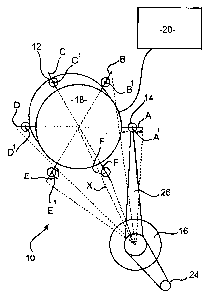
directed towards an ophthalmic camera apparatus.10, generally comprising a
camera 12 highly sensitive to low light {ie. somewhere in the range of <0.05
lux),
an illumination means in the form of a plurality of solid-state LEDs 14, and
an
ophthalmic lens 16 all contained within a housing (not shown). This
arrangement
is shown graphically in Figure 1.
The camera 12 has a camera lens 18 and so the ophthalmic lens constitutes a
second lens of the apparatus 10. Ideally, the camera lens 18 has a diameter of
5-
8mm. The camera lens 18 provides for an adjustable field of view.
The plurality of LEDs 14 surround the circumference of the camera lens 18 and
are linked to a control unit 20. The intensity of the light generated by LEDs
14 can
be varied by way of the control unit 20.
Each LED 14 is equidistant to its adjacent LEDs.14. Each LED 14 also has an
adjustable illumination angle.
As shown in Figures 1 a and 2, LEDs 14 are able to move linearly along their
respective radial axes (marked A through E), relative to the main optical axis
X.
As shown in Figures lb and 2, each LED 14 is also able to pivot about a pivot
point thereof (A' to E'), towards, or away from the camera lens 18, so that
the
illumination axis Y thereof can move either towards or away the point of
intersection of the optical axis X with the ophthalmic lens 16, in order to
compensate for linear movement of the LED along its respective radial axis A-E
and corresponding displacement of the illumination axis Y relative to the
optical
axis X.
The ophthalmic lens 16 has an inner convex surface 22 that opposes the camera
lens 18. The central axis of the came~q~&'~e@(ligns with the central axis of
4he
1PEA/AU
CA 02570933 2006-12-18
PCT/AU2005/000880
- 12 - Received 31 March 2006
inner convex surface 22 to form an alignment axis, which constitutes the main
optical axis X. Ideally, the ophthalmic lens 16 is of the same size as, or
smaller
than, the camera lens 18. An anti-reflective coating may be applied to the
ophthalmic lens 16.
The ophthalmic lens is typically in the range of 20 to 90 dioptres, with 40
dioptres
considered optimum. To allow for focusing of the ophthalmic lens 16, the
ophthalmic lens 16 is capable of linear movement along optical axis X of the
monochromatic camera 12.
The position of the LEDs 14, as well as the illumination angle of LEDs 14 and
field
of view of the camera lens 18, are all a function of the current setting of
the
ophthalmic camera apparatus 10. Each setting of the ophthalmic camera
apparatus 10 represents a range of sizes of a pupil 24 of a patient with
beirig
examined with the apparatus. To elaborate,
= setting I is used for pupils of size less than 3mm;
= setting 2 is used for pupils having a size between 3-4mm; and
= setting 3 is used for dilated pupils.
Upon choosing a setting:
= The illumination angle of a beam of light 26 generated by each LED
14 along its illumination axis Y is restricted or enlarged, as
appropriate, such that the circle generated by the beam of light 26 at
the point of intersection with the ophthalmic lens 16 is of a size that,
when the light is focused by the ophthalmic lens 16 onto the pupil 24,
the angle of the focused light 0 provides a wide field of view for the
appropriate pupil 24 size.
= LEDs 14 move linearly along their. respective radial axes (marked A
through E) and pivot about their respective pivot point (marked A'
Aniended Sheet
IPBA/AU
CA 02570933 2006-12-18
PCT/AU2005/000880
- 13 - Received 31 March 2006
through E') such that the centre of the circle generated by the beam
of light 26 at the point of intersection with the ophthalmic lens 16 can
be precisely adjusted with respect to the centre of the ophthalmic
lens 16.
= The field of view of camera lens 18 is restricted to substantially the
same size as the pupil 24 size associated with the setting.
This allows the same LEDs 14 to be used for pupils 24 of all sizes while
negating
the need to unnecessarily restrict the field of view of the camera lens 18 to
avoid
reflection from the cornea or iris. This also means that for larger size
pupils 24,
the angle of the focused light 0 along the illumination axis Y is greater than
the
angle of the focused light 0 generated in respect of smaller size pupils 24.
The second embodiment of the best m.ode is substantially similar to the first
embodiment, where like numerals reference like parts, but involves the use of
optical filters. As shown in Figure 3, a first filter 28 is located along the
optical axis
X of the camera 12 at a position in front of camera lens 18. A second filter
30 is
attached to each LED 14, such that the beam of light 26 emitted thereby passes
through the second filter 30.
The first filter 28 is oppositely polarised to second filter 30.
As the beam of light 26 reflects off the fundus 24 it enters the ophthalmic
lens 18.
On entering the ophthalmic lens 18, the polarisation of the beam of light 26
is
reversed. However, as the beam of light 26 enters the ophthalmic lens 18,
light
that reflects off the two walls of the ophthalmic lens 18 will not be captured
by the
camera 12 due to the cross-polarisation effect of first and second filters 28,
30.
The third embodiment of the best mode is shown in Figure 4, and is direeted
towards an ophthalmic camera apparatus 50 comprising a digital camera 52
highly sensitive to low light (ie. somewhere in the range of <0.05 lux), a
solid state
LED 54, an ophthalmic lens 56, a beamsplitter 58 and a light focusing lens 60,
all
contained within a housing (not shown).
Amended Sheet
IPEA/AU
CA 02570933 2006-12-18
PCT/AU2005/000880
- 14 - Received 31 March 2006
The digital camera 52 has a camera lens 62. Ideally, the camera lens 62 has a
diameter of 5-8mm. The camera lens 62 provides for an adjustable field of
view.
The ophthalmic lens 56 has an inner convex surface 64 that opposes the camera
lens 62. The centre of the camera lens 62 aligns with the centre of the inner
surface 64. Ideally, the ophthalmic lens 56 is of the same size as, or smaller
than,
the camera lens 62.
The ophthalmic lens 56 is typically in the range of 20 to 90 dioptres, with 40
dioptres considered optimum. To allow for focusing of the ophthalmic lens 56,
the
ophthalmic lens 56 is capable of linear movement along optical axis X of the
digital camera 52.
Opposite the outer convex surface 66 of the ophthalmic lens 56, but still
within the
optical axis X of the digital camera 52, is beamsplitter 58. In this
embodiment,
beamsplitter 58 is a 50/50 beamsplitter, but beamsplitters of other
proportions
may be used.
Located substantially at a right angle to the optical axis X of the digital
camera 52,
as taken at the point of intersection with beamsplitter 58, is illumination
axis Y.
Located on illumination axis Y are light focusing lens 60 and solid state LED
54.
Solid state LED 54 is capable of linear movement along illumination axis Y.
Solid
state LED 54 is also capable of pivotal movement about a pivot point that
permits
the illumination axis to be moved and adjusted relative to the optical axis X.
Solid state LED 54 has an adjustable illumination angle. The intensity of the
light
generated by the solid state LED 54 is also adjustable.
As with previous embodiments of the invention, the position of the solid state
LED
54, the illumination angle of LED 54 and the field of view of the camera Iens
62,
are all a function of the current setting of the ophthalmic camera apparatus
50.
Each setting of the ophthalmic camera 50 apparatus represents a range of sizes
for the pupil 70 of a patient being examined with the apparatus. To elaborate,
Amended Sheet
IP~A/AU
CA 02570933 2006-12-18
PCT/AU2005/000880
- 15 - Received 31 March 2006
= setting 1 is used for pupils 70 of size less than 3mm;
= setting 2 is used for pupils 70 having a size between 3-4mm; and
= setting 3 is used for dilated pupils 70.
Upon choosing a setting:
= The illumination angle of light emitted by solid state LED 54 is
restricted or enlarged, as appropriate, such that the circle of light
reflected by the beamsplitter 58 towards pupil 70 is of a size that the
angle of the focused light 0 provides a wide field of view for the
appropriate pupil 24 size..
= Solid state LED 54 moves linearly along illumination axis Y and
pivots about the plane that includes illumination axis Y and optical
axis X such that the centre of the circle of light reflected by the
beamsplitter 58 towards pupil 70 is substantially aligned with or
relative to the centre of the outer surface 66 of the ophthalmic lens
56.
= The field of view of camera lens 62 is restricted to substantially the
same size as the pupil 70 size associated with the setting.
This also means that for larger size pupils 70,.the angle of the focused light
0 is
greater than the angle of the focused light 0 generated in respect of smaller
size
pupils 70.
The fourth embodiment of the best mode is directed towards an ophthalmic
camera adaptor 100, The ophthalmic camera adaptor 100 is shown in Figures 5a
and 5b.
The ophthalmic camera adaptor 100 consists of a body 102. In the embodiment
being described, body 102 is substantially rectangular in shape and has a rear
face 104, two sides 106a, 106b and a front face 108.
Aniended Sheet
IPEA/AtJ
CA 02570933 2006-12-18
PCT/AU2005/000880
- 16 - Received 31 March 2006
Located centrally about rear face 104 is an aperture 110. Aperture 110 extends
through the ophthalmic adaptor 100 such that the aperture 110 is also located
centrally about front face 108. Situated adjacent aperture 110 is an interface
contact 112.
Adjacent face 104 are two snap clips 114a, 114b. Snap clip 114a extends from
side 106a, while snap clip 114b extends from side 106b. Each snap clip 114 has
an internal recess 116a, 116b positioned such that, when appropriate pressure
is
applied, the snap clips 114 can flex towards aperture 110. Snap clips 114a,
114b
are adapted to be releasably retained within grooves on the body of a camera
(not
shown) to which it is ultimately attached.
Surrounding front face 108, and extending along a portion of sides 106 towards
rear face 104, is a rubber overmoulding 118. Rubber overmoulding 118 covers a
portion 120 of each snap clip 114. Finger grips 122 are formed within the
external
surface 124 of rubber overmoulding 118 at a position substantially adjacent
portion 120.
The optics as described in any of the previous embodiments of the invention
can
be implemented in this ophthalmic adaptor 100 arrangement. The optics are
connected to the interface contact 112 such that control of the optics is
facilitated
through the interface contact 112.
It should be appreciated by the person skilled in the art that the present
invention
is not limited to the above embodiments and that variations and modifications
thereof are considered to be within the scope of the invention. In particular,
the
following modifications and variations fall within the scope of the invention:
= LEDs 14 may have a fixed illumination angle. In this arrangement, a
collimator, or other like device, may be positioned in front of each
LED 14. On choosing a setting, the collimator, or other like device,
will operate to restrict or enlarge, as.appropriate, the beam of light 26
generated by the LED 14 such that the circle generated by the beam
of light 26 at the point of intersection with the ophthalmic lens 16 is of
Amended Sheet
1PEA/AU
CA 02570933 2006-12-18
PCT/AU2005/000880
- 17 - Received 31 Rlarch 2006
substantially the same size as the pupil 24 size associated with the
setting. A similar collimator arrangement can be implemented in
respect of the third embodiment of the invention.
= The ophthalmic camera 10, 50 and ophthalmic camera adaptor 100
may include magnification lenses, Each magnification lens is
associated with at least one setting, such that, on choosing the
setting, the magnification lens is positioned within the optical axis X
of the monochromatic camera 12 and in-between the ophthalmic lens
16 and the camera lens 18.
= Camera 12 may be a monochromatic camera. Additionally, camera
12 may be a digital camera.
= LEDs 14, 54 can be replaced by a light focusing means and light bulb
arrangement.
= Beamsplitter 58 may be replaced with a prism arrangement.
= The ophthalmic lens 16 may be replaced with any other type of lens.
= The plurality of LEDs 14 may be replaced with a single LED 14
disposed about the circumference of the camera lens 18.
Alternatively, more or less LEDs 14 may be used than have been
described herein.
0 An alternate number of settings may be used than has been
described herein. Alternatively, rather than having settings that move
the LEDs 14 to predefined positions, the linear and pivotal movement
of LEDs 14 may be facilitated through separate manual controls.
Similarly, the illumination angle of beam of light 26 and the field of
view of camera lens 18 may be facilitated through separate manual
controls.
Amended "Sheet
IPEA/AU
CA 02570933 2006-12-18
PCT/AU2005/000880
Received 31 March 2006
= The linear and pivotal movement of LEDs 14, the illumination angle
of beam of light 26 and the field of view of camera lens 18 may be
facilitated through a single manual control.
= The intensity of the LEDs 14, 54 may be controlled by means of
settings representing the various pupil colours. Alternatively, the
intensity of the LEDs 14, 54 may be controlled by manual adjustment
across the spectrum of intensities.
= An iris structure may be used to assist in limiting the field of view of
the camera lens 18. The iris may be manually or automatically
controlled.
= Control unit 20 may be adapted to control the linear and pivotal
movement of LEDs 14, the illumination angle of beam of light 26 and
the field of view of camera lens 18 based on the determined size of
the pupil 24 to be examined.
= The linear movement of the ophthalmic lens 18 as a means of
focusing the image to be captured can be replaced by other focusing
structures.
= The adaptor structure mentioned above can be replaced with any
other structure incorporating the optical arrangement mentioned.
= The interface contact 112 may be omitted and in its place control unit
20 may be in-built into the adaptor.
It should be further appreciated by the person skilled in the art that
features and
modifications discussed above, not being alternatives or substitutes, can be
combined to form yet other embodiments that fall within the scope of the
invention
described.
Amended Sheet
1PEA/AZJ