Note: Descriptions are shown in the official language in which they were submitted.
CA 02571322 2006-12-15
WO 2006/002296 PCT/US2005/022195
SYSTEMS AND PROCESSES FOR DETERMINING PROPER
SUPERIOR-INFERIOR JOINT LINE POSITIONING
RELATED APPLICATION
This application claims priority to U.S. Application No. 10/873,041
filed June 22, 2004 entitled "Systems and Processes for Determining
Proper Superior-Inferior Joint Line Positioning," the contents of which are
hereby incorporated by this reference.
FIELD OF THE INVENTION
The invention relates generally to systems, methods, and
apparatuses related to prosthetic or orthopedic implants, and more
specifically to systems, methods, and apparatuses for determining proper
superior-inferior joint line positioning.
BACKGROUND
In a total knee arthroplasty, portions of the distal femur, and proximal
tibia are replaced by prosthetic components made of metal alloys, high-
grade plastics and polymeric materials. Much of the other anatomical
structure of the knee, such as the connecting ligaments, remain intact.
The human knee is a very complex joint because the surfaces must
roll and glide properly as the knee alternates from flexion to extension.
Prostheses attempt to conform to the complexity of the joint, and attempt to
replicate the more complicated motions and to take advantage of the
posterior cruciate ligament (PCL) and collateral ligaments for support.
Up to three bone surfaces may be replaced during a total knee
arthroplasty: the distal portion of the femur, encompassing the medial and
lateral condyles, the proximal portion of the tibia, and occasionally, the
posterior surtace of the patella. Components are usually designed so that
metal articulates against plastic, which provides smooth movement and
results in minimal wear.
CA 02571322 2006-12-15
WO 2006/002296 PCT/US2005/022195
The metal femoral component curves around the distal end of the
femur and has an anterior indentation so the patella can articulate smoothly
as the knee alternates between flexion and extension. Usually, one large
femoral component is applied the distal end of the femur. If only one
condylar portion of the femur is damaged, a smaller component may be
used. This is referred to as a unicompartmental knee replacement. Some
designs such as posterior stabilized designs, have an internal structure that
cooperates with corresponding structure on a tibia) component to help
prevent the femur from sliding anteriorly too far on the tibia when the knee
is placed in flexion. The tibia) component is typically a generally flat metal
platform with a polyethylene bearing. The bearing may be part of the
platform or separate with either a flat surface or a raised, sloping surface.
The patellar component is typically a dome-shaped piece of polyethylene
that duplicates the shape of the patella anchored with bone cement.
In a conventional total knee arthroplasty procedure, a patient's knee
is placed in flexion so that all surfaces to be replaced are patent and
accessible to a surgical team. A standard surgical approach is through a
sagittal incision on an anterior surface of the knee slightly medial to the
patella, although some surgeons will approach the joint from an incision
lateral and superior to the patella. The incision through the skin is usually
6" to 12" in length. The large quadriceps muscle and the patella are moved
to the side to expose the bone surtaces of the knee.
After taking several measurements to ensure that a new prosthetic
component will fit properly, the surgeon begins to resect portions of the
distal femur and / or proximal tibia. Depending on the type of implant used,
the surgeon may begin with either the femur or the tibia. Special
instrumentation such as cutting blocks can be used to ensure accurate
resection of the damaged surfaces at the distal portion of the femur. The
devices help shape the distal end of the femur so it conforms to the inside
surface of the new prosthesis. If it is necessary to remove portions of the
condyle or other distal portions of the femur, the surgeon typically uses
instrumentation which is connected to the femur in order to resect the
2
CA 02571322 2006-12-15
WO 2006/002296 PCT/US2005/022195
necessary portions of the femur so that the implant can be properly
positioned or oriented. In some cases however, such as a revision case,
the distal portion of the femur is so severely deteriorated that it requires
augmentation before the implant can be installed.
The tibia is then modified by making a transverse cut across the
bone and a central portion of the tibia is prepared. The surgeon removes
just enough of the tibia so that when the prosthesis is inserted, it recreates
the joint line at the same level as prior to surgery. If any ligaments around
the knee have contracted due to degenerative disease or injury before the
surgery, the surgeon carefully releases them so that they function as close
to the normal state as possible.
During the total knee arthroplasty, proper positioning of the
superior/inferior joint line between the femoral component and tibial '
component is critical to a successful operation. Joint line malpositioning
can adversely affect the patellafemoral mechanics and may lead to anterior
knee pain and may reduce range of motion. Proper superior/inferior joint
line positioning is equally critical in a revision total knee arthroplasty.
During a revision total knee arthroplasty however, determining the
superior/inferior joint line position is particularly difficult because there
is
often significant deterioration of the proximal fibia and distal femur and,
therefore, an absence of adequate anatomical landmarks for an accurate
joint line positioning. To aid in the determination of the proper
superior/inferior position of the joint line between the femoral component
and tibial component, a trial femoral component is often used. During the
replacement procedure, testing of proper positioning for the prosthetic
components can be conducted with the trial components in place without
exposing the actual components to potential wear or degradation.
Existing methods of determining proper superior/inferior joint line
positioning include ratios that are determined based upon the position of
the tibial plateau relative to the length of the patella tendon. Ratios
dependent on the tibial plateau position may be faulty because they must
assume the correct level of the tibial plateau which may not be achieved.
CA 02571322 2006-12-15
WO 2006/002296 PCT/US2005/022195
Ratios dependent on the length of the patella tendon can be time-
consuming, confusing and do not have a relationship to the total knee
arthroplasty instrumentation. A need exists, therefore, for a joint line
positioning apparatus that will help determine the proper superior/inferior
joint line position between the femoral component and the tibial component
during a total knee arthroplasty or a revision total knee arthroplasty when
existing anatomical landmarks or portions of the femur are damaged or
nonexistent.
SUMMARY
Systems, methods and devices according to embodiments of the
present invention are applicable to knee repair, reconstruction, and
replacement surgery and specifically to total knee arthroplasty and revision
total knee arthroplasty procedures. Methods and devices according to
certain embodiments of the present invention facilitate the proper
positioning of the superior/inferior joint line between the femoral
component and the tibial component during a revision total knee
arthroplasty procedure.
During a revision, total knee arthroplasty procedure, the surgeon must
remove the previously implanted femoral component. Often there is
significant deterioration of the proximal fibia and the distal femur and a
loss
of adequate anatomical landmarks for accurate joint line positioning.
Methods and devices according to one embodiment of the present
invention provide a device for determining proper superior-inferior joint line
positioning between a femoral component and a tibial component
characterized in that the device provides one or more referencing indicia
indicating at least one of a proper patellofemoral contact point in extension,
a native joint line, an implant joint line, or an instrument alignment
position.
According to one embodiment, the instrument alignment position indicates
a proper position for a femoral cutting block and a bone spike or other
suitable device can be used to mark the instrument alignment position.
4
CA 02571322 2006-12-15
WO 2006/002296 PCT/US2005/022195
According to other embodiments, the device can comprise a femoral
trial with an alignment indicator or mark on an anterior flange of the trial
indicating a proper joint line position. According to some embodiments, a
surgeon can then use the mark to assist in determining the proper
superior-inferior joint line position between the femoral component and the
tibial component.
According to one embodiment, the femoral trial component can
comprise additional indicia identifying a distance superior or inferior to the
first alignment indicator. According to other embodiments of the invention,
one or more of these marks can be referenced to determine a position of
an item relative to a desired alignment, such as a patellofemoral contact
position. According to one embodiment, the proper patellofemoral joint line
position can be determined at least in part on observing with which of the
additional indicia the item is aligned.
According to other embodiments, the joint line alignment indicator
can be configured to indicate a proper alignment with a surgical
component or an anatomical landmark. In one embodiment, the surgical
component can be a patellar component and the joint line alignment
indicator can indicate a proper patellofemoral contact point in extension. In
another embodiment, the surgical component can comprise a bone spike.
In another embodiment, the femoral trial component can comprise
additional indicia superior and inferior to the first joint line alignment
indicator and can also comprise distance indicators corresponding to the
additional indicia, wherein the distance indicators identify a distance
proximal or distal to the proper superior-inferior joint line position. In one
embodiment, the distance indicators can be equally spaced transverse
marks on the anterior flange of the femoral trial component.
In one embodiment, the femoral trial component can be adapted for
use in a revision total knee arthroplasty procedure or a primary total knee
arthroplasty procedure. In other embodiments, the first indicator can
identify a proper patellofemoral zone, the zone indicating an upper and
lower limit for a patellofemoral contact point in extension. In still another
5
CA 02571322 2006-12-15
WO 2006/002296 PCT/US2005/022195
embodiment, the first joint line alignment indicator or the additional indicia
can be transverse laser etched lines.
According to certain aspects and embodiments of the invention,
there is provided a method for determining proper superior-inferior joint line
positioning including providing a femoral trial component, installing a
femoral prosthetic component, and installing a tibial component, wherein
an anterior flange of the femoral trial component comprises at least one
mark indicating a desired patellofemoral contact point in extension,
wherein determination of the proper superior-inferior joint line position
between the tibial component and the femoral component is based at least
in part on the mark indicating the desired patellofemoral contact point in
extension, and wherein installing the femoral prosthetic component and the
tibial component is based on the determination of the proper superior-
inferior joint line position.
BRIEF DESCRIPTION OF THE DRAWINGS
Fig. 1 is an illustration of a trial prosthetic component to assist in
proper positioning of the superior-inferior joint line between the femoral
and tibial components according to one embodiment of the present
invention.
Fig. 2 is a lateral view of the trial prosthetic component illustrated in
Fig. 1 depicting some of the unique features of the prosthetic component
according to one embodiment of the present invention.
Fig. 3 is a more detailed view of a portion of the trial prosthetic
component illustrated in Fig. 2 depicting some of the unique features and
aspects of the trial prosthetic component according to one embodiment of
the present invention.
Fig. 4 shows a method for determining the proper superior-inferior
joint line between the femoral and tibial components during a total knee
orthroplasty procedure and for properly installing the femoral and tibial
components according to the proper superior-inferior joint line position in
accordance with one embodiment of the present invention.
6
CA 02571322 2006-12-15
WO 2006/002296 PCT/US2005/022195
DETAILED DESCRIPTION
Methods and devices according to certain embodiments of the
present invention assist a surgeon to determine a proper superior/inferior
position of the tibiafemoral joint line of the femoral component during a
revision total knee arthroplasty. The present invention can be equally
useful in a primary total knee arthroplasty, but by way of example, the
present invention will be described in the context of a revision total knee
arthroplasty.
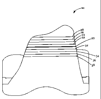
Referring now to Fig. 1, Fig. 1 shows one embodiment of the
present invention comprising a femoral trial 10. The femoral trial 10
comprises a mark 20 indicating a desired point on the femoral trial 10.
According to some embodiments, the desired point can be a proper
patellofemoral contact position in extension. In other embodiments, the
desired point could be other joint-line indicating features such as ligament
or bone landmarks or intermediate positioning features such as a bone
spike alignment. In other embodiments, the mark 20 could indicate a
range of proper patellofemoral contact positions. According to the
embodiment depicted, the mark is a transverse laser etch line on an
anterior flange of the femoral trial 10. In other embodiments, the mark
could be a mechanically etched line, an imprint from a mold, a drawn or
painted line, or any other mark.
The femoral trial 10 further comprises a plurality of additional marks
22-38 indicating a distance from the mark 20. According to the
embodiment depicted, the plurality of additional marks 22-38 are equally
spaced. In the embodiment depicted, the additional marks 22-28 are
located proximally to the mark 20 and the additional marks 32-38 are
located distally to the mark 20. According to some embodiments, the
additional marks 22-38 further comprise a plurality or numbers designating
a distance proximal or distal from the mark 20.
Referring now to Figs. 2 and 3, Fig. 2 illustrates a lateral view of the
femoral trial of Fig. 1. Fig. 3 illustrates a close up of a portion of Fig. 2.
7
CA 02571322 2006-12-15
WO 2006/002296 PCT/US2005/022195
According to the embodiment depicted, the anterior flange of the femoral
component 10 has a lateral side. The lateral side of the anterior flange
comprises extensions of the additional marks 20-38 shown in Fig. 1. The
lateral edge of the anterior flange depicted in Fig. 3 further comprises a
plurality of numbers 40-58 indicating a distance from a corresponding
additional mark to the mark 20. For example, the additional mark 22 in the
embodiment shown is two millimeters proximal to the mark 20. The
number 42 corresponding to the additional mark 20 thus indicates "+2."
Similarly, the additional mark 32 is two millimeters distal to the mark 20,
thus the number 52 corresponding to the additional mark 32 indicates "-2."
In the embodiment depicted, negative numbers indicate distance in
millimeters distal to the mark 20, and positive numbers indicate a distance
in millimeters proximal to the mark 20. In other embodiments, other units
or spacings can be used.
Fig. 4 illustrates a method in accordance with the present invention.
Although the method shown in Fig. 4 could be carried out with other
devices or other embodiments of the present invention, it will be described
using the embodiments depicted in Figs. 1-3. Fig. 4 illustrates a method in
accordance with certain embodiments of the present invention for placing a
femoral component using a femoral trial 10 comprising a mark indicating a
desired patellofemoral contact point in extension during a surgical
procedure. The method illustrated in Fig. 4 begins in block 402 wherein a
previously implanted femoral component is removed. Following removal of
the previously implanted tibial and femoral components, there is often
significant bone deterioration on the proximal tibia and the distal femur and
an absence of adequate anatomical landmarks to indicate accurate joint
line positioning. Once the previously implanted femoral component is
removed, the distal femur is prepared with the standard revision technique
and the method illustrated in Fig. 4 proceeds to block 404, wherein the trial
femoral component 10 is inserted onto the distal femur. Inserting the trial
femoral component 10 onto the distal femur allows the surgeon to assess
positioning and alignment of the femoral trial prior to placing a new femoral
8
CA 02571322 2006-12-15
WO 2006/002296 PCT/US2005/022195
component. According to one embodiment, the trial femoral component 10
can comprise a mark 20 indicating a proper patellofemoral contact point in
extension. According to one embodiment, the trial 10 can further comprise
a plurality of marks 28-38 as illustrated in Fig. 1. According to certain
embodiments, the marks can extend to a lateral or medial side of the
femoral trial 10 as illustrated in Figs. 2 and 3. According to one
embodiment, the femoral trial 10 can further comprise numbers 48-58
indicating a distance proximal to or distal to the mark 20 indicating the
proper patellofemoral contact point in extension.
Once the trial femoral component 10 is inserted onto the distal
femur, the method illustrated in Fig. 4 proceeds to block 406 wherein the
patient's leg is placed in full extension. When the patient's leg is placed in
full extension, the patellar component is brought in place above the
femoral trial component 10. Once the patellar component is in place
above the femoral trial component, the method proceeds to block 408
wherein it is determined whether the patella component is superior to,
inferior to, or aligned with the mark 20 indicating the desired patellafemoral
contact position on the femoral trial component. If the patellar component
is superior to the mark 20 on the femoral trial 10 indicating the desired
patellofemoral contact point in extension, the method illustrated in Fig. 4
proceeds to block 412 wherein the distal femur can be resected in order to
proximalize the distal femur component and to bring the patellar
component into proper alignment with the desired patellafemoral contact
position. According to one embodiment, a surgeon or other member of a
surgical team can examine the marks 22-28 and corresponding numbers
42-48 to assist in determining how much the resection should occur on the
distal femur. For example, the surgeon can determine that the patellar
component aligns with mark 24 on the femoral trial 10. Mark 24 on the
femoral trial 10 corresponds to number 42 which, according to the
embodiment depicted in Fig. 3, can indicate to the surgeon that the patellar
component is four millimeters superior to the desired patellofemoral
contact point 20. Thus, in this example, the surgeon can determine that
9
CA 02571322 2006-12-15
WO 2006/002296 PCT/US2005/022195
the distal femur should be resected sufficient to proximalize the trial
femoral component 10 a total of four millimeters.
If it is determined in block 408 that the patellar component is inferior
to the mark 20 indicating the desired patellofemoral contact point, the
femoral component 10 can be distalized through the use of femoral
augments. Distalizing the femoral trial 10 through the use of femoral
augments can function to bring the patellar component into proper
alignment with the desired joint line position between the femoral and tibial
prosthetic components as indicated by the mark 20 on the femoral trial 10.
According to one embodiment, the surgeon or other member of the
surgical team can compare a position of the patellar component with the
marks 32-38 and corresponding numbers 52-58 to assist in determining
the degree to which the femoral trial 10 should be distalized through the
use of femoral augments. For example, the surgeon can determine that
the patellar component aligns with mark 34 on the femoral trial 10. Mark
34 on the femoral trial 10 corresponds to number 54, which can indicate,
according to the example illustrated in Fig. 3, that the patellar component
is four millimeters distal to the desired patellofemoral contact point as
indicated by the mark 20 on the femoral trial 10. Thus, according to this
example, the surgeon could use sufficient femoral augments to distalize
the femoral trial 10 a total of four millimeters in order to establish the
correct joint line between the femoral and tibial prosthetic components.
Following block 412 or 410, the example method illustrated in Fig. 4
returns to block 408 wherein it is again determined whether the patellar
component is superior to, inferior to, or aligned with the mark. If it is
determined in block 408 that the patella component is aligned with the
mark indicating the desired point on the femoral trial component, the
method proceeds to block 414 wherein the procedure is completed
according to standard surgical technique. Completing the procedure
according to standard surgical technique includes, for example, placing the
femoral prosthetic component in proper position as determined by the
method described above to achieve the desired joint line between the
CA 02571322 2006-12-15
WO 2006/002296 PCT/US2005/022195
femoral and tibial prosthetic components. Completing the procedure
according to standard surgical technique can further comprise placing the
tibial prosthetic components in proper position as determined by the
method described above which will achieve the desired joint line between
the femoral and tibial prosthetic components.
According to other embodiments, the proper patellofemoral contact
position can be further identified by mating reference instrumentation to
existing anatomical landmarks, implanted components, or to other femoral
or tibial bone reactions. According to other embodiments, the proper
patellofemoral contact position can be further identified by marking a
medial and/or lateral sides of the femur at the patellofemoral contact point
when the leg is paced in extension prior to removal of the primary
components. Following the removal of the primary components, a
reference plate can be attached to a revision femoral cutting block. The
reference plate can then be compared to the mark on the medial and/or
lateral sides of the femur to further assist in initial femoral resection
alignment.
The foregoing description of embodiments of the invention has been
presented only for the purpose of illustration and description and is not
intended to be exhaustive or to limit the invention to the precise forms
disclosed. Numerous modifications and adaptations thereof will be
apparent to those skilled in the art without departing from the spirit and
scope of the present invention.
11