Note: Descriptions are shown in the official language in which they were submitted.
DEMANDE OU BREVET VOLUMINEUX
LA PRESENTE PARTIE DE CETTE DEMANDE OU CE BREVET COMPREND
PLUS D'UN TOME.
CECI EST LE TOME 1 DE 2
CONTENANT LES PAGES 1 A 38
NOTE : Pour les tomes additionels, veuillez contacter le Bureau canadien des
brevets
JUMBO APPLICATIONS/PATENTS
THIS SECTION OF THE APPLICATION/PATENT CONTAINS MORE THAN ONE
VOLUME
THIS IS VOLUME 1 OF 2
CONTAINING PAGES 1 TO 38
NOTE: For additional volumes, please contact the Canadian Patent Office
NOM DU FICHIER / FILE NAME:
NOTE POUR LE TOME / VOLUME NOTE:
CA 02571642 2006-12-20
WO 2006/017150 PCT/US2005/024193
IDENTIFICATION OF MARKERS IN LUNG AND BREAST CANCER
INVENTORS
Tony Godfrey
Liqiang Xi
William E. Gooding
Siva Raja
CROSS REFERENCE TO RELATED APPLICATIONS
This application claims priority to United States Provisional Patent
Application Nos. 60/586,599 and
60/587,019, both filed on July 9, 2004, each of which is incorporated herein
by reference in its entirety.
BACKGROUND
1. Field of the Invention
Provided are improved methods for diagnosing cancer cells in lymph nodes,
along with
compositions and apparatus useful in conducting those methods.
2. Description of the Related Art
Early detection of cancer typically leads to increased survival rates.
Metastatic lesions
commonly are detected by histological techniques, including
immunohistochemical techniques.
Metastasized cells typically infiltrate the lymph nodes, and, thus in most
instances, certain sentinel lymph
nodes (lymph nodes where metastasized cells typically first infiltrate), or
staging lymph nodes (lymph
nodes typically analyzed for presence of certain types of cancers), are
recognized for each cancer type
and are analyzed for the presence of lesions, including micrometastases.
Trained histologists often can
detect metastatic lesions visually after tissue from a sentinel or staging
lymph node is sectioned and
stained. Highly trained histologists often can visualize micrometasteses, but
the ability to visualize such
lesions varies from histologist-to-histologist.
In many surgical procedures to remove tumors, biopsies of sentinel lymph nodes
are taken. The
surgical procedure is then halted and the excised lymphatic tissue is then
analyzed. Once it is determined
that the tumor has metastasized, a second, more radical surgical procedure is
performed, removing
regional lymphatics. A rapid method for identifying tumors is therefore
warranted, not only because
more assays can be performed in a given time period, thereby increasing
laboratory turnaround, but
permitting accurate, intraoperative decisions to be made, rather than
conducting a second surgical
procedure. It is therefore desirable to identify useful diagnostics for
malignancies, especially that permit
rapid and/or intraoperative detection of lymphatic micrometastases.
SUMMARY
The present invention relates to a diagnostic method for detecting the
presence of cancer cells in
a patient by identifying the expression of certain markers indicative of the
presence of cancer cells.
1
CA 02571642 2006-12-20
WO 2006/017150 PCT/US2005/024193
In one embodiment, the present invention relates to a method of identifying
the expression of
markers indicative of the presence of breast cancer cells in a lymph node of a
patient. The method
comprises determining if an mRNA species specific to one or more of TACSTDI,
CK19, MGBl,
MGB2, PIP and CK7 is overabundant in an RNA sample prepared from the lymph
node. The
overabundance of the mRNA species is indicative of the presence of displaced
breast cells in the lymph
node.
In another embodiment, the present invention relates to a method for
identifying the expression
of markers indicative of the presence of lung cancer cells in a lymph node of
a patient. The method
comprises determining if an mRNA species specific to one or more of CEA, CK7,
CK 19, PVA,
SCCA1.2 (SCCA1 +SCCA2), SFTPB and TACSTD1 is overabundant in an RNA sample
prepared from
the lymph node. The overabundance of the mRNA species is indicative of the
presence of displaced lung
cells in the lymph node.
In still another embodiment, the present invention relates to an article of
manufacture comprising
packaging material and one or more nucleic acids specific to one or more of
CK7, CK19, MGB1, MGB2,
PIP and TACSTD1. The packaging material comprises an indicia, for example and
without limitation, a
writing, illustration, label, tag, book, booklet and/or package insert,
indicating that the one or more
nucleic acids can be used in a method of identifying expression of markers
indicative of the presence of
breast cancer cells in a lymph node of a patient.
In a further embodiment, the present invention relates to an article of
manufacture comprising
packaging material and one or more nucleic acids specific to one or more of
CEA, CK7, CK19, PVA,
SCCA1.2, SFTPB and TACSTDl. The packaging material comprises an indicia
indicating that the one
or more nucleic acids can be used in a method of identifying expression of
markers indicative of the
presence of lung cancer cells in a lymph node of a patient.
In a still further embodiment, the present invention relates to a composition
comprising one or
more primers or probes specific to one or more of CK7, CK19, MGB1, MGB2, PIP
and TACSTDI and
RNA extracted from the lymph node of a patient diagnosed with or suspected of
having breast cancer, or
a nucleic acid, or analog thereof, derived from the RNA.
In yet a further embodiment, the present invention relates to a composition
comprising one or
more primers or probes specific to one or more of CEA, CK7, CK19, PVA,
SCCA1.2, SFTPB and
TACSTDI and RNA extracted from the lymph node of a patient diagnosed with or
suspected of having
lung cancer, or a nucleic acid, or analog thereof, derived from the RNA.
BRIEF DESCRIPTION OF THE DRAWINGS
Figure 1 is a listing of a cDNA sequence of the cytokeratin 7 (CK7) marker
(SEQ ID NO: 1).
Figure 2 is a listing of a cDNA sequence of the cytokeratin 19 (CK19) marker
(SEQ ID NO: 2).
2
CA 02571642 2006-12-20
WO 2006/017150 PCT/US2005/024193
Figure 3 is a listing of a cllNA sequence of the mammaglobin 1(MGB 1) marker
(SEQ ID NO: 3).
Figure 4 is a listing of a eDNA sequence of the mammaglobin 2 (MGB2) marker
(SEQ ID NO: 4).
Figure 5 is a listing of a cDNA sequence of the prolactin-inducible protein
(PIP) marker
(SEQ ID NO: 5).
Figure 6 is a listing of a cDNA sequence of the pemphigus vulgaris (PVA)
marker
(SEQ ID NO: 6).
Figure 7 is a listing of a cDNA sequence of the squamous cell carcinoma
antigen 1(SCCAl)
marker (SEQ ID NO: 7).
Figure 8 is a listing of a cDNA sequence of the squainous cell carcinoma
antigen 2 (SCCA2)
marker (SEQ ID NO: 8).
Figure 9 is a listing of a cDNA sequence of the surfactant, pulmonary-
associated protein b
(SFTPB) marker (SEQ ID NO: 9).
Figure 10 is a listing of a cDNA sequence of the tumor-associated calcium
signal transducer 1
(TACSTD1) marker (SEQ ID NO: 10).
Figure 11 is a listing of a cDNA sequence of the carcinoembryonic antigen-
related cell adhesion
molecule 5 (CEA) marker (SEQ ID NO: 11).
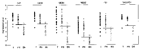
Figure 12 is a scatter plot showing the expression levels of CK7, CK19, MGB1,
MGB2, PIP and
TACSTDI in primary tumor, tumor-positive lymph nodes and benign lymph nodes of
a breast cancer
patient.
Figures 13 A-O provide scatter plots illustrating the ability of two-marker
systems to distinguish
between benign and malignant cells in a lymph node of a breast cancer patient
(negative - gray circle;
positive - black circle).
Figure 14 is a scatter plot showing the expression levels of CEA, CK7, CK19,
LUNX, PVA,
SCCA 1.2, SFTPB and TACSTDI in primary tumor, tumor-positive lymph nodes and
benign lymph
nodes of a lung cancer patient.
Figure 15 A-BB provide scatter plots illustrating the ability of two-marker
systems to distinguish
between benign and malignant cells in a lymph node of a lung cancer patient
(negative - circle; positive -
GL t_77).
Figure 16 is a plot of the best combination of three markers for detecting
lung cancer in different
histological types, plotting the Fold Change-Positive Lymph Nodes PLN vs.
Highest Benign Nodes BN
(PVA - asterix; SFTPB - circle; and TACSTDI- triangle).
3
CA 02571642 2006-12-20
WO 2006/017150 PCT/US2005/024193
Figure 17A provides data obtained from the secondary screening set of lymph
nodes on
individual gene expression observed in primary tumors, benign and positive
nodes. The horizontal line
indicates the most accurate cutoff value calculated by a receiver-operator
characteristic curve analysis.
Classification characteristics of the individual markers are reported in Table
1, below.
Figures 17B-E provide secondary screening set data on gene expression for
potential two-
marker combinations using a linear discriminator decision rule. As with the
individual markers, the black
line indicates the decision rule generated from the secondary screening set
data that produces the most
accurate characterization. Classification characteristics of the marker
combinations are reported in
Table I.
Figures 17F-I provide secondary screening set data on gene expression for
potential two-marker
combinations applying equal probability contour statistical analysis. Equal
probability curves were
generated around the mean expression value observed for the 2 markers in
benign lymph nodes. This
demonstrates that while the marker combination of CK 19 and MGB 1 accurately
characterizes the lymph
nodes (see Table 1), the wide distribution of expression observed in benign
nodes for these markers
increases optimism in applying the decision rule. By this method of analysis,
the marker combination of
TACSTDI and PIP more confidently characterizes the lymph nodes.
Figure 18A provides data obtained from the validation set of SLN on individual
gene expression
observed in negative and positive nodes. The horizontal line indicates the
decision rule calculated from
data obtained from the secondary screening set. Classification characteristics
of the individual markers
are reported in Table J.
Figures 18B-G provide validation set data on gene expression for two-marker
combinations
using linear discriminator decision rule for all potential marker pairs. As
with the individual markers, the
black line indicates the decision rule generated from the secondary screening
set data that produces the
most accurate characterization. Classification characteristics of the marker
combinations are reported in
Table J.
Figures 18H-M provide validation set data analyzed using the equal probability
contours
generated from the secondary screening set data. The relative levels of
expression observed for all but
one of the positive lymph nodes were well outside the 0.999 confidence
contour. Some of the positive
nodes are positive for only one marker (crosses located in the left upper or
right lower quadrants),
demonstrating that a 2 marker assay improves sensitivity while maintaining
high specificity.
Figures 19A-B provides results of a fully automated, 2-marker QRT-PCR analysis
of lymph
nodes. By eitlier linear decision rule analysis (Figure 19A) or equal
probability contour analysis (Figure
19B), the assay accurately characterized all 181ymph nodes (9 negative, 9
positive) evaluated.
4
CA 02571642 2006-12-20
WO 2006/017150 PCT/US2005/024193
DETAILED DESCRIPTION
Provided are methods and compositions useful in identifying breast cancer and
lung cancer cells,
including micrometastases, in lymph nodes. Early detection of metastases
typically is related to patient
survival. Very small metastases often go undetected in histological study of
lymph node biopsies,
resulting in false negative results that result in decreased chances of
patient survival. The nucleic acid
detection assays described herein are much more discriminating than are
histological studies in most
instances (a few, excellent histologists are capable of identifying
micrometastases in lymph node
sections), and are robust and repeatable in the hands of any minimally-trained
teclmician. Although the
methods and compositions described herein are necessarily presented comprising
expression of specific
mRNA markers, this should be understood that it shall not be deemed to exclude
metliods and
compositions comprising combinations of the specific markers and other markers
known in the art.
To this end, a number of molecular markers are identified, that are expressed
in certain cancer
types, including breast cancer and lung cancer. These markers are markers
specific to the tissue from
which the particular cancer type arises and typically are not expressed, at
least to the same levels, in
lymphoid tissue. The presence and/or elevated expression of one or more of
these markers in sentinel
lymph node tissue is indicative of displaced cells in the lymphoid tissue,
which correlates strongly with a
cancer diagnosis.
As used herein, the terms "expression" and "expressed" mean production of a
gene-specific
mRNA by a cell. In the context of the present disclosure, a "marker" is a gene
that is expressed
abnormally in a lymphatic biopsy. In one embodiment, the markers described
herein are mRNA species
that are expressed in cells of a specific tumor source at a significantly
higher level as compared to
expression in lymphoid cells.
Expression levels of mRNA can be quantifyd by a number of methods. Traditional
methods
include Northern blot analysis. More recently, nucleic acid detection methods
have been devised that
facilitate quantification of transcripts. Examples of PCR methods are
described in United States Patent
Application No. 10/090,326 (US 10/090,326), incorporated herein by reference
in its entirety. Other
methods for determining expression levels of a given mRNA include isothermic
amplification or
detection assays and array technologies, as are known in the art, such as,
without limitation, those
described below.
The improved PCR methods described herein as well as in US 10/090,326, and
other nucleic acid
detection and amplification methods described herein and as are known in the
art permit rapid detection
of cancer cells in lymph node tissue. These rapid methods can be used
intraoperatively, and also are
useful in detecting rare nucleic acid species, even in multiplexed PCR
reactions that concurrently detect a
more prevalent control nucleic acid.
CA 02571642 2006-12-20
WO 2006/017150 PCT/US2005/024193
A typical PCR reaction includes multiple amplification steps, or cycles that
selectively amplify a
target nucleic acid species. Because detection of transcripts is necessary,
the PCR reaction is coupled
with a reverse transcription step (reverse transcription PCR, or RT-PCR). A
typical PCR reaction
includes three steps: a denaturing step in which a target nucleic acid is
denatured; an annealing step in
which a set of PCR primers (forward and backward primers) anneal to
complementary DNA strands; and
an elongation step in which a thermostable DNA polymerase elongates the
primers. By repeating this
step multiple times, a DNA fragment is amplified to produce an amplicon,
corresponding to the target
DNA sequence. Typical PCR reactions include 30 or more cycles of denaturation,
annealing and
elongation. In many cases, the annealing and elongation steps can be performed
concurrently, that is at
the same temperature, in which case the cycle contains only two steps.
The lengths of the denaturation, annealing and elongation stages may be any
desirable length of
time. However, in attempting to shorten the PCR amplification reaction to a
time suitable for
intraoperative diagnosis, the lengths of these steps can be in the seconds
range, rather than the minutes
range. The denaturation step may be conducted for times of one second or less.
The annealing and
elongation steps optimally are less than 10 seconds each, and when conducted
at the same temperature,
the combination annealing/elongation step may be less than 10 seconds. Use of
recently developed
amplification techniques, such as conducting the PCR reaction in a Rayleigh-
Benard convection cell, also
can drainatically shorten the PCR reaction time beyond these time limits (see,
Krishnan, My et al., "PCR
in a Rayleigh-Benard convection cell." Science 298:793 (2002), and Braun, D.
et al., "Exponential DNA
Replication by Lominar Convection," Physical Review Letters, 91:158103).
As described in US 10/090,326, each cycle may be shortened considerably
witliout substantial
deterioration of production of amplicons. Use of high concentrations of
primers is helpful in shortening
the PCR cycle time. High concentrations typically are greater than about
400nM, and often greater than
about 800nM, though the optimal concentration of primers will vary somewhat
from assay-to-assay.
Sensitivity of RT-PCR assays may be enhanced by the use of a sensitive reverse
transcriptase enzyme
(described below) and/or high concentrations of reverse transcriptase primer
to produce the initial target
PCR template.
The specificity of any given PCR reaction relies heavily, but not exclusively,
on the identity of
the primer sets. The primer sets are pairs of forward and reverse
oligonucleotide primers that anneal to a
target DNA sequence to permit amplification of the target sequence, thereby
producing a target sequence-
specific amplicon. PCR primer sets can include two primers internal to the
target sequence, or one
primer internal to the target sequence and one specific to a target sequence
that is ligated to the DNA or
cDNA target, using a technique known as "ligation-anchored PCR" (Troutt, A.B.,
et al. (1992),
"Ligation-anchored PCR: A Simple Amplification Technique with Single-sided
Specificity," Proc. Natl.
Acad. Sci. USA, 89:9823-9825).
6
CA 02571642 2006-12-20
WO 2006/017150 PCT/US2005/024193
- ws usea nereIrr, -a""aet-ivanv'e" ora speciried oligonucleotide is an
oligonucleotide that binds to
the same target sequence as the specified oligonucleotide and amplifies the
same target sequence to
produce essentially the same amplicon as the specified oligonucleotide but for
differences between the
specified oligonucleotide and its derivative. The derivative may differ from
the specified oligonucleotide
by insertion, deletion and/or substitution of any residue of the specified
sequence so long as the
derivative substantially retains the characteristics of the specified sequence
in its use for the same
purpose as the specified sequence.
As used herein, "reagents" for any assay or reaction, such as a reverse
transcription and PCR, are
any compound or composition that is added to the reaction mixture including,
without limitation,
enzyme(s), nucleotides or analogs thereof, primers and primer sets, probes,
antibodies or other binding
reagents, detectable labels or tags, buffers, salts and co-factors. As used
herein, unless expressed
otherwise, a "reaction mixture" for a given assay or reaction includes all
necessary compounds and/or
compositions necessary to perform that assay or reaction, even if those
compounds or compositions are
not expressly indicated. Reagents for many common assays or reactions, such as
enzymatic reaction, are
known in the art and typically are provided and/or suggested when the assay or
reaction kit is sold.
As also described in US 10/090,326, multiplexed PCR assays may be optimized,
or balanced, by
time-shifting the production of amplicons, rather than by manipulating primer
concentrations. This may
be achieved by using two primer sets, each primer set having a different Tm so
that a two-stage PCR
assay can be performed, with different annealing and/or elongation
temperatures for each stage to favor
the production of one amplicon over another. This time and temperature
shifting method permits optimal
balancing of the multiplex reaction witliout the difficulties faced when
manipulation of primer
concentrations is used to balance the reaction. This technique is especially
useful in a multiplex reaction
where it is desirable to amplify a rare cDNA along with a control cDNA.
A quantitative reverse transcriptase polymerase chain reaction (QRT-PCR) for
rapidly and
accurately detecting low abundance RNA species in a population of RNA
molecules (for example, and
without limitation, total RNA or mRNA), includes the steps of: a) incubating
an RNA sample with a
reverse transcriptase and a high concentration of a target sequence-specific
reverse transcriptase primer
under conditions suitable to generate cDNA; b) subsequently adding suitable
polymerase chain reaction
(PCR) reagents to the reverse transcriptase reaction, including a high
concentration of a PCR primer set
specific to the cDNA and a thermostable DNA polymerase to the reverse
transcriptase reaction, and c)
cycling the PCR reaction for a desired number of cycles and under suitable
conditions to generate PCR
product ("ainplicons") specific to the cDNA. By temporally separating the
reverse transcriptase and the
PCR reactions, and by using reverse transcriptase-optimized and PCR-optimized
primers, excellent
specificity is obtained. The reaction may be conducted in a single tube (all
tubes, containers, vials, cells
and the like in which a reaction is performed may be referred to herein, from
time to time, generically, as
a "reaction vessel"), removing a source of contamination typically found in
two-tube reactions. These
7
CA 02571642 2006-12-20
WO 2006/017150 PCT/US2005/024193
readlion conditions'pe'r'mit'very rapTd QKT-YC:x reactions, typically on the
order of 20 minutes from the
beginning of the reverse transcriptase reaction to the end of a 40 cycle PCR
reaction.
The reaction c) may be performed in the same tube as the reverse transcriptase
reaction by
adding sufficient reagents to the reverse transcriptase (RT) reaction to
create good, or even optimal
conditions for the PCR reaction to proceed. A single tube may be loaded, prior
to the running of the
reverse transcriptase reaction, with: 1) the reverse transcriptase reaction
mixture, and 2) the PCR reaction
mixture to be mixed with the cDNA mixture after the reverse transcriptase
reaction is completed. The
reverse transcriptase reaction mixture and the PCR reaction mixture may be
physically separated by a
solid, or semi-solid (including amorphous, glassy substances and waxy) barrier
of a composition that
melts at a temperature greater than the incubation temperature of the reverse
transcriptase reaction, but
below the denaturing temperature of the PCR reaction. The barrier composition
may be hydrophobic in
nature and forms a second phase with the RT and PCR reaction mixtures when in
liquid form. One
example of such a barrier composition is wax beads, commonly used in PCR
reactions, such as the
AMPLIWAX PCR GEM products commercially available from Applied Biosystems of
Foster City,
California.
Alternatively, the separation of the reverse transcriptase and the PCR
reactions may be achieved
by adding the PCR reagents, including the PCR primer set and thermostable DNA
polymerase, after the
reverse transcriptase reaction is completed. Preferably the PCR reagents, are
added mechanically by a
robotic or fluidic means to make sample contamination less likely and to
remove human error.
The products of the QRT-PCR process may be compared after a fixed number of
PCR cycles to
determine the relative quantity of the RNA species as compared to a given
reporter gene. One method of
comparing the relative quantities of the products of the QRT-PCR process is by
gel electrophoresis, for
instance, by running the samples on a gel and detecting those samples by one
of a number of known
methods including, without limitation, Southern blotting and subsequent
detection with a labeled probe,
staining with ethidium bromide and incorporating fluorescent or radioactive
tags in the amplicons.
However, the progress of the quantitative PCR reactions typically is monitored
by determining
the relative rates of amplicon production for each PCR primer set. Monitoring
ainplicon production may
be achieved by a number of processes, including without limitation,
fluorescent primers, fluorogenic
probes and fluorescent dyes that bind double-stranded DNA. A common method is
the fluorescent 5'
nuclease assay. This method exploits the 5' nuclease activity of certain
thermostable DNA polymerases
(such as Taq or Tfl DNA polymerases) to cleave an oligomeric probe during the
PCR process. The
oligomer is selected to anneal to the amplified target sequence under
elongation conditions. The probe
typically has a fluorescent reporter on its 5' end and a fluorescent quencher
of the reporter at the 3' end.
So long as the oligomer is intact, the fluorescent signal from the reporter is
quenched. However, when
the oligomer is digested during the elongation process, the fluorescent
reporter no longer is in proximity
to the quencher. The relative accumulation of free fluorescent reporter for a
given amplicon may be
8
CA 02571642 2006-12-20
WO 2006/017150 PCT/US2005/024193
cornpared'Ito th6'ac:6iCrhul'atidYrof til'i6 s'aifib amplicons for a control
sample and/or to that of a control
gene, such as 0-actin or 18S rRNA to determine the relative abundance of a
given cDNA product of a
given RNA in a RNA population. Products and reagents for the fluorescent 5'
nuclease assay are readily
available commercially, for instance from Applied Biosystems.
Equipment and software also are readily available for monitoring amplicon
accumulation in PCR
and QRT-PCR according to the fluorescent 5' nuclease assay and other QPCR/QRT-
PCR procedures,
including the Smart Cycler, commercially available from Cepheid of Sunnyvale,
California, the ABI
Prism 7700 Sequence Detection System (TaqMan), commercially available from
Applied Biosystems. A
cartridge-based sample preparation system (GenXpert) combines a thermal cycler
and fluorescent
detection device having the capabilities of the Smart Cycler product with
fluid circuits and processing
elements capable of automatically extracting specific nucleic acids from a
tissue sample and performing
QPCR or QRT-PCR on the nucleic acid. The system uses disposable cartridges
that can be configured
and pre-loaded with a broad variety of reagents. Such a system can be
configured to disrupt tissue and
extract total RNA or mRNA from the sample. The reverse transcriptase reaction
components can be
added automatically to the RNA and the QPCR reaction components can be added
automatically upon
completion of the reverse transcriptase reaction.
Further, the PCR reaction may be monitored of production (or loss) of a
particular fluorochrome
from the reaction. When the fluorochrome levels reach (or fall to) a desired
level, the automated system
will automatically alter the PCR conditions. In one example, this is
particularly useful in the multiplexed
embodiment described above, where a more-abundant (control) target species is
amplified by the first,
lower Tm, primer set at a lower temperature than the less abundant species
amplified by the second,
higher Tm, primer set. In the first stage of the PCR amplification, the
annealing temperature is lower
than the effective Tm of the first primer set. The annealing temperature then
is automatically raised
above the effective Tm of the first primer set when production of the first
amplicon by the first primer set
is detected. In a system that automatically dispenses multiple reagents from a
cartridge, such as the
GeneXpert system, a first PCR reaction may be conducted at the first Tm and,
when the first PCR
reaction proceeds past a threshold level, a second primer with a different Tm
is added, resulting in a
sequential multiplexed reaction.
In the above-described reactions, the amounts of certain reverse transcriptase
and the PCR
reaction components typically are atypical in order to take advantage of the
faster ramp times of some
thermal cyclers. Specifically, the primer concentrations are very high.
Typical gene-specific primer
concentrations for reverse transcriptase reactions are less than about 20 nM.
To achieve a rapid reverse
transcriptase reaction on the order of one to two minutes, the reverse
transcriptase primer concentration
was raised to greater than 20 nM, preferably at least about 50 nM, and
typically about 100 nM. Standard
PCR primer concentrations range from 100 nM to 300 nM. Higher concentrations
may be used in
standard PCR reactions to compensate for Tm variations. However, the
referenced primer concentrations
9
CA 02571642 2006-12-20
WO 2006/017150 PCT/US2005/024193
are Yor b-ifrc'uiiistahoes'-ftere rro--ThrVomOnsation is needed.
Proportionately higher concentrations of
primers may be empirically determined and used if Tm compensation is necessary
or desired. To achieve
rapid PCR reactions, the PCR primer concentrations typically are greater than
200 nM, preferably greater
than about 500 nM and typically about 800 nM. Typically, the ratio of reverse
transcriptase primer to
PCR primer is about 1 to 8 or more. The increase in primer concentrations
permitted PCR experiments
of 40 cycles to be conducted in less than 20 minutes.
A sensitive reverse transcriptase may be preferred in certain circumstances
where either low
amounts of RNA are present or a target RNA is a low abundance RNA. By the term
"sensitive reverse
transcriptase," it is meant a reverse transcriptase capable of producing
suitable PCR templates from low
copy number transcripts for use as PCR templates. The sensitivity of the
sensitive reverse transcriptase
may derive from the physical nature of the enzyme, or from specific reaction
conditions of the reverse
transcriptase reaction mixture that produces the enhanced sensitivity. One
example of a sensitive reverse
transcriptase is SensiScript RT reverse transcriptase, commercially available
from Qiagen, Inc. of
Valencia, California. This reverse transcriptase is optimized for the
production of cDNA from RNA
samples of <50ng, but also has the ability to produce PCR templates from low
copy number transcripts.
In practice, in the assays described herein, adequate results were obtained
for samples of up to, and even
in excess of, about 400 ng RNA. Other sensitive reverse transcriptases having
substantially similar
ability to reverse transcribe low copy number transcripts would be equivalent
sensitive reverse
transcriptase for the purposes described herein. Notwithstanding the above,
the ability of the sensitive
reverse transcriptase to produce cDNA from low quantities of RNA is secondary
to the ability of the
enzyme, or enzyme reaction system to produce PCR templates from low copy
number sequences.
As discussed above, the procedures described herein also may be used in
multiplex QRT-PCR
processes. In its broadest sense, a multiplex PCR process involves production
of two or more amplicons
in the same reaction vessel. Multiplex amplicons may be analyzed by gel
electrophoresis and detection
of the amplicons by one of a variety of methods, such as, without limitation
ethidium bromide staining,
Southern blotting and hybridization to probes, or by incorporating fluorescent
or radioactive moieties into
the amplicons and subsequently viewing the product on a gel. However, real-
time monitoring of the
production of two or more amplicons is preferred. The fluorescent 5' nuclease
assay is the most common
monitoring method. Equipment is now available (for example, the above-
described Smart Cycler and
TaqMan products) that permits the real-time monitoring of accumulation of two
or more fluorescent
reporters in the same tube. For multiplex monitoring of the fluorescent 5'
nuclease assay, oligomers are
provided corresponding to each amplicon species to be detected. The oligomer
probe for each amplicon
species has a fluorescent reporter with a different peak emission wavelength
than the oligomer probe(s)
for each other amplicons species. The accumulation of each unquenched
fluorescent reporter can be
monitored to determine the relative amounts of the target sequence
corresponding to each amplicon.
CA 02571642 2006-12-20
WO 2006/017150 PCT/US2005/024193
Th fradition2l"milftipl8YQPCR1fftl QRT-PCR procedures, the selection of PCR
primer sets
having similar annealing and elongation kinetics and similar sized amplicons
are desirable. The design
and selection of appropriate PCR primer sets is a process that is well known
to a person skilled in the art.
The process for identifying optimal PCR primer sets, and respective ratios
thereof to achieve a balanced
multiplex reaction also is known. By "balanced," it is meant that certain
amplicon(s) do not out-compete
the other amplicon(s) for resources, such as dNTPs or enzyme. For instance, by
limiting the abundance
of the PCR primers for the more abundant RNA species in an RT-PCR experiment
will allow the
detection of less abundant species. Equalization of the Tm (melting
teinperature) for all PCR primer sets
also is encouraged. See, for instance, ABI PRISM 7700 Sequence Detection
System User Bulletin #5,
"Multiplex PCR with TaqMan VIC Probes", Applied Biosystems (1998/2001).
Despite the above, for very low copy number transcripts, it is difficult to
design accurate
multiplex PCR experiments, even by limiting the PCR primer sets for the more
abundant control species.
One solution to this problem is to run the PCR reaction for the low abundance
RNA in a separate tube
than the PCR reaction for the more abundant species. However, that strategy
does not take advantage of
the benefits of running a multiplex PCR experiment. A two-tube process has
several drawbacks,
including cost, the addition of more room for experimental error and the
increased chance of sample
contamination, which is critical in PCR assays.
A method has been described in WO 02/070751 for performing a multiplex PCR
process,
including QRT-PCR and QPCR, capable of detecting low copy number nucleic acid
species along with
one or more higher copy number species. The difference between low copy number
and high copy
number nucleic acid species is relative, but is referred to herein as a
difference in the prevalence of a low
(lower) copy number species and a high (higher) copy number species of at
least about 30-fold, but more
typically at least about 100-fold. For purposes herein, the relative
prevalence of two nucleic acid species
to be amplified is more salient than the relative prevalence of the two
nucleic acid species in relation to
other nucleic acid species in a given nucleic acid sample because other
nucleic acid species in the nucleic
acid sample do not directly compete with the species to be amplified for PCR
resources.
As used herein, the prevalence of any given nucleic acid species in a given
nucleic acid sample,
prior to testing, is unknown. Thus, the "expected" number of copies of a given
nucleic acid species in an
nucleic acid sample often is used herein and is based on historical data on
the prevalence of that species
in nucleic acid samples. For any given pair of nucleic acid species, one would
expect, based on previous
determinations of the relative prevalence of the two species in a sample, the
prevalence of each species to
fall within a range. By determining these ranges one would determine the
difference in the expected
number of target sequences for each species. An inRNA species is identified as
"overabundant" if it is
present in statistically significant amounts over normal prevalence of the
mRNA species in a sample from
a normal patient or lymph node. As is abundantly illustrated in the examples
and plots provided herein, a
11
CA 02571642 2006-12-20
WO 2006/017150 PCT/US2005/024193
person ot skili in the art WoulTtie'aCile to -ascertain statistically
significant ranges or cutoffs for
determining the precise definition of "overabundance" for any one or more mRNA
species.
The multiplex method involves performing a two- (or more) stage PCR
amplification, permitting
modulation of the relative rate of production of a first amplicon by a first
primer set and a second
amplicon by a second primer set during the respective amplification stages. By
this method, PCR
amplifications to produce amplicons directed to a lower abundance nucleic acid
species are effectively
"balanced" with PCR amplifications to produce amplicons directed to a higher
abundance nucleic acid
species. Separating the reaction into two or more temporal stages may be
achieved by omitting the PCR
primer set for any amplicons that are not to be produced in the first
amplification stage. This is best
achieved through use of automated processes, such as the GenXpert prototype
system described above.
Two or more separate amplification stages may be used to tailor and balance
multiplex assays, along
with, or to the exclusion of tailoring the concentration of the respective
primer sets.
A second method for temporally separating the PCR amplification process into
two or more
stages is to select PCR primer sets with variation in their respective Tm. In
one example, primers for a
lower copy number nucleic acid species would have a higher Tm (Tmi) than
primers for a higher
abundance species (Tm2). In this process, the first stage of PCR amplification
is conducted for a
predetermined number of cycles at a temperature sufficiently higher than TmZ
so that there is
substantially no amplification of the higher abundance species. After the
first stage of amplification, the
annealing and elongation steps of the PCR reaction are conducted at a lower
temperature, typically about
TmZ, so that both the lower abundance and the higher abundance amplimers are
amplified. It should be
noted that Tm, as used herein and unless otherwise noted, refers to "effective
Tm," which is the Tm for
any given primer in a given reaction mix, which depends on factors, including,
without limitation, the
nucleic acid sequence of the primer and the primer concentration in the
reaction mixture.
It should be noted that PCR amplification is a dynamic process. When using
temperature to
modulate the respective PCR reactions in a multiplex PCR reaction, the higher
temperature annealing
stage may be carried out at any temperature typically ranging from just above
the lower Tm to just below
the higher Tm, so long as the reaction favors production of the amplicon by
the higher Tm primer set.
Similarly, the annealing for the lower temperature reaction typically is at
any temperature below the Tm
of the low temperature primer set.
In the example provided above, in the higher temperature stage the amplicon
for the low
abundance RNA is amplified at a rate faster than that the amplicon for the
higher abundance RNA (and
preferably to the substantial exclusion of production of the second amplicon),
so that, prior to the second
amplification stage, where it is desirable that amplification of all amplicons
proceeds in a substantially
balanced manner, the amplicon for the lower abundance RNA is of sufficient
abundance that the
amplification of the higher abundance RNA does not interfere with the
amplification of the amplicon for
the lower abundance RNA. In the first stage of amplification, when the
amplicon for the low abundance
12
CA 02571642 2006-12-20
WO 2006/017150 PCT/US2005/024193
nucleic acin is preterenttally a1hiplitred; tTie annealing and elongation
steps may be performed above Tml
to gain specificity over efficiency (during the second stage of the
amplification, since there is a relatively
large number of low abundance nucleic acid amplicons, selectivity no longer is
a significant issue, and
efficiency of amplicon production is preferred). It, therefore, should be
noted that although favorable in
many instances, the temperature variations may not necessarily result in the
complete shutdown of one
amplification reaction over another.
In another variation of the above-described amplification reaction, a first
primer set with a first
Tm may target a more-abundant template sequence (for instance, the control
template sequence) and a
second primer set with a higher Tm may target a less-abundant template
sequence. In this case, the
more-abundant template and the less-abundant template may both be amplified in
a first stage at a
temperature below the (lower) Tm of the first primer set. When a tlireshold
amount of amplicon
corresponding to the more abundant template is reached, the annealing and/or
elongation temperature of
the reaction is raised above the Tm of the first primer set, but below the
higher Tm of the second primer
set to effectively shut down amplification of the more abundant template.
Selection of three or more sets of PCR primer sets having three or more
different Tms (for
instance, Tml > Tmz > Tm3) can be used to amplify sequences of varying
abundance in a stepwise
manner, so long as the differences in the Tms are sufficiently large to permit
preferential amplification of
desired sequences to the substantial exclusion of undesired sequences for a
desired number of cycles. In
that process, the lowest abundance sequences are amplified in a first stage
for a predetermined number of
cycles. Next, the lowest abundance and the lesser abundance sequences are
amplified in a second stage
for a predetermined number of cycles. Lastly, all sequences are amplified in a
third stage. As with the
two-stage reaction described above, the minimum temperature for each stage may
vary, depending on the
relative efficiencies of each single amplification reaction of the multiplex
reaction. It should be
recognized that two or more amplimers may have substantially the same Tm, to
permit amplification of
more than one species of similar abundance at any stage of the amplification
process. As with the two-
stage reaction, the three-stage reaction may also proceed stepwise from
amplification of the most
abundant nucleic acid species at the lowest annealing temperature to
amplification of the least abundant
species at the highest annealing temperature.
By this sequential amplification method, an additional tool is provided for
the "balancing" of
multiplex PCR reactions besides the matching of Tms and using limiting amounts
of one or more PCR
primer sets. The exploitation of PCR primer sets with different Tms as a
method for sequentially
amplifying different amplicons may be preferred in certain circumstances to
the sequential addition of
additional primer sets. However, the use of temperature-dependent sequencing
of multiplex PCR
reactions may be coupled with the sequential physical addition of primer sets
to a single reaction mixture.
An internal positive control that confirms the operation of a particular
amplification reaction for
a negative result also may be used. The internal positive controls (IPC) are
DNA oligonucleotides that
13
CA 02571642 2006-12-20
WO 2006/017150 PCT/US2005/024193
have'the sa'rrie prirr~er se'qu~n~es as~he"~~rget gene (CEA or tyrosinase) but
have a different internal probe
sequence. Selected sites in the IPC's optionally may be synthesized with
uracil instead of thymine so
that contamination with the highly concentrated mimic could be controlled
using uracil DNA
glycosylase, if required. The IPCs maybe added to any PCR reaction mastermix
in amounts that are
determined empirically to give Ct values typically greater than the Ct values
of the endogenous target of
the primer set. The PCR assays are then performed according to standard
protocols, and even when there
is no endogenous target for the primer set, the IPC would be amplified,
thereby verifying that the failure
to amplify the target endogenous DNA is not a failure of the PCR reagents in
the mastermix. hi this
embodiment, the IPC probe fluoresces differently than the probe for the
endogenous sequences. A
variation of this for use in RT-PCR reactions is where the IPC is an RNA and
the RNA includes an RT
primer sequence. In this embodiment, the IPC verifies function of both the RT
and PCR reactions. Both
RNA and DNA IPCs (with different corresponding probes) may also be employed to
differentiate
difficulties in the RT and PCR reactions.
The rapid QRT-PCR protocols described herein may be run in about 20 minutes.
This short time
period permits the assay to be run intraoperatively so that a surgeon can
decide on a surgical course
during a single operation (typically the patient will remain anesthetized
and/or otherwise sedated in a
single "operation", though there may be a waiting period between when the
sample to be tested is
obtained and the time the interoperative assay is complete), rather than
requiring a second operation, or
requiring the surgeon to perform unneeded or overly broad prophylactic
procedures. For instance, in the
surgical evaluation of certain cancers, including breast cancer, melanoma,
lung cancer, esophageal cancer
and colon cancer, tumors and sentinel lymph nodes are removed in a first
operation. The sentinel nodes
are later evaluated for micrometastases, and, when micrometastases are
detected in a patient's sentinel
lymph node, the patient will need a second operation, thereby increasing the
patient's surgical risks and
patient discomfort associated with multiple operations. With the ability to
determine the expression
levels of certain tumor-specific markers described herein in less than 30
minutes with increased accuracy,
a physician can make an immediate decision on how to proceed without requiring
the patient to leave the
operating room or associated facilities. The rapid test also is applicable to
needle biopsies taken in a
physician's office. A patient need not wait for days to get the results of a
biopsy (such as a needle biopsy
of a tumor or lymph node), but can now get more accurate results in a very
short time.
As used herein, in the context of gene expression analysis, a probe is
"specific to" a gene or
transcript if under reaction conditions it can hybrizide specifically to
transcripts of that gene within a
sample, or sequences complementary thereto, and not to other transcripts.
Thus, in a diagnostic assay, a
probe is specific to a gene if it can bind to a specific transcript or desired
family of transcripts in mRNA
extracted from a specimen, to the practical exclusion (does not interfere
substantially with the detection
assay) of other transcripts. In a PCR assay, primers are specific to a gene if
they specifically amplify a
sequence of that gene, to the practical exclusion of other sequences in a
sample.
14
CA 02571642 2006-12-20
WO 2006/017150 PCT/US2005/024193
Table S p'Wfflcs' 15riffier''ariti Oiobe sequences for the mRNA quantification
assays described and
depicted in the Examples and Figures. Figures 1-11 provide non-limiting
examples of cDNA sequences
of the various mRNA species detected in the Examples. Although the sequences
provided in Table B
were found effective in the assays described in the examples, other primers
and probes would likely be
equally suited for use in the QRT-PCR and other mRNA detection and
quantification assays, either
described herein or as are known in the art. Design of alternate primer and
probe sets for PCR assays, as
well as for other mRNA detection assays is well within the abilities of one of
average skill in the art. For
example and without limitation, a number of computer software programs will
generate primers and
primer sets for PCR assays from cDNA sequences according to specified
parameters. Non limiting
examples of such software include, NetPrimer and Primer Premier 5,
commercially available from
PREMIER Biosoft International of Palo Alto, California, which also provides
primer and probe design
software for molecular beacon and array assays. Primers and/or probes for two
or more different mRNAs
can be identified, for example and without limitation, by aligning the two or
more target sequences
according to standard methods, determining common sequences between the two or
more mRNAs and
entering the common sequences into a suitable primer design computer program.
As used herein, a "primer or probe" for detecting a specific mRNA species is
any primer, primer
set and/or probe that can be utilized to detect and/or quantify the specific
inRNA species. An "mRNA
species" can be a single mRNA species, corresponding to a single mRNA
expression product of a single
gene, or can be multiple mRNAs that are detected by a single common primer
and/or probe combination,
such as the SCCA1.2 and MAGEA136-plex pecies described below.
In the commercialization of the methods described herein, certain kits for
detection of specific
nucleic acids will be particularly useful. A test typically comprises one or
more reagents, such as,
without limitation, nucleic acid primers or probes, packaged in a container,
such as, without limitation, a
vial, tube or bottle, in a package suitable for commercial distribution, such
as, without limitation, a box, a
sealed pouch, a blister pack and a carton. The package typically contains an
indicia, for example and
without limitation, a writing, illustration, label, book, booklet, tag and/or
packaging insert, indicating that
the packaged reagents can be used in a method for identifying expression or
markers indicative of the
presence of cancer cells in a lymph node of a patient. As used herein,
"packaging materials" includes
any article used in the packaging for distribution of reagents in a kit,
including without limitation
cointainers, vials, tubes, bottles, pouches, blister packaging, labels, tags,
instruction sheets and package
inserts. One example of such a kit would include reagents necessary for the
one-tube QRT-PCR process
described above. In one example, the kit would include the above-described
reagents, including reverse
transcriptase, a reverse transcriptase primer, a corresponding PCR primer set,
a thermostable DNA
polymerase, such as Taq polymerase, and a suitable fluorescent reporter, such
as, without limitation, a
probe for a fluorescent 5' nuclease assay, a molecular beacon probe, a single
dye primer or a fluorescent
dye specific to double-stranded DNA, such as ethidium bromide. The primers may
be present in
quantities that would yield the high concentrations described above.
Thermostable DNA polymerases are
CA 02571642 2006-12-20
WO 2006/017150 PCT/US2005/024193
commonly arid c~fil'ir'i~~urb:lly''avaUla'ble-trom a variety of manufacturers.
Additional materials in the kit
may include: suitable reaction tubes or vials, a barrier composition,
typically a wax bead, optionally
including magnesium; reaction mixtures (typically 10X) for the reverse
transcriptase and the PCR stages,
including necessary buffers and reagents such as dNTPs; nuclease- or RNase-
free water; RNase
inhibitor; control nucleic acid(s) and/or any additional buffers, compounds,
co-factors, ionic constituents,
proteins and enzymes, polymers, and the like that may be used in reverse
transcriptase and/or PCR stages
of QRT-PCR reactions.
Components of a kit are packaged in any manner that is commercially
practicable. For example,
PCR primers and reverse transcriptase may be packaged individually to
facilitate flexibility in
configuring the assay, or together to increase ease of use and to reduce
containination. Similarly, buffers,
salts and co-factors can be packaged separately or together.
The kits also may include reagents and mechanical components suitable for the
manual or
automated extraction of nucleic acid from a tissue sample. These reagents are
known to those skilled in
the art and typically are a matter of design choice. For instance, in one
embodiment of an automated
process, tissue is disrupted ultrasonically in a suitable lysis solution
provided in the kit. The resultant
lysate solution is then filtered and RNA is bound to RNA-binding magnetic
beads also provided in the kit
or cartridge. The bead-bound RNA is washed, and the RNA is eluted from the
beads and placed into a
suitable reverse transcriptase reaction mixture prior to the reverse
transcriptase reaction. In automated
processes, the choice of reagents and their mode of packaging (for instance in
disposable single-use
cartridges) typically are dictated by the physical configuration of the
robotics and fluidics of the specific
RNA extraction system, for example and without limitation, the GenXpert
system. International Patent
Publication Nos. WO 04/4893 1, WO 03/77055, WO 03/72253, WO 03/55973, WO
02/52030, WO
02/18902, WO 01/84463, WO 01/57253, WO 01/45845, WO 00/73413, WO 00/73412 and
WO
00/72970 provide non-limiting examples of cartridge-based systems and related
technology useful in the
methods described herein.
The constituents of the kits may be packaged together or separately, and each
constituent may be
presented in one or more tubes or vials, or in cartridge form, as is
appropriate. The constituents,
independently or together, may be packaged in any useful state, including
without limitation, in a
dehydrated, lyophilized, a glassified or an aqueous state. The kits may take
the physical form of a
cartridge for use in automated processes, having two or more compartments
including the above-
described reagents. Suitable cartridges are disclosed for example in United
States Patent Nos. 6,440,725,
6,431,476, 6,403,037 and 6,374,684.
Array technologies also can facilitate determining the expression level of two
or more genes by
facilitating performance of the desired reactions and their analysis by
running multiple parallel reactions
at the same time. One example of an array is the GeneChip gene expression
array, commercially
available from Affymetrix, Inc. of Santa Clara, California. Patents
illustrating array technology and uses
16
CA 02571642 2006-12-20
WO 2006/017150 PCT/US2005/024193
theretor incluae~ w-ItIi-oiTt timrt2.tibYi; United States Patent Nos.
6,040,138, 6,245,517, 6,251,601,
6,261,776, 6,306,643, 6,309,823, 6,346,413, 6,406,844 and 6,416,952. A
plethora of other "array"
patents exist, illustrating the multitude of physical forms a useful array can
take. An "array", such as a
"microarray" can be a substrate containing one or more binding reagents,
typically in discrete physical
locations, permitting high throughput analysis of the binding of a sample to
the array. In the context of
the methods described herein, an array contains probes specific to transcripts
of one or more of the genes
described herein affixed to a substrate. The probes can be nucleic acids or
analogs thereof, as are known
in the art. An array also can refer to a plurality of discrete reaction
chambers, permitting multiple parallel
reactions and detection events on a miniaturized scale.
As mentioned above, PCR-based technologies may be used to quantify mRNA levels
in a given
tissue sample. Other sequence-specific nucleic acid quantification methods may
be more or less suited.
In one embodiment, the nucleic acid quantification method is a rolling circle
amplification method. Non-
limiting examples of rolling circle amplification methods are described in
U.S. Patents Nos. 5,854,003;
6,183,960; 6,344,329; and 6,210,884, each of which are incorporated herein by
reference to the extent
they teach methods for detecting and quantifying RNA species. In one
embodiment, a padlock probe is
employed to facilitate the rolling circle amplification process. (See Nilsson,
M. et al. (2002), "Making
Ends Meet in Genetic Analysis Using Padlock Probes," Hunaan Mutation 19:410-
415 and Schweitzer, B.
et al (2001), "Combining Nucleic Acid Amplification and Detection," Current
Opinion in Biotechnology,
12:21-27). A padlock probe is a linear oligonucleotide or polynucleotide
designed to include one target-
complementary sequence at each end, and which is designed such that the two
ends are brought
immediately next to each other upon hybridization to the target sequence. The
probe also includes a
spacer between the target-complementary sequences that includes a polymerase
primer site and a site for
binding to a probe, such as a molecular beacon probe, for detecting the
padlock probe spacer sequence.
If properly hybridized to an RNA template, the probe ends can then be joined
by enzymatic DNA ligation
to form a circular template that can be amplified by polymerase extension of a
complementary primer.
Thousands of concatemerized copies of the template can be generated by each
primer, permitting
detection and quantification of the original RNA template. Quantification can
be automated by use, for
example and witliout limitation, of a molecular beacon probe or other probe
capable of detecting
accumulation of a target sequence. By using padlock probes with different
spacers to bind different
molecular beacons that fluoresce a different color on binding to the amplified
spacer, this automated
reaction can be multiplexed. Padlock probe sequences target unique portions of
the target RNA in order
to ensure specific binding with limited or no cross-reactivity. RCA is an
isothermic method in that the
amplification is performed at one temperature.
Another isothermic method, for example and without limitation, is nucleic acid
sequence-based
amplification (NASBA). A typical NASBA reaction is initiated by the annealing
of a first
oligonucleotide primer to an RNA target in an RNA sample. The 3' end of the
first primer is
complementary to the target analyte; the 5' end encodes the T7 RNA polymerase
promoter. After
17
CA 02571642 2006-12-20
WO 2006/017150 PCT/US2005/024193
anndal'ing,"the pt"iirier"fs"~~te~izt~d l~~ r~~e'cse transcription (AMV-RT,
for example) to produce a cDNA.
The RNA is digested with RNase H, permitting a second primer (sense) to anneal
to the eDNA strand,
permitting the DNA polymerase activity of the reverse transcriptase to be
engaged, producing a double-
stranded cDNA copy of the original RNA template, with a functional T7 RNA
polymerase promoter at
one end. T7 polymerase is then used to produce an additional RNA template,
which is further amplified,
thougli in reverse order, according to the same procedure. A variety of other
nucleic acid detection
and/or amplification methods are known to those of skill in the art, including
variations on the isothermic
strand displacement, PCR and RCA methods described herein.
Example 1- General Materials and Methods
Identification of Potential Markers. An extensive literature and public
database survey was
conducted to identify any potential markers. Resources for this survey
included PubMed, OMIM,
UniGene (http://www.ncbi.nlm.nih.gov/), GeneCards
(http://bioinfo.weizmann.ac.il/cards), and CGAP
(http://cgap.nci.nih.gov). Survey criteria were somewhat flexible but the goal
was to identify genes with
moderate to high expression in tumors and low expression in normal lymph
nodes. In addition, genes
reported to be upregulated in tumors and genes with restricted tissue
distribution were considered
potentially useful. Finally, genes reported to be cancer-specific, such as the
cancer testis antigens and
hTERT, were evaluated.
Tissues and Pathological Evaluation. Tissue specimens were obtained from
tissue banks at the
University of Pittsburgh Medical Center through IRB approved protocols. All
specimens were snap
frozen in liquid nitrogen and later embedded in OCT for frozen sectioning.
Twenty 5-micron sections
were cut from each tissue for RNA isolation. In addition, sections were cut
and placed on slides for H&E
and IHC analysis at the beginning, middle (between the tenth and eleventh
sections for RNA), and end of
the sections for RNA isolation. All three H&E slides from each specimen
underwent pathological review
to confirm presence of tumor, percentage of tumor, and to identify the
presence of any contaminating
tissues. All of the unstained slides were stored at -20 C.
Immunohistochemistry evaluation was
performed using the AE1/AE3 antibody cocktail (DAKO, Carpinteria, CA), and
Vector Elite ABC kit
and Vector AEC Chromagen (Vecta Laboratories, Burlingame, CA). IHC was used as
needed as needed
to confirm the H&E histology.
Screening Approach. The screening was conducted in two phases. All potential
markers entered the
primary screening phase and expression was analyzed in 6 primary tumors and 10
benign lymph nodes
obtained from patients without cancer (5 RNA pools with 2 lymph node RNA's per
pool). Markers that
showed good characteristics for lymph node metastasis detection passed into
the secondary screening
phase. The secondary screen consisted of expression analysis on 20-25 primary
tumors, 20-25
histologically positive lymph nodes and 21 benign lymph nodes without cancer.
RNA Isolation and cDNA Synthesis. RNA was isolated using the RNeasy minikit
(Qiagen, Valencia,
CA) essentially as described by the manufacturer. The only modification was
that we doubled the
18
CA 02571642 2006-12-20
WO 2006/017150 PCT/US2005/024193
volume ot'lysis t''e8.gdrit,arid 1dad6d'"ChCco'lumn in two steps. This was
found to provide better RNA yield
and purity, probably as a result of diluting out the OCT in the tissue
sections. Reverse transcription was
performed in 100- 1 reaction volumes either with random hexamer priming or
sequence-specific priming
using a probe indicated in Table C, and Superscript II (Invitrogen, Carlsbad,
CA) reverse transcriptase.
For the primary screen, three reverse transcription reactions were performed,
each with 500ng of RNA.
The cDNA's were combined and QPCR was performed using the equivalent of 20ng
RNA per reaction.
For the secondary screen, the RNA input for primary tumors and positive nodes
was also 500ng. For
benign nodes however, the RNA input was 2000ng resulting in the equivalent of
80ng RNA per QPCR
reaction.
Quantitative PCR. All quantitative PCR was performed on the ABI Prism 7700
Sequence Detection
Instrument (Applied Biosystems, Foster City, CA). Relative expression of the
marker genes was
calculated using the delta-CT methods previously described and with ~-
glucuronidase as the endogenous
control gene. All assays were designed for use with 5' nuclease hybridization
probes although the
primary screening was performed using SYBER Green quantification in order to
save cost. Assays were
designed using the ABI Primer Express Version 2.0 software and where possible,
amplicons spanned
exon junctions in order to provide cDNA specificity. All primer pairs were
tested for amplification
specificity (generation of a single band on gels) at 60, 62 and 64 C annealing
temperature. In addition,
PCR efficiency was estimated using SYBER green quantification prior to use in
the primary screen.
Further optimization and more precise estimates of efficiency were performed
with 5'nuclease probes for
all assays used in the secondary screen.
A mixture of the Universal Human Reference RNA (Stratagene, La Jolla, CA) and
RNAs from
human placenta, thyroid, heart, colon, PCI13 cell line and SKBR3 cell line
served as a universal positive
expression control for all the genes in the marker screening process.
Quantification with SYBER Green (Primary Screen). For SYBR Green I-based QPCR,
each 50 1
reaction contained 1 x TaqMan buffer A (Applied Biosystems), 300nM each dNTP,
3.5mM MgC12, 0.06
units/ l Amplitaq Gold (Applied Biosystems), 0.25X SYBR Green I (Molecular
Probes, Eugene, OR)
and 200nM each primer. The amplification program comprised 2-stages with an
initial 95 C Taq
activation stage for 12 min followed by 40 cycles of 95 C denaturation for 15
s, 60 or 62 or 64 C
anneal/extend for 60 s and a 10 second data collection step at a temperature 2-
4 C below the Tn, of the
specific PCR product being amplified (Tom B. Morrison, et al, 1998). After
amplification, a melting
curve analysis was performed by collecting fluorescence data while increasing
the temperature from
60 C-95 C over 20 minutes.
Quantification with 5' Nuclease Probes (Secondary Screen). Probe-based QPCR
was performed as
described previously (Godfrey, et al., Clin Cancer Res. 2001 Dec., 7(12):4041-
8). Briefly, reactions were
performed with a probe concentration of 200nM and a 60 second anneal/extend
phase at 60 C, or 62 C,
19
CA 02571642 2006-12-20
WO 2006/017150 PCT/US2005/024193
or 64"C I he sequerme~-or p7rrYnersvana"'prones ~purcnased from IDT,
Coralville, IA) for genes evaluated
in the secondary screen are listed in Table B, below.
Data Analysis. In the primary screen, data from the melt curve was analyzed
using the ABI Prism 7700
Dissociation Curve Analysis 1.0 software (Applied Biosystems). The first
derivative of the melting cure
was used to determine the product Tm as well as to establish the presence of
the specific product in each
sample. In general, samples were analyzed in duplicate PCR reactions and the
average Ct value was used
in the expression analysis. However, in the secondary screen triplicate
reactions were performed for each
individual benign node and the lowest Ct value was used in the calculation of
relative expression in order
to obtain the highest value of background expression for the sample.
Cancer tissue-specific studies have been conducted, as described in the
Examples below, in
which a variety of molecular markers were identified as correlating with
pathological states in cancers
including breast cancer and lung cancer. Table A identifies genes used in the
following studies. Table B
provides PCR primer and TAQMAN probe sequences used in the quantitative PCR
and RT-PCR
amplifications described herein. Table C provides RT primer sequences as used
instead of random
hexamer primers. All PCR and RT-PCR reactions were conducted using standard
methods. For all
figures, T=primary tumor; PN=tumor-positive lymph nodes (by histological
screening, that is, by review
of H&E stained tissue and, when needed, by IHC, as described above); and
BN=benign lymph nodes (by
histological screening)
Table A
Marker Accession No./ Official Gene Official Gene Name Alternative Gene Alias
OIVIIM No.* Symbol Symbol CK7 NM_005556/148059 KRT7 keratin 7 K7, CK7, SCL,
Sarcolectin;
K2C7, MGC3625 cytokeratin 7;
type II mesothelial keratin K7;
keratin, type II cytoskeletal 7;
keratin, 55K type II cytoskeletal;
keratin, simple epithelial type I, K7
CK19 NM_002276/148020 KRT19 keratin 19 K19, CK19, K1CS, cytokeratin 19;
MGC15366 keratin, type I, 40-kd;
keratin, type I cytoskeletal 19;
40-kDa keratin intermediate filament
Ln
precursor gene p
w MGBI NM002411/605562 SCGB2A2 secretoglobin, family MGB1, UGB2 mammaglobin 1
N
2A, member 2
MGB2 NM_002407/604398 SCGB2A1 secretoglobin, family LPHC, MGB2, lipophilin C;
o
2A, member 1 UGB3 mammaglobin 2; 0)
mammaglobin B
PIP NM_002652/176720 PIP prolactin-induced GP 17, GCDFP- 15 prolactin-
inducible protein
protein
TACSTDl NM_002354/185535 TACSTDI tumor-associated EGP, KSA, M4S1, MK-1
antigen;
calcium signal MK-1, KS 1/4, antigen identified by monoclonal
transducer 1 EGP40, MIC18, antibody AUAI;
TROP 1, Ep-CAM, membrane component, chromosome 4,
CO17-lA, GA733-2 surface marker (35kD glycoprotein) PVA NM_001944/169615 DSG3
desmoglein 3 PVA, CDHF6 pemphigus vulgaris antigen;
(pemphigus vulgaris 130-kD pemphigus vulgaris antigen
antigen)
SCCA1 NM_006919/600517 SERPINB3 serine (or cysteine) SCC, T4-A, SCCA1,
squamous cell carcinoma antigen 1
proteinase inhibitor, SCCA-PD
clade B (ovalbumin),
member 3 carcinoma
antigen 1&2 SCCA2 NM_002974/600518 SERPINB4 serine (or cysteine) PIl 1, SCCA2,
leupin;
proteinase inhibitor, LEUPIN squamous cell carcinoma antigen 2;
clade B (ovalbumin), protease inhibitor (leucine-serpin)
member 4
SFTPB NM_000542/178640 SFTPB surfactant, SP-B, PSP-B, Pulmonary surfactant-
associated protein
pulmonary- SFTB3, SFTP3 B, l8kD
associated protein b
*Online Mendelian Inheritance in Man (www.ncbi.nlm.nih.gov).
~
0
N
LYI
F-'
0)
N N
N
0
0
0)
F-'
N
I
N
0
Table B - Oligonucleotide primer and probe sequences used in secondary marker
screening for all cancer types
Gene Oligonucleotide Sequence (5' -> 3') Sequence Reference 0
CK19 Forward primer AGATCGACAACGCCCGT SEQ ID NO: 12 a o\
Reverse primer AGAGCCTGTTCCGTCTCAAA SEQ ID NO: 13
Probe TGGCTGCAGATGACTTCCGAACCA SEQ ID NO: 2, bases 614 to 637
CK7 Forward primer CCCTCAATGAGACGGAGTTGA SEQ ID NO: 1, bases 807 to 827
Reverse primer CCAGGGAGCGACTGTTGTC SEQ ID NO: 14
Probe AGCTGCAGTCCCAGATCTCCGACACATC SEQ ID NO: 1, bases 831 to 858
MGB 1 Forward primer GTTGCTGATGGTCCTCATGCT SEQ ID NO: 3, bases 66 to 86
Reverse primer GGAAATCACATTCTCCAATAAGGG SEQ ID NO: 15
Probe AGCCAGAGCCTGCGTAGCAGTGCT SEQ ID NO: 16
MGB2 Forward primer ATGCCGCTGCAGAGGCTAT SEQ ID NO: 4, bases 222 to 240
Reverse primer CTGTCGTACACTGTATGCATCATCA SEQ ID NO: 17
Probe TCAAGCAGTGTTTCCTCAACCAGTCACA SEQ ID NO: 4, bases 249 to 276 Ln
PIP Forward primer CTGGGACTTTTACACCAACAGAACT SEQ ID NO: 5, bases 333 to 357
rn
Reverse primer GCAGATGCCTAATTCCCGAA SEQ ID NO: 18
W Probe TGCAAATTGCAGCCGTCGTTGATGT SEQ ID NO: 5, bases 359 to 383
0
PVA Forward primer AAAGAAACCCAATTGCCAAGATTAC SEQ ID NO: 6, bases 280 to 304 0
0)
Reverse primer CAAAAGGCGGCTGATCGAT SEQ ID NO: 19
Probe CCAAGCAACCCAGAAAATCACCTACCG SEQ ID NO: 6, bases 314 to340
SCCA1.2 Forward primer AAGCTGCAACATATCATGTTGATAGG SEQ ID NO: 7, bases 267 to
292
Reverse primer GGCGATCTTCAGCTCATATGC SEQ ID NO: 20
Probe TGTTCATCACCAGTTTCAAAAGCTTCTGACT SEQ ID NO: 7, bases 301 to 331
SFTPB Forward primer ACATGTGGGAGCCGATGAC SEQ ID NO: 9, bases 183 to 201
Reverse primer CCTCCTTGGCCATCTTGTTAAG SEQ ID NO: 21
Probe TGCCAAGAGTGTGAGGACATCGTCCAC SEQ ID NO: 9, bases 205 to 231
TACSTDl Forward primer TCATTTGCTCAAAGCTGGCTG SEQ ID NO: 10, bases 348 to 368
Reverse primer GGTTTTGCTCTTCTCCCAAGTTT SEQ ID NO: 22
Probe AAATGTTTGGTGATGAAGGCAGAAATGAATGG SEQ ID NO: 10, bases 371 to 402
A A universal primer set designed to recognize transcripts of both SCCAl AND
SCCA2.
CA 02571642 2006-12-20
WO 2006/017150 PCT/US2005/024193
Table C
Gene Sequence Reference
Marker RT Specific Primer ( 5' -> 3'
CEA GTGAAGGCCACAGCAT SEQ ID NO: 23
MGB 1 GGAAATCACATTCTCCAAT SEQ ID NO: 24
PIP GCAGATGCCTAATTCCC SEQ ID NO: 25
PVA TGTCAACAACAAAGATTCCA SEQ ID NO: 26
SCCA1.2 TCTCCGAAGAGCTTGTTG SEQ ID NO: 27
TACSTDI AGCCCATCATTGTTCTG SEQ ID NO: 28
Example 2 - Breast Cancer
Expression levels of CK7, CK19, MGB1, MGB2, PIP, and TACSTDI were determined
by the
methods described in Example 1. Figure 12 is a scatter plot showing the
expression levels of CK7,
CK19, MGB1, MGB2, PIP, and TACSTDI in primary tumor, tumor-positive lymph
nodes and benign
lymph nodes. Figures 13A-O provide scatter plots illustrating the ability of
two-marker systems to
distinguish between benign and malignant cells in a lymph node. Tables D and E
provide the raw data
from which the graphs of Figures 12 and 13A-O were generated. This data
illustrates the strong
correlation of expression of CK7, CK19, MGBl, MGB2, PIP and TACSTD1 markers,
alone or in
combination, in sentinel lymph nodes with the presence of malignant cells
arising from a breast cancer in
the sentinel lymph nodes.
24
CA 02571642 2006-12-20
WO 2006/017150 PCT/US2005/024193
Table D. Single Marker Prediction Characteristics for Breast Cancer
Marker Observed Data Parametric Bootstrap
Estimates*
Sensitivity Specificity Classification Sensitivity Specificity Classification
Classification
Accuracy Accuracy Bias
CK7 .889 .952 .917 .828 .909 .863 .054
CK 19 1.0 .952 .979 .997 .891 .951 .028
MGB 1 .926 .857 .896 .903 .748 .836 .060
MGB2 .963 .905 .938 .943 .834 .895 .043
PIP .852 .952 .896 .814 .892 .848 .048
TAC S TD 1 1.0 1.0 1.0 .999 .956 .980 .020
* 500 parametric bootstrap samples of 481yinph node expression levels (27
positive, 21 benign were
generated from the log-normal distribution and a new decision rule based on
the most accurate cutoff was
formulated each time (total of 500 bootstrap decision rules). The differences
between classifying the
original data and classifying the bootstrap data were averaged to form the
estimate of bias in the re-
substitution decision rule. The respective estimated bias was then subtracted
from the sensitivity,
specificity and classification of the original data to arrive at the bootstrap
estimates. The bias in the
estimated classification accuracy is shown in the last column
** Classification Bias = average difference in classification accuracies of
the bootstrap decision rule
applied to original data and the bootstrap decision rule applied to the
bootstrap data. This estimates the
optimism in using the original data to characterize the decision rule.
CA 02571642 2006-12-20
WO 2006/017150 PCT/US2005/024193
Table E. Two Marker Prediction Characteristics for Breast Cancer
Observed Data Parametric Bootstrap Estimates* Classification
Bias**
Sensitivity Specificity Classification Sensitivity Specificity Classification
Accurac Accurac
CK7 + CK19 1.0 .905 .958 .993 .868 .938 .020
CK7 + MGB 1 .963 1.0 .979 .954 1.0 .977 .002
CK7 + MGB2 1.0 1.0 1.0 1.0 1.0 1.0 .000
CK7 + PIP .963 1.0 .979 .963 1.0 .979 .000
CK7 + TACSTD 1 .963 1.0 .979 .928 1.0 .959 .020
CK 19 + MGB 1 1.0 1.0 1.0 .996 1.0 1.0 .000
CK19 + MGB2 .963 1.0 .979 .945 1.0 .975 .004
CK19 + PIP .926 1.0 .958 .900 1.0 .951 .007
CK 19 + TACSTD 1 .963 1.0 .979 .928 1.0 .960 .019
MGB 1 + MGB2 .889 .952 .917 .853 .925 .885 .032
MGB 1 + PIP .963 .905 .938 .963 .876 .934 .004
MGB 1+ TACSTD 1 .963 1.0 .979 .942 1.0 .967 .012
MGB2 + PIP .926 1.0 .958 .915 1.0 .953 .005
MGB2 + TACSTD 1 .963 1.0 .979 .930 1.0 .961 .018
PIP + TACSTD 1 .963 1.0 .979 .929 1.0 .960 .017
* 500 parametric bootstrap samples of 48 lymph node expression levels (27
positive, 21 benign were generated from the bivariate log-normal
distribution and a new decision rule and a new decision rule formulated each
time. The differences between classifying the original data and
classifying the bootstrap data were averaged to form the estimate of bias in
the re-substitution decision rule. The respective estimated bias was
then subtracted from the sensitivity, specificity and classification accuracy
of the original data to arrive at the bootstrap estimates. The bias in
the estimated classification accuracy is shown in the last column.
** Classification Bias = average difference in classification accuracies of
the bootstrap decision rule applied to original data and the bootstrap
decision rule applied to the bootstrap data. This estimates the optimism in
using the original data to characterize the decision rule.
26
CA 02571642 2006-12-20
WO 2006/017150 PCT/US2005/024193
Example 3 -"Lung Cancer
Expression levels of CEA, CK7, CK19, LIJNX, PVA, SCCA1.2, SFTPB, and TACSTDI
were
determined by the methods described in Example 1. Figure 14 is a scatter plot
showing the expression
levels of CEA, CK7, CK19, LUNX, PVA, SCCAI.2, SFTPB, and TACSTDI in primary
tumor, tumor-
positive lymph nodes and benign lymph nodes. Figures 15A-BB provide scatter
plots illustrating the
ability of two-marker systems to distinguish between benign and malignant
cells in a lymph node. Figure
16 is a plot of the best combination of three markers for detecting lung
cancer in different histological
types. Tables F and G provide the raw data from which the graphs of Figures 14
and 15A-BB were
generated. This data illustrates the strong correlation of expression of CEA,
CK7, CK19, PVA,
SCCA1.2, SFTPB, and TACSTDl markers, alone or in combination, in sentinel
lymph nodes with the
presence of malignant cells arising from a lung cancer in the sentinel lymph
node.
Table F. Single Marker Prediction Characteristics for Lung Cancer
Observed Data Cross Validation Estimates*
Sensitiv Specificit Classificati Sensitivit Specificit Classificati
ity y on y y on Bias*
CEA 1.0 .952 .976 .952 .905 .928 .048
CK7 .810 .952 .881 .762 .905 .833 .048
CK19 1.0 1.0 1.0 .952 .952 .952 .048
LUNX 1.0 .857 .929 .952 .857 .905 .024
PVA .667 1.0 .833 .619 .952 .786 .048
SCCA1.2 .810 .667 .738 .524 .524 .524 .214
SFTPB .619 .952 .786 .571 .762 .667 .119
TACSTD 1 1.0 1.0 1.0 1.0 1.0 1.0 0.0
*Leave-one-out cross validation
**Classification Bias = difference in classification accuracies between
observed data and cross validation
estimates. This estimates the optimism in using the original data to
characterize the decision rule.
27
CA 02571642 2006-12-20
WO 2006/017150 PCT/US2005/024193
Ta[ile'G. Two'1Vlarker'PrecTiction'L.'haracteristics for Lung Cancer
Observed Data Parametric Bootstrap Estimates*
Bias**
Classification Classification
Sensitivity Specificity Accuracy Sensitivity Specificity Accurac
CEA + CK7 1.0 .952 .976 .952 .952 .952 .024
CEA + CK19 1.0 .952 .976 1.0 .952 .976 0
CEA + LUNX .952 .952 .952 .952 .952 .952 0
CEA + PVA .952 1.0 .976 .952 1.0 .976 0
CEA + SCCA 1.2 .952 1.0 .976 .952 1.0 .976 0
CEA + SFTPB .905 1.0 .952 .905 1.0 .952 0
CEA+TACSTD1 1.0 1.0 1.0 1.0 1.0 1.0 0
CK7 + CK19 1.0 .952 .976 1.0 .952 .976 0
CK7 + LUNX .810 .952 .881 .810 .952 .881 0
CK7 + PVA .905 .952 .929 .905 .952 .929 0
CK7 + SCCA1.2 .952 .952 .952 .952 .952 .952 0
CK7 + SFTPB .810 .952 .952 .810 .952 .881 .071
CK7 + TACSTD 1 1.0 1.0 1.0 1.0 1.0 1.0 0
CK19 + LUNX 1.0 .952 .976 1.0 .952 .976 0
CK19 + PVA 1.0 .952 .976 1.0 .952 .976 0
CK19 + SCCA1.2 1.0 .952 .946 1.0 .952 .976 0
CK19 + SFTPB 1.0 1.0 1.0 1.0 .952 .976 .024
CK19 + TACSTDI 1.0 1.0 1.0 1.0 1.0 1.0 0
LUNX + PVA .857 .905 .881 .762 .905 .833 .048
LUNX + SCCA 1.2 .905 .905 .905 .857 .905 .881 .024
LUNX + SFTPB .762 .905 .833 .714 .905 .810 .023
LUNX + TACSTD 1 1.0 1.0 1.0 1.0 1.0 1.0 0
PVA + SCCA1.2 .571 .857 .714 .476 .810 .643 .071
PVA + SFTPB .762 1.0 .881 .762 1.0 .881 0
PVA+TACSTD1 1.0 1.0 1.0 1.0 1.0 1.0 0
SCCA1.2 + SFTPB .857 .952 .905 .810 .905 .857 .048
SCCAI.2+TACSTD 1 1.0 1.0 1.0 1.0 1.0 1.0 0
SFTPB + TACSTD 1 1.0 1.0 1.0 1.0 1.0 1.0 0
* Leave-one-out cross validation
** Classification Bias = average difference in classification accuracies of
the bootstrap decision rule applied to
original data and the bootstrap decision rule applied to the bootstrap data.
This estimates the optimism in using the
original data to characterize the decision rule.
28
CA 02571642 2006-12-20
WO 2006/017150 PCT/US2005/024193
Example 4- Follow-on stndy - Breast cancer
Materials and Methods
As outlined above in Example 2, an extensive literature and database survey
identified potential
mRNA markers for detection of lymph node metastases in breast cancer. A
primary screen analyzed the
relative expression of 43 potential markers in 6 primary breast tumors and 10
benign lymph nodes
obtained from patients without cancer. Six markers showed good characteristics
for lymph node
metastasis detection and entered a secondary screening phase where expression
was analyzed in 25
primary tumors, 27 histologically positive lymph nodes and 21 benign lymph
nodes from patients
without cancer (73 independent patients). Based on the classification
characteristics, 4 markers were
selected for an external validation study of 90 SLN from independent patients
with breast cancer using a
rapid, multiplex real-time PCR assay. Finally, 9 histologically negative and 9
histologically positive
lymph nodes were analyzed using a completely automated and rapid RNA isolation
and real-time PCR
assay on the GeneXpert .
Source of Tissues. Tissues for the marker screening and the GeneXpert study
were obtained from
tissue banks at the University of Pittsburgh Medical Center and SLN for the
marker validation study were
obtained from the Minimally Invasive Molecular Staging of Breast Cancer Trial
(MIMS) initiated at the
Medical University of South Carolina.
Tissue Preparation and Histologic Analysis. All tissues were snap-frozen in
liquid nitrogen and stored
at -80 C until use, at which time they were embedded in optimal cutting
temperature (OCT) compound
for frozen sectioning on a cryostat. For the marker screening and GeneXpert
studies, forty 5-micron
sections were cut for RNA isolation. Additional sections from the beginning,
middle and end of the
sections for RNA isolation were cut for H&E and IHC analysis. All three H&E
slides from each
specimen underwent pathological review by two pathologists. All unstained
slides were stored at -20 C
and used for IHC evaluation (with the AE1/AE3 pancytokeratin antibody
cocktail) as needed to confirm
the H&E histology.
For the validation study, 115 chronologically-obtained SLN specimens from
individual patients
were identified. Five-micron serial sections were cut from each tissue, and
the initial and final two tissue
sections were mounted on slides for histological analysis with H&E staining
and pancytokeratin IHC.
The intervening sections were distributed 4:1:4:1:4 etc., such that four
sections were immediately placed
in chaotropic lysis buffer for RNA isolation and every fifth section was
mounted on a slide for histology
review. The total number of sections cut was dependent on size of the SLN
(range 50-60). All
specimens were reviewed to confirm adequate preservation of histology for
pathologic analysis resulting
in the exclusion of 25 specimens. For the remaining 90 SLN, sections from
three levels (beginning,
middle and end) underwent pathologic review with both H&E and IHC staining,
and remaining slides
were reviewed as needed.
29
CA 02571642 2006-12-20
WO 2006/017150 PCT/US2005/024193
" -A11 specimens were independently evaluated by two pathologists with
extensive experience
interpreting breast cancer specimens. The pathologists determined the presence
of tumor, the percentage
of tumor, and the presence of any contaminating tissues (-e.g.. normal breast
tissue). Discordantly
interpreted specimens were noted, and then reviewed simultaneously and
consensus made.
RNA Isolation. For the screening and validation studies, RNA was isolated
using the RNeasy minikit
(Qiagen, Valencia, CA) as described by the manufacturer. The only modification
was that the volume of
lysis reagent was doubled and loaded on the column in two steps. All RNA's
were DNAse treated using
the DNA-free Kit from Ambion.
Quantitative RT-PCR Analysis. For the marker screening study, cDNA was
synthesized using random
hexamers. Quantitative real-time PCR was performed on the ABI Prism 7700
Sequence Detection
instrument and expression of each marker gene was measured relative to the
endogenous control gene (3-
glucuronidase using ACt calculations. To save cost, the primary screen was
performed using
quantification with SYBR green. In the secondary screen, 5' nuclease
hybridization probes were used to
increase assay specificity. All assays were designed using the ABI Primer
Express Version 2.0 software
and where possible, amplicons spanned exon junctions to provide cDNA
specificity. Negative controls
were included in each PCR plate. A mixture of the Universal Human Reference
RNA (Stratagene, La
Jolla, CA) and RNAs from human placenta, thyroid, heart, colon, PCI13 cell
line and SKBR3 cell line
served as a universal positive expression control for all the genes in the
marker screening process.
Analysis of the four genes in the marker validation study was performed using
rapid, multiplex
(endogenous control gene and target gene) QRT-PCR on the Cepheid SmartCyclerTM
(Cepheid,
Sunnyvale, CA). RNA input for each lymph node sample was 50-200 ng per QRT-PCR
reaction and all
reactions were performed in duplicate. Each reaction incorporated an internal
positive control (IPC)
oligonucleotide to demonstrate adequate assay sensitivity in the case of
negative results. Gene specific
reverse transcription primer sequences and PCR primer and probe sequences are
shown in Table B.
GeneXpert Analysis. Twenty-four, 5- m sections of OCT embedded tissue were
sectioned into 800 l
of GeneXpert lysis buffer (Cepheid, Sunnyvale, CA). The lysis buffer was
filtered through a 0.22- m
syringe filter (Osmonics Inc, West borough, MA), and loaded into a GeneXpert
cartridge. The
automated processes of RNA isolation, reverse transcription, and QRT-PCR on
the GeneXpert .
Statistical Analyses. The characteristics used to evaluate markers were
sensitivity, specificity,
classification accuracy and negative and positive predictive values. The
evaluation included
characterizing the distributions of the markers and testing the fit of the
data to the log-normal
distribution. For individual markers, a cutoff value was determined that
maximized the classification
accuracy. In cases where classification accuracy was 100%, the cutoff was set
at the midpoint between
the highest expressing benign node and the lowest expressing histologically
positive node. Markers were
also evaluated in paired combinations and a linear prediction rule was
generated for each pair. The rule
was equivalent to the linear predictor that equalized the fitted probabilities
above and below the linear
CA 02571642 2006-12-20
WO 2006/017150 PCT/US2005/024193
boundaiy: That is, points onIhe liouridary line had a predicted probability
midway between the numeric
scores assigned to positive and negative nodes.
Properties of single and paired marker prediction rules were also investigated
by examining the
distributional properties of the expression levels and by applying parametric
bootstrap validation. Data
were simulated from the lognormal and bivariate lognormal distributions using
moment estimators for
mean, variance and correlation between marker pairs. Five hundred parametric
samples of the original
data were obtained and the prediction for each bootstrap sample was applied to
the original data. Using
Efron's improved bootstrap for prediction error (Efron B, TR. An Introduction
to the Bootstrap. Boca
Raton: Chapman and Hall, 1993: 247-252), the difference between the observed
classification accuracy
and the average bootstrap classification accuracy was used to estimate the
optimism in the resubstitution
prediction rules. The single marker and double marker decision rules were then
applied to data from the
marker validation study and classification characteristics were calculated.
Prediction characteristics of marker combinations were also determined by
generating equal
probability contours. In this method, the joint distributions of marker pairs
were assumed to follow a
bivariate log-normal distribution. From estimates of the means, variances and
covariances of benign
nodes, equal probability contours were constructed around the estimated mean
values obtained for
relative level of expression in benign lymph nodes. Observed values were then
plotted against these
equal probability ellipsoids and compared to contours for the more extreme
quantiles, including the 95'h,
99th and 99.9t" percentiles. This method of analyzing the data attaches a
value to each point that is the
approximate probability that the plotted node is benign.
Results
Primary Marker Screen. Median relative expression in primary tumors and in
benign lymph nodes was
calculated for all 43 potential markers included in the primary screen (Table
H).
31
CA 02571642 2006-12-20
WO 2006/017150 PCT/US2005/024193
Table H - Relative expression in primary tumor and benign 1 m h node from
primary screening
Gene Accession Median Median Highest Lowest Median
Number Tumor Benign Benign Tumor/Highest Tumor/Highest
Node Node Benign Node Benign Node
TACSTDI NM004616 56.797758 0.009753 0.049549 643.6 1146.3
CK19 NM_002276 34.646797 0.003691 0.018136 1086.1 1910.4
CK7 NM_005556 14.725973 0.000953 0.003162 22.2 4657.3
CK18 NM_000224 6.956115 0.033609 0.073557 18.1 94.6
MMP7 NM002423 0.626631 0.009453 0.099098 1.6 6.3
MGBI NM002411 0.283272 0.000034 0.000207 10.1 1365.5
Survivin NM003317 0.186357 0.022251 0.113834 0.1 1.6
PIP NM_002652 0.117351 0.000519 0.001258 1.0 93.3
MGB2 NM002407 0.106771 -' - ao ao
c-MET NM000245 0.061745 0.020617 0.039692 0.3 1.6
PTHrP NM002820 0.024784 0.001186 0.004425 2.5 5.6
NIS NM000453 0.015681 0.003933 0.016232 0.02 1.0
TM4SF3 NM004616 0.013634 0.025295 0.136787 0.01 0.1
BHCG NM000737 0.007882 0.001893 0.009005 0.4 0.9
CEA NM_004363 0.007112 0.000175 0.000270 1.3 26.3
SCCA1.2 NM_006919 0.005290 0.000014 0.015571 0.04 0.3
MAGEA8 NM_005364 0.004843 -'' - 00 00
Villinl NM007127 0.003967 0.000288 0.000433 0.8 9.2
KRTHBI NM002281 0.002590 -b 0.000165 4.2 15.7
TITF1 NM_007127 0.001900 0.001320 0.007625 0.03 0.2
HTERT NM003219 0.001455 0.012648 0.026645 0.01 0.1
ITGB4 NM_000213 0.000799 0.000038 0.000063 3.2 12.6
STX NM_002354 0.000728 0.000030 0.000194 0.8 3.7
LDHC NM_017448 0.000487 -6 0.016402 0.00006 0.03
BAGE NM_001187 0.000445 -b 0.000152 0.007 2.9
CTAG1 N1V1_001327 0.000416 -6 0.002036 0.0005 0.2
NTS NM_006183 0.000305 0.532185 2.321408 0.0 0.001
MAGEA2 NM_005361 0.000183 -b 0.000279 0.004 0.7
CK20 NM_019010 0.000161 -' - 00 00
GAGE1 NM_001468 0.000144 -b 0.000703 0.001 0.2
SSX2 NM_006011 0.000136 - 00 00
MAGEA3 NM_005362 0.000116 0.000328 0.001271 0.001 0.1
SSXu NM_001169 0.000061 -C 00 00
BRDT NM001726 0.000037 0.000278 0.000350 0.003 0.1
SGY-1 NM014419 0.000024 0.000134 0.000605 0.002 0.04
GAGEu 0.000023 0.000010 0.000126 0.1 0.2
MAGEA12 NM_005367 0.000022 0.000103 0.000404 0.002 0.1
MAGEAl NM004988 0.000017 -' -' 00 00
MAGEA4 NM_002362 0.000011 -2 -' 00 00
CK14 NM_000526 - - -' 00 00
LUNX NM_130852 -h -' -' 00 00
MAGEAIO NM_021048 -' -' -' 00 00
TYR NM 000372 -b - 00 00
In addition, the ratio was calculated between the median expression in tumors
and the highest
expressing benign node and between the lowest expressing tumor and the highest
expressing benign
node. When using median expression in the tumors as the numerator, four genes,
TACSTDI, cytokeratin
7 (CK7), cytokeratin 19 (CK19), and mammoglobin 1(MGB1) stood out as having
tumor/benign node
32
CA 02571642 2006-12-20
WO 2006/017150 PCT/US2005/024193
ratfos"greater ih'ati"'1'0'biJ: -'T1-us,-jliee'4 niarkers were selected for
further evaluation. Mammoglobin 2
(MGB2) and prolactin inducible protein (PIP) were also selected based on the
primary screen data as well
as previously published data regarding these markers (Mitas M, Mikhitarian K,
Walters C, Baron PL,
Elliott BM, Brothers TE et al. Quantitative real-time RT-PCR detection of
breast cancer micrometastasis
using a multigene marker panel. Int J Cancer 2001; 93(2):162-171). The other
37 markers were excluded
from further evaluation.
Secondary Marker Screen. Histologic evaluation of the 25 primary breast cancer
specimens used in the
secondary screen revealed a median tumor percentage of 75% (range of 5-95%).
The median tumor
percentage in the 27 histologically positive nodes was 80% (range of 5-95%).
The relative expression of
the 6 markers included in the secondary screen in breast tumors, positive
lymph nodes, and benign lymph
nodes are shown in Figure 12. The classification characteristics of each
marker (compared with
pathology review) are summarized in Table I.
33
O
Table I - Single or two marker prediction characteristics in secondary
screening
Observed Data Parametric Bootstrap
Estimates*
Marker Classification Sensitivity Specificity Classification Classification
Sensitivity Specificity Accuracy Accuracy Bias**
CK7 .889 .952 .917 .828 .909 .863 .054
CK19 1.0 .952 .979 .997 .891 .951 .028
MGB 1 .926 .857 .896 .903 .748 .836 .060
MGB2 .963 .905 .938 .943 .834 .895 .043
PIP .852 .952 .896 .814 .892 .848 .048 0
TACSTD 1 1.0 1.0 1.0 .999 .956 .980 .020 Ln
~
rn
CK7 + CK19 1.0 .905 .958 .993 .868 .938 .020
CK7 + MGB 1 .963 1.0 .979 .954 1.0 .977 .002
CK7 + MG132 1.0 1.0 1.0 1.0 1.0 1.0 .000 00
CK7 + PIP .963 1.0 .979 .963 1.0 .979 .000 O1
CK7 + TACSTDI .963 1.0 .979 .928 1.0 .959 .020
CK19 + MGB 1 1.0 1.0 1.0 .996 1.0 1.0 .000
CK19 + MGB2 .963 1.0 .979 .945 .979 .975 .004
CK19 +PIP .926 1.0 .958 .900 1.0 .951 .007
CK19 + TACSTD 1 .963 1.0 .979 .928 1.0 .960 .019
MGB1 +MG132 .889 .952 .917 .853 .925 .885 .032
MGB 1 + PIP .963 .905 .938 .963 .876 .934 .004
MGB 1+ TACSTD 1 .963 1.0 .979 .942 1.0 .967 .012
MGB2 + PIP .926 1.0 .958 .915 1.0 .953 .005 ro
MGB2 +TACSTD1 .963 1.0 .979 .930 1.0 .961 .018
PIP + TACSTD1 .963 1.0 .979 .929 1.0 .960 .017
~
CA 02571642 2006-12-20
WO 2006/017150 PCT/US2005/024193
The observed classification accuracies ranged from 89.6% (MGB 1 and PIP) to
100%
(TACSTD 1). Parametric bootstrap analysis of this data is also shown in Table
I and the estimates of
classification bias ranged from 2% (TACSTDl) to 6% (MGB1). Thus, the relative
expression level cut-
offs established for each individual marker in the screening set should
accurately characterize
subsequently analyzed lymph nodes.
We also examined all possible combinations of marker pairs to determine if an
assay that
evaluates more than one marker produces a more robust lymph node
characterization. The relative
expression of each possible marker pairing was analyzed using a linear
decision rule that optimized
characterization accuracy and these decision rules were again internally
validated using a parametric
bootstrap analysis. This data is depicted in Figure 17A-17H and summarized in
Table I. Eleven of the
15 combinations provided 100% classification accuracy in the observed data but
only two combinations
retained 100% predicted accuracy in the bootstrap analysis. In general, the
use of a pair of markers
resulted in a reduction in classification bias (0-3.2%) confirming that a 2-
marker assay improved assay
classification confidence.
Since linear classification rules are not necessarily the best method for
lymph node classification
in the marker combination analysis, a novel classification method was
developed based on the observed
distribution of expression levels for each marker in a given pair. Equal
probability contours were
calculated around the mean values obtained for relative expression in benign
lymph nodes (Figure 17E-
17H). This metliod of analysis demonstrates that the distribution of relative
expression values obtained
from benign lymph nodes impacts the confidence for classifying a positive
lymph node. While
CK19/MGB1 provided the best classification based on a linear prediction rule,
the probability contour
plot clearly shows that the wide distribution of expression for both of these
markers in benign nodes
negatively impacts the confidence with which a positive node can be
identified. By this analysis, the
combinations of TACSTDI/PIP, CK19/TACSTD1, TACSTDI/MGB1 and TACSTDI/MGB2
provide
the best classification with all positive nodes correctly identified with
probabilities >0.99 and in most
cases >0.999. For all four of these combinations, all benign nodes fell within
the 0.99 probability
contour and all but one was within the 0.95 probability contour. Therefore, in
the screening data, four
marker combinations were capable of providing 100% sensitivity with >99%
specificity.
Validation of QRT-PCR classification in a Rapid, Multiplex format. To
externally validate the
classification accuracy of selected markers tested in the secondary screen, an
independent, validation set
of 90 breast cancer sentinel lymph nodes was prospectively analyzed (Figures
19A and 19B).
Furthermore, to demonstrate the potential for intraoperative analysis, this
study was performed on the
SmartCycler instrument (Cepheid) using rapid, multiplex QRT-PCR. Subtle
differences in calculated
relative expression values were observed (data not shown) from this change in
thermocycler platform, but
in an effort to indirectly evaluate the robustness of the QRT-PCR analysis,
the classification algorithms
from the secondary screen were applied to the validation set data without any
correction factors.
CA 02571642 2006-12-20
WO 2006/017150 PCT/US2005/024193
Patholbgic review identified 73 negative SLN's and 17 SLN's positive for
metastasis, with a
median tumor percentage in the positive lymph nodes of 60% (range 5% - 95%).
The relative expression
data for each of the 4 selected markers, and marker combinations, is shown in
Figure 18A-M, and
prospective classification accuracy for individual markers and all potential
marker pairs is reported in
Table J.
Table J Validation set results. Prospective classification characteristics of
QRT-PCR
assa s using single or paired markers
Marker/Combination) Sensitivity Specificity Accuracy NPV* PPV**
PIP .882 .959 .944 .972 .833
MGB1 .882 .890 .889 .970 .652
TACSTD1 .882 1.0 .978 .973 1.0
CK19 .941 .986 .941 .978 .986
PIP + MGB 1 .882 .944 .933 .971 .789
PIP + TACSTDI .882 1.0 .978 .973 1.0
PIP + CK19 .941 .986 .978 .986 .941
MGBI +TACSTD1 .823 1.0 .966 .960 1.0
MGBI + CK19 .941 .986 .978 .986 .941
TACSTD1 + CK19 .823 1.0 .966 .960 1.0
*NPV = negative predictive value; **PPV = positive predictive value.
When cut-off values (individual markers) or linear prediction rules (marker
combinations) from
the secondary screen were applied to the validation set data, overall
classification accuracy ranged from
89% (MGB1 alone) to 98% (TACSTDI alone, TACSTDI/PIP, PIP/CK19 and MGB1/CK19).
When
probability contours from the secondary screen were applied, several marker
combinations identified
16/17 (94%) of positive nodes with > 99.9% probability while all negative
nodes fell within the 99%
probability contour. One histologically positive sample was characterized as
negative with >95%
probability by analysis with a114 markers. In a post-analysis review, this
specimen was found to have
been discordantly interpreted by the two pathologists. A concurrent opinion
had been reached, based on
a very small focus of tumor in the first 2 serial sections that was not
present in the remaining 8 slides.
Thus, our finding that this specimen was consistently classified as negative
by QRT-PCR may represent
sampling error.
From our data, we conclude there are a number of mRNA markers and marker
combinations
capable of accurately detecting metastatic breast cancer in lymph nodes.
However, there are at least 3
pseudogenes for CK19 within the human genome that lack intronic sequence.
Thus, an mRNA-specific
primer set cannot be designed for CK19, and failure of DNAse treatment to
completely digest
contaminating genomic DNA within the sample could produce a false positive
result. Thus, the
combination that produces the highest accuracy without other potentially
negative attributes is the marker
pair of TACSTD1 and PIP.
36
CA 02571642 2006-12-20
WO 2006/017150 PCT/US2005/024193
Auto'riiafed Lyinpi- Node Anafyiis vvith the GeneXpert". Eighteen lymph node
specimens from
individual patients were evaluated witli fully automated, QRT-PCR assays for
the markers TACSTDI
and PIP (Figures 19A and 19B). Histologic review confirmed that this set
consisted of 9 positive (60-
95% tumor) and 9 negative lymph nodes. When prospectively analyzed by either a
linear decision rule or
equal probability contour analysis using decision rules based on data from the
secondary screen set, the
multiplex GeneXpert assay accurately (100%) characterized all 18 specimens
within 35 minutes per
assay. We conclude that a fully automated, rapid QRT-PCR assay accurately
characterizes lymph nodes
for the presence of metastatic breast cancer.
The above-described methods are seen to provide exceptional accuracy detecting
metastatic
disease within the SLN of breast cancer patients using a 2-marker QRT-PCR
assay compared to the
current methods of complete SLN analysis including histological and
immunohistochemical review.
Also demonstrated is the accurate classification of the lymph node specimens
obtained when the assay
was fully automated using the GeneXpert instrument. Thus, this assay
surpasses the accuracy of
current frozen section analysis of SLNB specimens, and is potentially superior
to complete histological
and IHC analysis in that: 1) it is fully automated, reducing the potential for
human error, 2) it uses
objective criteria, removing subjective analysis and improving
standardization, and 3) it is completed in
less than 35 minutes, facilitating intraoperative use and reducing anxious
apprehension for the patient.
Previous studies have aimed to determine if RT-PCR analysis of lymph nodes is
more sensitive
than IHC and thus capable of further improving the clinical staging of breast
cancer patients. The present
study differs from those studies in that the present aim was not to determine
if QRT-PCR identified
metastatic disease in definitively analyzed, histologically negative SLN, but
rather to surpass current
methods of analysis with regards to timeliness, reproducible objectivity, and
automation. However,
based on the published literature regarding sensitivity of QRT-PCR analyses
and the ability of this
automated assay to improve sampling by evaluating a larger percentage of the
LN (current SLNB
analysis examines less than 1.5% of the specimen), it is believed that this
assay may prove to be capable
of surpassing current techniques in this regard.
This assay ultimately may prove to be superior to conventional histological
analysis because of
the objective nature of the test, but this benefit is implied and has not yet
been scientifically proven. The
accurate histological analysis of lymph nodes for micrometastatic disease is
challenging under ideal
conditions, by nature subjective, and the interpretation of microscopic foci
of tumor cells has eclipsed
clinical outcome data. The AJCC Cancer Staging Manual, 6t" edition has
established definitions to
facilitate consistency in interpretation of these materials, yet these
definitions make further demands on
the pathologist's subjective interpretation of the lymph node. In the only
published study examining this
problem, Roberts, et al. found that when 10 pathologists evaluated 25 cases of
breast cancer SLNB
specimens, only 12% of the cases were correctly classified by all the
pathologists, and 80% of the IHC-
positive cases had at least one pathologist incorrectly characterize the case
(Roberts CA, Beitsch PD, Litz
37
CA 02571642 2006-12-20
WO 2006/017150 PCT/US2005/024193
(aJ; Hiiton ll~;"">/wing-GE, C;littord E et al. Interpretive disparity among
pathologists in breast sentinel
lymph node evaluation. Am J Surg 2003; 186(4):324-329). In contrast, as
demonstrate herein and
separately, the fully automated QRT-PCR assay is robust and objective. Thus, a
reproducible, fully
automated, objective analysis of SLNs has the potential to be superior to
current methods of analysis, and
a multi-center, prospective trial designed to make this comparison is
currently in development.
In summary, it has been shown that a 2-marker, QRT-PCR assay that is fully
automated and
completed in under 35 minutes can accurately characterize lymph nodes for the
presence of metastatic
breast cancer. This assay is clearly superior to current methods of
intraoperative analysis and is as
accurate as current methods of complete histological analysis including
immunohistochemical analysis.
Theoretical advantages to such an assay include improved standardization
across varying healthcare
environments, increased sampling of the lymph node, and reduced human error.
38
DEMANDE OU BREVET VOLUMINEUX
LA PRESENTE PARTIE DE CETTE DEMANDE OU CE BREVET COMPREND
PLUS D'UN TOME.
CECI EST LE TOME 1 DE 2
CONTENANT LES PAGES 1 A 38
NOTE : Pour les tomes additionels, veuillez contacter le Bureau canadien des
brevets
JUMBO APPLICATIONS/PATENTS
THIS SECTION OF THE APPLICATION/PATENT CONTAINS MORE THAN ONE
VOLUME
THIS IS VOLUME 1 OF 2
CONTAINING PAGES 1 TO 38
NOTE: For additional volumes, please contact the Canadian Patent Office
NOM DU FICHIER / FILE NAME:
NOTE POUR LE TOME / VOLUME NOTE: