Note: Descriptions are shown in the official language in which they were submitted.
CA 02572870 2007-01-04
WO 2006/003659 PCT/IL2005/000710
DELIVERY SYSTEM FOR TRANSDERMAL IMMUNIZATION
FIELD OF THE INVENTION
The present invention relates to a delivery system for transdermal
immunization.
More particularly, the invention relates to a delivery system for effective
topical
administration of antigenic agents in conjunction with an apparatus that
generates
micro-channels in the skin of a subject. The delivery system is useful for
immunization
against bacterial, viral, and fungal antigens and for treating tumors and
allergies.
BACKGROUND OF THE INVENTION
Vaccination can be achieved through various routes of administration,
including
oral, nasal, intramuscular (IM), subcutaneous (SC), and intradermal (ID). The
majority
of commercial vaccines are administered by IM or SC routes. In almost all
cases, they
are administered by conventional injection with a syringe and needle, though
high
velocity liquid jet-injectors have had some success.
The skin is a known immune organ. Pathogens entering the skin are confronted
with a highly organized and diverse population of specialized cells capable of
eliminating microorganisms through a variety of mechanisms. Epidermal
Langerhans
cells are potent antigen-presenting cells. Lymphocytes and dermal macrophages
can
penetrate to the dermis. Keratinocytes and Langerhans cells express or can be
induced
to generate a diverse array of immunologically active compounds. Collectively,
these
cells orchestrate a complex series of events that ultimately control both
innate and
specific immune responses.
The skin's primary barrier, the stratum comeum, is impermeable to hydrophilic
and high molecular weight drugs and macromolecules such as proteins, naked
DNA,
and viral vectors. Consequently, transdennal delivery has been generally
limited to the
passive delivery of low molecular weight compounds (<500 daltons) with limited
hydrophilicity.
A number of approaches have been evaluated in an effort to circumvent the
stratum corneum. Chemical permeation enhancers, depilatories and hydration
1
CA 02572870 2007-01-04
WO 2006/003659 PCT/IL2005/000710
techniques can increase skin permeability to macromolecules. However, these
methods
are relatively inefficient means of delivery. Furthermore, at nonirritating
concentrations,
the effects of chemical permeation enhancers are limited. Physical methods of
permeation enhancement have also been evaluated, including sandpaper abrasion,
tape
stripping, and bifurcated needles. While these techniques increase
permeability, it is
difficult to predict the magnitude of their effect on drug absorption. Laser
ablation may
provide more reproducible effects, but it is currently cumbersome and
expensive. Active
methods of transdermal delivery include iontophoresis, electroporation,
sonophoresis
(ultrasound), and ballistic delivery of solid drug-containing particles.
Delivery systems
using active transport (e.g., sonophoresis) are in development, and delivery
of
macromolecules is possible with such systems. However, at this stage, it is
not yet
known if these systems will allow successful and reproducible delivery of
macromolecules in humans.
U.S. Patent No. 5,980,898 discloses a patch for transcutaneous immunization
comprising a dressing, an immunizing antigen, and an adjuvant, whereby
application of
the patch to intact skin induces an immune response specific for the
immunizing
antigen. According to U.S. Patent No. 5,980,898, application of the patch
comprising
the antigen does not involve perforating the intact skin neither by sound nor
by
electrical energy. Yet, inducing the immune response against an immunizing
antigen,
particularly a protein, which is otherwise not immunogenic by itself when
placed on the
skin, requires the presence of an adjuvant. The adjuvant according to U.S.
Patent No.
5,980,898 is preferably an ADP-ribosylating exotoxin such as cholera toxin,
heat-labile
enterotoxin, or pertussis toxin.
U.S. Patent No. 6,706,693 discloses methods of non-invasively inducing a
systemic immune response comprising topically administering either a plasmid
DNA
and liposome complex vector or a DNA vector that encode a gene of interest and
express a protein encoded by the gene of interest, to the skin of a mammal to
induce
systemic immune response to the protein. According to U.S. Patent No.
6,706,693, the
DNA vectors may be adenovirus recombinants or DNA/adenovirus complexes.
U.S. Patent Publication No. 2001/0006645 discloses a method for the
transdermal
delivery of a selected drug comprising the steps of treating a skin area with
alpha
hydroxy acid to exfoliate the skin area, providing a patch containing the
selected drug
and a vehicle for enhancing the transdermal delivery of the selected drug, and
applying
the patch to the treated skin area. The method according to U.S. Patent
Publication No.
2
CA 02572870 2007-01-04
WO 2006/003659 PCT/IL2005/000710
2001/0006645 is useful particularly for immunization or vaccination against,
for
example, diphteria toxin, hepatitis B, polio, and chicken pox.
U.S. Patent Publication No. US 2002/0193729 discloses an intradermal vaccine
delivery device comprising a microprojection array having a plurality of
stratum
comeum piercing microprojections, which cut holes in the stratum corneum by
piercing
the skin to a depth of less than 500 m, and a reservoir containing an
antigenic agent
and an immune response augmenting adjuvant, the reservoir being positioned in
agent
and adjuvant transmitting relationship with the holes.
U.S. Patent No. 6,595,947 claims a method for a single and immediate delivery
of
a substance to the epidermal tissue of skin to enhance the immune response
comprising
simultaneously disrupting only the stratum corneum but not the epidermis of
the skin
and delivering the substance to the epidermal tissue of the skin. According to
U.S.
Patent No. 6,595,947, simultaneous delivery of a substance and abrasion of the
outer
layers of the skin by scraping or rubbing enhances an immune response to the
substance. The substance according to U.S. 6,595,947 can be a nucleic acid,
amino acid,
peptide or polypeptide.
U.S. Patent Publication No. 2004/0028727 discloses a patch for transcutaneous
immunization comprising a dressing, an antigen, and an adjuvant, wherein at
least one
of the antigen and the adjuvant ingredients is in dry form, and whereby
application of
the patch to intact skin induces an immune response specific for the antigen.
According
to U.S. Patent Publication No. 2004/0028727, the adjuvant is preferably an ADP-
ribosylating exotoxin.
PCT International Patent Applications WO 2004/039426; WO 2004/039427; and
WO 2004/039428, all assigned to the applicant of the present application,
disclose
systems and methods for transdermal delivery of pharrnaceutical agents.
Specifically
disclosed are hydrophilic anti-emetic agents, dried compositions comprising
polypeptides and proteins, and water-insoluble drugs. The systems and methods
disclosed in WO 2004/039426, WO 2004/039427, and WO 2004/039428 significantly
increased the permeation of the pharmaceutical compositions to the blood.
There is an unmet need for practical, reliable, and effective methods for
delivering antigens into or through the skin to induce immunization.
Particularly, there
is still an unmet need for methods, which do not require the use of hypodermic
needles,
permeation enhancers, adjuvants, or viral vectors and do not cause discomfort
due to
aggressive abrasion or piercing of the skin.
3
CA 02572870 2007-01-04
WO 2006/003659 PCT/IL2005/000710
SUMMARY OF THE INVENTION
The present invention relates to a transdermal delivery system for
immunization.
The transdermal delivery system comprises an apparatus that generates a
plurality of
micro-channels in an area of the skin of a subject and a composition
comprising an
antigenic agent.
Surprisingly, it is now disclosed that the transdermal delivery system of the
present invention does not require an adjuvant. The immunizing effect achieved
by the
system of the present invention is as efficient in the absence of an adjuvant
as in its
presence, and thus rescues the skin area to which the antigenic agent is
applied from
irritation, sensitization or toxic effects associated with the use of an
adjuvant. A
composition comprising an antigenic agent or a commercially available vaccine
can be
administered in conjunction with the apparatus of the present invention, as it
is shown
herein that the micro-channels generated by the apparatus of the present
invention
enable effective delivery of a vaccine into the subject's body and induction
of an
antigen-specific immune response.
It is further disclosed that the delivery system of the present invention is
highly
useful for inducing an immune response against high molecular weight
molecules. The
immune response induced is not limited to one antibody subtype, but rather can
include
the production of several antibody subtypes, i.e., IgM, IgG, and IgA.
It is further disclosed that treatment of an area of the skin of a subject
with the
apparatus of the present invention and subsequent topical application of an
antigenic
agent on the area of the skin of the subject, increases the IgA and the IgG
antibody titers
specific to the antigenic agent and these titers are comparable or even higher
than those
obtained by conventional immunization routes, i.e., subcutaneous or
intramuscular
routes. Thus, the present invention provides a system for immunization or
vaccination
that avoids the need for injections.
Unexpectedly, treatment of an area of the skin of a subject with the apparatus
of
the present invention and then topical application of an antigenic agent on
the area of
the skin of the subject results in earlier appearance of significant and
detectable titers of
IgG antibodies specific to the antigenic agent as compared to the time of
appearance of
antibodies subsequent to subcutaneous or intramuscular antigen administration.
Thus,
4
CA 02572870 2007-01-04
WO 2006/003659 PCT/IL2005/000710
for many applications, which require a rapid onset of immunity, the system of
the
present invention is specifically advantageous.
It is further disclosed that topical application of a solution comprising an
antigenic agent on an area of the skin of a subject, which has been treated
with the
apparatus of the present invention, elicits antigen specific IgG antibodies
more
efficiently than a patch comprising a dried antigenic agent that is applied on
skin treated
with said apparatus. However, treatment of skin with the apparatus of the
present
invention and then application of a patch comprising a dried antigenic agent
on the
treated skin is shown to be highly efficient in eliciting antigen specific IgA
antibodies as
compared to subcutaneous or intramuscular routes. Thus, the apparatus of the
present
invention in conjunction with a particular fomiulation of an antigenic agent
is useful for
manipulating the immune system.
It is explicitly intended that the present invention encompass a wide variety
of
bacterial antigens, viral antigens, fungal antigens and other high molecular
weight
agents capable of inducing an antigen-specific immune response. The principles
of the
present invention are exemplified herein below using ovalbumin, a 45 kDa
protein, and
inactivated influenza vaccine consisting of three strains originally isolated
from
humans.
According to one aspect, the present invention provides a transdermal delivery
system for inducing an antigen-specific immune response comprising an
apparatus for
facilitating transdermal delivery of an antigen through an area of the skin of
a subject,
wherein the apparatus capable of generating a plurality of micro-channels in
the area of
the skin of the subject other than by mechanical means, and a composition
comprising
an immunogenically effective amount of an antigen.
According to some embodiments, the present invention incorporates the
techniques for creating micro-channels by inducing ablation of the stratum
corneum by
electrical energy including the devices disclosed in U.S. Patents Nos.
6,148,232;
6,597,946; 6,611,706; 6,711,435; and 6,708,060; the contents of which are
incorporated
by reference as if fully set forth herein. It is, however, emphasized that
although some
preferred embodiments of the present invention relate to intradermal or
transdermal
antigen delivery obtained by ablating the skin by the aforementioned
apparatus,
substantially any method known in the art for generating micro-channels in the
skin of a
subject can be used, except of methods utilizing mechanical means.
According to some embodiments, the transdermal delivery system comprising the
5
CA 02572870 2007-01-04
WO 2006/003659 PCT/IL2005/000710
apparatus for facilitating transdermal delivery of an antigen through an area
of the skin
of a subject, said apparatus comprises:
a. an electrode cartridge comprising a plurality of electrodes;
b. a main unit comprising a control unit which is adapted to apply electrical
energy between the plurality of electrodes when said plurality of electrodes
are in vicinity of the skin, typically generating current flow or one or more
sparks, enabling ablation of stratum comeum in an area beneath the
electrodes, thereby generating the plurality of micro-channels.
According to additional embodiments, the control unit of the apparatus
comprises
circuitry to control the magnitude, frequency, and/or duration of the
electrical energy
delivered to the electrodes, so as to control the current flow or spark
generation, and
thus the width, depth and shape of the plurality of micro-channels.
Preferably, the
electrical energy is at radio frequency.
According to an exemplary embodiment, the electrode cartridge comprising the
plurality of electrodes generates a plurality of micro-channels having uniform
shape and
dimensions. According to some embodiments, the electrode cartridge is
removable. The
electrode cartridge can be discarded after one use, and as such it is designed
for easy
attachment to the main unit and subsequent detachment from the main unit.
According to some embodiments, the antigen is selected from the group
consisting of bacterial antigens, viral antigens, fungal antigens, protozoan
antigens,
tumor antigens, allergens, autoantigens, fragments, analogs and derivatives
thereof.
According to additional embodiments, the bacterial antigen is derived from a
bacterium selected from the group consisting of anthrax, Campylobacter, Vibrio
cholera, clostridia, Diphtheria, enterohemorrhagic E coli, enterotoxigenic E.
coli,
Giardia, gonococcus, Helicobacter pylori, Hemophilus influenza B, Hemophilus
influenza non-typeable, Legionella, meningococcus, Mycobacteria, pertussis,
pneumococcus, salmonella, shigella, staphylococcus, Group A beta-hemolytic
streptococcus, Streptococcus B, tetanus, Borrelia burgdorfi, and Yersinia.
According to other embodiments, the viral antigen is derived from a virus
selected from the group consisting of adenovirus, ebola virus, enterovirus,
hanta virus,
hepatitis virus, herpes simplex virus, human immunodeficiency virus, human
papilloma
virus, influenza virus, measles (rubeola) virus, Japanese equine encephalitis
virus,
papilloma virus, parvovirus B19, poliovirus, respiratory syncytial virus,
rotavirus, St.
6
CA 02572870 2007-01-04
WO 2006/003659 PCT/IL2005/000710
Louis encephalitis virus, vaccinia virus, yellow fever virus, rubella virus,
chickenpox
virus, varicella virus, and mumps virus.
According to other embodiments, the fungal antigen is derived from a fungus
selected from the group consisting of tinea corporis, tinea unguis,
sporotrichosis,
aspergillosis, and candida.
According to additional embodiments, the protozoan antigen is derived from
protozoa selected from the group consisting of Entamoeba histolytica,
Plasmodium, and
Leishmania.
According to some embodiments, the antigen is selected from peptides,
polypeptides, proteins, glycoproteins, lipoproteins, lipids, phospholipids,
carbohydrates,
glycolipids and conjugates thereof. It is to be understood that the
composition can
comprise two or more antigens.
According to yet other embodiments, the composition comprising the antigen of
the invention can be formulated in a dry formulation or liquid formulation.
According to
an exemplary embodiment, the dry formulation is a patch.
According to some embodiments, the composition comprising the antigen further
comprises an adjuvant.
According to another aspect, the present invention provides a method for
inducing transdermally an antigen-specific immune response in a subject
comprising:
(i) generating a plurality of micro-channels in an area of the skin of a
subject other than by mechanical means; and
(ii) topically applying a composition comprising an immunogenically
effective amount of an antigen and a pharmaceutically acceptable carrier
to the area of the skin in which the plurality of micro-channels are
present, thereby inducing an antigen-specific immune response.
According to some embodiments, the plurality of micro-channels are generated
by an apparatus comprising:
a. an electrode cartridge comprising a plurality of electrodes;
b. a main unit comprising a control unit which is adapted to apply electrical
energy between the plurality of electrodes when said plurality of electrodes
are in vicinity of the skin, typically generating current flow or one or more
sparks, enabling ablation of stratum corneum in an area beneath the
electrodes, thereby generating the plurality of micro-channels.
According to additional embodiments, the electrode cartridge comprising the
7
CA 02572870 2007-01-04
WO 2006/003659 PCT/IL2005/000710
plurality of electrodes is removable. According to further embodiments, the
electrical
energy is of radio frequency.
According to some embodiments, the method for inducing an antigen-specific
immune response comprises an antigen-specific antibody. According to
additional
embodiments, the antigen-specific immune response comprises an antigen-
specific
lymphocyte.
It is to be understood that as the method for transdermally inducing an immune
response according to the principles of the present invention enables
eliciting the
response against a variety of antigenic agents such as bacterial antigens,
viral antigens,
fungal antigens, protozoan antigens, tumor antigens, allergens, and
autoantigens, the
method of the present invention is useful for immunoprotection,
immunosuppression,
modulation of an autoimmune disease, potentiation of cancer
immunosurveillance,
prophylactic vaccination to prevent disease, and therapeutic vaccination to
treat or
reduce the severity and/or duration of established disease.
These and other embodiments of the present invention will be better understood
in
relation to the figures, description, examples and claims that follow.
BRIEF DESCRIPTION OF THE FIGURES
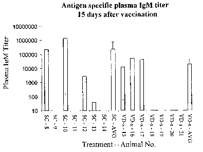
FIG. 1 shows IgM plasma titers in guinea pigs 15 days after either primary
subcutaneous immunization (S.C.) with ovalbumin or ViaDerm treatment followed
by
transdermal immunization with ovalbumin solution (VD-s).
FIG. 2 shows IgG plasma titers in guinea pigs 15 days after either primary
subcutaneous immunization (S.C.) with ovalbumin or ViaDerm treatment followed
by
transdermal immunization with ovalbumin solution (VD-s).
FIGs. 3A-B show IgA and IgG plasma titers in guinea pigs 6 days after boost
(day 36 after primary immunization). FIG. 3A shows IgA and IgG plasma titers 6
days
after boost (day 36 after primary immunization) by intramuscular immunization
with
ovalbumin solution (i.m.) or subcutaneous immunization (S.C.) with ovalbumin.
FIG.
3B shows IgA and IgG plasma titers 6 days after boost (day 36) by ViaDerm
treatment
followed by transdermal immunization with either ovalbumin solution (VD-s) or
ovalbumin powder (VD-p).
8
CA 02572870 2007-01-04
WO 2006/003659 PCT/IL2005/000710
FIG. 4 shows IgG plasma titers in guinea pigs 95 days after boost (125 days
after
primary vaccination) by either subcutaneous immunization (S.C.) with ovalbumin
or
ViaDerm treatment followed by transdermal immunization with ovalbumin solution
(VD-s).
FIG. 5 shows IgA plasma titers in guinea pigs 15 days after either primary
subcutaneous immunization (S.C.) with ovalbumin or ViaDerm treatment followed
by
transdermal immunization with ovalbumin solution (VD-s).
FIG. 6 shows IgA plasma titers in guinea pigs 12 days after boost (day 42
after
primary immunization) by either subcutaneous immunization (S.C.) with
ovalbumin or
ViaDerm treatment followed by transdermal immunization with ovalbumin solution
(VD-s).
FIG. 7 shows Trans Epidermal Water Loss (TEWL) values in guinea pigs treated
with either 50-micron or 100-micron length electrodes of ViaDerm and control
guinea
pigs.
FIG. 8 shows serum IgG antibody titers against A/Panama strain of influenza in
guinea pigs treated with either 50-micron or 100-micron length electrodes of
ViaDerm
and then immunized with the influenza vaccine patch in the absence or presence
of E.
coli heat labile enterotoxin (LT). A control group was immunized with the
influenza
vaccine patch in the absence or presence of LT. A group of guinea pigs
immunized
intramuscularly with the influenza vaccine and then boosted intramuscularly
with the
same vaccine is also shown.
FIG. 9 shows serum IgG antibody titers against A/Caledonia strain of influenza
in guinea pigs treated with either 50-micron or 100-micron length electrodes
of
ViaDerm and then immunized with the influenza vaccine patch in the absence or
presence of LT. A control group was immunized with the influenza vaccine patch
in the
absence or presence of LT. A group of guinea pigs immunized intramuscularly
with the
influenza vaccine and then boosted intramuscularly with the same vaccine is
also
shown.
FIG. 10 shows serum IgG antibody titers against B/Shangdong strain of
influenza
in guinea pigs treated with either 50-micron or 100-micron length electrodes
of
ViaDerm and then immunized with the influenza vaccine patch in the absence or
presence of LT. A control group was immunized with the influenza vaccine patch
in the
absence or presence of LT. A group of guinea pigs immunized intramuscularly
with the
9
CA 02572870 2007-01-04
WO 2006/003659 PCT/IL2005/000710
influenza vaccine and then boosted intramuscularly with the same vaccine is
also
shown.
DETAILED DESCRIPTION OF THE INVENTION
The present invention provides transdermal delivery system for inducing an
antigen-specific immune response comprising an apparatus for facilitating
transdermal
delivery of an antigenic agent through the skin of a subject, said apparatus
capable of
generating at least one micro-channel in an area on the skin of the subject
and a
composition comprising an immunogenically effective amount of at least one
antigenic
agent.
Antigen
The terms "antigenic agent" and "antigen", used interchangeably throughout the
specification and claims, refer to an active component of the composition,
which is
specifically recognized by the immune system of a human or animal subject
after
immunization or vaccination. The antigen can comprise a single or multiple
immunogenic epitopes recognized by a B-cell receptor (i.e., secreted or
membrane-
bound antibody) or a T cell receptor.
The antigenic agent according to the present invention is also an immunogenic
agent. An "immunogenic" agent refers to an agent that is capable of inducing
an antigen
specific immune response.
The temis "immunization" and "vaccination" refer to the induction of an
antigen
specific immune response and are used interchangeably throughout the
specification
and claims.
An antigen can be a peptide, a polypeptide, a protein, a glycoprotein, a
lipoprotein, a lipid, a phospholipid, a carbohydrate, a glycolipid, a mixture
or a
conjugate thereof, or any other material known to induce an immune response.
The
molecular weight of the antigen may be greater than 1 kilodalton (kDa), 10 kDa
or 100
kDa (including intermediate ranges thereof). An antigen can be conjugated to a
carrier.
An antigen can be provided as a whole organism such as, for example, a
bacterium or
virion; an antigen can be obtained from an extract or lysate of organisms,
e.g., from
whole cells or from membranes; an antigen can be provided as live organisms
such as,
CA 02572870 2007-01-04
WO 2006/003659 PCT/IL2005/000710
for example, live viruses or bacteria, attenuated live organisms such as, for
example,
attenuated live viruses or bacteria, or organisms that have been inactivated
by chemical
or genetic techniques; and an antigen can be chemically synthesized, produced
by
recombinant technology or purified from natural sources.
A "peptide" refers to a polymer in which the monomers are amino acids linked
together through amide bonds. Peptides are generally smaller than
polypeptides,
typically under 30-50 amino acids in total.
A "polypeptide" refers to a single polymer of amino acids, generally over 50
amino acids.
A "protein" as used herein refers to a polymer of amino acids typically over
50
amino acids comprising one or more polypeptide chains.
Antigenic peptides or polypeptides include, for example, natural, synthetic or
recombinant B-cell or T-cell epitopes, universal T-cell epitopes, and mixed T-
cell
epitopes from one organism or disease and B-cell epitopes from another.
Antigens
obtained through recombinant technology or peptide synthesis as well as
antigens
obtained from natural sources or extracts can be purified by purification
methods based
on the physical and chemical characteristics of the antigens, preferably by
fractionation
or chromatography. Peptide synthesis is well known in the art and is available
commercially from a variety of companies. A peptide or polypeptide can be
synthesized
using standard direct peptide synthesis (e.g., as summarized in Bodanszky,
1984,
Principles of Peptide Synthesis (Springer-Verlag, Heidelberg), such as via
solid-phase
synthesis (see, e.g., Merrifield, 1963, J. Am. Chem. Soc. 85:2149-2154).
Recombinant antigens can combine one or more antigens. An antigen
composition comprising one or more antigens can be used to induce an immune
response to more than one antigen at the same time. Such recombinant antigens
can be
made by ligating the appropriate nucleic acid sequences encoding the desired
amino
acid sequences to each other by methods known in the art, in the proper coding
frame,
and expressing the recombinant antigens by methods commonly known in the art
(see,
for example, Sambrook et al., 1989, Molecular Cloning: A Laboratory Manual, 2d
edition, Cold Spring Harbor Press). Additionally or alternatively, a
multivalent antigen
composition can be used to induce an immune response to more than one
immunogenic
epitope in one antigenic agent. Conjugates can also be used to induce an
immune
response to multiple antigens, to boost the immune response, or both. Such
conjugates
11
CA 02572870 2007-01-04
WO 2006/003659 PCT/IL2005/000710
can be made by protein synthesis, e.g., by use of a peptide synthesizer.
Fragments of
antigens can be also used to induce an immune response.
Many antigens can be used to vaccinate a subject and to induce an immune
response specific for the antigen. The antigen can be derived from a pathogen
that can
infect a subject. Thus, antigens can be derived from, for example, bacteria,
viruses,
fungi, or parasites. The antigen can be a tumor antigen. The antigen can be an
allergen
including, but not limited to, pollen, animal dander, mold, dust mite, flea
allergen,
salivary allergen, grass, or food (e.g., peanuts and other nuts). The antigen
can be an
autoantigen. The autoantigen can be associated with an autoimmune disease such
as, for
example, the pancreatic islet antigen.
Antigens can be derived from bacteria. Examples of bacteria include, but are
not
limited to, anthrax, Campylobacter, Vibrio cholera, clostridia including
Clostridium
difficile, Diphtheria, enterohemorrhagic E. coli, enterotoxgenic E. coli,
Giardia,
gonococcus, Helicobacter pylori, Hemophilus influenza B, Hemophilus influenza
non-
typeable, Legionella, meningococcus, Mycobacteria including those organisms
responsible for tuberculosis, pertussis, pneumococcus, salmonella, shigella,
staphylococcus, Group A beta-hemolytic streptococcus, Streptococcus B,
tetanus,
Borrelia burgdorfi, Yersinia, and a like. According to the present invention,
bacterial
antigens include, for example, toxins, toxoids (i.e., chemically inactivated
toxins, which
are less toxic but retain immunogenicity), subunits or combinations thereof,
and
virulence or colonization factors. Bacterial constituents, products, lysates
and/or
extracts can be used as a source for bacterial antigens.
Antigens can be derived from viruses. Viruses include, but are not limited to,
adenovirus, dengue serotypes 1 to 4 virus, ebola virus, enterovirus, hanta
virus, hepatitis
virus serotypes A to E, herpes simplex virus 1 or 2, human immunodeficiency
virus,
human papilloma virus, influenza virus, measles (rubeola) virus, Japanese
equine
encephalitis virus, papilloma virus, parvovirus B 19, poliovirus, rabies
virus, respiratory
syncytial virus, rotavirus, St. Louis encephalitis virus, vaccinia virus,
yellow fever virus,
rubella virus, chickenpox virus, varicella virus, and mumps virus. Viral
constituents,
products, lysates and/or extracts can be used as a source for the viral
antigens.
Antigens can be derived from fungi. Fungi include, but are not limited to,
tinea
corporis, tinea unguis, sporotrichosis, aspergillosis, candida, and other
pathogenic fungi.
Fungal constituents, products, lysates and/or extracts can be used as a source
for the
fungal antigens.
12
CA 02572870 2007-01-04
WO 2006/003659 PCT/IL2005/000710
Antigens can be produced from protozoans. Protozoans include, for example,
Entamoeba histolytica, Plasmodium, and Leishmania. Protozoan constituents,
products,
lysates and/or extracts can be used as a source for the protozoan antigens.
Vaccination can be also used as a treatment for cancer, allergies, and
autoimmune
diseases. For example, vaccination with a tumor antigen (e.g., HER2, prostate
specific
antigen) can induce an immune response in the form of antibodies and
lymphocyte
proliferation, which allows the body's immune system to recognize and kill
tumor cells.
Tumor antigens useful for vaccination are known in the art and include, for
example,
tumor antigens of leukemia, lymphoma, and melanoma.
Vaccination with T-cell receptors or autoantigens (e.g., pancreatic islet
antigen)
can induce an immune response that halts progression of an autoimmune disease.
It is to be understood that the present invention encompasses fragments,
derivatives, and analogs of the antigenic agents so long as the fragments,
derivatives,
and analogs being immunogenic and thereby capable of inducing an antigen
specific
immune response.
Fragments of an antigenic agent can be produced by subjecting the antigen to
at
least one cleavage agent. A cleavage agent can be a chemical cleavage agent,
e.g.,
cyanogen bromide, or an enzyme, e.g., endoproteinase, exoproteinase, or
lipase.
Derivatives of the antigenic agents are also included in the scope of the
present
invention. Thus, protein antigenic agents can be modified by derivatization
reactions
including, but not limited to, oxidation, reduction, myristylation, sulfation,
acylation,
ADP-ribosylation, amidation, cyclization, disulfide bond formation,
hydroxylation,
iodination, methylation, glycosylation, deglycosylation, phosphorylation,
dephosphorylation or any other derivatization method known in the art. Such
alterations, which do not destroy the immunogenic epitope of an antigen can
occur
anywhere in the antigen. It will be appreciated that one or more modifications
can be
present in the same antigen.
The term "analog" as used herein refers to antigenic agents comprising altered
sequences by amino acid substitutions, additions or deletions.
Adjuvant
The present invention provides highly effective systems and methods for
transdermal delivery of antigenic agents without the use of adjuvants.
However, the
present invention also encompasses compositions comprising an antigen and an
13
CA 02572870 2007-01-04
WO 2006/003659 PCT/IL2005/000710
adjuvant. Generally, activation of antigen presenting cells by an adjuvant
occurs prior to
presentation of an antigen. Alternatively, an antigen and an adjuvant can be
separately
presented within a short interval of time but targeting the same anatomical
region.
The term "adjuvant" refers to a substance that is used to specifically or
nonspecifically potentiate an antigen-specific immune response. The term
"adjuvant
activity" is the ability to increase the immune response to an antigen (i.e.,
an antigen
which is a separate chemical structure from the adjuvant) by inclusion of the
adjuvant in
a composition.
Adjuvants include, but are not limited to, an oil emulsion (e.g., complete or
incomplete Freud's adjuvant), chemokines (e.g., defensins, HCC-1, HCC-4, MCP-
1,
MCP-3, MCP-4, MIP-l(x, MIP-1(3, MIP-18, MIP-3 a, and MIP-2); other ligands of
chemokine receptors (e.g., CCR-1, CCR-2, CCR-5, CCR-6, CXCR-1); cytokines
(e.g.,
IL-1, IL-2, IL-6, IL-8, IL-10, IL-12, IFN-y; TNF- a, GM-CSF); other ligands of
receptors for these cytokines, immunostimulatory CpG motifs of bacterial
DNA or oligonucleotides; muramyl dipeptide (MDP) and derivatives thereof
(e.g.,
murabutide, threonyl-MDP, muramyl tripeptide); heat shock proteins and
derivatives
thereof; Leishmania homologs and derivatives thereof; bacterial ADP-
ribosylating
exotoxins, chemical conjugates and derivatives thereof (e.g., genetic mutants,
A and/or
B subunit-containing fragments, chemically toxoid versions); or salts (e.g.,
aluminum
hydroxide or phosphate, calcium phosphate).
Most ADP-ribosylating exotoxins (bARE) are organized as A:B heterodimers
with a B subunit containing the receptor binding activity and an A subunit
containing
the ADP-ribosyltransferase activity. Exemplary bARE include cholera toxin
(CT), E.
coli heat-labile enterotoxin (LT), diphtheria toxin, Pseudomonas exotoxin A
(ETA),
pertussis toxin (PT), C. botulinum toxin C2, C. botulinum toxin C3, C. limosum
exoenzyme, B. cereus exoenzyme, Pseudomonas exotoxin S, S. aureus EDIN, and B.
sphaericus toxin. Mutant bARE containing mutations of the trypsin cleavage
site or
mutations affecting ADP-ribosylation may be used.
It is to be understood that adjuvants such as bARE are known to be highly
toxic
when injected or given systemically. But if placed on the surface of intact
skin or
penetrate to the epidermis, they can provide adjuvant effects without systemic
toxicity
(see, for example, U.S. Patent Application Publication Nos. 2004/0258703 and
2004/0185055, incorporated by reference as if fully set for the herein).
14
CA 02572870 2007-01-04
WO 2006/003659 PCT/IL2005/000710
Adjuvant can be chosen to preferentially induce specific antibodies (e.g.,
IgM,
IgD, IgAl, IgA2, IgE, IgGl, IgG2, IgG3, and/or IgG4), or specific T-cell
subsets (e.g.,
CTL, Thl, and/or Th2).
Unmethylated CpG dinucleotides or similar motifs are known to activate B
lymphocytes and macrophages. Other forms of bacterial DNA can be used as
adjuvants.
It is to be understood that bacterial DNA belongs to a class of structures,
which have
patterns allowing the immune system to recognize their pathogenic origin and
to
stimulate the innate immune response leading to adaptive immune responses.
These
structures are called pathogen-associated molecular patterns (PAMP) and
include
lipopolysaccharides, teichoic acids, unmethylated CpG motifs, double stranded
RNA,
and mannins. PAMP induce endogenous signals that can mediate the inflammatory
response and can act as co-stimulators of T-cell function.
Adjuvants can be biochemically purified from a natural source, can be produced
synthetically or recombinantly produced. The adjuvants according to the
present
invention include truncations, substitutions, deletions, and additions of the
natural
occurring adjuvants so long as the adjuvant activity is retained.
Compositions
Currently, licensed vaccines are delivered in an aqueous solution or
suspension,
and administered by the intramuscular or oral route during immunization. The
drawbacks of mixing vaccine components with water or buffers under conditions
of
questionable sterility and the possibility that antigens in solution will
break down are
well known and, in part, has led to the need for cold storage of vaccine
components.
Vaccine components in the presence of water are chemically less stable and
more prone
to contamination through the provision of an aqueous medium for the growth of
bacteria. The stringent requirement for cold storage during transport and
storage of
vaccines has led to the 'cold chain', indicating that at all times after
manufacture of the
vaccine, the vaccine is kept in proper cold storage conditions. This increases
the
complexity of storing vaccine, creates logistical problems when transporting
vaccine,
and adds greatly to the expense of vaccination.
The compositions useful for immunization or vaccination according to the
present
invention contain an immunogenically effective amount of at least one
antigenic agent
and a pharmaceutically acceptable carrier or vehicle in order to provide
pharmaceutical-
acceptable compositions suitable for administration to a subject (i.e., human
or animal).
CA 02572870 2007-01-04
WO 2006/003659 PCT/IL2005/000710
The term "pharmaceutically acceptable" means approved by a regulatory agency
of the Federal or a state government or listed in the U. S. Pharmacopeia or
other
generally recognized pharmacopeia for use in animals, and more particularly in
humans.
The term "carrier" refers to a diluent, excipient, or vehicle with which the
therapeutic
compound is administered. Thus, according to the invention, antigens can be
solubilized
in a buffer or water, or incorporated in emulsions, lipid micelles or
vesicles. Suitable
buffers include, but are not limited to, phosphate buffered saline (PBS),
phosphate
buffered saline Ca++/Mg++ free, normal saline (150 mM NaCI in water), Hepes or
Tris
buffer. Antigens, which are not soluble in neutral buffer, can be solubilized
in 10 mM
acetic acid and then diluted to the desired volume with a neutral buffer such
as PBS. In
the case of an antigen, which is soluble only at acidic pH, acetate-PBS at
acidic pH can
be used as a diluent after solubilization in dilute acetic acid. Other useful
carriers
include, for example, ethanol, ethylene glycol, propylene glycol, butane-1, 3-
diol,
isopropyl myristate, isopropyl palmitate, or mineral oil. Methodology and
components
for formulation of pharmaceutical compositions are well known, and can be
found, for
example, in Remington's Pharmaceutical Sciences, Eighteenth Edition, A. R.
Gennaro,
Ed., Mack Publishing Co. Easton Pa., 1990.
Optionally, components like stabilizers, colorings, humectants, preservatives,
adhesives, plasticizers, tackifiers, and thickeners can be included in the
composition.
Stabilizers include, but are not limited to, dextrans and dextrins, glycols,
alkylene
glycols, polyalkane glycols, polyalkylene glycols, sugars, starches, and
derivatives
thereof. Preferred additives are non-reducing sugars and polyols. In
particular, glycerol,
trehalose, hydroxymethyl or hydroxyethyl cellulose, ethylene or propylene
glycol,
trimethyl glycol, vinyl pyrrolidone, and polymers thereof can be added. Alkali
metal
salts, ammonium sulfate, and magnesium chloride can stabilize proteinaceous
antigens.
A polypeptide can also be stabilized by contacting it with a sugar such as,
for example,
a monosaccharide, disaccharide, sugar alcohol, and mixtures thereof (e.g.,
arabinose,
fructose, galactose, glucose, lactose, maltose, mannitol, mannose, sorbitol,
sucrose,
xylitol). Polyols can also stabilize a polypeptide. Various other excipients
can also
stabilize polypeptides including amino acids, phospholipids, reducing agents,
and metal
cheating agents.
The compositions of the invention can be formulated as a dry or liquid
formulation. A dry formulation is more easily stored and transported than
conventional
liquid vaccines, as it breaks the cold chain required from the vaccine's place
of
16
CA 02572870 2007-01-04
WO 2006/003659 PCT/IL2005/000710
manufacturing to the location where vaccination occurs. In addition, a dry
formulation
can be more advantageous than liquid formulations since high concentrations of
a dry
active component of the composition (e.g., one or more antigens) can be
achieved by
solubilization directly at the site of immunization over a short time span.
Moisture from
the skin and an occlusive backing layer can hasten this process.
The composition can be provided as a liquid formulation including, but not
limited to, solution, suspension, emulsion, cream, gel, lotion, ointment,
paste, or other
liquid forms. The composition can be provided as a dry formulation. Dry
formulations
include, but not limited to, fine or granulated powders, uniform films,
pellets, tablets
and patches. The formulation may be dissolved and then dried in a container or
on a flat
surface (e.g., skin), or it may simply be dusted on the flat surface. It may
be air dried,
dried with elevated temperature, freeze or spray dried, coated or sprayed on a
solid
substrate and then dried, dusted on a solid substrate, quickly frozen and then
slowly
dried under vacuum, or combinations thereof. If more than one antigenic agent
is
included in a composition, the antigenic agents can be mixed in solution and
then dried,
or mixed in a dry form only.
The composition can be provided in a form of a patch. A "patch" refers to a
product, which comprises an antigenic agent and a solid substrate, typically a
backing
layer, which fiinctions as the primary structural element of the patch (see,
for example,
WO 02/074244 and WO 2004/039428, incorporated by reference as if fully set
forth
herein). A patch can further comprise an adhesive and/or a microporous liner
layer.
Typically, the microporous liner layer is a rate-controlling matrix or a rate-
controlling
membrane that allow extended release of the antigenic agent.
A liquid formulation can be incorporated in a patch (i.e., a wet patch). The
liquid
formulation can be held in a reservoir or can be mixed with the contents of a
reservoir.
A wet patch can contain a single reservoir containing one antigenic agent, or
multiple
reservoirs to separate individual antigenic agents.
A patch can also be a dry patch. A dry patch can be a powder patch such as,
for
example, a printed patch as disclosed in WO 2004/039428 or any other dry patch
known
in the art (see Examples herein below); applying a powder patch allows control
over the
time and rate of the dissolution of the antigenic agent. A dry patch can
include one or
more dried antigenic agents such that application of a patch, whether a wet or
dry patch,
comprising multiple antigens induces an immune response to the multiple
antigens. In
such a case, antigens can or cannot be derived from the same source, but will
have
17
CA 02572870 2007-01-04
WO 2006/003659 PCT/IL2005/000710
different chemical structures so as to induce an immune response specific for
the
different antigens.
The backing layer can be non-woven or woven (e.g., gauze dressing). It may be
non-occlusive or occlusive, but the latter is preferred. The optional release
liner
preferably does not adsorb significant amounts of the composition. The patch
is
preferably hermetically sealed for storage (e.g., foil packaging). The patch
can be held
onto the skin and components of the patch can be held together using various
adhesives.
One or more of the antigens may be incorporated into the substrate or adhesive
parts of
the patch. Generally, patches are planar and pliable, and they are
manufactured with a
uniform shape. Optional additives are plasticizers to maintain pliability of
the patch,
tackifiers to assist in adhesion between patch and skin, and thickeners to
increase the
viscosity of the formulation at least during processing.
Metal foil, cellulose, cloth (e.g., acetate, cotton, rayon), acrylic polymer,
ethylenevinyl acetate copolymer, polyamide (e.g., nylon), polyester (e.g.,
polyethylene
naphthalate, ethylene terephthalate), polyolefin (e.g., polyethylene,
polypropylene),
polyurethane, polyvinyl alcohol, polyvinyl pyrrolidone, polyvinylidene
chloride
(SARAN), natural or synthetic rubber, silicone elastomer, and combinations
thereof are
examples of patch materials (e.g., backing layer, release liner).
The adhesive may be an aqueous-based adhesive (e.g., acrylate or silicone).
Acrylic adhesives, available from several commercial sources, are sold under
the trade
names AROSET, DUROTAK, EUDRAGIT, GELVA, and NEOCRYL.
For the purpose of increasing or decreasing the water absorption capacity of
an
adhesive layer, the acrylic polymer may be co-polymerized with hydrophilic
monomer,
monomer containing carboxyl group, monomer containing amide group, monomer
containing amino group, and the like. Rubbery or silicone resins may be
employed as
the adhesive resin; they may be incorporated into the adhesive layer with a
tackifying
agent or other additives.
Alternatively, the water absorption capacity of the adhesive layer can be also
regulated by incorporating therein highly water-absorptive polymers, polyols,
and
water-absorptive inorganic materials. Examples of the highly water-absorptive
resins
may include mucopolysaccharides such as hyaluronic acid, chondroitin sulfate,
dermatan sulfate and the like; polymers having a large number of hydrophilic
groups in
the molecule such as chitin, chitin derivatives, starch and carboxy-
methylcellulose; and
18
CA 02572870 2007-01-04
WO 2006/003659 PCT/IL2005/000710
highly water-absorptive polymers such as polyacrylic, polyoxyethylene,
polyvinyl
alcohol, and polyacrylonitrile.
The plasticizer may be a trialkyl citrate such as, for example, acetyl-
tributyl
citrate (ATBC), acetyl-triethyl citrate (ATEC), and triethyl citrate (TEC).
Exemplary
tackifiers are glycols (e.g., glycerol, 1,3 butanediol, propylene glycol,
polyethylene
glycol). Succinic acid is another tackifier.
Thickeners can be added to increase the viscosity of an adhesive or
immunogenic
composition. The thickener may be a hydroxyalkyl cellulose or starch, or water-
soluble
polymers: for example, poloxamers, polyethylene oxides and derivatives
thereof,
polyethyleneimines, polyethylene glycols, and polyethylene glycol esters.
However, any
molecule which serves to increase the viscosity of a solution may be suitable
to improve
handling of a formulation during manufacture of a patch.
Gel and emulsion systems can be incorporated into patch delivery systems, or
be
manufactured separately from the patch, or added to the patch prior to
application to the
human or animal subject. Gels or emulsions may serve the same purpose of
facilitating
manufacture by providing a viscous formulation that can be easily manipulated
with
minimal loss. The term "gel" refers to covalently cross-linked, non cross-
linked
hydrogel matrices. Hydrogels can be formulated with at least one antigenic
agent.
Additional excipients may be added to the gel systems that allow for the
enhancement
of antigen delivery, skin hydration, and protein stability. The term
"emulsion" refers to
formulations such as water-in-oil creams, oil-in-water creams, ointments, and
lotions.
Emulsion systems can be either micelle-based, lipid vesicle-based, or both
micelle- and
lipid vesicle-based.
A liquid formulation may be applied directly to the skin and allowed to air
dry or
held in place with a dressing, patch, or absorbent material. The formulation
may be
applied in an absorbent dressing or gauze. The formulation may be covered with
an
occlusive dressing such as, for example, AQUAPHOR (an emulsion of petrolatum,
mineral oil, mineral wax, wool wax, panthenol, bisabol, and glycerin from
Beiersdorf),
plastic film, COMFEEL (Coloplast) or VASELINE petroleum jelly; or a non-
occlusive
dressing such as, for example, TEGADERM (3M), DUODERM (3M) or OPSITE
(Smith & Napheu).
The relative amount of an antigenic agent within a composition and the dosing
schedule can be adjusted appropriately for efficacious administration to a
subject (e.g.,
human or animal). This adjustment may depend on the subject's particular
disease or
19
CA 02572870 2007-01-04
WO 2006/003659 PCT/IL2005/000710
condition, whether therapy or prophylaxis is intended, the administration
route, the
physical condition and of the subject. To simplify administration of a
composition to a
subject, each unit dose can contain one or more antigenic agents in
predetermined
amounts for a single round of immunization. The amount of an antigenic agent
in the
unit dose can range from about 0.1 g to about 10 mg.
The compositions of the present invention can be manufactured under good
manufacturing practices regulated by government agencies (e.g., Food and Drug
Administration) for biologicals and vaccines.
Devices for transdermal immunization
The system of the present invention comprises an apparatus for enhancing
transdermal immunization. According to the principles of the present invention
the
apparatus is used to generate at least one micro-channel in an area on the
skin of a
subject through which a composition comprising an antigenic agent is delivered
efficiently.
The term "micro-channel" as used in the context of the present invention
refers to
a pathway, generally extending from the surface of the skin through all or
significant
part of the stratum corneum, through which molecules can diffuse.
According to some embodiments of the present invention, the apparatus for
facilitating transdermal movement of an antigenic agent is as disclosed in one
or more
of the U.S. Pat. Nos. 6,148,232; 6,597,946; 6,611,706; 6,711,435; 6,708,060;
and
6,615,079, the contents of which is incorporated by reference as if fully set
forth herein.
Typically, the apparatus comprises an electrode cartridge comprising a
plurality of
electrodes, and a main unit comprising a control unit adapted to apply
electrical energy
between the plurality of electrodes when the electrodes are in vicinity of the
skin,
typically generating current flow or one or more sparks, enabling ablation of
stratum
comeum in an area beneath the electrodes, thereby generating at least one
micro-
channel. The main unit loaded with the electrode cartridge is also denoted
herein
ViaDerm.
According to some enlbodiments, the control unit of the apparatus comprises
circuitry to control the magnitude, frequency, and/or duration of the
electrical energy
delivered to the electrodes, so as to control the current flow or spark
generation, and
thus the width, depth and shape of the one or more formed micro-channels.
Preferably,
the electrical energy applied by the control unit is at radio frequency (RF).
CA 02572870 2007-01-04
WO 2006/003659 PCT/IL2005/000710
The micro-channels formed by the apparatus of the present invention are
hydrophilic and typically have a diameter of about 10 to about 100 microns and
a depth
of about 20 to about 300 microns, thus facilitating the diffusion of antigenic
agents
through the skin.
According to the principles of the present invention, the electrode cartridge
comprises a plurality of electrodes thus forming an electrode array, which
generates
upon application of an electrical energy a plurality of micro-channels within
the
subject's skin. Typically, however, the overall area of micro-channels
generated in the
stratum corneum is small compared to the total area covered by the electrode
array. It
will be understood that the term "plurality" refers herein to two or more
elements, e.g.,
two or more electrodes or two or more micro-channels.
According to additional embodiments, the pressure obtained while placing the
apparatus of the present invention on a subject's skin activates the
electrical energy
delivered to the electrodes. Such mode of action ensures that activation of
electrodes
occurs only in a close contact with the skin enabling the desired formation of
the micro-
channels.
The number and dimension of micro-channels may be adjusted to the amount of
the antigenic agent desired to be delivered into the skin.
The electrode cartridge is preferably removable. According to certain
embodiments, the electrode cartridge is discarded after one use, and as such
is designed
for easy attachment to the main unit and subsequent detachment from the main
unit.
According to the present invention, application of current to the skin causes
ablation of the stratum comeum, which results in the formation of micro-
channels.
Spark generation, cessation of spark generation, or a specific current level
can be used
as a form of feedback, which indicates that the desired depth has been reached
and
current application should be terminated. For these applications, the
electrodes are
preferably shaped and/or supported in a cartridge that is conducive to
facilitate
formation of micro-channels in the stratum corneum to the desired depth, but
not
beyond that depth. Alternatively, the current can be configured so as to form
micro-
channels in the stratum comeum without the generation of sparks. The resulted
micro-
channels are uniform in shape and size.
According to the present invention, the electrodes can be maintained either in
contact with the skin, or in vicinity of the skin, up to a distance of about
500 microns
therefrom. According to some embodiments, ablation of the stratum corneum is
21
CA 02572870 2007-01-04
WO 2006/003659 PCT/IL2005/000710
performed by applying electrical current having a frequency between about 10
kHz and
about 4000 kHz, preferably between about 10 kHz and about 500 kHz, and more
preferably at 100 kHz.
Methods for transdermal immunization
The present invention further provides a method for inducing an antigen-
specific
immune response using a transdermal delivery system of the invention.
Typically, the
procedure for inducing an antigen-specific immune response comprises a step of
placing
over the skin the apparatus for generating at least one micro-channel.
Preferably, prior
to generating the micro-channels the treatment sites will be swabbed with pads
comprising sterile alcohol. Preferably, the site should be allowed to dry
before
treatment.
In exemplary embodiments of the present invention, the apparatus containing
the
electrode array is placed over the site of treatment, the array is energized
by RF energy,
and treatment is initiated. In principle, the ablation and generation of micro-
channels is
completed within seconds. The apparatus is removed after micro-channels are
generated
at limited depth. A composition according to the invention is applied to the
area of the
treated skin where micro-channels are present.
The present invention thus provides a method for inducing an antigen-specific
immune response by transdermal delivery system comprising the steps of:
generating at
least one micro-channel in an area of the skin of a subject, and applying a
composition
comprising an immunogenically effective amount of an antigenic agent to the
area of
skin in which the at least one micro-channel is present, thereby inducing an
antigen-
specific immune response.
The term "transdermal" delivery refers to delivery of an antigenic agent into
or
through the dermal layers of the skin, i.e., the epidermis or dermis, beneath
the stratum
corneum, or into or through the subcutaneous layers of the skin. Thus, an
antigen can be
delivered into the skin or through the skin into the blood or lymphatic
system. The term
transdermal is therefore meant to include also transcutaneous delivery.
The term "immunogenically effective amount" is meant to describe the amount of
an antigenic agent, which induces an antigen-specific immune response.
The immune response induced by the composition of the present invention can
comprise humoral (i.e., antigen-specific antibody such as IgM, IgD, IgAl,
IgA2, IgE,
IgGl, IgG2, IgG3, and/or IgG4) and/or cellular (i.e., antigen-specific
lymphocytes such
22
CA 02572870 2007-01-04
WO 2006/003659 PCT/IL2005/000710
as CD4+ T cells, CD8+ T cells, cytotoxic lymphocytes, Thl cells, and/or Th2
cells)
effector arms. Moreover, the immune response may comprise NK cells that
mediate
antibody-dependent cell-mediated cytotoxicity (ADCC). The antibody isotypes
(e.g.,
IgM, IgD, IgAl, IgA2, IgE, IgGl, IgG2, IgG3, and IgG4) can be detected by
immunoassay techniques as known in the art (see also the Examples herein
below)
and/or by a neutralizing assay. The terms "inducing an immune response",
"vaccination", and "immunization" are meant to describe the induction of an
immune
response, whether humoral or cellular, and are used interchangeably throughout
the
specification and claims of the present invention.
In a neutralization assay, for example in a viral neutralization assay, serial
dilutions of sera are added to host cells, which are then observed for
infection after
challenge with infectious virus. Alternatively, serial dilutions of sera can
be incubated
with infectious titers of virus prior to inoculation of an animal, and the
inoculated
animals are then observed for signs of infection.
The transdermal immunization system of the invention can be evaluated using
challenge models in either animals or humans, which evaluate the ability of
immunization with an antigenic agent to protect the subject from a disease.
Such
protection would demonstrate an antigen-specific immune response.
According to the principles of the present invention, induction of an immune
response is useful for treating a condition or disease in a subject. Thus,
induction of an
immune response by the systems and methods of the present invention provides
immunoprotection, immunosuppression, modulation of an autoimmune disease,
potentiation of cancer immunosurveillance, prophylactic vaccination to prevent
disease,
and/or therapeutic vaccination to treat or reduce the severity and/or duration
of
established disease. When the antigen is derived from a pathogen, for example,
the
treatment may vaccinate the subject against infection by the pathogen or
against its
pathogenic effects such as those caused by toxin secretion.
A method "induces" an immune response when it causes a statistically
significant
change in the magnitude or kinetics of the immune response, change in the
induced
elements of the immune system (e.g., humoral and/or cellular), effect on the
number
and/or the severity of disease symptoms, effect on the health and well-being
of the
subject (i.e., morbidity and mortality), or combinations thereof.
It will be appreciated that the application site can be protected with anti-
inflammatory corticosteroids or non-steroidal anti-inflammatory drugs (NSAIDs)
to
23
CA 02572870 2007-01-04
WO 2006/003659 PCT/IL2005/000710
reduce possible local skin reaction or modulate the type of immune response.
Similarly,
anti-inflammatory steroids or NSAIDs can be included in the patch material, in
creams,
ointments, and a like or alternatively corticosteroids or NSAIDs may be
applied after
application of the formulation of the invention. IL-10, TNF-a, or any other
immunomodulator can be used instead of the anti-inflammatory agents.
Alternatively or
additionally, pimecrolimus, tacrolimus, aloevera or any other agent known in
the art to
reduce local skin reaction can be applied to the treated skin area or included
in the
patch.
Vaccination has also been used as a treatment for cancer and autoimmune
diseases. For example, vaccination with a tumor antigen (e.g., prostate
specific antigen)
can induce an immune response in the form of antibodies, CTLs and lymphocyte
proliferation, which allows the body's immune system to recognize and kill
tumor cells.
Tumor antigens useful for vaccination have been described for melanoma,
prostate
carcinoma, and lymphoma.
Vaccination with T-cell receptor oligopeptide can induce an immune response
that halts the progression of autoimmune disease. U.S. Pat. No. 5,552,300
describes
antigens suitable for treating autoimmune disease.
It is to be understood that transdermal immunization may be followed with
enteral, mucosal, and/or other parenteral techniques for boosting immunization
with the
same or altered antigens. Immunization by an enteral, mucosal, and/or other
parenteral
route may be followed with transdermal immunization for boosting immunization
with
the same or altered antigens.
EXAMPLES
Transdermal vaccination using an apparatus that generates micro-channels in
the
skin of a subject, which is denoted herein ViaDerm, was compared to the widely
used
subcutaneous (SC) and intramuscular (IM) vaccination routes in order to
establish its
usefulness as a potential vaccine administration system.
Ovalbumin (OA) and trivalent influenza virus (TIV) were used as exemplary
antigens to establish the efficacy of the system of the present invention to
induce
antigen-specific immune response.
24
CA 02572870 2007-01-04
WO 2006/003659 PCT/IL2005/000710
EXAMPLE 1
Transdermal immunization with ovalbumin
Materials
A solution of ovalbumin (50 g/ml water; Sigma) was used for IM and SC
injections.
A solution of ovalbumin (10 mg/ml) was used for solution transdermal
administration (VD-s).
Ovalbumin powder (2 mg) was used for powder transdermal administration (VD-
p).
A solution pouch was prepared as follows: a 300 m thick layer of adhesive
(Durotac 2516, National starch, Netherlands) was evenly spread over a silicone
sheet
(Sil-k Degania Silicone, Israel). The sheet was cut into 4X4cm squares. A
square hole
(1.57X1.57cm) was cut in the middle of each of the 4X4 squares. A piece of Sil-
k
silicone 2X2cm is adhered to the 4X4cm silicone square over the 1.57X1.57cm
hole
using 7701 primer and 4011 glue (Loctite, Ireland). The final product was a
pouch of
250 1 volume.
Powder patch was prepared as follows: ovalbumin powder was distributed on the
skin and then covered with a fixing patch containing BLF 2080 liner (Dow, USA)
covered with a layer of Durotak 2516 adhesive (National starch, Netherlands)
or
alternatively with TegadermTM (3M).
Procedure
Blood was collected intracardially or by abdominal Vena Cava venipuncture
immediately prior to immunization and at weekly intervals starting 8 days post
immunization. Each sample contained 1.3 ml of blood in Heparin anticoagulant
tubes.
The blood samples were centrifuged at 6000 rpm and the plasma was collected.
Group 1: Intramuscular injection
Guinea pigs, males, 600-650gr, Dunkin Hartley (7 animals) were anesthetized
and
blood (1.3 ml) was collected immediately prior to immunization. Ovalbumin
solution
was then injected (5 g; 0.1m1 of 50 g/ml) to the Quadriceps muscle of the
right hind
leg. Blood was drawn from each animal at days 8, 15, 22, and 30 after
immunization. At
day 30, the animals were injected again to the Quadriceps muscle of the right
hind leg
CA 02572870 2007-01-04
WO 2006/003659 PCT/IL2005/000710
(boost-5 g; 0.1m1 of 50 g/ml). Blood was collected at days 36, 42, 50, and
125 days
after immunization.
Group 2: Subcutaneous immunization
Guinea pigs, males, 600-650gr, Dunkin Hartley (7 animals) were anesthetized
and
blood (1.3 ml) was collected immediately prior to immunization. Ovalbumin
solution
was then injected (5 g; 0.lml of 50 g/ml) subcutaneously to the dorsal neck
area.
Blood was drawn from each animal at days 8, 15, 22, and 30 after immunization.
At day
30, the animals were injected again (boost-5 g; 0.lml of 50 g/ml)
subcutaneously to
the dorsal neck area. Blood was collected at days 36, 42, 50, and 125 days
after
immunization.
Grogp 3: Transdermal immunization by qpplication of an ovalbumin solution
pouch to
ViaDerm treated skin
Guinea pigs, males, 600-650gr, Dunkin Hartley (7 animals) were anesthesized
and blood (1.3 ml) was collected immediately prior to immunization. The
animals were
treated with a device, denoted herein ViaDerm, which utilizes electrical
energy at radio
frequency and consists of an array of electrodes, to generate micro-channels
in the skin
of the guinea pigs (see, for example, WO 2004/039426; WO 2004/039427; and WO
2004/039428 incorporated by reference as if fully set forth herein). ViaDerm
Operating
Parameters: burst length ( sec) - 700; starting amplitude - 330V; number of
bursts - 5;
2 applications on the same skin area (200 pores/cm2). Ovalbumin solution pouch
(2 mg;
0.2m1 of 10 mg/ml) was placed on the treated skin area. Twenty-four hours post
application, the pouch was removed. Blood was drawn from each animal at days
8, 15,
22, and 30 after immunization. At day 30, the animals were immunized again by
ViaDerm treatment as described above, i.e., burst length ( sec) - 700;
starting
amplitude - 330V; number of bursts - 5; 2 applications on the same skin area
(200
pores/cm2), followed by transdermal application of an ovalbumin solution pouch
(2 mg;
0.2ml of 10 mg/hnl). Blood was collected at days 36, 42, 50, and 125 days
after
immunization.
Group 4: Transdermal immunization by apnlication of an ovalbumin powder patch
to
ViaDerm pretreated skin
Guinea pigs, males, 600-650gr, Dunkin Hartley (7 animals) were anesthesized
and blood (1.3 ml) was collected immediately prior to the immunization. The
animals
were treated with ViaDerm. ViaDerm Operating Parameters: burst length ( sec) -
700;
26
CA 02572870 2007-01-04
WO 2006/003659 PCT/IL2005/000710
starting amplitude - 330V; number of bursts - 5; 2 applications on the same
skin area
(200 pores/cm2). Ovalbumin powder (2 mg) was evenly distributed with a spatula
on
the treated skin area and then covered with a fixing patch. Twenty-four hours
post
application, the patch was removed. Blood was drawn from each animal at days
8, 15,
22, and 30 after immunization. At day 30, the animals were immunized again by
ViaDerm treatment as described above, i.e., burst length ( sec) - 700;
starting
amplitude - 330V; number of bursts - 5; 2 applications on the same skin area
(200
pores/cm2), followed by transdermal application of ovalbumin powder (2 mg;
0.2ml of
mg/ml) as described above. Blood was collected at days 36, 42, 50, and 125
days
10 after immunization.
Detection of anti-ovalbumin antibodies in guinea-pig plasma samples:
Ninety six-well plates (Maxisorp; Nunc, Denmark) were coated with ovalbumin
(100 1 of a solution of 200 g/ml). Coating was conducted for 16-18 hours at 4
C.
Unbound ovalbumin was removed by washing three times with a wash solution (PBS
containing 0.05% Tween 20). Remaining adsorption sites were blocked with a
diluent/blocker solution (PBS containing 0.05% Tween 20 and 4% skim milk) for
one
hour at room temperature, followed by three washes with the wash solution.
Guinea pig's plasma samples, serially diluted with the diluent/blocker, were
added to the ovalbumin-coated plates in triplicates and incubated for one hour
at 22 C.
Unbound antibodies were washed three times with the wash solution. In order to
detect
guinea pig IgG antibodies, the wells were incubated for one hour at 22 C with
horseradish-peroxidase (HRP) conjugated goat-anti guinea pig IgG antibody
diluted in
the diluent/blocker solution (Jackson Immunoresearch Laboratories, 0.8mg/ml,
1:10,000), and then washed three times with the wash solution. In order to
detect IgA or
IgM guinea pig antibodies, the wells were incubated for one hour at 22 C with
rabbit
anti-guinea pig IgA or rabbit anti-guinea pig IgM, respectively (both were
purchased
from I.C.L; 1:5,000 dilution). Unbound antibodies were washed three times with
the
wash solution. Then, horseradish-peroxidase (HRP) conjugated donkey anti-
rabbit IgG
diluted in the diluent/blocker solution (Jackson Immunoresearch laboratories;
1:5,000)
was incubated for one hour at 22 C, followed by three washes as described
above.
HRP substrate (Substrate-chromogen, TMB- ready to use, DAKO) was then
added and incubated for 30 minutes at 22 C. The reaction was stopped with 1 M
H2S04.
The signal was detected in a spectrophotometer at 405 nm and the background at
595
nm.
27
CA 02572870 2007-01-04
WO 2006/003659 PCT/IL2005/000710
Titer Calculation: The average (AVG) optical density (O.D.) data was
calculated
for every duplicate/triplicate of the samples. Similarly, AVG O.D.s were
obtained from
equivalent dilutions of normal plasma samples (from naive non-immunized
animals).
The AVG O.D.s obtained from non-immunized animals were subtracted from the
O.D.s
obtained from the immunized animals.
The data obtained for an internal standard (animal #27 at day 36) was plotted
in a
logarithmic scale. Using this plot, the linear-power regression range was
determined.
The end point titer (titer) is calculated using the regression formula
obtained from the
linear range. The cut-off O.D. (y axis - "noise" cut-off) data was calculated
as 5 times
blank STD.
Results
Trans epidermal water loss (TEWL; DERMALAB Cortex Technology,
Hadsund, Denmark) measurements were used to verify the efficacy of micro
channel
formation by measuring TEWL levels on potential treatment sites before ViaDerm
application (BVD) and after ViaDerm application (AVD). Only sites that were
within
the TEWL specification (i.e., TEWL before treatment < 8.5 g/h/m2; A TEWL > 20
g/h/m2) were approved for testing. The results are presented in Tables 1 and
2.
Table 1: TEWL of primary immunization.
TEWL
Guinea TEWL AVD
Group BVD
Pig (g/h/mz)
(g/h/m2)
15 3.1 46
16 5.3 34.9
17 4.3 39
18 5 36.5
Transdermal, 2 mg 19 4.9 37.5
OVA solution 20 4.9 40.8
21 4.6 47.6
AVG 4.59 40.33
STDEV 0.73 4.82
28
CA 02572870 2007-01-04
WO 2006/003659 PCT/IL2005/000710
22 5.7 44.9
23 5.1 33.7
24 6.1 36.9
25 5.5 35.8
Transdermal, 2 mg 26 4.7 41.7
OVA powder 27 6.3 38.3
28 4.3 46.9
AVG 5.39 39.74
STDEV 0.73 4.90
Table 2: TEWL of boost immunization.
TEWL
Guinea TEWL AVD
Group BVD
Pig (g/h/ml)
(g/h/m2)
5.7 44.9
16 5.5 38.7
17 5.8 34.9
Transdermal, 2 mg 20 5 43.8
OVA solution 21 3.8 39.8
AVG 5.16 40.42
STDEV 0.82 4.04
29
CA 02572870 2007-01-04
WO 2006/003659 PCT/IL2005/000710
22 4.2 34.2
23 2.7 33.1
25 3 24.7
26 1.6 33.5
TD, 2mg OVA
27 0.8 31.1
powder
28 3.8 38.7
AVG 2.68 32.55
STDEV 1.29 4.59
IgM:
IgM antibodies 15 days after primary immunization represent the earliest
response to antigen presentation. As shown in FIG. 1, the group of animals
injected
subcutaneously (SC) with ovalbumin and the group of animals treated with
ViaDerm
and thereafter immunized against ovalbumin by the ovalbumin solution pouch (VD-
s)
showed induction of ovalbumin specific IgM antibodies, though the IgM antibody
titer
detected in the SC group was higher than in the VD-s group. In addition, both
groups
demonstrated similar incidence of "non-responder" animals, e.g., animals that
did not
show detectable titer of IgM antibodies.
I2G:
The appearance of antigen specific IgG antibodies following antigen
presentation
express the maturation of the antigen specific immune response.
FIG. 2 presents the IgG plasma titers 15 days post immunization. There was a
significant difference between the VD-s group and the SC group. As shown in
FIG. 2,
generation of micro-channels by ViaDerm treatment and subsequent application
of the
ovalbumin solution pouch (VD-s) resulted in significantly higher IgG titers at
day 15
compared to the titers obtained by SC injection. These results clearly
indicate that
ViaDerm treatment can shorten the time for IgG antibodies appearance. This
effect is
highly advantageous as IgG antibodies are the most important antibody subtype
in an
antigen specific immune response.
CA 02572870 2007-01-04
WO 2006/003659 PCT/IL2005/000710
FIG. 2 also shows that all the animals in the VD-s and SC groups were found to
be positive for antigen specific plasma IgG antibodies. FIG. 2 further shows
that there
was low variability between the individual animals. The single animal of the
VD-s
group that did not show detectable titer of IgG (Animal No. 19) was found to
be in a
bad physical condition at the time of bleeding and died the next day. Animal
No. 19 did
not have any detectable antigen specific IgM and IgA.
Six days after the boost (FIG. 3), there was a strong IgG antibody secondary
response in both the VD-s and the SC groups, with plasma titers that were 3.5
and 4.1
greater (for VD-s and SC, respectively) over the titers observed 15 days post
immunization. It should be also noted that the IgG titers in the VD-s group
were
approximately 5 times higher than in the SC group, indicating the efficacy of
this
method in eliciting an antigen specific IgG antibodies. The IgG titers in the
intramuscularly (IM) injected group were very low compared to all other
groups,
including the SC group immunized with the same ovalbumin dose.
A comparison of the two transdermal formulations revealed that the IgG titer
for
the VD-s was 9.5 times greater than the VD-p group (using the same dose). The
IgG
titer for the VD-p group was lower than that of the SC group, which received a
lower
dose.
Ninety-five days after boost administration (FIG. 4), only 1.3% and 6% of the
IgG antibody titer were detected in the VD-s and SC groups, respectively.
IgA:
The antigen specific plasma IgA titer was determined in the SC and the VD-s
groups at 15 days post primary antigen presentation (FIG. 5). Only 2 out of 7
animals in
the SC group demonstrated detectable IgA titers compared to 4 animals out of 6
in the
VD-s group. This superiority of the VD-s treatment compared to the SC
injection was
fiuther demonstrated six days after boost administration (FIG. 6). The animals
that had
no detectable specific IgA response after boost administration (animals Nos. 9
& 11)
had neither IgA nor IgM at 15 days post immunization. All animals (SC and VD-
s)
were IgA positive 12 days after antigen boosting (FIG. 6).
The higher titers in VD groups as well as the high frequency of sero-positive
individual animals indicate the usefulness of transdermal immunization using
ViaDerm.
The time for appearance of significant titers of IgG and IgA antibodies was
shorter in
the ViaDerm treated groups compared to that of the well-established and widely
31
CA 02572870 2007-01-04
WO 2006/003659 PCT/IL2005/000710
accepted SC and IM routes, thus indicating the efficacy of this transdermal
route of
immunization.
The significant immune response following VD antigen presentation included all
the important plasma antibody isotypes: IgM, IgG and IgA, thus indicating
efficient
isotype switching. There was no correlation between IgG and IgM antibody
titers in the
VD-s vs. S.C. groups. Thus, while higher IgG titers were observed in the VD-s
group
vs. SC group, higher IgM titers were observed in the SC group vs. VD-s group
during
the primary response. Without being bound to any theory, this phenomenon may
be
explained by a very efficient cellular response, which takes place following
VD
application. This data is supported by previous observations performed by the
applicant
of the present invention demonstrating that shortly after VD application there
is a strong
leukocyte infiltration around the micro channels. As isotype switching is a
process
involving antigen presentation and extended support by T helper lymphocytes
existing
mainly in the peripheral lymph nodes (PLN), it can be speculated that VD
treatment can
activate local "professional" antigen presenting dendritic cells (APDC).
Shortly after
VD antigen presentation, APDC can activate lymphocytes locally, following
their
infiltration to the inflamed micro channel site. Yet, it will be understood
that the
majority of these interactions are normally taking place in the PLN, the
natural target
for activated APDC migration.
While the route of antigen presentation is an important parameter for inducing
antigen-specific immune response, the use of antigen-formulation and adjutants
can be
equally important. In the present example, VD treatment was used with two
ovalbumin
formulations, i.e., powder (VD-p) and solution (VD-s), at the same dose and in
the
absence of any adjuvant. The lower IgG titer in the VD-p vs. VD-s emphasized
that
antigen-formulation is critical for successful vaccine development. The
impressive IgA
titer in the VD-p group compared to the poor IgG titer strongly indicates that
antigen
formulation can play a significant role in manipulating the immune response as
desired.
Because specific antibody isotypes are often more important than others in a
given
condition, it can be very useful to utilize this phenomenon. For example, in
diseases of
mucous menlbranes application of a dry antigen with the apparatus of the
present
invention can be advantageous in order to elicit IgA antibodies, which are
secreted from
these membranes.
Thus, transdermal immunization using ViaDerm technology is highly efficient
and can provide an alternative technique for the traditional vaccination
routes.
32
CA 02572870 2007-01-04
WO 2006/003659 PCT/IL2005/000710
EXAMPLE 2
Transdermal immunization with trivalent influenza vaccine
Materials
Female Hartley guinea pigs (>350 g), >7 weeks old (Charles River).
Inactivated influenza vaccine: A/Panama/2007/99, A/New Caledonia/20/99 and
B/Shangdong/7/97,1ot#001, 2.046 mg/ml, diluted to 0.2046 mg/ml for use.
E. coli heat labile enterotoxin (LT): FIN0023, 1.906mg/ml.
One-layer rayon square patch 1 cm2.
ViaDerm: Length of electrodes 50 and 100 m, cylinder shape.
Tegaderm 1624W: 3M, NDC 8333-1624-05, 6cmX7cm size
Adhesive tape: 3M
Hydration solution: 10% Glycerol/saline
Immunization
Before immunization, the guinea pigs were shaved and sedated with ketamine and
xylazine. All animals were bolus intramuscular injected with 0.5 g HA (0.17
g HA
each strain) in 100ul 1xDPBS on study day 1.
Pretreatment
Guinea pigs were shaved on the abdomen one day before immunization and re-
shaved immediately before patch application on study day 22. The immunization
site
was marked with a permanent marker and the shaven skin was pretreated as
follows:
= Groups 1-2 were hydrated with 10% glycerol/saline;
= Groups 3-4 were pretreated with the ViaDerm device <50 m twice on dry,
shaven
skin hydrated with 10% glycerol/saline;
= Groups 5-6 were pretreated with the ViaDerm device <50 m twice on dry,
shaven
skin without hydration;
= Groups 7-8 were pretreated with the ViaDerm device 100 m twice on dry,
shaven
skin without hydration;
TEWL measurements were done before and immediately following pretreatment as
known in the art (see, for example, WO 2004/039426; WO 2004/039427; and WO
2004/039428).
33
CA 02572870 2007-01-04
WO 2006/003659 PCT/IL2005/000710
Patch application
A 1 cm2 rayon patch containing 15 g HA (5 g HA each strain) alone (no LT) or
with 1 g LT in 15 l 1xDPBS were applied immediately after the pretreatment.
To
insure proper patch adherence, patches were covered with a modified Tegaderm
overlay. The patch was wrapped with adhesive tape. Patches were applied for 18-
24
hr, removed, and the skin was rinsed with warm water.
Serum collection
Pre-immune (prior to immunization) and post immune (day 22 and 36) blood
samples
were collected from the orbital plexus using standard methods. Serum was
collected by
centrifugation of whole blood and the cell free serum transferred to a labeled
tube and
stored frozen at -20 C.
ELISA
Sera was evaluated for total IgG titers to A/Panama, A/New Caledonia, and
B/Shangdong using an ELISA method known in the art (see, for example, US
Patent
Application Publication No. 2004/018055 incorporated by reference as if fully
set forth
herein). Antibody titers were presented as ELISA Units (EU), which is the
serum
dilution equal to 1 O.D. at 405 nm.
Results
FIG. 7 shows the TEWL values of non-treated or ViaDerm treated guinea pigs.
As shown in FIG. 7, TEWL values obtained in guinea pigs treated with 100-
micron
length electrodes of ViaDerm or 50-micron length electrodes of ViaDerm were
significantly higher than those obtained from non-treated guinea pigs. These
results
confirni that micro channels were generated in the skin of the guinea pigs.
FIG. 8 shows serum IgG antibody titers against A/Panama influenza strain in
the
absence or presence of E. coli heat labile enterotoxin (LT) as an adjuvant in
guinea pigs
treated with ViaDerm and immunized by a patch containing the trivalent
influenza
vaccine. As shown in FIG. 8, ViaDerm treatment of guinea pigs either with 50-
micron
or 100-micron length electrodes followed by influenza patch application
significantly
increased the IgG antibody titers against A/Panama influenza strain as
compared to
guinea pigs, which were not treated with ViaDerm but administered with
influenza
patch. Addition of LT as an adjuvant did not improve the IgG antibody titer
against this
strain of influenza. As a comparison, guinea pigs were immunized
intramuscularly (IM)
with 0.5 g of the trivalent influenza vaccine at day 1, and boosted IM with
the same
34
CA 02572870 2007-01-04
WO 2006/003659 PCT/IL2005/000710
vaccine (15 g) at day 22. As shown in FIG. 8, the IgG antibody titers in the
ViaDerm
treated groups were comparable, or even higher, than those of the IM injected
guinea
pigs, indicating that transdermal immunization using ViaDerm is as efficient
as IM
immunization.
FIGs. 9 and 10 show similar results when serum IgG antibody titers against
A/New Caledonia strain and B/Shangdong strain of influenza were detennined. As
shown in FIGs. 9 and 10, the IgG antibody titers against each of these strains
was
significantly higher in the ViaDerm treated guinea pigs that were then
administered
with the influenza patch as compared to guinea pigs not treated with ViaDerm
but
administered with the influenza patch. Addition of LT as an adjuvant did not
improve
the IgG antibody titers. The IgG antibody titers in ViaDerm treated animals
were
comparable to those obtained in guinea pigs injected intramuscularly with the
trivalent
influenza vaccine.
It will be appreciated by persons skilled in the art that the present
invention is not
limited by what has been particularly shown and described herein above. Rather
the
scope of the invention is defined by the claims that follow.