Note: Descriptions are shown in the official language in which they were submitted.
= CA 02574432 2007-01-19
WO 2006/012413
PCT/US2005/025875
Enriched Antibody for Detecting Mycobacterial Infection, Methods of Use and
Diagnostic Test Employing Same
Technical Field
The present invention relates to diagnostic tests for detecting microbial-
based diseases
and conditions, and more particularly for diagnostic tests and methods for
detecting
tuberculosis.
=
Background
During the last decades TB has evolved from a predominantly pulmonary
infection into
a multifaceted pathology with a growing rate of extrapulmonary cases. Until to
date effective
TB prevention programs are hampered by the absence of a rapid and field
adapted screening
assay. In high-income countries mycobacterial culture remains the diagnostic
standard, but it is
time-consuming and relatively expensive. Ideally, sputum microscopy based on
three sputum
smears can identify up to 67% of culture positive cases. HIV co-infection has
been reported to
impair the demonstration of Mycobacterium tuberculosis in sputa, although some
investigators
do not report any influence of the HIV serostatus on the AFB diagnosis. The
higher percentage
of extrapulmonary TB in HIV positive TB patients additionally increases the
rate of APB-
negative TB cases. This renders tuberculosis an increasing diagnostic
challenge and underlines
an urgent need for improved laboratory tools for its diagnosis.
Current approaches for diagnosing TB are not satisfactory. The sputum test for
pulmonary TB is not always effective, particularly if there are no detectable
bacteria in the
sputum, or no sputum sample can be obtained. In addition, this diagnostic test
requires
microscopy and/or culture of the bacteria to confirm the diagnosis;neither of
which is
especially suitable to diagnosis in the field. Using cerebrospinal fluid for
diagnosis of TB-
meningitis is also problematic, particularly in the field since, once again,
microscopy and/or
culture of the bacteria and/or an ELISA test is usually required to confirm
the diagnosis.
Blood tests for TB are also known, but have a poor track record, being complex
and
unreliable. Urine tests are simpler and more reliable, but current methods
require processing
of the urine before performing the diagnostic test ¨ such processing usually
involving
concentration of the urine.
CA 02574432 2007-01-19
WO 2006/012413
PCT/US2005/025875
Among the newly developed methods antibody tests against a number of
mycobacterial
antigens have been developed, but none of these tests has so far reached the
needed specificity
for routine diagnostic purpose. The drop of sensitivity in HIV positive cases
is also a major
constraint. A different approach is to measure immune responses to
Mycobacterium
tuberculosis specific antigens like ESAT-6, but so far the differentiation
between latent TB
infection and TB disease is not possible.
Tuberculosis is an extremely complex pathology existing in multiple forms, but
always
starting as an airborne infection. Pulmonary tuberculosis occurs immediately
at the entry point
of the microorganism and extrapulrnonary tuberculosis is the result of further
penetration into
body of the patient with the most widespread examples of tuberculous
meningitis and bone
tuberculosis. Complexity of the pathology determines multitude of various
approaches tried
during this century of modem medicine. Furthermore clinical and radiographic
manifestations
of HIV-related pulmonary tuberculosis are dramatically altered by
immunodeficiency. These
factors severely limit our capability of early symptomatic recognition of
tuberculosis in
HIV/TB patients and also increase the danger of TB transmission to relatives
and caregivers of
such patients.
Mycobacteria can potentially be recovered from a variety of clinical
specimens,
including upper respiratory collections (sputum, bronchial washes,
bronchoalveolar lavage,
bronchial biopsies and such); urine, feces, blood, cerebrospinal fluid (CSF),
tissue biopsies,
and deep needle aspirations of virtually any tissue or organ. Bacterial
culture remains the gold
standard in the diagnosis of tuberculosis, but it can take up to 6-8 weeks to
make a conclusive
diagnosis. There are three major technologies used for rapid (faster than
bacterial culture)
diagnosis of the mycobacterial infections:
= Direct microscopy of sputum smears;
= PCR-based assays;
= Immunodiagnostic methods.
Direct microscopy of sputum smears. More than a century ago, Robert Koch
identified the etiologic agent of tuberculosis by staining it and culturing it
from clinical
specimens. Today, the diagnosis of tuberculosis is usually established using
staining and
culturing techniques that do not differ substantially from those that Koch
used. Direct
microscopy of sputum is the norm for the diagnosis of tuberculosis in
developing countries
today and it is the benchmark against which the efficiency of any new test
must be assessed. It
is applied to pulmonary tuberculosis, but is not very useful for children or
for patients with
initial stages of pulmonary tuberculosis.
2
= CA 02574432 2007-01-19
WO 2006/012413 PCT/US2005/025875
PCR-based assays. A comparative study of the performance of PCR tests in seven
laboratories has shown high levels of false-positive PCR-results, ranging from
3% to 20%
(with an extreme of 77% in one laboratory). This relatively poor performance
resulted from
lack of monitoring of each step of the procedure and underscores the necessity
for careful
quality control during all stages of the assay.
Inununodiagnostic methods.
The Tuberculosis Skin Test. This is the probably oldest immunological test for
tuberculosis. A small amount of substance called PPD Tuberculin is placed just
under the top
layer of the skin on the forearm with a small needle. The test is read 48 to
72 hours after it has
been given. Generally, a swelling of 10 mm. or more is considered positive.
Many developing
countries use BCG vaccination to protect against TB. After BCG vaccination,
the PPD skin
test usually becomes positive. Results of the skin test vary dependent on the
quality of the PPD
antigen, reactivity of the immune system and probably even race of the
individual. This test
also does not provide an unequivocal indication about the stage and location
of the infection.
Serological tests for M. tuberculosis. This approach, based on the detection
of antibody
immune response to mycobacterial antigens is one of the most widely used in
research and
clinical environments. All serological tests have approximately the same
sensitivity and
specificity if they use purified antigens. The sensitivity of the best tests
is in a range of 80%
for smear-positive cases and 60-70% for smear negative cases. The reported
specificity is
generally high and is in a range of 95-100%.
Currently existing technologies are limited in their performance in several
ways.
Most widely accepted rapid microscopic test requires several hours to
complete,
skillful technician and clinical laboratory environment. Test interpretation
is far too difficult
compared to current standards of rapid Point of Care testing in the infectious
diseases area. Real cost
of one analysis per one patient runs in the range of $100-150 for a US
hospital. Clinical specificity of
the test is very good, but any improvements in sensitivity will be more than
welcome.
Skin test has sufficient sensitivity, but takes a long time and does not
provide
information about stage of pathological process and does not sufficiently
differentiate infected
and vaccinated individuals.
Serological tests usually do not have sufficient sensitivity. Test results
vary with
variations in the individual immune response to TB antigens. These tests
practically do not
work in HIV patients infected by M. tuberculosis. This factor severely limits
their
3
CA 02574432 2007-01-19
WO 2006/012413
PCT/US2005/025875
applicability in Africa and many Asian countries. In the US this group of
patients constitutes
the majority of TB infected patients as well.
PCR tests are widely used in developed countries, but are complex, expensive
and are
not sensitive enough to justify their use as a screening test in developing
countries.
A preferred method for rapid diagnosis of infectious diseases is based on the
detection
of a bacterial antigen in the patient sample, that provides unequivocal proof
of active
infectious process caused by specific pathogen. The concept of using a direct
antigen test for
detection of mycobacterial infections was described in several publications.
For example, the development of one of the first direct antigen assay for M.
tuberculosis was reported in 1982 - a radioimmunoassay for the detection M.
tuberculosis
antigens in sputum of patients with active pulmonary tuberculosis, using a
rabbit antibody
specific to the whole cells of M.bovis (BCG vaccine). Autoclaved and sonicated
sputum was
used as a sample. The assay detected antigen in 38 of 39 sputum samples from
patients with
active tuberculosis pulmonary tuberculosis.
Later studies reported the development of the ELISA system for the detection
of
mycobacterial antigens in the cerebrospinal fluid of patients with tuberculous
meningitis, also
using antibodies specific to the whole cells of Mbovis. Both systems showed
surprisingly high
specificity. Despite the fact that LAM was the major antigen responsible for
the detection, it
was reported that M. kansasii showed 5% cross-reactivity, and M.
intracellulare, M. avium, M.
fortitum, and M. vaccae cross-reacted only at 2%. Others reported detection,
by ELISA, of
mycobacterial antigen in the CSF of nine of 12 patients with tuberculous
meningitis,
corresponding to the sensitivity of 81.25%. Specificity of the test was equal
to 95%.
Practically all previous attempts to develop a test for diagnosis of
tuberculosis have
focused on the detection of the pulmonary form of the disease. Extrapulmonary
forms, which
are notoriously difficult to diagnose, attracted relatively little attention
due to low prevalence
rate compared to pulmonary forms. Until the 1950s and 1960s, extrapulmonary TB
cases
comprised only around 10% of all tuberculosis cases. The onset of the HIV/AIDS
pandemic
has changed the situation completely. These two diseases eventually merged
into a new
complex public health problem. Now fully 60 % of untreated HIV patients
develop active TB
during their lifetime and up to 70% of TB patients are HIV infected in sub-
Saharan Africa and
Asia. Superimposition of HIV and TB changed not only the epidemiology of
tuberculosis, but
also the course of the disease itself. During the last decades TB has evolved
from
predominantly a pulmonary infection into a multifaceted pathology with an ever
growing
prevalence of extrapulmonary forms. It is estimated that extrapulmonary TB
cases currently
comprise up to 30% of all cases of tuberculosis; this number might even be an
underestimation
4
CA 02574432 2007-01-19
WO 2006/012413
PCT/US2005/025875
due to the lack of tools for rapid screening and diagnosis of extrapulmonary
forms of
tuberculosis. Moreover, even pulmonary tuberculosis in HIV patients frequently
exhibits
atypical symptoms. For example, such patients typically do not produce sputum.
These factors
severely limit our capability of early symptomatic recognition of tuberculosis
in 111V/TB
patients and also increase the danger of TB transmission to relatives and
caregivers of such
patients. An easy to use screening test, capable of detecting a broad spectrum
of pathologies
due to M. tuberculosis infection, is urgently needed, including a test for
extrapulmonary forms
of TB. Such a need has long been discussed with no progress towards realising
goal. Today
the need has became a public health care emergency.
In other cases of pulmonary bacterial infections, the current screening
process of
choice is based on the detection of polysaccharide antigens secreted in the
patient's urine.
Bacterial polysaccharides are composed of monosaccharides uncommon to humans
and
therefore resistant to cleavage by human enzymes. This enables their secretion
in urine in
immunochemically intact forms suitable for detection by a polysaccharide-
specific
immunoassay. Extremely low concentrations of bacterial polysaccharides
secreted in urine
require very high sensitivity of the immunoassay in order to use it as a
screening procedure.
Collaborating research groups from Sweden and Norway attempted development of
a
LAM-specific ELISA system detecting LAM antigen in patient urine. The system
used
antigen capture for detecting tuberculosis from urine based on
lipoarabinomannan, a
polysaccharide present on the surface of Mycobacterium tuberculosis, the
organism
responsible for causing tuberculosis in humans, as disclosed in PCT
application no.
W097/34149 to Svenson. The disclosed diagnostic procedure detected the
presence of LAM
in pkient urine in 81.3% of AFB-positive patients and 57.4% of AFB-negative
patients and
demonstrated utility of the detection of mycobacterial LAM antigen for
diagnosis of
mycobacterial infections. At the same time the system failed to demonstrate
utility of the
disclosed process for screening purposes. Despite use of the affinity purified
rabbit polyclonal
antibody specific to LAM antigen, the procedure lacked sufficient sensitivity
to be used on
non-processed un-concentrated urine samples. The diagnostic procedure required
approximately 24-48 hrs of sophisticated manipulations in a biochemical lab
focused on
concentrating patient urine and preparing it for analysis by ELISA test.
Overall, the sensitivity
of the Svenson assay is not sufficient for practical use of the disclosed
method. The
complexity and length of the immunoassay also prevents its practical use as a
screening test
for detection of mycobacterial infections because it proved too cumbersome for
use in a
clinical setting, where speed, ease of use, and high sensitivity are all
critically important for
diagnostic tests used to detect disease conditions.
5
CA 02574432 2007-01-19
WO 2006/012413
PCT/US2005/025875
Summary of the Invention
In a first embodiment of the invention there is provided an antigenically
active isoform
of lipoarabinomannan from mycobacterium tuberculosis, prepared by oxidation of
LAM using
mild oxidation methods such as treatment with low concentrations of NaI04. In
other
embodiments, the antigenically active isoform of LAM, generated by mild
oxidation methods,
is used to prepare highly specific, highly pure antibodies to inactivated
mycobacterium, more
particularly to surface polysaccharides such as LAM, for use in the detection
of
polysaccharides (e.g. LAM) in urine, sputum, blood, tissue or other samples
from patients of
interest. Other embodiments use the highly specific, highly pure antibody
raised to the
antigenically active form of LAM to diagnose tuberculosis in patients of
interest.
In another particular embodiment, there is provided an enriched antibody
population
highly specific for an antigen of a surface polysaccharide from a
mycobacterium. In this
embodiment, the enriched antibody population may be enriched by having been
raised in an
environment that maintains antigenically active antigen. Alternatively or in
addition, the
antibody is enriched by exclusion of antibodies that recognize relatively
inactive antigen. In
some embodiments, the mycobacterium may be Mycobacterium tuberculosis.
Similarly, the
surface polysaccharide may be lipoarabinomannan (LAM).
In another embodiment, there is provided a process for producing an enriched
antibody
highly specific to an antigen of a mycobacterium. In this embodiment, the
process comprises
raising and isolating antibody to antigen from mycobacteria; and separating
from the isolated
antibodies that population of antibodies which is specific to relatively
inactive antigen to
produce isolated enriched antibody.
In another embodiment, there is provided a process for producing an enriched
antibody
highly specific to an antigen of a mycobacterium. In this embodiment, the
process comprises
isolating antigen from mycobacteria under conditions maintaining antigenic
activity; and
raising antibodies to the isolated antigen while maintaining its antigenic
activity.
In still another embodiment, there is provided a process for producing an
enriched
antibody highly specific to an antigen of a mycobacterium. In this embodiment,
the process
comprises applying sera from a mammal inoculated with mycobacteria to a first
affinity matrix
prepared with isolated antigen from mycobacterium such that antibody specific
to the isolated
antigen is retained by the first affinity matrix; isolating antibody specific
to the isolated antigen
from the first affinity matrix; applying the isolated antibody to a second
affinity matrix
prepared with modified antigen from mycobacterium such that antibody specific
to the
modified antigen is retained by the second affinity matrix, wherein the
modified antigen has
6
CA 02574432 2007-01-19
, WO 2006/012413
PCT/US2005/025875
been treated with an agent to deactivate it relative to the isolated antigen;
isolating enriched
antibody specific to the isolated antigen by collecting effluent from the
second affinity matrix,
so that the enriched antibody is more highly specific and displays higher
sensitivity to
mycobacterium antigen than non-enriched antibody.
In further related embodiments, the mycobacterium may be Mycobacterium
tuberculosis.
and the surface polysaccharide may be lipoarabinomannan (LAM).
In other embodiments, the agent for modifying the antigen from mycobacterium
is
sodium periodate. In other related embodiments, the surface polysaccharide may
be isolated
from Freund's adjuvant.
Still another embodiment provides a method for detecting a mycobacterial
infection in a
sample from a subject. In this embodiment, the method comprises providing an
immunoreactive environment, such environment developed from the enriched
antibody as
described above; and reacting the sample in the immunoreactive environment so
as to detect the
mycobacterial infection.
Optionally the mycobacterial infection may be M. tuberculosis or Johne's
disease.
Similarly, the surface polysaccharide may be lipoarabinomannan (LAM). In
further related
embodiments, the immunoreactive environment comprises an ELISA, and may be
implemented as a strip test. In related embodiments, the mycobacterial
infection may be a
pulmonary Mycobacterium tuberculosis infection or an extra-pulmonary
Mycobacteriwn
tuberculosis infection, and the sample may be any of sputum, blood, urine,
tissue or other
suitable sample. In a related particular embodiment, the sample may be non-
processed
unconcentrated urine.
Other embodiments provide a kit for detecting a mycobacterial infection in a
sample,
the kit comprising an assay providing an immunoreactive environment wherein
the
environment comprises an enriched antibody as described above. In related
embodiments, the
immunoreactive environment comprises an ELISA, and may be implemented as a
strip test. In
further related embodiments, the mycobacterial infection may be Mycobacterium
tuberculosis
and the surface polysaccharide may be lipoarabinomannan (LAM).
7
CA 02574432 2007-01-19
WO 2006/012413 PCT/US2005/025875
Brief Description of the Drawings
The foregoing features of the invention will be more readily understood by
reference to
the following detailed description, taken with reference to the accompanying
drawings, in
which:
Figure 1 shows a structural model of mycobacterial ManLAM, PILAM, and AraLam.
Figure 2 shows a comparison of serological activity for LAM experiments.
Figure 3 shows the efficiency of LAM-specific Ab preparations in capture
ELISA.
Figure 4A. Sensitivity of the LAM ELISA for different concentrations of LAM in
urine,
wherein solid circles represent ELISA results using LAM from M tuberculosis,
and open circles
represent the control ELISA results. The cut off was the Optical Density of
the Negative Control + 0.1,
resulting in a minimal detection limit of 0.25 ng/ml.
Figure 4B. Binding of LAM-specific antibodies in the ELISA to non-
mycobacterial antigens
was excluded for the following bacterial species:
Klebsiella pneumoniae, Streptococcus agalactiae, Streptococcus pneumoniae
14/I2F,
Pseudomonas aeruginosa, Staphylococcus aureus 25923/43300, Proteus vulgaris,
E. coli 8739,
Neisseria meningitidis A/B/13102,Haemophilus influenzae A/B/D, as can be seen
by comparing the
ELISA results for M tuberculosis (open triangles) with the ELISA results for
other bacterial species
tested (solid triangles, open and solid diamonds, open and solid circles)
depicted in Fig. 4B.
Figure 4C. Sensitivity of the LAM ELISA for various mycobacterial strains. LAM
of M
bovis and M tuberculosis are detected most sensitively.
Figure 5. Correlation between the microscopic mycobacterial density of AFB
positive
patients and their antigen concentration measured by the LAM ELISA in
unprocessed urine. AFB +
(light microscopy 1000 x magnification: 4-90 acid fast bacilli/100 fields) 28
cases. AFB ++ (1-9/field)
23 cases. AFB I __ I I (-10/field) 20 cases. Box plot showing 10th, 25th,
50th, 75th, - -th
9u percentile and the
mean antigen concentration.
Figure 6 shows a schematic of an antigen purification process in accordance
with particular
embodiments of the claimed invention.
Figure 7 shows a schematic for preparing affinity columns in accordance with
particular
embodiments of the claimed invention.
Figure 8 shows a schematic of an antibody purification process in accordance
with particular
embodiments of the present invention.
Figure 9 shows a schematic of a conjugate preparation, in accordance with
particular
embodiments of the present invention.
Detailed Description of Specific Embodiments
8
CA 02574432 2007-01-19
WO 2006/012413
PCT/US2005/025875
Definitions
The following terms shall have the meanings indicated unless the context
otherwise
requires:
"Immunoreactive environment" as used herein means, an environment supportive
of
immunoassays, immunoreactions, immunochemistry, and any process, assay,
methodology or
system which involves, relates to or relies on an immunological reaction to
achieve a desired
result. Examples of immunoreactive environments are those detailed in US
Patent No.
5,073,484 to Swanson et al.; and US Patent Nos. 5,654,162 and 6,020,147 to
Guire et al,
disclosing method and apparatus for quantitatively determining an analyte in a
liquid, wherein
particular embodiments employ immunochemical reactions in which the analyte
and the
reactant represent different parts of a specific ligand (antigen) - antibody
(anti-ligand) binding
pair. These patents relate to technology that has been implemented as what we
call in this
description and the following claims as a "strip test."
"Freund's adjuvant" is from Sigma, USA.
We have developed a high-sensitivity method for detecting the presence of
mycobacterium antigens, particularly M. tuberculosis antigens, such as the
surface
polysaccharides lipoarabinomannan (LAM) and related species, in bodily fluids
including but
not limited to urine. Heretofore, tests of this nature lacked sensitivity and
were not operable for
unprocessed urine samples or for detecting extrapulmonary TB infections. In
particular, we
have developed enriched antibodies raised to antigen from mycobacteria wherein
the antibody
is enriched by having been raised in an environment that maintains
antigenically active antigen.
We call the method for producing this first class of enriched antibody the
"direct method,"
which is described in further detail below.
We have also developed antibody that is enriched by exclusion of antibodies
that
recognize relatively inactive antigen. The method for producing this class of
antibodies begins
by following the "direct method" to obtain enriched antibodies, but then also
operates by
excluding antibodies that recognize relatively inactive antigen. We call the
method for
producing this second class of enriched antibody the "enhanced method," which
is also
described in further detail below.
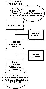
Figure 8 is a schematic depiction showing the steps involved in practicing an
embodiment of the enhanced method. Because the enhanced method builds on the
direct
method, Figure 8 also illustrates the direct method, if one stops after the
first affinity column.
Below we show how these enriched antibodies of either or both classes can be
used to
detect pulmonary and extrapulmonary infections of TB in a variety of samples,
including but
9
CA 02574432 2007-01-19
WO 2006/012413
PCT/US2005/025875
not limited to untreated (i.e. non-concentrated) urine samples. (Other
potential sources of
sample include sputum, cerebrospinal fluid, blood, tissue, lavages.) In the
examples which
follow, the enriched antibodies are raised to an epitope of lipoarabinomannan
(LAM) in an
environment which maintains its antigenic activity.
Prior methods for detecting surface polysaccharides (LAM) using different body
fluids
such as serum, urine or sputum have been investigated, but have proven
problematic. In
serum, the detection of LAM seems to be disturbed by immune complex formation.
Detection
of LAM in sputum is possible only in the samples of the patients with
pulmonary TB because
extra-pulmonary infections often do not provide sputum containing
mycobacterial antigens.
Prior studies with urine required extensive sample processing and
manipulation, limiting such
methOdologies in the field. None were effective for diagnosing extra-pulmonary
mycobacterial infections such as those on the rise in REV-positive subjects.
Embodiments of the present invention overcome difficulties in the prior art by
providing enriched antibodies that may be used for detecting mycobacterial
antigens in a wide
range of sample types from a subject. These sample types include sera, blood,
sputum, lavages,
tissue, and unprocessed, non-concentrated urine, among others.
Lipoarabinomannan (LAM) is a 17500 mol wt lipopolysaccharide specific for the
genus
mycobacterium. Lipoarabinomannan is a complex polysaccharide antigen composed
of
mannose and arabinose residues forming a highly branched and complex
structure. Despite
more than four decades of structural studies of polysaccharide antigens of
mycobacteria, those
in the art still speak only about fragments of the structure or structural
motifs and composite
models. The most recent composite model of LAM structure is presented in
Fig.1, below.
As part of the outer cell wall of mycobacteria, LAM is released from
metabolically
active or degenerating bacterial cells. It is assumed that in active TB
infection LAM leaks into
the circulation, passes through the kidneys and can therefore be detected in
the urine reflecting
the level of mycobacterial burden. Since LAM is a carbohydrate antigen with
glycosidic
linkages for which no human degrading glycosidases exist, the antigen occurs
in the urine in
intact form.
LAM antigen of mycobacteria is composed of three major structural domains: the
mannosyl-phospahtidyl-myo-inositol (MIP) anchor, containing variable number of
fatty acids
with variable chain length; mannan core polysaccharide variable in number of
mannose
residues; and branched arabinan polysaccharide chains connected to mannan
core. Despite
many efforts, the attachment site(s) for arabinan chains on the mannan core
remain unknown.
Arabinan polysaccharide chains are, capped by mannose oligosaccharides,
consisting of mono-
CA 02574432 2013-06-17
(al-2)-di- and (al-2)-tri-mannosyl units variable in their length (capping
motifs). Capping
degree is variable from strain to strain and possibly is also dependent from
growth conditions.
Extremely high structural complexity and variability of mycobacterial LAM lead
to
very complex spectrum of antigenic epitopes. Complexity of the selected
diagnostic antigen
forces us to use affinity purified polyclonal antibody as a main immunoassay
reagent. Only use
of polyclonal antibody allows one to cover the full spectrum of antigenic
specificities
potentially associated with LAM present in clinical samples. In order to
achieve the highest
possible assay sensitivity of sandwich immunoassay, we use the highest
concentration of
antigen-specific antibody in the 'capture zone and also as the labeled
antibody. Antigen-specific
affinity purification is known to produce such an antibody.
To prepare the antigen-based affinity column, we developed a process for
antigen
isolation and coupling to the solid phase support. The process of LAM antigen
isolation is
based, with some minor modifications, on the methods of isolation of other
bacterial
polysaccharides described in the literature and well-known to those in the
art, and described
below.
Previous LAM-based direct antigen immunoassay described in the literature used
polyclonal antibody purified by antigen-specific affinity chromatography using
a LAM-
Sepharos e column. The prior art approach to the synthesis of the affinity
matrix was based on
the partial NaI04-oxidation of LAM polysaccharide with subsequent coupling to
NH2-
Sepharose. Surprisingly, our experiments have shown that Nalaroxidation
reduces antigenic activity
of LAM polysaccharide, as can be seen from the Fig.2.
Because coupling efficiency of oxidized polysaccharide to NH2-solid support is
proportional to the degree of oxidation, we have coupled to Sepharose support
via
functionalized BSA-spacer molecule LAM antigen oxidized with 50 rriM Nal04. At
this level
of oxidation LAM polysaccharide still retains some antigenic activity, as
described below, but
provides high coupling efficiency. Application of the immune serum to such
affinity matrix
resulted in the isolation with high yield of the fraction of rabbit antibody.
Testing of such
antibody in the plate ELISA immunoassay format as a capture antibody showed
some
functional activity, but not at the level sufficient to be used in the high
sensitivity
immunoassay necessary for screening applications using non-concentrated urine
samples.
These data explain results obtained in the literature previously, where LAM-
specific affinity
purified antibody was used, but it was still necessary to concentrate urine
samples in order to
detect Lam present in the samples.
11
*Registered Trade-mark of GE Healthcare Bio-Sciences AB LLC
CA 02574432 2007-01-19
WO 2006/012413
PCT/US2005/025875
Unexpectedly, by changing the LAM coupling chemistry to a milder non-
destructive
process, based on polysaccharide activation with cyanogen bromide (CNBr)
resulted in the
purification of a much better quality of LAM specific antibody, as can be seen
in Fig.3.
Then, surprisingly, passing the antibody purified on the column with intact
LAM
(CNBr-activation process) through a column with LAM antigen after deep, strong
Na104
oxidation (see below) produced a relatively small fraction of antibody,
approximately 7-10%
of the applied amount, with very high activity in the LAM-specific direct
antigen
immunoassay. Fig.3 shows the efficiency of such antibody as a capture
antibody. When such
antibody was labeled with horse radish peroxidase (HRP) and used as a labeling
antibody, it
also demonstrated activity higher than any other antibody tested or known. The
ELISA system
based on such antibody has shown extremely high sensitivity and proven to be
useful in testing
non-concentrated urine samples. This enabled us to produce a screening LAM-
specific
immunoassay with performance characteristics suitable for rapid screening, in
the field, or
both pulmonary and extra-pulmonary TB cases, a feat unattainable by others
before. Thus,
although LAM has been described in the frozen urine of TB patients, the assay
for such reports
requires an extensive sample preparation and therefore is not field adapted.
PROTOCOLS
In this section we describe protocols suitable for practicing the "direct
method" and the
"enhanced method" defined above. This discussion is not sorted strictly
according to the
direct method and the enhanced method per se, but describes specifically
methods of preparing
columns suitable for use in either or both methods, depending upon the
context. Figure 8
shows a schematic of the direct and enhanced methods.
Isolation of dry cells of M. tuberculosis from Freund's Adjuvant
First, allow cells with Freund's Adjuvant to settle for a minimum of 1 week at
room
temperature before use. Remove caps from adjuvant vials and without disturbing
cells settled
on the bottom of the vial, pull off the bulk mineral oil. A small amount of
mineral oil may be
left in the vial as a precaution to avoid drawing cell precipitate. Discard
mineral oil and then
mix 6.0 L of ethanol and 6.0 L diethyl ether, and add 5mL ethanol:diethyl
ether mixture to
each vial.
Next, close the vial, vortex and quickly transfer the suspension into a 1-L
Erlenmyer
flask. Avoid letting cells resettle in the vial during this step. When the 1-L
Erlenmeyer flask is
filled to the 1-L line, let the cells settle for 1-1.5 hr. Next, gently decant
solvent from the
Erlenmeyer flask into a clean 1-L beaker. Avoid disturbing settled cells and
moving them with
12
CA 02574432 2013-06-17
solvent. If solvent decanted into beaker is clear, discard it. If a
significant amount of cells were
decanted with the solvent, return the decanted solvent to the Erlenmeyer flask
and repeat
settling step. Using 20-30 ml aliquots of ethanol: diethyl ether mixture,
transfer cells onto
glass sintered
Wash the cells with 500 mL of an ethanol- diethyl ether mixture, then wash
with 200
ml of diethyl ether. Next, air-dry cells on a filter using a vacuum of 100
mmHg +1- 10 (low
vacuum) Occasionally mix and homogenize the cell mass, then cover the filter
with a porous
material (such as a Kim-wipe) and leave in hood until dry (approximately 15
hours, i.e.
overnight).
Weigh and record the total weight and calculate the dry weight of the cells.
Then tightly
seal with rubber lined cap and store at 15-30 C.
Phenol extraction of crude LAM antigen
Place the dry cells of M. tuberculosis into a 250-mL Pyrex media bottle and
add warm
deionized water to the cells. Vortex and pulse sonicate (¨ 20 second pulses)
the suspension in
the ultrasonic water bath until suspension is homogeneous.
Phenol extract the cells, then ethanol precipitate and place the precipitated
cells in the
refrigerator (2-8 C) overnight (¨ 16 hours) to allow the precipitate to
settle. Being very careful
not to disturb precipitate, gently draw off the supernatant until about 100 mL
of supernatant is
left covering the precipitate. Gently swirl to mix, then transfer the
remaining suspension into
teflon centrifuge tubes and centrifuge at 12,000rpm for 20 minutes. Draw off
as much
supernatant as possible from all tubes with out disturbing the pellet, add 5
ml of deionized
water to each tube and, using vortexing and pulse sonication, dissolve pellet
in water. Combine
all the fractions with the dissolved pellet and place in a 500-mL flask (Note:
do not exceed
1/10 of the flask capacity/volume). Rotary evaporate to minimal volume, but
avoid
caramelizing the sample. Redissolve film with approximately 50 ni.L of water
and repeat
drying and redissolving until sample has been dried 3 times. Redissolve in 50
mL of water and
lyophilize.
Purification of LAM antigen by Sephadex G-25 chromatography
Dissolve 800 mg of crude LAM Ag in 15 mL of 0.25% acetic acid solution. Vortex
and
sonicate in ultrasonic bath to achieve complete dissolution. Centrifuge in a
microcentrifuge at
5000 rpm for 5 min. Collect the supernatant in a 20-mL glass vial, divide the
supernatant into
3 equal parts for separate chromatographic runs, and then gently apply 1/3rd
of the LAM Ag
supernatant collected above onto the chromatographic column. After a volume of
100 mL has
13
*Registered Trade-mark of Kimberly-Clark Corp.
**Registered Trade-mark of GE Healthcare Bio-Sciences AB Limited Liability
Company
CA 02574432 2007-01-19
=
WO 2006/012413
= PCT/US2005/025875
flowed through the column, begin collecting fractions. Continue collecting
fractions until 350
mL of mobile phase has passed since the start of chromatography. Cover all
fractions and
store at 2-8 C. Rotary evaporate in a 250-mL evaporation flask (no volumes
greater than 25
mL). Evaporate to minimal volume, but avoid caramelizing the sample. Dilute
evaporated
material in 20 mL of water, sonicate, vortex until complete dissolution and
then lyophilize
(approximately 8 hours). Scrape dried material with a spatula into a tared
glass vial and weigh.
The foregoing steps are depicted schematically in Figure 6.
Coupling LAM antigen to BSA-spacer by CNBr activation.
First, prepare 0.5 M sodium bicarbonate and 1 M potassium carbonate solutions.
Then dissolve 30.0 mg of purified LAM Ag in 1.5 mL of deionized water. Use
pulse sonication
(10-20 sec pulses) and vortexing to completely dissolve the LAM Ag.
Dissolve 300 mg of BSA-hydrazine ligand in 15 mL of deionized water. Pulse
sonicate
(10-20 sec pulses) and vortex to dissolve completely, then place in microfuge
tubes and
centrifuge in a microcentrifuge for 10 minutes at 10,000 rpm. Using a Pasteur
pipette carefully
collect and pool the clear supernatant from each tube and transfer into a 20-
mL vial. Avoid
disturbing any pellet that may form. Add 1.0 mL of 0.5M sodium bicarbonate to
the vial and
mix well by shaking. Add 150 AL of chilled 1M potassium carbonate to the LAM
solution and
mix well by brief vortexing. Place obtained solution in ice/water bath.
= Prepare 5 mg/mL CNBr in acetonitrile for immediate use and add 180 [EL of
the
cyanogen bromide solution to the LAM solution. Mix by vortexing and place on
ice for
approx. 15 minutes. Add this solution to the BSA-Hydrazine ligand solution
(above) with a
Pasteur pipette. Mix well and incubate overnight (16 ¨24 hours), at 2-8 C, in
tightly sealed
vial.
Coupling of LAM antigen to BSA-spacer by Na104 activation
Dissolve the LAM antigen in 1.25 mL of deionized water in a 3-4-mL vial. Pulse
sonicate and vortex to dissolve completely. Prepare a 0.1M sodium periodate
solution: (in
Na0Ac buffer, pH 4.0). Add 1.25 mL of the 0.1M Na104 to the solution 1.25 mL
LAM solution.
Vortex to mix. Cover the vial with aluminum foil; place it on the rocking
platform and mix for
1 hour +1-5 minutes at ambient temperature.
Dissolve 250 mg of BSA-hydrazine ligand in 12.5 mL of deionized water in a 25-
40
mL glass serum vial. Use pulse sonication and vortexing to dissolve
completely, then
centrifuge for approx. 10 minutes at 10,000 rpm. Using a Pasteur pipette
collect the
supernatant from each tube and pool into a 25-40 mL glass vial. Avoid
disturbing the pellet.
14
CA 02574432 2007-01-19
, WO 2006/012413
PCT/US2005/025875
Add 12.5mL of 0.1M sodium phosphate (pH 6.8) to the vial and mix well by brief
vortexing.
Coupling process:
To the BSA-hydrazine solution add the oxidized LAM solution and vortex. Add
100
mg of sodium cyanoborohydride and seal. Sample 10 p.L of the final solution
and dilute with
90 uL 1X PBS buffer (QC solution) and retain for further analysis (LAM
concentration will be
approximately 0.75 mg/mL).
Activation of Sepharose by NaIO4
Measure an aliquot of suspension of Sepharose 4B-CL corresponding to 80 ml of
settled gel and transfer onto a sintered glass filter. Wash with 500 mL water
and drain using
low vacuum (approx 300 mmHg) until the granular structure of the gel surface
becomes
visible. Avoid formation of the air cracks in the gel layer. Prepare a 0.1 M
sodium acetate
buffer, pH 4.0 solution and use to prepare a 30 mM solution of NaI04 in 0.1 M
Na0Ac.
Add 250 mL of 30 mM Nalat to the gel and thoroughly mix. Cover the mixture
with
aluminum foil and place at a 450 angle on a rocker platform at medium speed
for 1.5 hours
10 minutes at ambient temperature. Transfer to the sintered glass filter and
wash with 1 L of
water using low vacuum (approx 300 mmHg). The activated gel must be prepared
within a
maximum of 4 hours of use.
/0
Coupling of BSA-LAM ligand to activated Sepharose (For Synthesis of first and
second
affinity columns)
Preparation of matrix
Prepare a 0.1% sodium azide solution in 1X PBS (phosphate buffered saline).
Measure
a suspension of activated Sepharose corresponding to 60 ml of the settled gel
(or other suitable
matrix) and transfer it onto a sintered glass filter. Drain gel using low
vacuum (300 mm Hg)
until the gel packs and granular structure becomes visible, but avoid
formation of cracks on the
gel surface.
BSA-LAM Ligand Solution:
In a 250-m.L media bottle dilute approximately 17 to 20 mL of the solution of
BSA-
LAM ligand to 90 mL with sodium phosphate buffer (pH 6.8). Add 90 mg of
crystalline
sodium cyanoborohydride to the solution. Tightly close the bottle using the
supplied plastic
cap. Mix well by vortexing briefly. The solution may appear opalescent but
there should be no
CA 02574432 2007-01-19
WO 2006/012413
PCT/US2005/025875
precipitate. Microscopic gas bubbles formed by the sodium cyanoborohydride may
be visible.
Coupling step:
To the LAM solution prepared above add the drained activated Sepharose gel.
Tightly
close and thoroughly mix the suspension using gentle vortexing. Incubate for
approx 4 hours
at 37 C+/- 2 C, mixing (by inversion) the reaction mixture every hour. Add 4.5
mL of 1.5 M
Tris buffer and tightly close cap again. Continue incubating at 37 C +/- 2 C
for approximately
16 hours (overnight).
Transfer the reaction mixture onto a sintered glass filter and collect the
liquid phase
into a clean 100 - 200 mL Bunzen flask by applying low vacuum (300mm Hg). Wash
the
LAM ¨ Sepharose gel on the filter with 400 ml of deionized water and continue
washing with
600 mL of 1X PBS
Packing and storage of Column:
In a 250 mL beaker add 100 mL of 1X PBS to the prepared gel. Stir manually
into a
slurry. Pack into a column according to standard procedures, using
1X PBS. Equilibrate the column with 1X PBS plus 0.1% sodium azide.
Generic Coupling of -LAM ligand to activated Sepharose (for Preparation of
affinity columns I and II)
Measure suspension of Activated Sepharose corresponding to 100 ml of the
settled gel
and transfer it onto a sintered glass filter. Drain gel using low vacuum (300
mm Hg) until the
gel packs and granular structure becomes visible, but avoid formation of
cracks on the gel
surface. Retain drained gel for later use.
BSA-LAM ligand Solution:
In 250 mL Pyrex media bottle dilute approx. 27.5mL solution of BSA-LAM ligand
to
100 mL with the sodium phosphate buffer (pH 6.8). Add 100 mg of crystalline
sodium
cyanoborohydride to the solution. Tightly close the bottle using the supplied
plastic
cap. Mix well by briefly vortexing. The solution may appear opalescent but
there should be no
precipitate. Microscopic gas bubbles formed by sodium cyanoborohydride may be
visible.
Coupling step:
16
CA 02574432 2007-01-19
WO 2006/012413
PCT/US2005/025875
To the LAM solution prepared above add the drained activated Sepharose gel.
Tightly
close with the supplied plastic cap. Thoroughly mix the suspension using
gentle vortexing
(medium speed) and incubate for approx 4 hours at 37 C 2 C, mixing the
reaction mixture
(by inversion) every hour. Add 7.5mL of 1.5 M Tris buffer and tightly close.
Continue
incubating at 37 C 2 C for approximately 16 hours (overnight).
Transfer the reaction mixture onto a sintered glass filter and collect the
liquid phase
into a clean 100-200 mL Bunzen flask by applying low vacuum (300mm Hg). Wash
the LAM-
Sepharose gel on the filter with approx 800 ml of deionized water. Continue
washing with
approx 1.2L of 1X PBS.
Packing and storage of Column:
In a 250-mL beaker add approximately 160 mL of 1X PBS to the gel above. Stir
manually (with spatula/glass rod) into a slurry. Pack into a column according
to standard
procedures using 1X PBS. Equilibrate the column with 1X PBS with 0.1% sodium
azide.
The foregoing steps involving use of purified LAM and preparation of affinity
columns
I and 11 are depicted schematically in Figure 7. =
Isolation of antibody by affinity chromatography-I (the "Direct Method").
Prepare the following stock solutions:
1 liter of 0.1M glycine buffer and adjust the pH to 2.5 with 1M HC1.
1 liter of 3x PBS solution (dilute a 10x PBS stock solution with deionized
water) and check the
pH, and re-adjust to 7.2 to 7.4, if needed, with 1M HC1 or 1M NaOH.
200 mL of a 1X PBS plus 0.1% sodium azide solution.
100 mL of a 0.5 M disodium hydrogen phosphate (Na21-11)04) solution
Serum Preparation
Slow-thaw frozen serum in the refrigerator (approx 16 hours/overnight) until
completely thawed. Measure sera volume and weigh 2.9g of sodium chloride for
every 100 mL
of serum and add to the sera. Swirl gently until completely dissolved: the
final concentration
will be 0.5M NaCl.
Centrifuge (4-8 C) at ¨ 8000 g for 20 minutes. Draw off supernatant from all
centrifuge tubes with Pasteur pipette. Do not to disturb the pellet. Filter
supernatant through a
cotton-plugged funnel and collect the filtrate. Collected filtrate should be
slightly opalescent,
but should not contain any particulate materials. Place filtered serum in the
refrigerator until
17
CA 02574432 2007-01-19
WO 2006/012413
PCT/US2005/025875
Serum Application:
Prepare column I (non-modified LAM coupled to column material) for serum
application by equilibrating with 1X PBS. Adjust the flow rate to 2.0 mL/min
and continue
applying 1X PBS until the baseline remains stable for at least 1 hour. Adjust
zero for the
recorder and detector as needed. Once the baseline is stable, adjust the flow
rate to 0.5 to 0.6
mL/min. and then apply the serum prepared above to the LAM Affinity Column I
at the 0.5 ¨
0.6 ml/min flow rate. Collect void volume eluant (it will be approximately 30%
of the column
volume). Monitor fractions by UV detection at 260-280 nm and when an increase
in signal
occurs, begin collecting serum passed through the column in a 500- 1000 mL
serum. After the
entire volume of serum has been applied to the column, briefly stop the column
flow, apply 3X
PBS buffer, and then resume liquid flow. Continue to wash column with 3X PBS
until the
signal decreases to approximately 50% of baseline. At this point stop
collection of serum and
save all collected fractions. Change the flow rate to 2.0mL/min and continue
washing the
column with 3X PBS until baseline is approximately 10-15%. Discard flow-
through. Replace
3X PBS buffer with 1X PBS buffer and wash with approx 2 - 2.5 column volumes
at a flow
rate of 2.0 mL/min. Discard flow-through.
Elution of Antibodies Step:
Adjust flow rate to 1.0 mL/min. Replace 1X PBS with cold 0.1M Gly-HCI buffer
prepared above, and start elution of the adsorbed antibody. When the signal
increases rapidly
and gains about 10-15% of the full scale, begin collecting eluent column into
15 ml conical
tubes placed in ice-water bath (0 C). Collect 5-ml fractions.
Continue collecting antibodies in Gly-HC1 buffer until the signal begins to
decrease
rapidly. Stop fraction collection when the signal drops to the signal level of
the beginning of
collection (10-15% of full scale). Neutralize the collected antibody solution
by to each 5-mL
fraction 0.5mL of 0.5M Na 2HPO4 in 0.1-ml increments. The total volume added
should be
equal to 10% of the fraction volume before neutralization.
Gently mix solution during addition of Na2HPO4 buffer and pool the neutralized
fractions. Measure the O.D. of antibodies at 280 nm against a blank containing
only 0.1M
Gly-HC1 buffer and calculate the antibody concentration. Place the antibody
collected at 2-8 C
for a minimum of 3 days to allow crashing and shedding.
Column Care:
18
CA 02574432 2007-01-19
WO 2006/012413
PCT/US2005/025875
Equilibrate the column with 1X PBS until neutral (pH 7). During non-use,
equilibrate
the column with the 1X PBS plus 0.1% sodium azide solution and store the
column at 4-8 C
until future use.
Dialysis
Centrifuge prepared antibodies at 10,000G for a minimum of 5 minutes. Transfer
the
supernatant to 12-14 mol. wt. cut-off dialysis tubing and dialyze against 1X
PBS for 2-3 days
with a minimum of 4 changes of buffer, with a ratio of Ab solution to total
volume of ?. 1:20.
Remove antibodies from dialysis. Measure volume of antibody solution using
glass graduated
cylinder. If there is any additional crashing/shedding (in the form of a
precipitate) centrifuge
the antibody solution again at 10,000G for a minimum of 5 minutes. Measure the
O.D. of
antibodies at 280 nm after blanking the spectrophotometer with 1X PBS buffer.
Calculate the
concentration in mg/mL and place for storage at 4-8 C.
Isolation of antibody by affinity chromatography-II (the "Enhanced Method")
Purification of Highly Specific Antibodies
Apply 1X PBS to the LAM Affinity Column 2 prepared above, (LAM modified by
strong oxidation, coupled to column material using NaI04), at a 2.0 mL/min
flow rate until the
baseline remains stable for at least 15 minutes. Adjust the recorder and
detector to Zero, as
required. Continue to monitor the baseline for the next 30 minutes and once
stable, apply
antibody to the column. Adjust the flow rate to 0.5-0.6 mL/min and apply a
volume of
antibody, as prepared above, corresponding to ~100-150 mg of Ab to the LAM
Affinity
Column 2 using an Econo pump or similar device. Collect void volume eluate (It
will be
approximately 30% of the column volume) at 280 nm. Begin collecting antibodies
as the signal
increases to about 10-15% above baseline in a clean serum bottle. When the
total antibody
volume has been applied, briefly stop the liquid flow, apply 1X PBS buffer and
resume liquid
flow at 0.5-0.6 mL/min. Continue to collect material flowing through column at
280 nm.
When the signal drops to 10-15% above the start of collection (30-50% above
baseline), stop
collecting the solution.
Measure the O.D. of highly specific antibodies at 280 mu after and calculate
the
antibody concentration. Immediately place antibody solution at 4-8 C for
temporary storage.
Column Wash
19
CA 02574432 2007-01-19
, WO 2006/012413
PCT/US2005/025875
Continue to wash the column with 1X PBS at a flow rate of 2.0-2.5 ml/min. Pass
minimum 3 column volumes of 1X PBS. Elute material absorbed onto column with
cold
0.1M Gly-HC1 buffer, prepared above. Collect material eluted in glass vials.
When the
monitor/signal drops to ¨10-15% of baseline, stop collection. Neutralize the
collected
Antibody solution by adding 10% of total volume of 0.5M sodium phosphate,
prepared above,
by adding in 0.5 mL increments. Measure the O.D. of antibodies at 280 and
calculate the
antibody concentration. Immediately place the collected antibodies solution at
4-8 C and
retain until the analysis of antibodies collected in step above is complete.
If the concentration
of antibodies above is less than 0.3 mg/mL, concentrate.
Wash the column with a minimum of 3 column volumes of 1X PBS at a 2.0-2.5
mL/min flow rate. Wash the column again with 1 column volume of 1X PBS plus
0.1%
sodium azide, and store at 4-8 C until future use.
The foregoing steps showing isolation of enriched antibodies from affinity
columns I and ll
using the direct and enhanced methods are depicted schematically in Figure 8.
ELISA Plate coating process.
Set-up of the Moduline 300 System.
The Ab coating must be completed within maximum 8 hours from end
of preparation of the coating solution M815. The Antibody coating solution
must be kept in
on ice (0 C) during the coating process.
Step One
Pre-weigh and inspect empty plates and discard any broken plates. Dispense 100
pi of
MTB-LAM specific Ab coating solution into each well of each strip plate using
a Moduline
300 System. Visually check all the 96 wells in each plate for uniformity of
well filling during
coating process. Save unused Ab solution and store at (2-8 C) until the
complete lot of plates
are processed and passed for use. Stack plates with dispensed Ab in stacks of
10 plates each
and cover the top plate with an empty plate used as a cover. Label each stack
cover plate from
1 to 18. Refrigerate the stacked plates at 2-8 C and incubate overnight (14-
18 hrs).
Step Two:
Set-up of the Moduline 300 System to perform 3-times wash cycles followed by
immediate
dispense cycle of 312 uL Block Solution. Block Solution must be used within
maximum 24 hours from end of preparation.
CA 02574432 2007-01-19
WO 2006/012413
PCT/US2005/025875
Remove plates from refrigerator and remove the covering plates from stacks as
they are being
placed on the Moduline and place them aside. Set the timer for 6 hours. Set
blocked plates
coming from the conveyor, on sequentially numbered trays and block for 5 to 6
hours at ambient temperature(20-28 C).
Place the plates on trays in the Drying Chamber and incubate at 20-23 C and
20-22%
relative humidity for 24 -72 hr. Remove dry plates from drying chamber.
MTB-Ab Preparation for Conjugation to HRP
(MTB-Ab solution preparation should be performed at least 7 days before
conjugation
procedure.)
Dialysis:
Dialyze the necessary amount of MTB-LAM-Ab solution against 1X PBS
for minimum 48 h with minimum 4 changes, at 2-8 C . Use dialyzing tubing with
MWCO
12-14,000. After dialysis centrifuge Ab solution at 12,000 rpm for 10 min. and
carefully
aspirate supernatant into the 15 ml graduated centrifuge tube.
Measure optical density of Ab solution after dialysis at 280 nm andCalculate
Ab
concentration after dialysis. If Ab solution after dialysis has OD zao nin >
2.8,
make a 1:7 dilution of Ab solution in 1XPBS.
Concentration:
Prewash an Amicon Ultrafree-15 centrifugal filter device with 1X PBS. Place
approx.
15 mL of 1XPBS solution into device and centrifuge at 3500 rpm for
approximately
5 min. Discard all the solution from device units. Concentrate the above Ab
solution after
dialysis with Ultrafree-15 centrifugal filter devices to 4.5 ¨5.5 mg/ml by
centrifugation on
Bench-top centrifuge (bucket rotor) at 3500 rpm for approx. 5 min. x 3.
Carefully aspirate the
concentrated Ab solution from the Filter Unit of the Amicon device into a 15
mL tube. To
maximize recovery, remove concentrated sample immediately after centrifugation
and
resuspend concentrate volume several times with a pipette to ensure proper
mixing before Ab
aspiration.
Centrifuge the concentrated Ab solution at 10000 rpm for approx. 15 min. and
aspirate
the Ab supernatant into a 15 mL tube. Measure the 0D280 of Ab solution
at 1:20 dilution in 1XPBS and calculate concentration of Ab.
Sample 0.1 ml of Ab solution for ELISA analysis. Store at 2-8 C.
21
CA 02574432 2007-01-19
, WO 2006/012413 PCT/US2005/025875
MTB-LAM-Ab-HRP Conjugate Preparation
Wash all glass vials and stir bars for conjugation steps and glass vials for
conjugate
storage with }LSO,' solution and thoroughly rinse them with tap water and
deionized H20.
Preparing the Sephadex G-25 column for chromatography:
Obtain a column (1.5 x 30 cm) with approximately V = 50 ml packed with
Sephadex
G-25 (Fine). Pack the column as described above. Set the following
chromatography
conditions to equilibrate the column with the 1 mM Sodium
= Acetate Buffer, pH 4.4.
= UV Monitor wavelength for 280 nm
= Monitor Sensitivity: 0.2 OD
= Chart recorder speed: 2 nun/min.
=
= Pump Flow rate for column washing: 60 ml/h
Wash the column with approx 100-150 ml of 1 mM Sodium Acetate, pH 4.4 and
adjust the UV
monitor baseline to 0-position. Make sure that established base line is stable
for approx. 30
min. Calculate the amount of MTB-LAM-Ab solution necessary for conjugation and
centrifuge Ab
at 12,000 rpm for approx. 10 mM. Carefully aspirate the Ab supernatant into a
clean glass vial.
Measure the OD280 nm of Ab solution at 1: 20 dilution in 1XPBS and Calculate
concentration of
the undiluted Ab solution. Store the Ab solution at 2-8 C until use.
=
Oxidation of HRP (horse radish peroxidase) with Na104:
Weigh 8 mg of HRP in V-shaped glass vial. Add 2.0 ml of deionized1-190. Gently
stir
the solution for approx. 2-3 mM. until all the HRP has dissolved. Make sure
there are no
undissolved HRP particles on the glass vial walls left.
Prepare a fresh solution of 0.1 M Na104, pH 4.4 for use within a maximum of 5
minutes and protect from light. Add 0.4 ml of 0.1 M NaI04 to the HRP solution
prepared
above, while stirring. Cover the vial with aluminum foil to protect the
mixture from light.
Incubate the mixture for 20 min. with stirring at ambient temperature. Add 4
drops of ethylene
glycol to the reaction mixture and stir for approximately 2 min.
Chromatography and Concentration of Oxidized HRP:
Immediately after completing the above step 5.4.7 purify the oxidized HRP by
gel-filtration on
Sephadex G-25 (Fine) column. Set the pump flow rate for sample elution to
approximately 50
ml/h. Carefully apply the total volume of the oxidized HRP prepared above onto
the dry gel
bed but take care not to disturb the gel bed. Do not over dry gel. Collect all
oxidized HRP
22
CA 02574432 2007-01-19
=
, WO 2006/012413
PCT/US2005/025875
(colored solution) into one 15 ml tube. ¨ 1St peak on the chromatography Chart
(0D28onal
>0.05). After chromatography is completed, empty the column of Sephadex 0-25
and discard
the gel and record the volume of HRP solution after
chromatography:
Concentrating Oxidized HRP
Prew ash Ultrafree-15 centrifugal filter devices with 1 mM Sodium Acetate, pH
4.4
with approximately 15 mL of 1 mIV1 Sodium Acetate, pH 4.4, and centrifuge the
filter unit for
approx. 5 min. at 3500 rpm using a bench-top centrifuge (bucket rotor). Then
discard all
solutions from the Filter Unit. Immediately after chromatography, concentrate
the oxidized
HRP solution (from above) to approx. 2 + 0.2 ml with an Ultrafree-15
centrifuge filter unit
(Biomax-10K membrane) by centrifugation at 3500rpm for approx. 5 min.
Carefully aspirate
the concentrated HRP solution from the Filter Unit of the device into the
clean glass vial,
measure and record the volume, and store at 2-8 C.
Conjugation HRP to MTB-LAM-Ab:
Calculate the amount of MTB-LAM-Ab solution necessary for conjugation to HRP.
Place the MTB-LAM-Ab (from above) into a V-shaped glass vial with triangular
stir bar,
without leaving drops of the Ab solution on the vial walls. Add 1/2 volume of
oxidized IMP
solution (above) to the MTB-LAM-Ab solution, cover the vial with aluminum foil
to protect
reaction mixture from the light and stir reaction mixture in the glass vial
for 30 mm at room
temperature. Avoid foaming.
Add 1 M Carbonate-HC1, to pH 9.5and stir at room temperature for two hr.
Protect
from the light and avoid foaming.
Prepare 4 mg,/m1 Sodium Borohydride (NaBH4) immediately before use and protect
from the light with the aluminum foil. Immediately add the calculated amount
of NaBH4
required to the MTB-LAM-Ab solution prepared above, and incubate the reaction
mixture
at approx. 2-8 C for 2 hr. Dialyze reaction mixture against 1 X PBS for
minimum 48 h at 2-8
C with a minimum of 4 buffer changes at 8-16 hours intervals.Use 12-141cDa cut-
off dialyzing
tubing for dialysis.
Conjugate Storage and Analysis:
23
CA 02574432 2007-01-19
WO 2006/012413
PCT/US2005/025875
After dialysis, centrifuge the conjugate solution at 4000 rpm for approx. 4
min.
Carefully withdraw supernatant and place conjugate solution into the clean 6
ml glass vial.
Measure 18 ml of Gardian Peroxidase Conjugate Stabilizer/ Diluent into the 50
ml
glass bottle with magnetic stir bar. Add 2 ml of MTB-Ab-HRP conjugate and
stir the mixture for approx. 10 min. Store at 2-8 C, and protect from light.
The foregoing steps relating to MTh conjugate preparation are depicted
schematically
in Figure 9.
RESULTS
Below we present data from the evaluation of a direct antigen ELISA which
detects
LAM in unprocessed, non-concentrated urine using the "direct method" for
enriched antibody
production. (It is believed that even better data will result by using
enriched antibodies
produced using the "enhanced method" described above.) The studies producing
these data
were carried out in the Mbeya region that is located in the Southwestern
highlands of Tanzania
in collaboration with the Regional TB and Leprosy Programme and the Mbeya
Medical
Research Project (MMRP). In the Mbeya Region approximately 3,500 new TB cases
are
diagnosed annually and treatment is conducted according to the national DOTS
strategy.
Initiation of every therapy is initiated at a central facility at the Mbeya
Referral Hospital. The
TB cure rate was 72.3% in 2002. The aim of the study was to evaluate the
performance of a
commercially available LAM-capture ELISA in clinical practice and to compare
the results
with the gold standard for TB diagnosis: Sputum microscopy, TB-culture, chest
radiography
and clinical investigation.
MATERIAL AND METHODS
LAM-ELISA Description
The MTB-ELISA direct antigen sandwich immunoassay (MTB-ELISA, Chemogen,
So.
Portland, ME, USA) is a LAM-ELISA similar to assay developed by others. The
immune sera
were harvested from white New Zealand rabbits that were immunized with
inactivated whole
cells of M. tuberculosis H37Rv. Polyclonal LAM- specific antibodies were
isolated by affinity
chromatography using immobilized LAM as a ligand. The test kit consists of an
96-well
ELISA plate pre-coated with LAM-specific antibody, blocked and sealed in a
plastic pouch
with desiccant; a vial with LAM-specific HRP-conjugated LAM-specific
polyclonal antibody;
a vial with TMB single component chromogenic substrate; a vial with the
negative control
solution, and three vials with calibrators corresponding to 0.5 ng/ml, 1.5
ng/ml and 4.5 ng/ml
24
CA 02574432 2013-06-17
of LAM in urinary samples. Urine samples were considered positive in the ELISA
when the
obtained optical density at 450 nm was at least 0.1 above signal of the
negative control
(>2SD).
A patient urine sample of 0.1 ml is placed in duplicates on the ELISA plate,
incubated
for
1 hour and washed with 0.05% Tween-20/ PBS (PBST) solution. 0.1 ml of LAM-
specific
IMP-conjugate are added. After 1 hour incubation the plate is washed with PBST
solution and
0.1 ml of TMB substrate are added. After 10 minutes of incubation time the
substrate reaction
is stopped by adding 0.1 ml of 1M H2SO4 and the color development is read at
450 nm.
In other embodiments, the specific isoform of lipoarabinomannan (LAM)
determined
to contain the antigenic activity is used to generate highly specific, highly
purepolyclonal
antibodies for use in the detection of mycobacterium lipoarabinomannan in the
urine of
patients to be screened for active tuberculosis, using protocols similar to
that described above.
The antigenically active isoform of LAM was identified using selective
oxidation of LAM,
wherein two isoforms were readily identifiable and distinguishable (data not
shown). One
contained portions sensitive to high concentrations of sodium periodate
(Na104) such that at
high concentrations of sodium periodate the serological activity of the LAM
was destroyed.
The other isoform maintained serological activity, even when subjected to high
concentrations
of sodium periodate. A comparison of two methods of oxidation of LAM, using
either mild
oxidizing agents or low concentrations of Na104preserved the antigenic
activity of the LAM.
Oxidation by high concentrations of NaI04, however, resulted in loss of
antigenic activity of
the LAM.
Therefore, only LAM activated with CNBr, or oxidized with mild oxidizing
agents or
low concentrations of Na104 is used to generate highly antigenic LAM for use
in the
preparation of highly specific, highly pure polyclonal antibodies for use in
detecting LAM in
urine samples for diagnosing TB in patients of interest.
These results are completely unexpected compared to the detection methods
disclosed
by Svenson et al. (see e.g. W097/34149) which used only high concentrations of
NaI04 to
oxidize the mycobacterial LAM, and consequently destroyed antigenic activity
of the LAM
used to generated the antibodies. Not knowing that there was more than one
isoform of the
LAM to be detected, it was not possible to in the earlier disclosure to
prepare highly specific
antibodies to the antigenically active form of LAM, because no one prior to
these studies even
knew that a separate isoform existed that contained the antigenic activity, or
that such activity
was lost during standard means of oxidation, namely, treatment with high
concentrations of
Na.I04.
CA 02574432 2007-01-19
WO 2006/012413
PCT/US2005/025875
Clinical Site Description.
Within eight weeks 242 suspected TB patients were recruited at the outpatient
departments of 5 clinical centers in Mbeya, Tanzania. The standard protocol of
investigation
included clinical assessment, chest radiography, ESR, white blood cell x count
and HIV test, 3
x AFB staining (Ziehl Neelson) of sputum at day 1, 2 and 3, 2 sputum culture
on Loewenstein
Jenssen medium and LAM-ELISA in urine and serum.
All patients had clinical signs of TB (cough > 4 weeks, night sweats, weight
loss, loss
of
appetite). One hundred thirty-seven of these had laboratory confirmed
pulmonary TB (PTB), 9
had high radiological suspicion of PTB (pleural effusions or enlarged hilar
lymph nodes), and
8 showed clinical and radiological signs of military TB. Consenting patients
were tested for
their HIV status and 70% were confirmed as HIV-positive. Data were handled
confidentially.
The study was approved by the local Institutional review board and the
national ethical
committee of the Republic of Tanzania.
All laboratory procedures were performed in the laboratory facilities of the
Mbeya
Medical Research Project.
Microscopy and Culture of Sputum Samples
Ziehl Neelson staining and microscopy was done by an experienced and well
qualified
lab technician. After decontamination sputum samples were cultured on
Loewenstein Jenssen
medium in duplicates. Cultures were examined weekly for growth for 8 weeks.
Urine Specimens
From each patient 30 ml of urine were collected in a sterile plastic
container, which
was
labeled with the code number of the respective patient's data form. 100 p.1 of
fresh and
unprocessed urine was added to the wells of the ELISA plate in duplicate.
Negative controls,
low, medium and high positive controls were also added to each plate in
duplicates. Specimens
were processed within 24 h and then stored at -20 C for future testing in
Germany.
Control Groups from Tanzania and USA
Urine samples of 23 staff members of the Mbeya Referral Hospital, of 20 staff
members
26
=
CA 02574432 2007-01-19
_
WO 2006/012413
PCT/US2005/025875
,*
of Chemogen, Inc. and of 200 patients from 2 clinics in New York were tested
in the LAM
ELISA. All of them appeared healthy in clinical examination and did not have
any signs of
respiratory infections.
RESULTS
Preclinical Evaluation of the ELISA System.
Fig. 4A shows the dose response curve using different concentrations of LAM in
urine wherein
solid circles represent ELISA results using LAM from M tuberculosis, and open
circles represent the
control ELISA results.. The optimal cut off value was defined according to
this curve as LAM
concentration producing an optical density (OD) exceeding OD of negative
control by 0.1 OD, that
corresponds to more than 2 standard deviations above the signal of the
negative control sample. All
samples with an optical density above this cut off were considered as ELISA
positive. The cut off was
equal to approximately 0.25 ng/ml of LAM in untreated fresh urine.
The MTB-ELISA was evaluated for cross-reactivity with other species and genera
of various
Gram-positive and Gram-negative bacteria typical for urinary tract infections
and bacterial pneumonia.
None of the tested species has shown any reactivity in the evaluated LAM-ELISA
system even at the
highest tested concentrations as can be seen by comparing the ELISA results
for M tuberculosis (open
triangles) with the ELISA results for other bacterial species tested (solid
triangles, open and solid
diamonds, open and solid circles) depicted in Fig. 4B. An analysis of whole
cells of various species of
mycobacteria in the LAM-ELISA system shows cross-reactivity with all tested
species of
mycobacteria (M) (Fig.4C), however, M tuberculosis H37Rv and M bovis are
detected most
sensitively. Both species are very close form the immunochemical standpoint,
but M bovis is rarely a
cause of mycobacterial infection in humans.
Study Participant Data
According to table 1 the 242 TB suspects were divided into 3 major categories:
(1)
pulmonary TB patients with confirmed microscopic and/or culture diagnosis, (2)
patients
with typical clinical and radiographic signs and (3) patients with clinical
symptoms of TB, that
were not considered TB patients as all available diagnostic tools
(radiography, sputum
microscopy and culture) were negative.
Group one included 137 patients that had a laboratory confirmed pulmonary TB.
132
were confirmed by Loewenstein Jenssen culture and five had a negative culture
but positive
AFB-stain. Out of the 132 culture positive cases 62.12 % were AFB positive.
Group two comprised an additional 17 patients that were enrolled into the DOTS
therapy
program based on radiographic and clinical findings (Table 1). The 88 patients
of group
three were sputum negative and did not present specific radiological signs of
pulmonary TB
and were therefore not enrolled in the DOTS program.
27
CA 02574432 2007-01-19
= WO 2006/012413
*
PCT/US2005/025875
The mean age of the participants was 34 years. The female male ratio was
41:59. The
overall HIV prevalence among the 223 patients that agreed to be tested for HIV
was
69.1 % (see Table 2). The HIV prevalence was 73.2% among patients with and
60.8%
among patients without confirmed TB.
Clinical Evaluation of the ELISA
Of the 137 patients with confirmed pulmonary TB (culture or AFB positive) 111
were
LAM-ELISA positive (sensitivity 81.02 %) for the predefmed cut off (optical
density (OD) of
negative control + 0.1). The mean OD increment (= absolute mean OD - OD of
negative
control) for the smear and culture positive group (82) was 0.604. For smear
negative and
culture positive cases (50) the mean OD increment was 0.293 and for smear
positive, but
culture negative cases (5) 0.249.
Of the 17 patients in group two that were culture and AFB negative, but had
typical
radiological and clinical signs for TB 13 (76.47%) had a positive LAM-ELISA
test results
with a mean OD increment of 0.183. 13 (76.47%) of them were HIV positive.
The remaining 88 patients that came to the special TB clinic with clinical
signs
suggestive of pulmonary TB were culture and AFB negative and had no specific
radiographic
findings for TB. Of these 13 (14.77%) had a positive LAM-ELISA test (mean OD
increment
0.184).
Based on the known concentration in the low, medium and high positive control
that
were included on each plate, it was possible to determine the approximate LAM
concentration
of each urine sample based on the OD value of the ELISA. Whether the LAM
concentration
correlates to the individual burden of tubercle bacteria was assessed in AFB
positive patients.
While patients with a low density of tubercle bacteria in microscopy (AFB +)
had a mean
LAM antigen concentration of 0.93 ng/ml in the urine, patients with an
intermediate density of
acid fast bacilli (AFB ++) had a mean antigen concentration of 1.74 ng/ml in
their urine and
AFB +++ patients 2.02 ng/ml (Fig. 5). The later value is lower than the real
concentration of
LAM in urine of AFB+++ patients because the ELISA reader used in the Tanzania
lab could
not read signals above two corresponding to about 4 ng/ml.
The HIV serostatus did not influence the sensitivity of the LAM-ELISA in
confirmed
pulmonary TB patients. Of 124 patients with known HIV serostatus and positive
TB culture
and/or A113 stain 73 of 89 HIV infected patients (82.0%) were positive in the
LAM-ELISA
compared to 26 out of 35 uninfected individuals (74.3%). Similarly, the
sensitivity of the AFB
was not compromised by HIV serostatus. The sensitivity was 61.2% and 58.8% in
HIV infected
and negative individuals, respectively.
The specificity of the assay was assessed using the urine of healthy Tanzanian
and US
28
CA 02574432 2007-01-19
,* WO 2006/012413
PCT/US2005/025875
volunteers. The urine of 23 healthy hospital staff members of Tanzanian origin
was analyzed.
None of the samples was tested positive in LAM-ELISA (-0.047 mean relative OD,
specificity
100%). Urine samples of 220 healthy volunteers from US were collected and
analyzed. All but
4 had an optical density below the cut off 0.1 (specificity 98.18%).
DISCUSSION
The classical tools for the diagnosis of TB, sputum culture and smear
microscopy, have
obvious limitations. Both methods only detect cases of open pulmonary TB. This
significantly
impairs the possibility of the detection of all cases of active TB regardless
of the organ
manifestation. Therefore multiple new methods have been evaluated in the past
that could
supplement the classic tools, especially in resource poor settings. The
criteria that were set
for such a new assay are a) a higher sensitivity than microscopy, b)
comparable specificity, c)
a limited additional workload, d) the possibility to diagnose sputum-negative
TB and e) a
sensitivity that is not impaired by HIV co-infection.
In this first evaluation, the sensitivity of the LAM-ELISA (81% of culture
positives)
was
superior to AFB-stain (69%). Sensitivity can be further improved by
concentrating fresh urine,
which would however result in an additional effort for a lab technician. The
detection rate of
the LAM-ELISA for cases with radiological confirmed military TB (87.5%) as
well as for
sputum negative cases with typical radiological signs of pulmonary TB (67%)
was
encouraging, although the case numbers were not high enough to allow a final
conclusion. For
healthy individuals the specificity of the ELISA was high (98.18% in US and
100% in
Tanzania). HIV co-infection in culture positive TB cases did not influence the
sensitivity of
the LAM-ELISA.
In comparison to previous published results of the LAM-ELISA the new test
detects
LAM at lower concentrations (0.2 ng/ml) than former tests. The sensitivity of
the new test was
82.9 % (of AFB +) for unconcentrated and fresh urine compared to a sensitivity
of 81.3 % for
the previous test using processed and frozen urine. The test specificity was
98.36% in this
study compared to 86.9% in the previous study.
The limitation of this cross sectional TB study was the fact that a certain
proportion of
TB suspected patients remained ambiguous in terms of their TB status (group 2
and 3). To
acknowledge this problem we have created three major categories for analysis:
Group 1:
laboratory confirmed TB, Group 2: clinically and radiological diagnosed TB,
Group 3: no
laboratory or radiological proof of TB. While we are confident that
participants in category 1
are true TB cases, we cannot exclude that category 2 and 3 contain some
wrongly categorized
29
" CA 02574432 2007-01-19
=
f WO 2006/012413 PCT/US2005/025875
4
patients. We therefore excluded them from our sensitivity and specificity
calculation.
Diagnosis of TB often requires the longitudinal follow-up of patients.
Especially sputum
negative patients with unusual radiological features would have needed several
follow-up
consultations in order to re-question their TB status. In a longitudinal study
clinical as well as
diagnostic reevaluation and TB treatment outcome would have given important
additional
information to classify group 2 and 3 in TB and non TB patients.
Of major interest is the question if there is a quantitative correlation
between the
bacterial
burden of M. tuberculosis and the amount of LAM detected in urine. The only
way to address
this question in a cross sectional study format was to correlate the AFB
sputum staining score
with concentration of LAM in urine. As shown in Figure 5 there was an obvious
positive
correlation of antigen concentration in urine and tubercle bacteria density in
sputum. Such a
correlation opens up several additional applications for the LAM assay. The
monitoring of
treatment success and the early recognition of relapses after completion of
treatment are of
immediate practical relevance. The combination of a sensitive urine assay with
the capacity to detect
extrapulmonary and AFB-negative TB renders the LAM assay a potent tool in an
environment with a
growing prevalence of extrapulmonaty forms of TB and pulmonary forms with
atypical clinical
symptoms. The LAM-ELISA could not only be used for the diagnosis of patients
with clinical
symptoms, but also for screening HIV positive patients and other high risk
groups. Early case
detection of active TB and effective treatment are the two pillars in a
successful fight against
TB. To further explore the role of the LAM assay in this fight we are
currently planning
several prospective and multicenter studies.
In summary, the LAM-ELISA can be easily integrated in the routine diagnostic
procedures of laboratories of both, developed and developing countries. It is
an easy to use and
robust assay. Completion of the ELISA requires only 2 1/2 hr and many samples
can be
analyzed at the same time. As the antigen Lipoarabinomannan is stable, it was
possible to keep
the urine refrigerated for 3 days without significant drop in optical density.
The newly
developed MTB-ELISA for detection of LAM in unprocessed urine has the
potential of a
screening test to be used also under field conditions in developing countries.
30
= CA 02574432 2007-01-19
= WO
2006/012413 PCT/US2005/025875
Study Group TB Diagnosis Participants LAM
+ LAM -
1: U +
and/or AFB 137 111 (81.02%) 26
Laboratory Confirmed
TB
LJ + and AFB + 82 68(82.9%) 14(17.1 %)
Only Li + 50 38 (76%) 12
(24%)
Only AFB + 5 5(100%) 0(0%)
2: Military TB 8
7 1
Clinically and (87.5 %)
(12.5 %)
Radiologic-ally
=
diagnosed TB
Pleural effusion or 9 6 3
_enlarged hi lar lvmnh
(67 %) (33 %)
3:
No laboratory or Only clinical signs of 88
13 75
radiological proof of
TB.
TB
No enrollment in DOTS (14.77%) (85.23%)
4: No clinical signs of TB
243 4 239
Negative Control
(1.64%) (98.36%)
Group
Table 1. Analysis of urinary LAM excretion in the 242 patients coming to the
OPD with
clinical suspicion for TB and the 243 clinically healthy controls. The cut off
value for
LAM-ELISA positivity is 0.1 above the mean optical density of the negative
control on the
plate. + = positive.
31
CA 02574432 2007-01-19
WO 2006/012413
PCT/US2005/025875
HIV +
All TB suspects 69.1 %
Culture + (119) 71. %
AFB + (77) 72.7%
EXPTB (17) 76.47%
Table 2. Proportion of HIV positive patients in the different groups. 223 of
242
patients consented to be tested for HIV.
32