Note: Descriptions are shown in the official language in which they were submitted.
DEMANDE OU BREVET VOLUMINEUX
LA PRESENTE PARTIE DE CETTE DEMANDE OU CE BREVET COMPREND
PLUS D'UN TOME.
CECI EST LE TOME 1 DE 2
CONTENANT LES PAGES 1 A 40
NOTE : Pour les tomes additionels, veuillez contacter le Bureau canadien des
brevets
JUMBO APPLICATIONS/PATENTS
THIS SECTION OF THE APPLICATION/PATENT CONTAINS MORE THAN ONE
VOLUME
THIS IS VOLUME 1 OF 2
CONTAINING PAGES 1 TO 40
NOTE: For additional volumes, please contact the Canadian Patent Office
NOM DU FICHIER / FILE NAME:
NOTE POUR LE TOME / VOLUME NOTE:
CA 02574530 2007-01-19
WO 2006/008542 PCT/GB2005/002884
Cell Cycle Phase Markers
Technical Field
The present invention relates to cell cycle phase-specific markers and
methods for determining the transition between different phases of the cell
cycle
in mammalian cells.
Background of the Invention
Eukaryotic cell division proceeds through a highly regulated cell cycle
comprising consecutive phases termed G1, S, G2 and -M. Disruption of the cell
cycle or cell cycle control can result in cellular abnormalities- or disease
states
such as cancer which arise from multiple genetic changes that transform growth-
limited cells into highly invasive cells that are unresponsive to normal
control of
growth. Transition of normal cells into cancer cells can arise though loss of
correct function in DNA replication and DNA repair mechanisms. All dividing
cells are subject to a number of control mechanisms, known as cell-cycle
checkpoints, which maintain genomic integrity by arresting or inducing
destruction of aberrant cells. Investigation of cell cycle progression and
control
is consequently of significant interest in designing anticancer drugs (Flatt,
P.M.
and Pietenpol, J.A. Drug Metab. Rev., (2000), 32(3-4), 283-305; Buolamwini,
J.K. Current Pharmaceutical Design, (2000), 6, 379-392).
Cell cycle progression is tightly regulated by defined temporal and spatial
expression, localisation and destruction of a number of cell cycle regulators
which exhibit highly dynamic behaviour during the cell cycle (Pines, J.,
Nature
Cell Biology, (1999), 1, E73-E79). For example, at specific cell cycle stages
some proteins translocate from the nucleus to the cytoplasm, or vice versa,
and
some are rapidly degraded. For details of known cell cycle control components
and interactions, see Kohn, Molecular Biology of the Cell (1999), 10, 2703-
2734.
Accurate determination of cell cycle status is a key requirement for
investigating cellular processes that affect the cell cycle or are dependent
on cell
1
CA 02574530 2007-01-19
WO 2006/008542 PCT/GB2005/002884
cycle position. Such measurements are particularly vital in drug screening
applications where:
i) substances which directly or indirectly modify cell cycle progression are
desired, for example, for investigation as potential anti-cancer treatments;
ii) drug candidates are to be checked for unwanted effects on cell cycle
progression; and/or
iii) it is suspected that an agent is active or inactive towards cells in a
particular phase of the cell cycle.
Traditionally, cell cycle status for cell populations has been determined by
flow cytometry using fluorescent dyes which stain the DNA content of cell
nuclei
(Barlogie, B. et al, Cancer Res., (1983), 43(9), 3982-97). Flow cytometry
yields
quantitative information on the DNA content of cells and hence allows
determination of the relative numbers of cells in the GI, S and G2+M phases of
the cell cycle. However, this analysis is a destructive non-dynamic process
and
requires serial sampling of a population to determine cell cycle status with
time.
A further disadvantage of flow cytometry techniques relates to the indirect
and
inferred assignment of cell cycle position of cells based on DNA content.
Since
the DNA content of cell nuclei varies through the cell cycle in a reasonably
predictable fashion, ie. cells in G2 or M have twice the DNA content of cells
in
GI, and cells undergoing DNA synthesis in S phase have an intermediate
amount of DNA, it is possible to monitor the relative distribution of cells
between
different phases of the cell cycle. However, the technique does not allow
precision in determining the cell cycle position of any individual cell due to
ambiguity in assigning cells to G2 or M phases and to further imprecision
arising
from inherent variation in DNA content from cell to cell within a population
which
can preclude precise discrimination between cells which are close to the
boundary between adjacent phases of the cell cycle. Additionally, variations
in
DNA content and DNA staining between different cell types from different
tissues or organisms require that the technique is optimised for each cell
type,
and can complicate direct comparisons of data between cell types or between
experiments (Herman, Cancer (1992), 69(6), 1553-1556). Flow cytometry is
therefore suitable for examining the overall cell cycle distribution of cells
within a
2
CA 02574530 2007-01-19
WO 2006/008542 PCT/GB2005/002884
population, but cannot be used to monitor the precise cell cycle status of an
individual cell over time.
EP 798386 describes a method for the analysis of the cell cycle of cell
sub-populations present in heterogeneous cell samples. This method uses
sequential incubation of the sample with fluorescently labelled monoclonal
antibodies to identify specific cell types and a fluorochrome that
specifically
binds to nucleic acids. This permits determination of the cell cycle
distribution of
sub-populations of cells present in the sample. However, as this method
utilises
flow cytometry, it yields only non-dynamic data and requires serial
measurements to be performed on separate samples of cells to determine
variations in the cell cycle status of a cell population with time following
exposure
to an agent under investigation for effects on cell cycle progression.
A number of researchers have studied the cell cycle using traditional
reporter enzymes that require the cells to be fixed or lysed. For example
Hauser
& Bauer (Plant and Soil, (2000), 226, 1-10) used P-glucuronidase (GUS) to
study
cell division in a plant meristem and Brandeis & Hunt (EMBO J., (1996), 15,
5280-5289) used chloramphenical acetyl transferase (CAT) fusion proteins to
study variations in cyclin levels. US 6048693 describes a method for screening
for compounds affecting cell cycle regulatory proteins, wherein expression of
a
reporter gene is linked to control elements which are acted on by cyclins or
other
cell cycle control proteins. In this method, temporal expression of a reporter
gene product is driven in a cell cycle specific fashion and compounds acting
on
one or more cell cycle control components may increase or decrease expression
levels.
US 6159691 describes nuclear localisation signals (NLS) derived from the
cell cycle phase-specific transcription factors DP-3 and E2F-1 and claims a
method for assaying for putative regulators of cell cycle progression. In this
method, nuclear localisation signals (NLS) derived from the cell cycle phase
specific transcription factors DP-3 and E2F-1 may be used to assay the
activity
3
CA 02574530 2007-01-19
WO 2006/008542 PCT/GB2005/002884
of compounds which act to increase or decrease nuclear localisation of
specific
NLS sequences from DP-3 and E2F-1 fused to a detectable marker.
Jones et al (Nat Biotech., (2004), 23, 306-312) describe a fluorescent
biosensor of mitosis based on a plasma membrane targeting signal and an
SV40 large T antigen NLS fused to EYFP. Throughout the cell cycle the
reporter resides in the nucleus but translocates to the plasma membrane during
mitosis, between nuclear envelope breakdown and re-formation.
WO 03/031612 describes DNA reporter constructs and methods for
determining the cell cycle position of living mammalian cells by means of cell
cycle phase-specific expression control elements and destruction control
elements.
Gu et al. (Mol Biol Cell., 2004, 15, 3320-3332) have recently investigated
the function of human DNA helicase B (HDHB) and shown that it is primarily
nuclear in G1 and cytoplasmic in S and G2 phases, that it resides in nuclear
foci
induced by DNA damage, that the focal pattern requires HDHB activity,A,and
that
HDHB localization is regulated by CDK phosphorylation.
None of the preceding methods specifically describe sensors which can
be stably integrated into the genome and used to indicate G1, S and G2 phases
of the cell cycle. Consequently, methods are required that enable these phases
of the cell cycle to be determined non-destructively in a single living
mammalian
cell, allowing the same cell to be repeatedly interrogated over time, and
which
enable the study of the effects of agents having potentially desired or
undesired
effects on the cell cycle. Methods are also required that permit the parallel
assessment of these effects for a plurality of agents.
Summary of the Invention
The present invention describes a method which utilises key components
of the cell cycle regulatory machinery in defined combinations to provide
novel
4
CA 02574530 2007-01-19
WO 2006/008542 PCT/GB2005/002884
means of determining cell cycle status for individual living cells in a non-
destructive process providing dynamic read out.
The present invention further provides proteins, DNA constructs, vectors,
and stable cell lines expressing such proteins, that exhibit translocation of
a
detectable reporter molecule in a cell cycle phase specific manner, by direct
linkage of the reporter signal to a G1/S cell cycle phase dependent location
control sequence. This greatly improves the precision of determination of cell
cycle phase status and allows continuous monitoring of cell cycle progression
in
individual cells. Furthermore, it has been found that key control elements can
be isolated and abstracted from functional elements of the cell cycle control
mechanism to permit design of cell cycle phase reporters which are dynamically
regulated and operate in concert with, but independently of, endogenous cell
cycle control components, and hence provide means for monitoring cell cycle
position without influencing or interfering with the natural progression of
the cell
cycle.
According to a first aspect of the present invention, there is provided a
polypeptide construct comprising a detectable live-cell reporter molecule
linked
via a group having a molecular mass of less than 112, 000 Daltons to at least
one cell cycle phase-dependent location control element, the location of which
said element changes during G1 and S phase, wherein the translocation of said
construct within a mammalian cell is indicative of the cell cycle position.
It will be understood that translocation is defined as the detectable
movement of the reporter from one sub-cellular location to another, typically
from the nucleus to the cytoplasm or vice versa. It will be further understood
that the term 'live cell', as it relates to a reporter molecule, defines a
reporter
molecule which produces a detectable signal in living cells, or a reporter,
such
as an antigenic tag, that is expressed in living cells and can be detected
after
fixation through immunological methods, and is thus suitable for use in
imaging
systems, such as the IN Cell Analyzer (GE Healthcare).
CA 02574530 2007-01-19
WO 2006/008542 PCT/GB2005/002884
Suitably, said group has a molecular mass of less than 100,000 Daltons.
Suitably, the group has a molecular mass of less than 50,000 Daltons.
Suitably, the group has a molecular mass of less than 25,000 Daltons.
Suitably, the group has a molecular mass of less than 10,000 Daltons.
Suitably, the group has a molecular mass of less than 1,000 Daltons.
Suitably, the group has a molecular mass of less than 700 Daltons.
Suitably, the group has a molecular mass of less than 500 Daltons.
Preferably, the group is a polypeptide. The polypeptide group should be
relatively small and comprise amino acids that allow flexibility and/or
rotation of
the reporter molecule relative to the cell cycle phase-dependent location
control
element. More preferably, the polypeptide group is a heptapeptide. Most
preferably, said heptapeptide group is Gycine-Asparagine- Glycine-Glycine-
Asparagine-Alanine-Serine (GNGGNAS). As stated above, any amino acids
which allow flexibility and/or rotation of the reporter molecule relative to
the
location control element may be used in the polypeptide.
Suitably, the cell cycle phase-specific dependent location control element
is selected from the group of peptides consisting of Rag2, Chaf1B, Fen1,
PPP1 R2, helicase B, sgk, CDC6 or motifs therein such as the phosphorylation-
dependent subcellular localization domain of the C-terminal special control
region of helicase B (PSLD). Helicase B is known to cause uncontrolled DNA
licensing and may be detrimental to cell survival when over-expressed.
Therefore, preferably, the cell cycle phase-dependent location control element
is
the phosphorylation-dependent subcellular localization domain of the C-
terminal
spacial control region of helicase B (PSLD).
6
CA 02574530 2007-01-19
WO 2006/008542 PCT/GB2005/002884
A human helicase B homolog has been reported and characterised
((Taneja et al J. Biol. Chem., (2002), 277, 40853-40861); the nucleic acid
sequence (NM 033647) and the corresponding protein sequence are given in
SEQ ID No. 1 and SEQ ID No. 2, respectively. The report demonstrates that
helicase activity is needed during G1 to promote the G1/S transition. Gu et al
(Moi. Biol. Cell., (2004), 15, 3320-3332) have shown that a small C-terminal
region of the helicase B gene termed the phosphorylation-dependent subcellular
localization domain (PSLD) is phosphorylated by Cdk2/cyclin E and contains
NLS and NES sequences. Gu et al (Mol. Biol. Cell., (2004), 15, 3320-3332)
carried out studies on cells that had been transiently transfected with
plasmid
encoding an EGFP ,flGal-PSLD fusion (beta-galactosidase (flGal) was included
in the construct as an inert group to make the whole fusion protein similar in
size
to the complete helicase B) expressed from a CMV promoter. Cells in G1
exhibited EGFP signal predominantly in the nucleus, whilst cells in other
phases
of the cell cycle exhibited predominantly cytoplasmic EGFP signal. These
researchers concluded that the PSLD was directing translocation of the
reporter
from the nucleus to the cytoplasm around the G1/S phase transition of the cell
cycle.
Suitably, the live-cell reporter molecule is selected from the group
consisting of fluorescent protein, enzyme and antigenic tag. Preferably, the
fluorescent protein is derived from Aequoria Victoria, Renilla reniformis or
other
members of the classes Hydrozoa and Anthozoa (Labas et al., Proc.Natl.Acad.
Sci, (2002), 99, 4256-4261). More preferably, the fluorescent protein is EGFP
(BD Clontech), Emerald (Tsien, Annu. Revs. Biochem., (1998), 67, 509-544) or
J-Red (Evrogen). Most preferably, the fluorescent protein is selected from the
group consisting of Green Fluorescent Protein (GFP), Enhanced Green
Fluorescent Protein (EGFP), Emerald and J-Red.
Suitably, the reporter is an enzyme reporter such as halo-tag (Promega).
Suitably, the reporter molecule is EGFP or J-Red and the cell cycle
phase-dependent location control element is PSLD.
7
CA 02574530 2007-01-19
WO 2006/008542 PCT/GB2005/002884
Suitably, the reporter molecule is tandemized (i.e. present as a tandem
repeat).
A polypeptide construct comprising the amino acid sequence of SEQ ID
No. 5.
According to a second aspect of the present invention, there is provided a
nucleic acid construct encoding any of the polypeptide constructs as
hereinbefore described.
Suitably, said nucleic acid construct additionally comprises and is
operably linked to and under the control of at least one cell cycle
independent
expression control element.
The term, 'operably linked' indicates that the elements are arranged so
that they function in concert for their intended purposes, e.g. transcription
initiates in a promoter and proceeds through the DNA sequence coding for the
reporter molecule of the invention.
Suitably, the expression control element controls transcription over an
extended time period with limited variability in levels of transcription
throughout
the cell cycle. Preferably, the expression control element is the ubiquitin C
or
CMV I/E promoter which provide transcription over an extended period which is
required for the production of stable cell lines.
Preferably, the nucleic acid construct comprises a Ubiquitin C promoter,
and sequences encoding PSLD and EGFP or J-Red.
Optionally, the nucleic acid construct comprises a CMV promoter, and
sequences encoding PSLD and EGFP or J-Red.
8
CA 02574530 2007-01-19
WO 2006/008542 PCT/GB2005/002884
In a third aspect of the present invention, there is provided a vector
comprising any of the nucleic acid constructs as hereinbefore described.
Suitably, said vector is either a viral vector or a plasmid. Suitably, said
viral
vector is an adenoviral vector or a fentiviral vector.
Optionally, the vector additionally contains a drug resistance gene that is
functional in eukaryotic cells, preferably a drug resistance gene that is
functional
in mammalian cells.
Expression vectors may also contain other nucleic acid sequences, such
as polyadenylation signals, splice donor/splice acceptor signals, intervening
sequences, transcriptional enhancer sequences, translational enhancer
sequences and the like. Optionally, the drug resistance gene and reporter gene
may be operably linked by an internal ribosome entry site (IRES), (Jang et
al., J.
Virology, (1988), 62, 2636-2643) rather than the two genes being driven by
separate promoters. The pIRES-neo and pIRES vectors commercially available
from Clontech may be used.
In a fourth aspect of the present invention, there is provided a host cell
transfected with a nucleic acid construct as hereinbefore described. The host
cell into which the construct or the expression vector containing such a
construct
is introduced may be any mammalian cell which is capable of expressing the
construct.
The prepared DNA reporter construct may be transfected into a host cell
using techniques well known to the skilled person. These techniques may
include: electroporation (Tur-Kaspa et al, Mol. Cell Biol. (1986), 6, 716-
718),
'calcium phosphate based methods (eg. Graham and Van der Eb, Virology,
(1973), 52, 456-467), direct microinjection, cationic lipid based methods (eg.
the
use of Superfect (Qiagen) or Fugene6 (Roche) and the use of bombardment
mediated gene transfer (Jiao et al, Biotechnology, (1993), 11, 497-502). A
further alternative method for transfecting the DNA construct into cells,
utilises
the natural ability of viruses to enter cells. Such methods include vectors
and
9
CA 02574530 2007-01-19
WO 2006/008542 PCT/GB2005/002884
transfection protocols based on, for example, Herpes simplex virus (U.S. Pat
5288641), cytomegalovirus (Miller, Curr. Top. Microbiol. Immunol., (1992),
158,
1), vaccinia virus (Baichwal and Sugden, 1986, in Gene Transfer, ed. R.
Kucheriapati, New York, Plenum Press, p117-148), and adenovirus and adeno-
associated virus (Muzyczka, Curr. Top. Microbiol. Immunol., (1992), 158, 97-
129).
Examples of suitable recombinant host cells include HeLa cells, Vero
cells, Chinese Hamster ovary (CHO), U2OS, COS, BHK, HepG2, NIH 3T3
MDCK, RIN, HEK293 and other mammalian cell lines that are grown in vitro.
Preferably the host cell is a human cell. Such cell lines are available from
the
American Tissue Culture Collection (ATCC), Bethesda, Maryland, U.S.A. Cells
from primary cell lines that have been established after removing cells from a
mammal followed by culturing the cells for a limited period of time are also
intended to be included in the present invention.
In a preferred embodiment, the cell line is a stable cell line comprising a
plurality of host cells according to the fourth aspect.
Cell lines which exhibit stable expression of a cell cycle position reporter
may also be used in establishing xenografts of engineered cells in host
animals
using standard methods. (Krasagakis, K.J et al, Cell Physiol., (2001), 187(3),
386-91; Paris, S. et al, Clin.Exp.Metastasis, (1999), 17(10), 817-22).
Xenografts
of tumour cell lines engineered to express cell cycle position reporters will
enable establishment of model systems to study tumour cell division, stasis
and
metastasis and to screen new anticancer drugs.
In a fifth aspect of the present invention, there is provided the use of a
polypeptide as hereinbefore described for determining the cell cycle position
of a
mammalian cell.
Use of engineered cell lines or transgenic tissues expressing a cell cycle
position reporter as allografts in a host animal will permit study of
mechanisms
CA 02574530 2007-01-19
WO 2006/008542 PCT/GB2005/002884
affecting tolerance or rejection of tissue transplants (Pye & Watt, J. Anat.,
(2001), 198 (Pt 2), 163-73; Brod, S.A. et al, Transplantation (2000), 69(10),
2162-6).
According to a sixth aspect of the present invention, there is provided a
method for determining the cell cycle position of a mammalian cell, said
method
comprising:
a) expressing in a cell a nucleic acid construct as hereinbefore described;
and
b) determining the cell cycle position by monitoring signals emitted by the
reporter molecule.
To perform the method for determining the cell cycle position of a cell
according to the sixth aspect, cells transfected with the DNA reporter
construct
may be cultured under conditions and for a period of time sufficient to allow
expression of the reporter molecule at a specific stage of the cell cycle.
Typically, expression of the reporter molecule will occur between 16 and 72
hours post transfection, but may vary depending on the culture conditions. If
the
reporter molecule is based on a green fluorescent protein sequence the
reporter
may take a defined time to fold into a conformation that is fluorescent. This
time
is dependent upon the primary sequence of the green fluorescent protein
derivative being used. The fluorescent reporter protein may also change colour
with time (see for example, Terskikh, Science, (2000), 290, 1585-8) in which
case imaging is required at specified time intervals following transfection.
If the reporter molecule produces a fluorescent signal in the method of
the sixth aspect, either a conventional fluorescence microscope, or a confocal
based fluorescence microscope may be used to monitor the emitted signal.
Using these techniques, the proportion of cells expressing the reporter
molecule,
and the location of the reporter can be determined. In the method according to
the present invention, the fluorescence of cells transformed or transfected
with
the DNA construct may suitably be measured by optical means in for example; a
spectrophotometer, a fluorimeter, a fluorescence microscope, a cooled charge-
11
CA 02574530 2007-01-19
WO 2006/008542 PCT/GB2005/002884
coupled device (CCD) imager (such as a scanning imager or an area imager), a
fluorescence activated cell sorter, a confocal microscope or a scanning
confocal
device, where the spectral properties of the cells in culture may be
determined
as scans of light excitation and emission.
In the embodiment of the invention wherein the nucleic acid reporter
construct comprises a drug resistance gene, following transfection and
expression of the drug resistance gene (usually 1- 2 days), cells expressing
the
modified reporter gene may be selected by growing the cells in the presence of
an antibiotic for which transfected cells are resistant due to the presence of
a
selectable marker gene. The purpose of adding the antibiotic is to select for
cells that express the reporter gene and that have, in some cases, integrated
the
reporter gene, with its associated promoter, into the genome of the cell line.
Following selection, a clonal cell line expressing the construct can be
isolated
using standard techniques. The clonal cell line may then be grown under
standard conditions and will express reporter molecule and produce a
detectable
signal at a specific point in the cell cycle.
Cells transfected with the nucleic acid reporter construct according to the
present invention may be grown in the absence and/or the presence of a test
agent to be studied and whose effect on the cell cycle of a cell is to be
determined. By determining the proportion of cells expressing the reporter
molecule and the localisation of the signal within the cell, it is possible to
determine the effect of a test agent on the cell cycle of the cells, for
example,
whether the test system arrests the cells in a particular stage of the cell
cycle, or
whether the effect is to speed up or slow down cell division.
Thus, according to a seventh aspect of the present invention, there is
provided a method of determining the effect of a test agent on the cell cycle
position of a mammalian cell, the method comprising:
a) expressing in the cell in the absence and in the presence of the test agent
a nucleic acid reporter construct as hereinbefore described; and
12
CA 02574530 2007-01-19
WO 2006/008542 PCT/GB2005/002884
b) determining the cell cycle position by monitoring signals emitted by the
reporter molecule wherein a difference between the emitted signals
measured in the absence and in the presence of the test agent is
indicative of the effect of the test agent on the cell cycle position of the
cell.
The term 'test agent' should be construed as a form of electromagnetic
radiation or as a chemical entity. Preferably, the test agent is a chemical
entity
selected from the group consisting of drug, nucleic acid, hormone, protein and
peptide. The test agent may be applied exogenously to the cell or may be a
peptide or protein that is expressed in the cell under study.
In an eighth aspect of the present invention, there is provided a method of
determining the effect of a test agent on the cell cycle position of a
mammalian
cell, the method comprising:
a) expressing in said cell in the presence of said test agent a nucleic acid
reporter construct as hereinbefore described;
b) determining the cell cycle position by monitoring signals emitted by the
reporter molecule, and
c) comparing the emitted signal in the presence of the test agent with a
known value for the emitted signal in the absence of the test agent;
wherein a difference between the emitted signal measured in the presence of
the test agent and the known value in the absence of the test agent is
indicative
of the effect of the test agent on the cell cycle position of the cell.
In a ninth aspect of the present invention, there is provided a method of
determining the effect of a test agent on the cell cycle position of a
mammalian
cell, the method comprising:
a) providing cells containing a nucleic acid reporter construct as
hereinbefore described;
b) culturing first and second populations of the cells respectively in the
presence and absence of a test agent and under conditions permitting
expression of the nucleic acid reporter construct; and
13
CA 02574530 2007-01-19
WO 2006/008542 PCT/GB2005/002884
c) measuring the signals emitted by the reporter molecule in the first and
second cell populations;
wherein a difference between the emitted signals measured in the first and
second cell populations is indicative of the effect of the test agent on the
cell
cycle position of the cell.
According to a tenth aspect of the present invention, there is provided a
method of determining the effect of the mammalian cell cycle on a cellular
process measurable by a first detectable reporter which is known to vary in
response to a test agent, the method comprising:
a) expressing in the cell in the presence of the test agent a second nucleic
acid reporter construct as hereinbefore described;
b) determining the cell cycle position by monitoring signals emitted by the
second reporter molecule; and
c) monitoring the signals emitted by the first detectable reporter,
wherein the relationship between cell cycle position determined by step b) and
the signal emitted by the first detectable reporter is indicative of whether
or not
said cellular process is cell cycle dependent.
In an eleventh aspect of the present invention, there is provided the use
of a polypeptide as hereinbefore described for measuring CDK2 activity in a
cell.
According to a twelfth aspect of the present invention, there is provided a
method for measuring CDK2 activity in a cell, said method comprising the steps
of
a) expressing a nucleic acid construct in a cell as hereinbefore described'
and
b) determining CDK2 activity by monitoring signals emitted by the reporter
molecule.
According to a thirteenth aspect of the present invention, there is provided
a method for determining the effect of a test agent on CDK2 activity of a
mammalian cell, said method comprising:
14
CA 02574530 2007-01-19
WO 2006/008542 PCT/GB2005/002884
a) expressing in said cell in the absence and in the presence of said test
agent a nucleic acid construct as hereinbefore described; and
b) determining CDK2 activity by monitoring signals emitted by the reporter
molecule wherein a difference between the emitted signals measured in
the absence and in the presence of said test agent is indicative of the
effect of the test agent on the activity of CDK2.
In a fourteenth aspect of the present invention, there is provided a
method of determining the effect of a test agent on CDK2 activity of a
mammalian cell, said method comprising:
a) expressing in said cell in the presence of said test agent a nucleic acid
construct as hereinbefore described; and
b) determining the cell cycle position by monitoring signals emitted by the
reporter molecule,
c) comparing the emitted signal in the presence of the test agent with a
known value for the emitted signal in the absence of the test agent;
wherein a difference between the emitted signal measured in the presence of
the test agent and said known value in the absence of the test agent is
indicative
of the effect of the test agent on the CDK2 activity of the cell.
Brief Description of the Drawings
The invention is further illustrated by reference to the following examples
and figures in which:
Figure 1- Localisation of HDHB in the nucleus or cytoplasm.
(A) Cytoplasmic and nuclear extracts of U2OS cells were analyzed by
denaturing gel electrophoresis and western blotting with antibody against
recombinant HDHB, a -tubulin, and PCNA. immunoreactive proteins were
detected by chemiluminescence.
(B) GFP-tagged HDHB microinjected and transiently expressed in U2OS cells
were visualized by fluorescence microscopy. Nuclei were stained with Hoechst
dye. Bar, 10 pm.
CA 02574530 2007-01-19
WO 2006/008542 PCT/GB2005/002884
(C) FLAG-tagged HDHB microinjected and transiently expressed in U2OS cells
were visualized by fluorescence microscopy.
Figure 2- The subcellular localization of GFP-HDHB is cell cycle-dependent.
(A) Subcellular localization of transiently expressed GFP-tagged HDHB in
asynchronous, GI, and S phase U2OS cells was quantified. The number of
GFP-positive cells with a given distribution pattern was expressed as a
percentage of the total number of GFP-positive cells (>100 cells).
(B) Cytoplasmic and nuclear extracts of synchronized U2OS cells (G1 and S
phase) were analyzed by denaturing gel e(ectrophoresis and western bfotfing
with antibody against recombinant HDHB, a-tubulin, and PCNA.
Immunoreactive proteins-were detected by chemiluminescence.
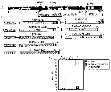
Figure 3 - Identification of a domain required for nuclear localization of
HDHB.
(A) Schematic representation of the HDHB protein showing seven potential
phosphorylation sites for CDK (SP or TP), the putative subcellular
localization
domain (SLD) and phosphorylated SLD (PSLD), the Walker A and Walker B
helicase motifs. Amino acid residue numbers are indicated below protein.
(B) GFP- and FLAG-tagged HDHB and C-terminal truncation mutants generated
in study. The C terminus of HDHB SLD (residues 1040-1087) and PSLD
(residues 957-1087) was fused to a GFP aGal reporter to create GFP-,6 Gal-
SLD and GFP-fl Gal-PSLD respectively.
(C) The subcellular localization of transiently expressed GFP-HDHB-ASLD in
asynchronous, GI, and S phase U2OS cells was quantified and expressed as a
percentage of the total number of GFP-positive cells.
Figure 4 - GFP-,6 Gal-PSLD subcellular localization pattern varies with the
cell
cycle. o
(A) The subcellular localization of transiently expressed GFP- fl Gal, GFP- fl
Gal-SLD, and GFP-a Gal-PSLD in asynchronous, G1, and S phase U2OS cells
was quantified and expressed as a percentage of the total number of GFP-
positive cells.
16
CA 02574530 2007-01-19
WO 2006/008542 PCT/GB2005/002884
Figure 5 - Identification of a functional rev-type nuclear export signal (NES)
in
SLD of HDHB.
(A) Alignment of the putative NES in HDHB with those identified in other cell
cycle-related proteins (Henderson and Eleftheriou, 2000; Fabbro and
Henderson, 2003). Superscripts above the amino acid sequence indicate
residue numbers. Thick arrows point to the conserved aliphatic residues in the
NES. Two pairs of residues in the putative NES in HDHB were mutated to
alanine as indicated by the thin arrows to create Mut1 and Mut2.
(B) GFP- and FLAG-tagged HDHB were transiently, expressed in
asynchronously growing U2OS cells with (+) or without (-) LMB to inhibit CRM1-
mediated nuclear export. The subcellular localization of GFP-HDHB and FLAG-
HDHB in asynchronous, G1, and S phase cells was quantified and expressed as
a percentage of the total number of GFP-positive cells in that sample.
(C) The subcellular localization of wild type and mutant GFP-HDHB and GFP-
Gal-PSLD in asynchronous U2OS cells was quantified and expressed as a
percentage of the total number of GFP-positive cells in that sample.
Figure 6- Cell cycle-dependent phosphorylation of FLAG-HDHB in vivo.
(A) U2OS cells transiently expressing FLAG-HDHB (lane 1) and its truncation
mutants 1-1039 (lane 2) and 1-874 (lane 3) were labeled with [32P] ortho-
phosphate. Cell extracts were immunoprecipitated with anti-FLAG resin. The
precipitated proteins were separated by 7.5% SDS-PAGE, transferred
to a PVDF membrane, and detected by autoradiography (top) or western blotting
(bottom). The positions of marker proteins of known molecular mass are
indicated at the left.
(B) FLAG-HDHB expressed in U2OS cells was immunoprecipitated with anti-
FLAG resin, incubated with (+) or without (-).i-phosphatase (a-PPase) in the
presence (+) or absence (-) of phosphatase inhibitors, as indicated, and
analyzed by SDS-PAGE and immunoblotting with anti-HDHB antibody.
(C) U2OS cells expressing FLAG-HDHB were arrested at G1/S (top) or at
G2/M(bottom), and then released from the block. FLAG-HDHB was harvested at
the indicated time points, immunoprecipitated with anti-FLAG resin, treated
with
(+) or without (-) A-PPase, and analyzed as in (B).
17
CA 02574530 2007-01-19
WO 2006/008542 PCT/GB2005/002884
Figure 7 - Identification of S967 as a major in vivo phosphorylation site in
HDHB.
(A) Phosphoamino acid markers (left) and phosphoamino acids from in vivo
32P-labeled FLAG-HDHB (right) were separated in two dimensions and
visualized by autoradiography. Some incompletely hydrolyzed phosphopeptides
remained near the origin (+).
(B) Wild type and mutant FLAG-HDHB proteins were radiolabeled with
orthophosphate in vivo, immunoprecipitated, separated by SDS-PAGE,
and analyzed by autoradiography (top) and immunoblotting with anti-HDHB
(bottom).
(C) Tryptic phosphopeptides of 32P-Iabeled wild type and S967A mutant FLAG-
HDHB were separated in two dimensions and visualized by autoradiography.
Figure 8 - Identification of cyclin E/CDK2 as the potential G1/S kinase of
HDHB
S967.
(A) Tryptic phosphopeptides from FLAG-HDHB phosphorylated in vivo as in Fig.
7C, or recombinant HDHB phosphorylated in vitro by purified cyclin E/CDK2 or
cyclin A/CDK2, were separated in two dimensions, either individually or as a
mixture, and visualized by autoradiography.
(B) Proteins that co-immunoprecipitated with FLAG vector (lanes 1, 4) or FLAG-
HDHB (lanes 2, 5) expressed in U2OS cells were analyzed by immunoblotting
with antibodies against HDHB (lanes 1-6), cyclin E (lanes 1-3), or cyclin A
(lanes
4-6). One tenth of the cell lysate used for immunoprecipitation was analyzed
in
parallel as a positive control (lanes 3, 6).
Figure 9 - The subcellular localization of HDHB is regulated by
phosphorylation
of S967.
(A) Subcellular (ocafization of GFP-HDHB S967A and S967D expressed in
asynchronous, GI, and S phase U2OS cells was quantified.
Figure 10 - Localisation of EGFP-PSLD in asynchronous U2OS cells exhibiting
stable expression of the pCORON1002-EGFP-CI-PSLD vector is cell cycle
18
CA 02574530 2007-01-19
WO 2006/008542 PCT/GB2005/002884
dependent. Fluorescence microscopy of the same partial field of cells in which
(A) nuclei were stained with Hoechst dye, (B) EGFP-PSLD was visualised, (C)
nuclei were exposed to BrdU for 1 hour exposure prior to fixation and
detection
with Cy-5 labelled antibody to indicate cells in S-phase. (D) A graph of
nuclear
fluorescent intensity in both the red (Cy-5 immunofluorescent detection of
BrdU)
and green (EGFP-PSLD) for individual cells present in a full field of view.
Figure 11 - Vector map of pCORON1002-EGFP-CI-PSLD.
Figure 12 - Vector map of pCORON1002-EGFP-C1-/3Gal-PSLD
Figure 13 - Flow cytometry data comparing brightness and homogeneity of
signal for representative stable cell lines developed with pCORON1002-EGFP-
C1-PSLD, pCORON1002-EGFP-C1 /3Gal-PSLD and the parental U2OS cell
line.
Detailed Description of the Invention
Methods
Plasmids
pGFP-HDHB and mutant derivatives (see Figs 4 and 6) were created by
inserting full-length HDHB cDNA as a Bglll/Notl fragment (Taneja et al., J.
Biol.
Chem., (2002) 277, 40853-40861) into the Notl site of the pEGFP-C1 vector
(Clontech, Palo Alto, CA). pFLAG-HDHB was constructed by inserting a
Hindlll/Notl fragment containing full-length HDHB cDNA into the Notl site of
pFlag-CMV2 vector (Eastman Kodak Co., Rochester, NY). Tagged HDHB-SLD
(1-1039) was constructed by cleaving the tagged HDHB plasmid with Nrul
following the coding sequence for residue 1034 and with Noti in the polylinker
and replacing the small fragment by a duplex adaptor oligonucleotide with a
blunt end encoding residues 1035 to 1039, a stop codon, and an overhanging
Notl-compatible 5' end. To create pFLAG-HDHB (1-874), Stul-digested pFLAG-
HDHB DNA was treated with Klenow polymerase to generate blunt ends and
ligated into the pFLAG-CMV2 vector. To generate pEGFP-RGaI, a DNA
19
CA 02574530 2007-01-19
WO 2006/008542 PCT/GB2005/002884
fragment encoding E. coli (i-galactosidase (PGal) was amplified by PCR from
p(iGal-control (Clontech) and inserted at the 3' end of the GFP coding
sequence
in pEGFP-C1, using the Hindlll site. The HDHB sequence for amino acid
residues 1040-1087(SLD) and 957-1087(PSLD) were PCR amplified and
inserted at the 3' end of the RGal cDNA in pEGFP-RGaI to create pGFP-(iGal-
SLD and pGFP- OGal-PSLD respectively. The NES mutants and
phosphorylation site mutants were created in the HDHB cDNA by site-directed
mutagenesis (QuikChange, Stratagene, La Jolla, CA).
pCORON1002-EGFP-CI-PSLD was constructed by PCR amplification of
the 390 bp PSLD region from the DNA construct pGFP-CI /3Gal-PSLD.
Introduction of 5' Nhel and 3' Sall restriction enzyme sites to the PSLD
fragment
allowed sub-cloning into the vector pCORON1002-EGFP-C1 (GE Healthcare,
Amersham, UK). The resulting 6704 bp DNA construct pCORON1002-EGFP-
C1-PSLD, contains an ubiquitin C promoter, a bacterial ampicillin resistance
gene and a mammalian neomycin resistance gene (Figure 11). The nucleic acid
sequence of the vector is shown in SEQ ID No. 3. Three further versions of
this
vector were created using standard cloning techiques (Sambrook, J. et al
(1989)); the EGFP gene was first replaced with J-Red (Evrogen), the neomycin
resitance gene was replaced with hygromycin resistance gene and the ubiquitin
C promoter was replaced with the CMV I/E promoter.
pCORON1002-EGFP-C1-,8GaI-PSLD was constructed by Nhel and Xmal
restriction enzyme digest of pEGFP-CI ;l3Gal-PSLD and insertion of the 4242 bp
EGFP;8Gal-PSLD fragment into pCORON1002 vector (GE Healthcare). The
resulting 9937 bp DNA construct pCORON1002-EGFP-CI-flGal-PSLD (Figure
12) contains an ubiquitin C promoter, a bacterial ampicillin resistance gene
and
a mammalian neomycin resistance gene. The nucleic acid sequence of the
vector is shown in SEQ ID No. 4.
The protein and nucleic acid sequence for the EGFP-PSLD fusion protein
are shown in SEQ ID No. 5 and 6, respectively.
CA 02574530 2007-01-19
WO 2006/008542 PCT/GB2005/002884
The correct DNA sequence of all constructs and substitution mutations
was confirmed by DNA sequencing.
Antibodies
Anti-HDHB antibody was generated against purified recombinant HDHB
(Bethyl Laboratories, Montgomery, TX) and affinity-purified on immobilized
HDHB (Harlow & Lane, Antibodies: A laboratory manual. Cold Spring Harbor
Laboratory).
Cell culture, synchronization, microiniection electroporation, transfection
and
stable cell line generation
U2OS cells were cultured as exponentially growing monolayers in
Dulbecco-modified Eagle medium (DMEM) (Gibco BRL Lifetechnologies,
Carlsbad, CA) supplemented with 10% fetal bovine serum (FBS) (Atlanta
Biologicals, Norcross, GA) at 37 C. Exponentially growing U2OS cells were
arrested at G1/S by incubation in DMEM containing 5 mM thymidine (Sigma-
Aldrich, St. Louis, MO), for 24 h. To release the cells into S phase, the
medium
was aspirated and the cells washed three times with warm DMEM plus 10%
FBS, and incubated in fresh DMEM plus 10% FBS. Exponentially growing U2OS
cells were arrested in G2/M for 16 h in DMEM containing 30 ng/mi nocodazole
(Sigma-Aldrich). To release cells into GI, mitotic cells were collected by
gently
shaking them off, washed three times with DMEM plus 10% FBS, and then
plated on glass coverslips for microinjection, or in culture dishes for
further
manipulation.
Cell cycle synchronization was verified by flow cytometry as described
previously (Taneja et al., J. Biol. Chem., (2002) 277, 40853-40861). In
experiments to block nuclear protein export, cells were cultured for 3 h in
DMEM
containing 10 ng/ml of leptomycin B (LMB) and 10 pM cycloheximide
(Calbiochem, San Diego, CA) to prevent new protein synthesis. Cells plated on
glass coverslips were microinjected as described (Herbig et al., 1999) except
that plasmid DNA rather than protein was injected.
21
CA 02574530 2007-01-19
WO 2006/008542 PCT/GB2005/002884
For electroporation, asynchronously growing U2OS cells (5 x10⁶) were
trypsinized, collected by centrifugation, and resuspended in 800 pl of 20 mM
HEPES (pH 7.4), 0.7 mM Na2HPO4/NaH2PO4, 137 mM NaCI, 5 mM KCI, 6 mM
glucose at a final pH of 7.4. Ten pg of DNA was added, transferred to a 0.4 cm
electroporation cuvette (BioRad, Hercules, CA) and electroporation performed
using Gene Pulser li apparatus (BioRad). Cells were plated in tissue culture
dishes for I h, washed with fresh medium and cultured for another 23 h.
Working with transiently transfected cells proved difficult in multiwell plate
format due to low transfection efficiency, heterogeneity of expression and
problems arising from the high throughput analysis of such data. Screening for
the effects of large numbers of siRNA or agents upon the cell cycle therefore
required production of a homogenous stable cell line. Due to the toxic effects
of
HDHB when overexpressed for long periods a stable cell line was generated
with the PSLD region linked to a reporter. U-20S cells were transiently
transfected with pCORON1002-EGFP-CI-PSLD (Figure 11), pCORON1002-
EGFP-C1-flGal-PSLD (Figure 12) or J-Red derivatives of the above vectors.
Stable clones expressing the recombinant fusion proteins were selected using 1
mg/ml G418 (Sigma) or hygromycin, where appropriate. Isolated primary clones
(-60 per construct) were analysed by flow cytometry to confirm the level and
homogeneity of expression of the sensor and where appropriate secondary
clones were developed using methods above.
Fluorescence microscopy
For indirect immunofluorescence staining, cells were washed three times
with phosphate buffered saline (PBS), fixed with 3.7% formaldehyde in PBS for
20 min, permeabilized for 5 min in 0.2% Triton X-100, and incubated with 10%
FBS in PBS for 45 min. FLAG-HDHB was detected with mouse monoclonal anti-
FLAG antibody (Sigma-Aldrich), 1:100 in PBS plus 10% FBS for 2 h at room
temperature. After washing, cells were incubated with Texas Red-conjugated
goat anti-mouse secondary antibody (Jackson ImmunoResearch Laboratories,
West Grove, PA) at 1:100 in PBS plus 10% FBS for I h at room temperature.
22
CA 02574530 2007-01-19
WO 2006/008542 PCT/GB2005/002884
After three washes, cells were incubated for 10 min with Hoechst 33258 (2 pM
in
PBS). Coverslips were mounted in ProLong Antifade (Molecular Probes,
Eugene, OR). Images were obtained with a Hamamatsu digital camera using the
Openlab 3.0 software (Improvision, Lexington, MA) on the Zeiss Axioplan 2
Imaging system (Carl Zeiss Inc.). The number of cells that exhibited each
pattern of subcellular localization was counted and expressed as a percentage
of the total number of cells scored (100 to 150 cells in each experiment). The
subcellular distribution of each protein was quantitatively evaluated in at
least
two independent experiments.
For GFP fluorescence, cells were washed three times with phosphate-
buffered saline (PBS), fixed with 3.7% formaldehyde containing 2 pM Hoechst
33258 for 20 min and imaged and evaluated as above.
For Triton X-100 extraction, cells were washed twice with cold
cytoskeleton buffer (CSK, 10 mM HEPES [pH 7.4], 300 mM sucrose, 100 mM
NaCi, 3 mM MgC12), and extracted for 5 min on ice with 0.5% Triton X-100 in
CSK buffer (supplemented with 1X protease inhibitors) and then fixed as
described above.
Where appropriate, for high throughput imaging, kinetic imaging (24 hr)
and analysis in multiwell plate format of stable cell lines flourescence
microscopy was conducted using a higti throughput confocal imaging system (IN
Cell Analyzer 1000 or IN Cell Analyzer 3000, GE Healthcare, Amersham, UK) on
cells transfected with pCORON1002-EGFP-CI-PSLD, pCORON1002-EGFP-C1-
flGal-PSLD or redFP derivatives of these vectors. Images were analysed using
the cell cycle phase marker algorithm (GE Health Care).
Metabolic phosphate labeling
U2OS cells (2.5x10⁶) were transiently transfected with wild type or mutant
FLAGHDHB. After 24 h, cells were incubated in phosphate-depleted DMEM
(Gibco BRL Lifetechnologies) for 15 min and radiolabeled with 32P-H3P04
23
CA 02574530 2007-01-19
WO 2006/008542 PCT/GB2005/002884
(0.35 mCi/mI of medium; ICN Pharmaceuticals Inc., Costa Mesa, CA) for 4 h.
Phosphate-labeled FLAG-HDHB was immunoprecipitated from extracts,
separated by 7.5% SDS/PAGE, and transferred to a polyvinylidene difluoride
(PVDF) membrane as described below.
Cell extracts, immunoprecipitation, and western blotting
At 24 h after transfection, FLAG-HDHB-transfected cultures to be
analyzed by immunoprecipitation and immunoblotting were lysed in lysis buffer
(50 mM Tris-HCI pH 7.5, 10% glycerol, 0.1 % NP-40, 1 mM DTT, 25 mM NaF,
100 tag/ml PMSF, I tag/ml aprotinin, I pg/ml leupeptin) (0.5 ml per 35 mm or I
ml per 60 mm dish or 75 cm flask). The extract was scraped off the dish,
incubated for 5 min on ice, and centrifuged for 10 min at 14 000 g. Samples of
the supernatant (0.5 to 1 mg of protein) were incubated with 10 pl anti-FLAG
agarose (Sigma) on a rotator for 2 h at 4 C. The agarose beads were washed
three times with lysis buffer. lmmunoprecipitated proteins were transferred to
a
PVDF membrane and analyzed by western blotting with anti-HDHB-peptide
serum (1:5000), anti-cyclin E antibody (1:1000), and anticyclin A antibody
(1:1000) (Santa Cruz Biotechnology Inc., Santa Cruz, CA), and
chemiluminescence (SuperSignal, Pierce Biotechnology Inc., Rockford, IL).
For selective nuclear and cytoplasmic protein extraction, 80-90%
confluent U2OS cells were harvested by trypsinization and washed with PBS.
They were resuspended and lysed in 10 mM Tris-HCI [pH 7.5], 10 mM KCI, 1.5
mM MgC12, 0.25 M sucrose, 10% glycerol, 75 pg/mi digitonin, 1 mM DTT, 10
mM NaF, 1 mM Na3VO4, 100 lag/mI PMSF, 1 pg/mI aprotinin, and I pg/mi
leupeptin for 10 min on ice, and centrifuged at 1000 x g for 5 min. The
supernatant fraction was collected as the cytosolic extract. The pellet was
washed, resuspended in high salt buffer (10 mM Tris-HCI [pH 7.51, 400 mM
NaCI 1 mM EDTA, 1 mM EGTA, 1 mM DTT, 1% NP-40, 100 pg/ml PMSF, I
pg/ml aprotinin, and I lag/ml leupeptin), and rocked for 10 min at 4 C. After
sonication, the suspended material, containing both soluble and chromatin-
bound protein, was analyzed as nuclear extract. Proteins in the nuclear and
cytoplasmic extracts were analyzed by 8.5% SDS-PAGE, followed by western
24
CA 02574530 2007-01-19
WO 2006/008542 PCT/GB2005/002884
blotting with antibodies against a-tubulin, PCNA (both Santa Cruz
Biotechnology), and recombinant HDHB.
Protein phosphatase reactions
FLAG-HDHB bound to anti-FLAG beads was incubated with 100 U of A-
phosphatase (New England Biolabs, Beverly, MA) in phosphatase buffer (50 mM
Tris-HCI [pH 7.5], 0.1 mM EDTA, 0.01 % NP-40) for I h at 30 C. The reaction
was carried out in the presence or absence of phosphatase inhibitors (5 mM
Na3VO4, 50 mM NaF). The proteins were separated by 7.5% SDSPAGE
(acrylamide-bisacrylamide ratio, 30:0.36) and HDHB was detected by western
blotting with anti-HDHB-peptide serum and chemiluminescence.
Tryptic peptide mapping and phosphoamino acid analysis
At 24 h after transfection, radiolabeled FLAG-HDHB-transfected cultures
to be used for immunoprecipitation and phosphoamino acid or phosphopeptide
mapping were processed as above, except that lysis buffer was substituted by
RIPA buffer (50 mM Tris-HCI [pH7.5], 150 mM NaCI, 1% NP-40, 0.5%
deoxycholic acid, 1% SDS, 50 mM NaF, 1 mM EDTA, 5 mM Na3VO4, 100
tag/mf PMSF, 1(ag/mf aprotinin, and 1 pg/ml leupeptin). Immunoprecipitated
proteins were separated by 7.5% SDS-PAGE and transferred to PVDF
membranes. The membranes containing radio(abe(ed HDHB were rinsed well
with deionized H20 twice before visualization of phosphoproteins by
autoradiography. The phosphoproteins were then excised, and the membrane
pieces were re-wet with methanol followed by water. The membranes were
blocked with 50 mM NH4HCO3 containing 0.1 % Tween 20 (Sigma-Aldrich) for
30 min at room temperature and washed three times with 50 mM NH4HCO3
before enzymatic cleavage of phosphoproteins from the PVDF with L-
(tosyfamido-2-pheny() ethyl chloromethyl ketonetreated bovine pancreatic
trypsin
(Worthington, Lakewood, NJ). The peptides were then subjected to two-
dimensional phosphopeptide mapping or phosphoamino acid analysis as
described in detail elsewhere (Boyle et al., Meth. Enzymology, (1991), 201,
110-
149).
CA 02574530 2007-01-19
WO 2006/008542 PCT/GB2005/002884
Cyclin-dependent kinase reactions in vitro
Kinase reactions using purified cyclin/CDK (200 pmol/h) (provided by R.
Otf and C. Voitenleitner) and purified recombinant HDHB (Taneja et al., J.
Biol.
Chem., (2002) 277, 40853-40861) as the substrate were performed as
described previously (Voitenleitner et al., Mol. Cell. Biol., (1999), 19, 646-
56).
BrdU labelling, identification of chemical cell cycle blocks and RNAi
experiments
on stable cell lines
Stable cells expressing the pCORON1002-EGFP-CI-PSLD construct,
were seeded at 0.3 x10⁵ /ml in 96-well Greiner plates using antibiotic-free
medium (1001aI/well) and incubate for 16 hours.
To demonstrate the distribution of EGFP-PSLD in S-phase, stable cells
were marked with BrdU for 1 hr using the cell proliferation kit (Amersham
Biosciences, GE Health Care). Cells were fixed in 2% formalin and incorporated
BrdU was detected by immunofluorescence with a Cy-5 labelled secondary
antibody system (Cell proliferation kit; GE Health Care). Nulcei were stained
with hoechst (2 pM).
For chemical block studies (Table 1), stable cells were exposed to
olomoucine, roscovitine, nocodazole, mimosine, coicemid or colchicine (Sigma).
Cells were fixed in 2% formalin and nulcei stained with hoechst (2 pM).
For siRNA studies, siRNA pools (Dharmacon) against certain cyclins,
MCM proteins, CDKs, polo-like kinase (PLK), and a random control duplex
(Table 2) were diluted in lipofectamine/optimem I(Invitrogen) to 25 nM and
added to stable cells for 4hrs. The medium was replaced and plates incubated
for 48hr. Cells were fixed in 2% formalin and nuicei stained with hoechst (2
pM).
After highthroughput imaging and analysis on the IN Cell Analyzer system
(GEHC), data for average nuclear intensity and N:C ratio (EGFP signal),
nuclear
size (hoescht signal) and, where appropriate, nuclear signal intensity (BrdU)
were obtained for for the total number of individual cells in a field of view
using
26
CA 02574530 2007-01-19
WO 2006/008542 PCT/GB2005/002884
hoescht as a nuclear mask and the IN Cell Analyzer 3000 cell cycle phase
marker algorithm (GEHC). For each well, the total number of cells per field of
view were catagorised into G1-phase (predominantly nuclear EGFP distribution;
high EGFP-PSLD nuclear intensity and N:C ratio), S-phase (nuclear BrdU signal
>3SDs above background; EGFP-PSLD N:C ratio around 1) and G2-phase
(large nuclear size; low EGFP-PSLD N:C ratio). Although it was possible to
differentiate M-phase cells (based on small nuclear size and very intense EGFP
signal) very few such cells were seen in wells fixed with formalin since they
were
removed during the washing and fixation process.
Results
HDHB resides in nuclear foci or in the c o lasm
To determine the subcellular localization of endogenous HDHB, nuclear
and cytoplasmic proteins were selectively extracted from human U2OS cells,
separated by denaturing gel electrophoresis, and analyzed by western blotting
(Fig. 1). The presence of PCNA and a-tubulin in each extract was first
monitored
to assess the extraction procedure. PCNA was enriched in the nuclear extract
and not in the cytoplasmic fraction, while a-tubulin was found primarily in
the
cytoplasmic fraction, validating the fractionation. HDHB was detected in both
the
nuclear and cytoplasmic fractions (Fig. 1). The cytoplasmic HDHB migrated
more slowly than the nuclear fraction (Fig. 1), suggesting the possibility of
post-
translational modification.
These results could indicate either that HDHB was distributed throughout
the cell, or that a mixed population of cells contained HDHB in either the
nucleus
or the cytoplasm. To distinguish between these alternatives, HDHB was
localized in situ in single cells; GFP- and FLAG-tagged HDHB were expressed in
human U2OS cells by transient transfection. Since prolonged over-expression of
tagged or untagged HDHB was cytotoxic, all experiments were conducted in the
shortest time period possible (usually 24 h). Tagged HDHB localization was
analyzed in individual cells by fluorescence microscopy. Both GFP-HDHB and
FLAG-HDHB displayed two major patterns of localization, either in the nucleus
in
27
CA 02574530 2007-01-19
WO 2006/008542 PCT/GB2005/002884
discrete foci or in the cytoplasm (Fig. 1). GFP-HDHB transiently expressed in
primary human fibroblasts was also observed in either the nucleus or the
cytoplasm.
Identification of a cell cycle-dependent subcellular localization domain in
HDHB
U2OS cells were arrested in G2/M with nocodazole, released
into GI for three hours, and then microinjected with pGFP-HDHB DNA into their
nuclei. GFP-HDHB expression was easily detectable six hours later, when
approximately 70% of GI phase cells had accumulated the fusion protein
primarily in the nuclei (Fig. 2). In contrast, when cells were synchronized at
G1/S
with thymidine, released into S phase, and then microinjected with pGFP-HDHB
DNA, more than 70% of S phase cells had accumulated the fusion protein
predominantly in the cytoplasm (Fig. 2). Selective extraction of U2OS cells in
G1
and S phase revealed that endogenous HDHB was mostly nuclear in G 1 and
cytoplasmic in S phase (Fig. 2b). However, endogenous HDHB was clearly
detectable in both subcellular fractions. The mobility of the S phase HDHB was
slightly retarded compared to the G1 phase protein. These results indicate
that
the subcellular localization of HDHB is regulated in the cell cycle and that
GFP-
tagged HDHB reflects the localization of the endogenous untagged helicase.
Prompted by the identification of C-terminal nuclear location signals in
Bloom's syndrome helicase and other RecQ-family helicases (Hickson, Nature
Rev. Cancer, (2003) 3, 169-178), a possible subcellular localization domain
(SLD) was identified at the extreme C-terminus of HDHB (Fig. 3). To determine
whether this putative SLD was important for HDHB localization, a truncation
mutant of HDHB (GFP-HDHB-.SLD) was generated that lacks the C-terminal 48
residues containing the SLD (Fig. 3). The expression vector was microinjected
into U2OS cells in G1 or S phase and the subcellular localization of the
fusion
protein was examined by fluorescence microscopy six hours later. Over 95% of
the cells accumulated the fusion protein in the cytoplasm, regardless of the
cell
cycle timing of HDHB expression (Fig. 3c). This result suggests that HDHB may
carry a NLS that is impaired or abolished by the C-terminal deletion in GFP-
HDHB-OSLD.
28
CA 02574530 2007-01-19
WO 2006/008542 PCT/GB2005/002884
To determine whether the C-terminal domain of HDHB was sufficient for
nuclear localization, a bacterial R-galactosidase (RGal) was used as a
reporter
protein because it has a molecular mass (112 kDa) close to that of HDHB and
does not contain subcellular localization signals (Kaideron et al., Cell,
(1984),
39, 499-509). As a control, a GFP-PGaI expression vector (Fig. 3) was created
and the subcellular localization of the fusion protein monitored after
microinjection of the expression vector into U2OS cells. As expected, GFP-PGaI
protein accumulated primarily in the cytoplasm (Fig. 4). In contrast, GFP-pGal-
SLD was found in both the nucleus and cytoplasm in asynchronous or
synchronized U2OS cells (Fig. 4), suggesting that SLD contains a NLS, but was
not sufficient for nuclear localization of the reporter protein. Reasoning
that
perhaps the neighboring potential CDK phosphorylation sites might affect
subcellular localization in the cell cycle (Fig. 3), a GFP-PGaI-PSLD was
constructed, in which the C-terminal 131 residues of HDHB, containing the
putative SLD and the cluster of potential CDK phosphorylation sites, were
appended to the C-terminus of GFP-PGaI (Fig. 3). When the GFP-PGai-PSLD
plasmid DNA was transiently expressed in asynchronous and synchronized
U2OS cells, GFP-pGal-PSLD was found in the nucleus in over 90% of GI phase
cells, and in the cytoplasm in more than 70% of S phase cells (Fig. 4). In
contrast with the focal pattern observed for nuclear GFP-HDHB in G1, GFP-
RGaI-PSLD and EGFP-PSLD proteins were distributed evenly throughout the
nucleus in G1, sparing only the nucleoli. Analysis of stable cell lines
expressing
pCORON1002-EGFP-CI-PSLD that have been marked with BrdU emphasized
that cells in S-phase (equal to approx 60% of the asychronous population)
exhibit equidistribution or predominantly cytoplasmic distribution of the EGFP-
PSLD signal (Figure 10). S-phase cells do not show a predominantly nuclear
distribution of EGFP-PSLD associated with GI cells. Some cells were seen to
exhibit absolute nuclear exclusion of the EGFP-PSLD reporter (Figure 10)
however these cells did not incorporate BrdU. We hypothesised that cells
demonstrating absolute clearance of EGFP-PSLD from the nucleus were in G2.
Kinetic imaging of the EGFP-PSLD stable cell lines over 24 hours showed that
29
CA 02574530 2007-01-19
WO 2006/008542 PCT/GB2005/002884
EGFP-PSLD is predominantly nuclear in G1 after mitosis, exhibits a rapid
nuclear to cytoplasmic movement around the G1/S transition (-3.5 hours after
cytokinesis) and further progressive translocation from the nucleus to the
cytoplasm from G1/S through to the end of G2 (approx 19 hours); at this point
cell rounding occured prior to re-division. These observations seem to confirm
the possibility that G2 cells exhibit an absolute cytoplasmic distribution of
the
EGFP-PSLD reporter. Stable expression of the EGFP-PSLD fusion was not
found to affect the total length of the cell cycle (approx 24 hours) when
compared to U2OS cells or the G2M cell cycle phase marker cell line (GEHC).
Taken together, these data suggest that the subcellular localization of HDHB
is
dependent on the cell cycle, that the C-terminal PSLD domain of HDHB plays a
major role in regulating the subcellular localization of the protein in a cell
cycle
dependent manner and that HDHB is nuclear in GI but progressively
translocates to the cytoplasm during S-phase and possibly G2.
Identification of a functional rev-type NES in HDHB
A number of proteins that shuttle between the nucleus and cytoplasm
have been demonstrated to contain a NES similar to the prototype NES of HIV
rev protein (Fig. 5). Proteins containing a rev-type NES require the export
factor
CRM1 (also called exportin 1) to bind and transport proteins from the nucleus
to
the cytoplasm (reviewed by Weis, Cell, (2003), 112, 441-451). Leptomycin B
(LMB), specifically inhibits CRM1 activity in nuclear protein export (Wolff et
al.,
Chem. Biol., (1997), 4 139-147; Kudo et al., Exp. Cell. Res., (1998), 242, 540-
547). Inspection of the PSLD sequence in HDHB revealed a putative rev-type
NES (LxxxLxxLxL; Fig. 5). To determine whether the cytoplasmic localization of
HDHB requires a functional NES, expression plasmids for GFP-HDHB or FLAG-
HDHB DNA were microinjected into asynchronous, G1, and S phase cells in the
presence and absence of LMB. The focalization of the fusion proteins was
examined by fluorescence microscopy and quantified. In the presence of LMB,
both fusion proteins accumulated in the nucleus independently of the cell
cycle
(Fig. 5), consistent with the possibility that HDHB contains a rev-type NES
that
functions through CRM1. However it is also possible that HDHB may not be a
direct cargo of CRMI and that its export may be indirectly mediated through
CA 02574530 2007-01-19
WO 2006/008542 PCT/GB2005/002884
some other protein(s). To assess whether the putative NES in HDHB was
functional, we mutated Val/Leu and Leu/Leu of the NES motif to alanine to
create NES mutants I and 2 (Fig. 5). GFP-HDHB and GFP-pGaI-PSLD
harboring these NES mutations were transiently expressed in either
asynchronous or synchronized U2OS cells. Both NES mutant fusion proteins
accumulated in the nucleus in more than 80% of cells, no matter when they
were expressed in asynchronous or synchronized cells (Fig. 5). The results
indicate that the NES mutations specifically impaired the export of both GFP-
HDHB and GFP-pGal-PSLD, arguing that the PSLD region of HDHB contains a
functional NES.
FLAG-HDHB is phosphorylated in a cell cycle-dependent manner in vivo.
The cluster of potential CDK phosphorylation sites in the PSLD domain of
HDHB (Fig 3) suggested that phosphorylation of HDHB might regulate its
subcellular localization in the cell cycle. If so, one would expect the PSLD
region
of HDHB to be phosphorylated in a cell cycle-dependent manner. To test
whether HDHB undergoes phosphorylation in PSLD, U2OS cells were
transiently transfected with expression plasmids for wild type and C-
terminally
truncated forms of FLAG-HDHB, radiolabeled with phosphate, and then FLAG-
HDHB was immunoprecipitated from cell extracts. Immunoprecipitated proteins
were analyzed by denaturing gel electrophoresis, immunoblotting, and
autoradiography (Fig. 6). A radiolabeled band of FLAG-HDHB was detected at
the same position as the immunoreactive HDHB band (Fig. 6A, lanes 1).
Truncated FLAG-HDHB lacking SLD was also robustly phosphorylated in vivo
(lanes 2), while truncated FLAG-HDHB (1-874) lacking PSLD was not
significantly phosphorylated (lanes 3). These results demonstrate that SLD is
not
required for HDHB phosphorylation, while PSLD is required, and suggest that
the phosphorylation sites probably reside in PSLD.
To examine the timing of HDHB phosphorylation in the cell cycle, it would
be convenient to detect phosphorylation without the use of radiolabeling.
Since
phosphorylation often reduces the electrophoretic mobility of a protein in
denaturing gels, transiently expressed FLAG-HDHB was immunoprecipitated
31
CA 02574530 2007-01-19
WO 2006/008542 PCT/GB2005/002884
and its mobility examined before and after treatment with A-phosphatase (,\-
PPase) (Fig. 6B). Without A-PPase treatment, FLAG-HDHB was detected in
western blots in two very closely migrating bands (lane 1), while
dephosphorylated FLAG-HDHB migrated as a single band at the mobility of the
faster band of the doublet (lane 2). When \-PPase inhibitors were present in
the
reaction, FLAG-HDHB migrated as a doublet identical to the mock-treated
protein (lane 3). These data suggest that the electrophoretic mobility of FLAG-
HDHB was reduced by phosphorylation and that this assay may be suitable to
track HDHB phosphorylation in the cell cycle.
To determine whether HDHB is phosphorylated in a cell cycle-dependent
manner, U2OS cells transiently expressing FLAG-HDHB were arrested in G1/S
by adding thymidine to the medium or in G2/M by adding nocodazole to the
medium. The cells were released from the blocks for different time periods,
and
FLAG-HDHB was immunoprecipitated from cell extracts.
The immunoprecipitated material was incubated with or without A-PPase
and then analyzed by denaturing gel electrophoresis and western blotting (Fig.
6C). The mobility of FLAG-HDHB from cells arrested at G1/S was increased by
A-PPase treatment, suggesting that the protein was phosphorylated at G1/S
(Fig. 6C, upper panel). A similar mobility shift was detected after
phosphatase
treatment of FLAG-HDHB for at least nine hours after release from the G1/S
block (upper panel), as well as in cells arrested at G2/M (Fig. 6C, lower
panel).
However, after the cells were released into G1 for four and eight hours, FLAG-
HDHB migrated as a single band that was much less affected by phosphatase
treatment (Fig. 6C, lower panel). By twelve hours after release from the G2/M
block, when most of the cells were entering S phase (data not shown), the
mobility of FLAG-HDHB was again increased by phosphatase treatment,
restoring the pattern observed in nocodazole-arrested cells (lower panel).
These
results strongly suggest that phosphorylation of FLAG-HDHB is cell cycle-
dependent, with maximal phosphorylation from G1/S through G2/M and minimal
phosphorylation during GI.
32
CA 02574530 2007-01-19
WO 2006/008542 PCT/GB2005/002884
Serine 967 is the maior phosphorylation site of ectopically expressed HDHB.
To map the phosphorylation sites in FLAG-HDHB, we first wished to
determine what arriino acid residues were modified. Phosphoamino acid
analysis of in vivo radiolabeled FLAG-HDHB revealed that phosphoserine(s)
was the major phosphoamino acid of FLAG-HDHB in vivo (Fig. 7A). Assuming
that the cell cycle-dependent phosphorylation sites of HDHB are located in
PSLD between residues 874 and 1039 (Fig. 3A), that these sites are modified by
CDKs, and that phosphoserine is the major amino acid modified (Fig. 7A), only
four of the seven potential CDK sites would remain as candidate sites. To test
each of these sites individually, FLAG-HDHB expression plasmids with the
corresponding serine to alanine mutations were constructed. Cells transiently
transfected with these plasmids were radiolabeled with orthophosphate in vivo
and FLAG-HDHB was immunoprecipitated and analyzed by autoradiography
and western blotting (Fig. 7B). The results showed that FLAG-HDHB and three
of the mutant proteins were phosphorylated approximately equally, while the
S967A mutant protein was only weakly phosphorylated (Fig. 7B). This result
suggested that S967 might be the primary site of HDHB phosphorylation in vivo.
Consistent with this interpretation, an electrophoretic mobility shift after
phosphatase treatment of immunoprecipitated FLAG-HDHB was detected with
three of the mutant proteins, but not with S967A protein.
To confirm that S967 was the major phosphorylation site in HDHB in vivo,
tryptic phosphopeptide mapping was carried out with wild type and S967A
mutant FLAG-HDHB that had been metabolically radiolabeled with
orthophosphate (Fig. 7C). One predominant radiolabeled peptide and a weakly
labeled peptide were observed with the wild type protein (left panel). The
predominant phosphopeptide was absent in the S967A protein, but the weakly
labeled peptide remained detectable (Fig. 7C, right panel). The results
provide
additional evidence that serine 967 is a prominent phosphorylation site in
HDHB
in vivo.
33
CA 02574530 2007-01-19
WO 2006/008542 PCT/GB2005/002884
Identification of cyclin E/CDK2 as a kinase that potentially modifies HDHB in
G1/S
To test whether CDKs can actually modify HDHB, as suggested by the
timing of HDHB phosphorylation in the cell cycle and the identification of
S967
as a primary site of modification, purified cyclin E/CDK2 or cyclin A/CDK2
were
incubated with purified recombinant HDHB and radiolabeled ATP in vitro. After
the kinase reactions, the proteins were separated by denaturing gel
electrophoresis, transferred to a PVDF membrane, and detected by
autoradiography. The results revealed that recombinant HDHB could be
phosphorylated strongly by both cyclin E/CDK2 and cyclin A/CDK2. The
radiolabeled HDHB bands were then further processed for tryptic
phosphopeptide mapping. Peptides from each digestion were separated in two
dimensions, either individually or after mixing with tryptic peptides from in
vivo
phosphorylated FLAG-HDHB, and visualized by autoradiography (Fig. 8A).
HDHB peptides phosphorylated by cyclin E/CDK2 and cyclin A/CDK2 yielded
patterns essentially identical to those observed in the in vivo labeled
peptide
map, with one major spot and one minor spot (Fig. 8A). When the in vitro and
in
vivo labeled peptides were mixed and separated on one chromatogram, they co-
migrated (Fig. 8A, right). These data argue that the major phosphopeptides
modified in vitro by cyclin E/CDK2 and cyclin A/CDK2 in purified recombinant
HDHB were the same ones modified in vivo in FLAG-HDHB.
Since cyclin E activity in human cells rises in late G1, while cyclin A
activity rises later coincident with the onset of S phase (Pines, 1999;
Eriandsson
et al., 2000), it was important to try to distinguish whether one of these
kinases
might preferentially modify HDHB. Cyclin subunits frequently form a complex
with the substrate proteins that they target for phosphorylation (Endicott et
al.,
1999; Takeda et al., 2001). To test whether cyclin E or cyclin A could
associate
with HDHB, FLAG-HDHB and associated proteins were immunoprecipitated
from extracts of cells transfected with either FLAG-HDHB expression vector or
empty FLAG vector as a control. The cell extracts and the immunoprecipitated
material were analyzed by western blotting (Fig. 8B). Cyclin E clearly co-
precipitated with FLAG-HDHB, but cyclin A did not (Fig. 8B, lanes 2 and 5),
34
CA 02574530 2007-01-19
WO 2006/008542 PCT/GB2005/002884
suggesting that FLAG-HDHB may interact preferentially with cyclin E in vivo.
It is
conceivable that this interaction may be required for phosphorylation of HDHB
by cyclin E/CDK2 in vivo, and if so, mutations in HDHB that prevent its
association with cyclin E would abrogate phosphorylation by cyclin E/CDK2. To
test the possibility that the FLAG-HDHB mutant S967A was not phosphorylated
in vivo (Fig. 7B, C) due to an inability to bind to cyclin E, FLAG-HDHB-S967A
and associated proteins were immunoprecipitated from extracts of transfected
cells and analyzed by western blotting. Co-precipitation of cyclin E with the
mutant protein was as robust as with wild type FLAG-HDHB.
Phosphorylation of serine 967 is critical for regulation of HDHB localization.
The data above indicate that subcellular localization and phosphorylation
of ectopically expressed HDHB were regulated in a cell cycle-dependent manner
with maximal phosphorylation from G1/S to G2/M, coinciding with the period
when HDHB accumulated in the cytoplasm. These results, together with the
identification of S967 as the major in vivo phosphorylation site in HDHB,
suggest
that phosphorylation of S967 may regulate the subcellular localization of
HDHB.
To test this idea, expression plasmids for wild type GFP-HDHB and the mutants
S967A, S984A, S1005A, and S1021A were microinjected into synchronized
U2OS cells. Wild type GFP-HDHB accumulated in nuclear foci of cells in G1, but
in the cytoplasm of cells in S phase as expected. However, regardless of cell
cycle timing, GFP-HDHB-S967A localized in nuclear foci in about 70% of the
fluorescent cells (Fig. 9). The other three substitution mutants localized in
either
the nucleus or the cytoplasm like wild type GFP-HDHB. In an attempt to mimic
the phosphorylation of S967, serine 967 was mutated to aspartic acid, GFP-
HDHB-S967D was expressed in asynchronous and synchronized U2OS cells,
and the subcellular distribution of the mutant fusion protein was examined.
About 60% of the cells expressing GFP-HDHB-S967D displayed
cytoplasmic fluorescence in asynchronous, GI phase, and S phase cells (Fig.
9A), demonstrating that the S967D mutation mimicked phosphorylated S967.
The data strongly suggest that phosphorylation of serine 967 is critical in
regulating the subcellular localization of HDHB.
CA 02574530 2007-01-19
WO 2006/008542 PCT/GB2005/002884
A C-terminal domain of HDHB confers cell cycle-dependent localization
A 131-residue domain, PSLD, is sufficient to target HDHB, EGFP or a
pGal reporter to either the nucleus or the cytoplasm in a cell cycle-dependent
manner (Fig. 4 and 10). A rev-type NES resides in this domain (Fig. 5), but
its
activity or accessibility to the nuclear export machinery depends on
phosphorylation of PSLD, primarily on serine 967, at the G1/S transition (Fig.
6-
9). S967 is a perfect match to the consensus CDK substrate recognition motif
(S/T)PX(K/R). Both cyclin EICDK2 and cyclin A/CDK2 can modify HDHB in vitro,
but the ability of cyclin E/CDK2 to complex with HDHB in cell extracts
suggests
that it may be the initial kinase that modifies HDHB at the G1/S transition
(Fig.
8). Addition of olomoucine and roscovitin, known Cdk2 inhibitors (Table 1), or
siRNA toward cyclin E (Table 2) resulted in predominantly nuclear distribution
of
EGFP-PSLD and arrest in G1 for EGFP-PSLD stable cell lines, further
supporting the possibility that Cdk2/cyclin E is responsible for control of
the
observed cell-cycle based phosphorylation-dependent subcellular localisation.
Phosphorylation of PSLD appears to persist through the latter part of the cell
cycle, correlating well with the predominantly cytoplasmic localization of
HDHB
in S and G2. Kinetic imaging of stable cell lines treated with olomoucine over
24
hours showed that, for cells arrested in G2 the EGFP-PSLD signal redistributes
from the cytoplasm to the nucleus over -4-8 hours (without the cell passing
through mitosis) suggesting that in the absence of cdk2 activity the EGFP-PSLD
either becomes dephosphorylated and re-enters the nucleus, or is destroyed
and newly synthesised protein is not phosphorylated due to cdk2 inhibition and
therefore locates in the nucleus.
36
CA 02574530 2007-01-19
WO 2006/008542 PCT/GB2005/002884
Table 1
%
Total
Compound S GI G2 cells
Colcemid (0.3,uM) 41 16 43 490
Coicemid (1.2 /jM) 32 8 59 450
Colchicine (4,uM) 36 9 55 467
Colchicine (100 NM) 32 12 57 439
L-mimosine (2 mM) 68 6 26 1710
Olomoucine (500,uM) 33 63 4 600
Roscovitin (100,uM) 36 52 13 693
Nocodazole (3,uM) 33 6 61 606
Control 61 17 22 2137
Table 2
%
siRNA S GI G2 Total
cells
PLK 53 9 38 66
MCM7 58 13 29 231
MCM6 64 14 22 166
MCM5 63 17 20 260
MCM4 56 20 24 223
MCM3 59 23 19 188
MCM2 50 24 26 266
Cyclin B1 49 36 15 280
V2
Cyclin B1 60 24 17 203
V1l
CDK8 50 23 27 299
CDK7 56 18 26 354
CDK6 58 22 20 328
Cyclin A2 61 13 26 319
Cyclin A1 66 10 24 298
Cyclin T2b 57 12 31 267
Cyclin T2b 55 22 23 355
cyclinTl 60 20 20 260
cyclinE1 49 27 24 272
Control 69 10 20 262
It was not possible to distinguish whether HDHB undergoes
dephosphorylation at the M/G 1 transition (Fig. 6C) or is perhaps targeted for
proteolysis and rapidly re-synthesized in early G1, when it would enter the
37
CA 02574530 2007-01-19
WO 2006/008542 PCT/GB2005/002884
nucleus. However, kinetic imaging of stable cell lines over 24 hours showed
that
the EGFP-PSLD signal is not greatly reduced during M phase or at the M/G1
boundary, but becomes predominantly nuclear approximately 30 minutes after
cytokinesis (this state then persists for -3 hours during G1), coincident with
nuclear membrane formation. This indicates that the EGFP-PSLD construct is
dephosphorylated rather than undergoing significant destruction around the
M/G1 boundary.
These data provide strong evidence that the PSLD contains active
targeting signals that are independent of protein context (Fig. 2-5, 10).
Since
mutant HDHB with an inactivated NES is nuclear even when it is expressed
during S phase and thus presumably phosphorylated (Fig. 5), it is probable
that
the NLS is not inactivated by phosphorylation and that the primary target of
CDK
regulation is the NES. Extending this reasoning, the NES may be masked during
G1 when the CDK motifs in PSLD are unmodified, and that the NES is liberated
when S967 becomes phosphorylated, leading to NES recognition by nuclear
export factors (Fig. 3-5). Structural studies of a rev-type NES have shown
that it
forms an amphipathic a-helix, with the leucines aligned on one side of the
helix
and charged residues on the other side (Rittinger et al., Mol. Cell. Biol.
(1999), 4,
153-166). Since the SLD of HDHB contains both the rev-type NES and an NLS,
and the basic residues likely to serve as the NLS are interspersed through the
NES, the NES and NLS may reside on opposite faces of an amphipathic helix.
Additional sequences in PSLD would mask the NES intramolecularly, allowing
only the NLS to be recognized. Phosphorylation of S967 would alter the
conformation of the mask in PSLD to expose the NES, without affecting
exposure of the NLS.
High throughput screening for inhibitors of the cell cycle with EGFP-PSLD
stable
cell lines
As stated above, working with transiently transfected cells proved difficult
in multiwell plate format due to low transfection efficiency, heterogeneity of
expression and problems arising from the high throughput analysis of such
data.
Screening for the effects of large numbers of siRNA or agents upon the cell
38
CA 02574530 2007-01-19
WO 2006/008542 PCT/GB2005/002884
cycle therefore required production of a homogenous stable cell line. A stable
cell line was generated with the PSLD region linked to a reporter (EGFP) via a
flexible seven amino acid linker (using pCORON1002-EGFP-CI-PSLD). As can
be seen from Figure 13, the fluorescent signal generated by the stable cell
lines
developed with pCORON1002-EGFP-C1 ,l3Gal-PSLD was significantly smaller
(approximately ten-fold) than that produced by cells lines having the flexible
seven amino acid linker. This is probably due to the size of theflGal protein
placing large demands upon the transcriptional and translational machinery of
the cell.
A stable cell line developed with pCORON1002-EGFP-CI-PSLD (see
figure 13) was homogeneous (average total cell RFU 435, SD 58; n=271; see
Fig 10) in nature and provided sensitive, stable and uniform assays for
investigating the cell cycle and for rapidly screening the effect of agents
upon
the cell cycle in mutliwell, plate format (Tables I and 2; and Figure 10).
Certain aspects of the invention disclosed hereinabove has been
published in Molecular Biology of the Cell (15: 3320-3332, July 2004) and
electronically published as MBC in press, 10.1091/mbc.E04-03-0227 on May 14,
2004, under the title of "Cell Cycle-dependent Regulation of a Human DNA
Helicase That Localizes in DNA Damage Foci", the disclosure of which is
incorporated herein by reference in its entireties.
The foregoing is illustrative of the present invention and is not to be
construed as limiting thereof. Although a few exemplary embodiments of this
invention have been described, those skilled in the art will readily
appreciate that
many modifications are possible in the exemplary embodiments without
materially departing from the novel teachings and advantages of this
invention.
Accordingly, all such modifications are intended to be included within the
scope
of this invention as defined in the claims. Therefore, it is to be understood
that
the foregoing is illustrative of the present invention and is not to be
construed as
limited to the specific embodiments disclosed, and that modifications to the
disclosed embodiments, as well as other embodiments, are intended to be
39
CA 02574530 2007-01-19
WO 2006/008542 PCT/GB2005/002884
included within the scope of the appended claims. The invention is defined by
the following claims, with equivalents of the claims to be included therein.
DEMANDE OU BREVET VOLUMINEUX
LA PRESENTE PARTIE DE CETTE DEMANDE OU CE BREVET COMPREND
PLUS D'UN TOME.
CECI EST LE TOME 1 DE 2
CONTENANT LES PAGES 1 A 40
NOTE : Pour les tomes additionels, veuillez contacter le Bureau canadien des
brevets
JUMBO APPLICATIONS/PATENTS
THIS SECTION OF THE APPLICATION/PATENT CONTAINS MORE THAN ONE
VOLUME
THIS IS VOLUME 1 OF 2
CONTAINING PAGES 1 TO 40
NOTE: For additional volumes, please contact the Canadian Patent Office
NOM DU FICHIER / FILE NAME:
NOTE POUR LE TOME / VOLUME NOTE: