Note: Descriptions are shown in the official language in which they were submitted.
DEMANDE OU BREVET VOLUMINEUX
LA PRESENTE PARTIE DE CETTE DEMANDE OU CE BREVET COMPREND
PLUS D'UN TOME.
CECI EST LE TOME 1 DE 2
CONTENANT LES PAGES 1 A 35
NOTE : Pour les tomes additionels, veuillez contacter le Bureau canadien des
brevets
JUMBO APPLICATIONS/PATENTS
THIS SECTION OF THE APPLICATION/PATENT CONTAINS MORE THAN ONE
VOLUME
THIS IS VOLUME 1 OF 2
CONTAINING PAGES 1 TO 35
NOTE: For additional volumes, please contact the Canadian Patent Office
NOM DU FICHIER / FILE NAME:
NOTE POUR LE TOME / VOLUME NOTE:
CA 02576142 2007-01-31
WO 2006/024541 PCT/EP2005/009475
DNA decontamination method
Fie1d of the invention
The invention is related to a method for amplifying a methylated target
nucleic acid
in a sample while avoiding amplification of a non-methylated target nucleic
acid by
inactivating it. This is accomplished by a restriction enzyme digest after
bisulfite
treatment of the target nucleic acid. The invention is further related to the
use of a
restriction enzyme to avoid amplification of a non-methylated target nucleic
acid
while amplifying a methylated target nucleic acid in a sample and kits for
performing the methods according to the invention.
Bac{eround of the invention
Genes constitute only a small proportion of the total mammalian genome, and
the
precise control of their expression in the presence of an overwhelming
background
of noncoding deoxyribonucleic add (DNA) presents a substantial problem for
their
regulation. Noncoding DNA, containing introns, repetitive elements, and
potentially active transposable elements requires effective mechanisms for its
long
term silencing. Mammals appear to have taken advantage of the possibilities
afforded by cytosine methylation to provide a heritable mechanism for altering
DNA-protein interactions to assist in such silencing. DNA methylation is
essential
for the development of mammals; and plays a potential role during aging and
cancer. The involvement of methylation in the regulation of gene expression
and as
an epigenetic modification marking imprinted genes is well established. In
mammals, methylation occurs only at cytosine residues and more specifically
only
on cytosine residues adjacent to a guanosine residue, i.e. at the sequence CG.
The
detection and mapping of DNA methylation sites are essential steps towards
understanding the molecular signals which indicate whether a given sequence is
methylated.
This is currently accomplished by the so-called bisulfite method described by
Prommer, M., et al., Proc. Natl. Acad. Sci. USA 89 (1992) 1827-1831 for the
detection of 5-methyl-cytosines. The bisulfite method of mapping 5-
methylcytosine
uses the effect that sodium hydrogen sulfite reacts with cytosine but not or
only
poorly with 5-methyl-cytosine. Cytosine reacts with bisulfite to form a
sulfonated
CA 02576142 2007-01-31
WO 2006/024541 PCT/EP2005/009475
-2-
cytosine reaction intermediate being prone to deamination resulting in a
sulfonated
uracil which can be desulfonated to uracil under alkaline conditions. It is
common
knowledge that uracil has the base pairing behavior of thymine different to
the
educt cytosine whereas 5-methylcytosine has the base pairing behavior of
cytosine.
This makes the discrimination of methylated or non-methylated cytosines
possible
by e.g. bisulfite genomic sequencing (Grigg, G., and Clark, S., Bioessays 16
(1994)
431-436; Grigg, G.W., DNA Seq. 6 (1996) 189-198), methylation specific PCR
(MSP) disclosed in US 5,786,146 or by the use of blocking probes in PCR
reactions
(W02002/072880). Oakeley, E.J., (Pharmacology & Therapeutics 84 (1999) 389-
400. DNA methylation analysis: a review of current methodologies) reviews
current
methodologies of DNA methylation analysis.
There are various documents addressing specific aspects of the bisulfite
reaction
(Benyajati, C., et al., Nucleic Adds Res. 8 (1980) 5649-5667) make general
investigations to the bisulfite modification of 5-methyl-deoxycytosine and
deoxycytosine. Olek, A., et al., Nucleic Acids Res. 24 (1996) 5064-5066
disclose a
method for bisulfite base sequencing whereby bisulfite treatment and
subsequent
PCR steps are performed on material embedded in agarose beads. In the
bisulfite
method as disclosed by Clark, S. J., et al., Nucleic Acids Res. 22 (1994) 2990-
2997,
the sample is desalted after deamination. Raizis, A.M., et al., Anal. Biochem.
226
(1995) 161-166 disclose a bisulfite method of 5-methylcytosine mapping that
minimizes template degradation. They investigate the influence of pH,
temperature
and time of reaction. Similar investigations have been made by Grunau, C., et
al.,
Nucleic Acids Res. 29 (2001) E65-5 or Warnecke, P.M., et al., Methods 27
(2002)
101-107. Different additional components in the bisulfite mixture are
disclosed by
WO 01/98528 or by Paulin, R. et al., Nucleic Acids Res. 26 (1998) 5009-5010.
An additional bisulfite step after bisulfite treatment and PCR is disclosed in
WO 02/31186. Komiyama, M., and Oshima, S., Tetrahedron Letters 35 (1994)
8185-8188 investigate the catalysis of bisulfite-induced deamination of
cytosine in
oligodeoxyribonucleotides. A specific bisulfite protocol is disclosed by
WO 2004/067545. A variation of the bisulfite genomic sequencing method is
disclosed by Feil, R., et al., Nucleic Adds Res. 22 (1994) 695-696, whereby
the
genomic DNA is bound to glass beads after deamination and washed. After
elution
the nucleic acid is desulfonated. EP 1 394 173 discloses a bisulfite method
whereby
the DNA is bound to the glass surface of a solid phase. Kits for performing
bisulfite
treatments are commercially available from Intergen, distributed by
Serologicals
Corporation, Norcross, GA, USA, e.g. CpGenomeTM DNA modification kit.
CA 02576142 2007-01-31
WO 2006/024541 PCT/EP2005/009475
-3-
The polymerase chain reaction (PCR) as described in U.S. Pat. No. 4,683,202 is
also
used in the field of the analyis of methylated nucleic acids. This method is
even able
to amplify analyte nucleic acids, e.g. of HCV, that are present in the
smallest
concentrations to such an extent that they become accessible to those nucleic
acid
tests which have been restricted to highly concentrated analytes. However,
over
time it has turned out that the laboratories in which the amplifications were
carried
out have in the meantime already become so strongly contaminated with the
amplified nucleic acids that tests in samples which in fact do not contain the
low
concentrated nucleic acid at all lead to false-positive results since the
samples have
become contaminated by the environment with nucleic acids from previous
amplifications (cross-contaminations): The high sensitivity of the
amplification-
based nucleic acid tests enables the detection of even the slightest
contaminations
and hence simulates the presence of the analyte in the sample (false-positive
results).
EP-A-0 401 037 describes a method which partially remedies the described
deficiency. In this method mononucleotides that are not naturally present in
the
nucleic acid to be detected are incorporated during the amplification into the
amplificate of each analyte nucleic acid. Before a subsequent amplification is
carried
out, the sample together with the reagents used are subjected to a
pretreatment in
which all imported amplificates from earlier amplifications are enzymatically
degraded. Uracil-N-glycosylase (UNG) is an example of a degradation reagent
and
dUTP is an example of a modified building block for the amplificates.
An alternative method utilizes primers containing uracil instead of
mononucleotides containing uracil. Such a method in which the primer binding
sites are degraded on amplificates generated earlier is described in EP-A-0
415 755.
The mechanism of this decontamination method is based on the specific
recognition of uracil-containing amplificates which are degraded by the
enzyme.
In the preparation of the amplification reaction UNG is added to the sample
and
usually already together with the master mix which contains all reagents
necessary
for the amplification. The aforementioned degradation reaction takes place in
a
brief incubation step before the subsequent amplification. If the reaction
mixture is
subsequently heated to a temperature above ca. 40 C, then UNG is inactivated.
This is necessary to ensure that the UNG does not degrade the newly
synthesized
DNA which accumulates during the course of the amplification.
CA 02576142 2007-01-31
WO 2006/024541 PCT/EP2005/009475
-4-
There are several documents disclosing further methods on the decontamination
of
mixtures used for this type of reaction. Klaschik, S. et al. (Molecular
Biotechnology
22 (2002) 231-242. Comparison of different decontamination methods for
reagents
to detect low concentrations of bacterial 16S DNA by real-time PCR) disclose a
comparison of decontamination methods using restriction enzyme cleavage
compared with other methods. There is no methylation-specific cleavage and not
in
combination with bisulfite method. Only a DNAse digestion and not a
restriction
enzyme digestion was regarded to be effective. Abravaya, K. et al. (Lee, H.H.
et al.
(Ed.), Nucleic Add Amplification Technologies 1997, 125-133. Strategies to
avoid
amplicon contamination) review techniques developed to prevent carryover
contamination by contaminant DNA in DNA amplification procedures. The review
includes pre-amplification decontamination using endonucleases but there is no
disclosure of bisulfite modification or use thereof in methylation detection.
US 5,683,896 discloses another process for controlling contamination of
nucleic
acid and amplification reactions. US 2004/0005555 discloses the detection of
bacteremia in emergency department patients at risk for infective endocarditis
using universal 16S rRNA primers in a decontaminated PCR assay. The background
DNA present in all PCR reagents is eliminated using a restriction endonuclease
AluI
digestion having multiple digestion sites in the amplicon but not in the
primer sets.
The restriction enzyme AluI enzyme is inactivated by heating to a temperature
which inactivates AluI but not Taq polymerase. The method is not disclosed in
combination with the bisulfite method and cannot be used when target DNA is
present. DeFilippes, F.M. (Biotechniques 10 (1991) 26-30. Decontaminating the
polymerase chain reaction) discloses the addition of template DNA to a
modified
PCR mixture to simulate contamination. The template DNA was inactivated by
restriction enzyme digestion. After inactivation of restriction enzymes
additional
DNA template, buffer and Taq polymerase were added to the reaction and PCR
proceeded. Limitations of this method are discussed. There is no disclosure of
the
bisulfite treatment or restriction enzyme digestion.
Several documents describe the general combination of bisulfite technology and
restriction enzyme cleavage in the field of detection of methylated nucleic
acids.
Sadri, R. et al. (Nucleic Acids Res. 24 (1996) 5058-5059, Rapid analysis of
DNA
methylation using new restriction enzyme sites created by bisulfite
modification)
disclose the bisulfite treatment of DNA and subsequent amplification. Changes
in
restriction enzyme sites are detected. Velinov, M. et al. (Methods in
Molecular
Biology 217 (2003) 209-216, PCR-based strategies for the diagnosis of Prader-
CA 02576142 2007-01-31
WO 2006/024541 PCT/EP2005/009475
-5-
Willi/Angelman syndromes) disclose a PCR-based methylation test using
methylation-specific digestion of the amplified, bisulfite-treated DNA.
Velinov, M.
et al. (Molecular Genetics and Metabolism 69 (2000) 81-83. The feasibility of
PCR-
based diagnosis of Prader-WiIli and Angelman syndromes using restriction
analysis
after bisulfite modification of genomic DNA) disclose a PCR-based methylation
test
using methylation-specific digestion of the amplified, bisulfite-treated DNA.
Xiong,
Z. et al. (Nucleic Acids Res. 25 (1997) 2532-2534. COBRA: a sensitive and
quantitative DNA methylation assay) uses restriction enzyme digestion to
reveal
methylation-dependent sequence differences in PCR products of sodium bisulfite
treated DNA. W02003/000926 discloses the bisulfite treatment of DNA and
subsequent amplification. Thereafter, the amplificate is digested with
restriction
endonucleases. The enzyme resistant fraction of digested DNA is then amplified
in
a further PCR. In all these documents, restriction enzyme cleavage is
performed
after PCR amplification.
Several other documents describe restriction enzyme digestions in connection
with
the bisulfite method. Fojtova, M. et al. (Plant Science 160 (2001) 585-593.
Cytosine
methylation of plastid genome in higher plants. Fact or artifact?) disclose
bisulfite
genomic sequencing performed on EcoRII-restricted DNA difference. The
restriction enzyme digestion is performed before bisulfite treatment. Clark,
S.J. et
al. (Ed. G.R: Taylor, Laboratory Methods for the Detection of Mutations and
Polymorphisms in DNA (1997) 151-162, Publisher CRC, Boca Raton, Fla) review
current methods available for the study of cytosine methylation in genomic
DNA.
The restriction enzyme digestions and the bisulfite method are described
independently similarly to W02003/064701. Jang, K.-H. et al. (J. Microbiology
and
Biotechnology 11 (2001) 819-824. Identification of a sequence containing
methylated cytidine in Corynebacterium glutamicum and Brevibacterium flavum
using bisulfite DNA derivatization and sequencing) disclose the bisulfite
treatment
of DNA and subsequent amplification, restriction enzyme digestion to remove
not
fully converted DNA and another round of amplification. Then, the DNA sequence
is determined.
Other documents describe the use of restriction enzyme digests for the
analysis of
methylations. Kaneda, S.A. et al. (Molecular Medicine 39 (2002) 824-832)
review
various methods for analysis of DNA methylation including restriction enzyme
digestions. W02003/027259 discloses assays for detecting DNA methylation
associated with diseases in mouse and their use in diagnosis. The technique to
_. ~.
..,~...'..~,
CA 02576142 2007-01-31
WO 2006/024541 PCT/EP2005/009475
-6-
detect the extent of DNA methylation entails generating DNA fragments of a
test
sample by cleaving at methylation sites that are not methylated while sparing
methylation sites in the DNA that are methylated. The method is not used in
combination with the bisulfite method. Moore, T. (Methods in Molecular Biology
181 (2002) 193-203. Southern analysis using methyl-sensitive restriction
enzymes)
discloses the use of methyl-sensitive restriction enzymes and Southern
analysis but
not in combination with the bisulfite method. EP 0 976 835 discloses the
detection
of nucleic acid methylation using amplification fragment length polymorphism
which is not used in combination with the bisulfite method. Pogribny, I. et
al.
(Biochem. Biophys. Res. Commun. 262 (1999) 624-628. Sensitive new method for
rapid detection of abnormal methylation patterns in global DNA and within CpG
islands) disclose a method based on the use of methylation-sensitive
restriction
endonucleases that leave a 5'-guanine overhang after DNA cleavage, with
subsequent single nucleotide extension with radiolabeled [3H)dCTP. The method
is not combined with the bisulfite method or PCR amplification. Kupper, D. et
al.
(Biotechniques 23 (1997) 843-847. Reliable detection of DNA CpG methylation
profiles by the isoschizomers MspI/HpaII using oligonucleotide stimulators)
disclose a protocol for detecting CpG methylation by the isoschizomeric
restriction
endonucleases MspI/HpaII but not in combination with the bisulfite method.
Watts, G.S. et al. (Nucleic Acids Res. 23 (1995) 4740-4741. Detecting
differences in
5-methylcytosine using restriction enzyme isoschizomers: an endogenous control
for complete digestion) disclose the southern blot analysis of genomic DNA cut
with methylation-sensitive isoschizomers like Mspl/HpaII but not in
combination
with the bisulfite method. Chang, S. et al. (Plant Molecular Biology Reporter
10
(1992) 362-366. PCR amplification following restriction to detect site-
specific
methylation) disclose a procedure to test for DNA methylation at sites
recognized
by methylation-sensitive restriction endonucleases. The procedure is based on
the
assumption that PCR will amplify sequences between two primers only if target
DNA is intact after digestion. The method is not used in combination with the
bisulfite method. Szyf, M. et al. (Nucleic Acids Res. 10 (1982) 7247-7259.
Studies on
the biological role of DNA methylation: V - The pattern of E. coli DNA
methylation) disclose an analysis of the state of the methylation of GATC
sites in
newly replicating DNA using the restriction enzyme DpnI but not in combination
with the bisulfite method. Youssoufian, H. et al. (J. Mol. Biol. 150 (1981)
133-136).
Detection of methylated sequences in eukaryotic DNA with the restriction
endonucleases SmaI and XmaI) disclose two isoschizomers which either digest
specific CpG sites methyl-sensitively or not. The restriction enzymes are not
used in
CA 02576142 2007-01-31
WO 2006/024541 PCT/EP2005/009475
-7-
combination with the bisulfite method. Cedar, H. et al. (Nucleic Acids Res. 6
(1979)
2125-2132. Direct detection of methylated cytosine in DNA by use of
restriction
enzyme MspI) disclose the analysis of the state of methylation of CCGG sites
using
MspI/HpaII restriction enzymes and subsequent gel analysis but not in
combination with the bisulfite method.
Sug;mar,v of the invention
The widely-used method for decontamination of PCR mixtures by employing
uracil-N-glycosylase cannot be used together with the bisulfite method which
generates uracil from cytosine bases. Therefore, there is a need to provide a
method
that can be used for the decontamination of PCR mixtures in the field of the
detection of methylated nucleic acids to avoid false positive results by the
detection
of contaminating nucleic acids.
Therefore, it is an object of the invention to provide a method which digests
non-
methylated target nucleic acid after the bisulfite treatment of DNA that can
lead to
wrong results. In more detail, an embodiment of the invention is a method for
amplifying a methylated target nucleic acid in a sample while avoiding
amplification of a non-methylated target nucleic acid whereby the methylated
target nucleic acid comprises a nucleotide with a methylcytosine base in the
recognition nucleic acid sequence of a restriction enzyme wherein the
recognition
nucleic acid sequence is in the amplified reaction product, the method
comprising
the steps of:
(a) converting a non-methylated cytosine base in the methylated target
nucleic acid in the sample into an uracil base while not converting the
methylcytosine base;
(b) adding a restriction enzyme to the sample which digests the non-
methylated target nucleic acid whereby the methylated target nucleic
acid is not digested by the restriction enzyme;
(c) inactivating the restriction enzyme; and
(d) amplifying the methylated target nucleic acid.
In another embodiment, the invention is related to a method for controlling
contamination in sequential target nucleic acid amplification processes
comprising
a first and a second nucleic acid amplification process to amplify a target
nudeic
CA 02576142 2007-01-31
WO 2006/024541 PCT/EP2005/009475
-8-
acid in a first and second sample, respectively, which comprises carrying out
the
first nucleic acid amplification process on the target nucleic acid sequence
in the
first sample prior to carrying out the second amplification process on the
target
nucleic acid in the second sample according to the invention.
In another embodiment of the invention, a restriction enzyme is used to digest
non-methylated target nucleic acid in a sample comprising a methylated target
nucleic acid that was not present in the sample during the conversion of a non-
methylated cytosine base in the methylated target nucleic acid into an uracil
base
while not converting the methylcytosine.
In another embodiment of the invention, a restriction enzyme is used to avoid
amplification of a non-methylated target nucleic acid present in a sample
while
amplifying a methylated target nucleic acid present in the sample.
In another embodiment of the invention, a kit is provided comprising a
restriction
enzyme, a compound comprising sulfite ions and a solid phase comprising a
glass
surface.
In another embodiment of the invention, a kit is provided comprising a
restriction
enzyme, a compound comprising sulfite ions, a solid phase comprising a glass
surface, a pair of primers, a probe and a DNA polymerase.
A "nucleic acid" is a polymeric compound of "nucleotides" as known to the
expert
skilled in the art. It is used herein to denote a"nucleic acid" in a sample
which
should be analyzed, i.e. the presence, non-presence or amount thereof in a
sample
should be determined. Therefore, in other words the "nucleic acid" is the
target and
can therefore be also denoted as "target nucleic acid". For example, if it has
to be
determined whether blood contains the human immunodeficiency virus, the
"target nucleic acid" is the nucleic acid of the human immunodeficiency virus
or
more specifically the nucleic acid sequence, i.e. the order of the bases
adenine,
guanine, cytosine or thymine, that is determined. More specifically in the
context of
the invention, the "target nucleic acid" is genomic DNA that may comprise
methylated-cytosine bases in CpG sites. After "bisulfite treatment" the
nucleic acid
sequence of the genomic DNA is changed depending on methylation as non-
methylated bases are converted to uracil bases and the changed nudeic acid
sequence is determined.
CA 02576142 2007-01-31
WO 2006/024541 PCT/EP2005/009475
-9-
The "methylated target nucleic acid" is according to the invention a target
nucleic
acid that comprises (after bisulfite treatment) a nucleotide with a methylated
cytosine base in the recognition nudeic acid sequence of a restriction enzyme,
particularly the restriction enzyme used in the present invention, more
specifically
the restriction enzyme chosen or used in step (b) of the method. The "non-
methylated target nucleic acid" is according to the invention a "target
nucleic acid"
that comprises a nucleotide with a (non-methylated) cytosine base in the
recognition nucleic acid sequence of a restriction enzyme, particularly the
restriction enzyme used in the present invention, more specifically the
restriction
enzyme chosen or used in step (b) of the method. As understood by the
invention,
the "non-methylated target nucleic acid" may even comprise a nucleotide with a
methylcytosine base but not in the recognition nucleic acid sequence of a
restriction
enzyme, particularly the restriction enzyme used in the present invention,
more
specifically the restriction enzyme chosen or used in step (b) of the method.
The
nucleic acid sequence of the "non-methylated target nucleic acid" is identical
to the
"methylated target nucleic acid" and the said nucleic acid molecules only
differ in
their methylation of a cytosine base or cytosine bases not affecting hydrogen
bonding to complementary nucleic acids. According to the invention, the "non-
methylated target nucleic acid" is a "contaminating nucleic acid" or
"contaminating
target nucleic acid" that makes the sample impure or pollutes the sample. It
should
not be present in the amplification step of the method as it may lead to false
results.
As is known in the art, a"nucleoside" is a base-sugar combination. The base
portion of the nucleoside is normally a heterocyclic base. The two most common
classes of such heterocyclic bases are the purines and the pyrimidines, in
more
detail the adenine (A), guanine (G), thymine (T) or cytosine (C) base. The
uracil
base is naturally contained in the ribonucleic acid. Another naturally
occurring base
is 5-methyl-cytosine or methyl-cytosine, which is cytosine which is
substituted by a
methyl group at the 5-position of the aromatic ring of the base.
"Nucleotides" are "nucleosides" that further include a phosphate group
covalently
linked to the sugar portion of the nucleoside. For those "nucleosides" that
include a
pentofuranosyl sugar, the phosphate group can be linked to either the 2', 3'
or 5'
hydroxyl moiety of the sugar. A"nucleotide" is the "monomeric unit" of an
"oligonucleotide", more generally denoted herein as an "oligomeric compound",
or
a "polynucleotide", more generally denoted as a "polymeric compound". Another
CA 02576142 2007-01-31
WO 2006/024541 PCT/EP2005/009475
-10-
general expression therefor is deoxyribonudeic acid (DNA) and ribonucleic acid
(RNA).
According to the invention, an "oligomeric compound" is a compound consisting
of "monomeric units" which may be "nucleotides" alone or "non-natural
compounds" (see below), more specifically "modified nucleotides" (or
"nucleotide
analogs") or "non-nucleotide compounds", alone or combinations thereof.
"Oligonucleotides " and "modified oligonucleotides" (or "oligonucleotide
analogs")
are subgroups of "oligomeric compounds" in the context of the invention.
In the context of this invention, the term "oligonucleotide" refers to
"polynudeotides" formed from a plurality of "nucleotides" as the "monomeric
unit", i.e. an "oligonucleotide" belongs to a specific subgroup of a
"oligomeric
compound" or "polymeric compound" of ribonucleic acid (RNA) or
deoxyribonucleic acid (DNA) with "monomeric units". The phosphate groups are
commonly referred to as forming the internucleoside backbone of the
"oligonucleotide". The normal linkage or backbone of RNA and DNA is a 3' to 5'
phosphodiester linkage.
"Oligonudeotides" and "modified oligonucleotides" (see below) according to the
invention may be synthesized as principally described in the art and known to
the
expert in the field. Methods for preparing oligomeric compounds of specific
sequences are known in the art, and include, for example, cloning and
restriction of
appropriate sequences and direct chemical synthesis. Chemical synthesis
methods
may include, for example, the phosphotriester method described by Narang,
S.A., et
al., Methods Enzymol. 68 (1979) 90-98, the phosphodiester method disclosed by
Brown, E.L., et al., Methods Enzymol. 68 (1979) 109-151, the phosphoramidite
method disclosed in Beaucage, S.L., and Caruthers, M.H., Tetrahedron Lett. 22
(1981) 1859-1862, the H-phosphonate method disclosed in Garegg, P.J., et al.,
Chem. Scr. 25 (1985) 280-282 and the solid support method disclosed in US
4,458,066.
As said above, a "nucleic acid" as well as the "target nucleic add" is a
polymeric
compound of "nucleotides" as known to the expert skilled in the art. It is
used
herein to denote a"nucleic acid" in a sample which should be analyzed, i.e.
the
presence, non-presence or amount thereof in a sample should be determined.
CA 02576142 2007-01-31
WO 2006/024541 PCT/EP2005/009475
-11-
The term "primer" is used herein as known to the expert skilled in the art and
refers
to "oligomeric compounds" primarily to "oligonucleotides" but also to
"modified
oligonucleotides" that are able to "prime" DNA synthesis by a template-
dependent
DNA polymerase, i.e. the 3'-end of the e.g. oligonucleotide provides a free 3'-
OH
group whereto further "nucleotides" may be attached by a template-dependent
DNA polymerase establishing 3' to 5' phosphodiester linkage whereby
deoxynucleoside triphosphates are used and whereby pyrophosphate is released.
Therefore, there is - except for the intended function - no fundamental
difference
between a"primer", an "oligonucleotide" or a "probe" according to the
invention.
The term õprobe" refers to synthetically or biologically produced nudeic acids
(DNA or RNA) which, by design or selection, contain specific nucleotide
sequences
that allow them to hybridize under defined predetermined stringencies
specifically
(i.e., preferentially) to "target nucleic acids". Aõprobe" can be identified
as a
õcapture probe" meaning that it "captures" the target nucleic acid so that it
can be
separated from undesirable materials which might obscure its detection. Once
separation is accomplished, detection of the captured "target nucleic acid"
can be
achieved using a suitable procedure. õCapture probes" are often already
attached to
a solid phase. A specific example therefor is the microarray situation wherein
a
multitude of "capture probes" are attached to a "solid phase" which "capture"
labeled cRNA or cDNA.
According to the invention the term a "bisulfite reaction", "bisulfite
treatment" or
"bisulfite method" shall mean a reaction for the conversion of a cytosine
base, in
particular cytosine bases, in a nucleic acid to an uracil base, or bases,
preferably in
the presence of bisulfite ions whereby preferably 5-methyl-cytosine bases are
not
significantly converted. This reaction for the detection of methylated
cytosines is
described in detail by Frommer et al., supra and Grigg and Clark, supra. The
bisulfite reaction contains a deamination step and a desulfonation step (see
Figure
1; Grigg and Clark; supra). The statement that 5-methyl-cytosine bases are not
significantly converted shall only take the fact into account that it cannot
be
excluded that a small percentage of 5-methyl-cytosine bases is converted to
uracil
although it is intended to convert only and exclusively the (non-methylated)
cytosine bases (Frommer et al., supra).
The terms "methyl-cytosine base", "methylcytosine base", "methylated cytosine
base" and "5-methyl-cytosine base" are used interchangeably throughout the
CA 02576142 2007-01-31
WO 2006/024541 PCT/EP2005/009475
-12-
application and shall denote the derivative of a cytosine base whereby a
methyl
group is attached to the C5 atom of the cytosine ring. A cytosine base is
shown in
the left part of Fig. 1. The term "non-methylated cytosine base" shall denote
an
underivatized cytosine base whereby no methyl group is attached to the C5 atom
of
the cytosine ring in contrast to the "methyl-cytosine base".
õRestriction enzymes" are endonucleases (restriction endonucleases) that are
capable of recognizing a specific nucleotide or nucleic acid sequence in a
deoxyribonucleic acid (DNA) molecule and cleaving the double-stranded DNA at
specific sites. According to the invention, the "recognition nucleic acid
sequence of
a restriction enzyme" is the specific nucleic acid sequence recognized by the
restriction enzyme. The restriction enzymes recognize specific short DNA
sequences four to eight nucleotides long, and cleave DNA at a site within this
sequence. In the context of the present disclosure, restriction enzymes may be
used
to deave DNA molecules at sites corresponding to various restriction-enzyme
recognition sites. Bacteria contain over 400 such enzymes that recognize and
cut
over 100 different DNA sequences. An isoschisomer (isoschizomer,
isoschisomere,
isoschizomere) is one of several restriction enzymes or endonucleases which
are
isolated from different sources but which break a DNA molecule at the same
recognition site
"Digestion" of DNA refers to catalytic "cleavage" of the DNA with a
restriction
enzyme that acts only at certain sequences in the DNA. The various restriction
enzymes used herein are commercially available and their reaction conditions,
cofactors and other requirements are used as would be known to the ordinarily
skilled artisan.
The term "restriction site" refers to a "recognition (nucleic acid) sequence"
or
"restriction nudeic acid sequence" that is necessary for the manifestation of
the
action of a restriction enzyme, and includes a site of catalytic cleavage.
When an
enzyme (e.g. a restriction enzyme) is said to "cleave" a polynucleotide or
nucleic
acid, it is understood to mean that the restriction enzyme catalyzes or
facilitates a
cleavage of a polynudeotide or nucleic acid.
CA 02576142 2007-01-31
WO 2006/024541 PCT/EP2005/009475
-13-
Detailed descrintion of the invention
Conventional techniques of molecular biology and nucleic acid chemistry, which
are within the ski11 of the art, are explained in the literature. See, for
example,
Sambrook, J., et al., Molecular Cloning: A Laboratory Manual, Cold Spring
Harbor
Laboratory Press, Cold Spring Harbor, New York, 1989, Gait, M.J., ed., 1984;
Nucleic Acid Hybridization, Hames, B.D., and Higgins, S.J., eds., 1984; and a
series,
Methods in Enzymology, Academic Press, Inc., all of which are incorporated
herein
by reference. All patents, patent applications, and publications mentioned
herein,
both supra and infra, are incorporated herein by reference.
In an embodiment of the invention, a method is provided to avoid (or to
inhibit or
to prevent) the amplification of a non-methylated target nucleic acid by
performing
a restriction enzyme digest after the bisulfite treatment which digests non-
methylated target nucleic acid after the bisulfite treatment of DNA that can
lead to
wrong results and can therefore be regarded as a contaminating nucleic acid or
a
contamination in general. Potential contaminations of the sample are
ubiquitous,
possible at any step of a method, difficult to avoid and can originate from
diverse
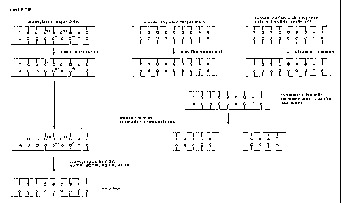
sources (see Fig. 2 and 3 for an exemplary survey of the method). The
bisulfite
reaction is the step wherein non-methylated cytosine bases are converted to
uracil
bases whereas 5-methylcytosine bases are not converted:
From Figures 2 and 3, it becomes apparent that a non-methylated target nucleic
acid may be introduced before, after or during the bisulfite reaction. If the
non-
methylated target nucleic acid is present in the bisulfite step, a non-
methylated
cytosine base will be converted to an uracil base, the nucleic acid sequence
will be
changed and not be amenable to nucleic acid amplification or restriction
digestion.
In consequence, it will not disturb the amplification as the primers and
probes are
sequence-specific and are chosen by the person skilled in the art and not lead
to
false results, Hence the bisulfite reaction itself is already a
decontamination of
previously amplified target nucleic acids present during the bisulfite
reaction when
all non-methylated cytosine bases are converted to uracil bases. Iri
consequence, the
bisulfite step will generally convert a non-methylated cytosine base in the
methylated target nucleic and in the non-methylated target nucleic acid in the
sample into an uracil base while not converting a methyl-cytosine base or the
methylcytosine base in the recognition nucleic acid sequence of the
restriction
CA 02576142 2007-01-31
WO 2006/024541 PCT/EP2005/009475
-14-
enzyme wherein the recognition nucleic acid sequence is in the amplified
reaction
product.
The first source for the non-methylated target nucleic acid present after
bisulfite
treatment can be non-methylated target nucleic acid inadvertently introduced
into
the sample after the bisulfite treatment and stemming e.g. from previous
(independent) amplifications reactions of the methylated target nucleic acid.
This
target nucleic acid does not comprise methylated cytosine bases as non-
methylated
cytosines are normally employed in amplification reactions producing non-
methylated amplification reaction products or generally non-methylated target
nucleic acids, i.e. non-methylated copies of the (originally) methylated
target
nucleic acid. The nucleic acid sequence of the non-methylated target nucleic
acid,
i.e. the amplification product of the methylated target nucleic acid, is
identical to
the methylated target nucleic acid and the said nucleic acid (molecule) only
differs
in its methylation of cytosine bases not affecting hydrogen bonding to
complementary nucleic acids. The non-methylated target nucleic acid does not
contain a nudeotide with a methyl-cytosine base in the recognition sequence of
the
restriction enzyme according to the invention.
A second possible source for "contaminating nucleic acid" or "non-methylated
target nucleic acid" can be samples from other sources or more specifically
human
patients comprising such nucleic acid and being prepared in spatial proximity
to
the sample being analysed.
The third source of "contaminating nucleic acid" or "non-methylated target
nucleic
acid" according to the invention can be "target nucleic add" not fully
converted in
bisulfite treatment, i.e. it comprises a nucleotide with a cytosine base in
the
recognition nucleic acid sequence of a restriction enzyme, particularly the
restriction enzyme chosen or used in step (b) of the method, but may contain a
nucleotide with a methylcytosine base that is not in the recognition nucleic
acid
sequence of a restriction enzyme. According to the invention, this is also a
"contaminating nucleic acid" although it is more generally a target nucleic
acid
comprising a methylcytosine base, but more specifically comprising a
methylcytosine base that is not in the recognition nucleic acid sequence of a
restriction enzyme used in step (b) of the method according to the invention
and
comprising a (non-methylated) cytosine base in the recognition nucleic acid
sequence of a restriction enzyme used in step (b) of the method according to
the
CA 02576142 2007-01-31
WO 2006/024541 PCT/EP2005/009475
-15-
invention. According to the invention, this is understood to be a non-
methylated
target nucleic acid.
The fourth source of contaminating nucleic acid can be non-methylated target
nucleic acid from other previous (independent) amplification reactions of the
methylated target nucleic acid that was present during the bisulfite treatment
and
that was not fully converted during bisulfite treatment, i.e. a non-methylated
cytosine base in the recognition nucleic acid sequence of a restriction
enzyme,
particularly the restriction enzyme chosen or used in step (b) of the method,
was
not converted to an uracil base. In other words, it comprises after bisulfite
treatment a nucleotide with a (non-methylated) cytosine base in the
recognition
nucleic acid sequence of a restriction enzyme, particularly the restriction
enzyme
chosen or used in step (b) of the method.
Therefore, it is an embodiment of the invention to provide a method for
amplifying
a methylated target nucleic acid in a sample while avoiding amplification of a
non-
methylated target nucleic acid whereby the methylated target nucleic acid
comprises a nucleotide with a methylcytosine base in the recognition nucleic
acid
sequence of a restriction enzyme wherein the recognition nucleic acid sequence
is in
the amplified reaction product the method comprising, preferably in the
following
order, the steps of
(a) converting a non-methylated cytosine base in the methylated target
nucleic acid in the sample into an uracil base while not converting the
methylcytosine base;
(b) adding a restriction enzyme to the sample which digests the non-
methylated target nucleic acid whereby the methylated target nucleic
acid is not digested by the restriction enzyme;
(c) inactivating the restriction enzyme; and
(d) amplifying the methylated target nucleic acid.
Preferably, the method according to the invention consists of the specified
steps.
As said above, for example a previously amplified target nucleic acid not
fully
converted, i.e. containing cytosine bases not converted to uracil bases, may
lead to
false results as well as non-methylated target nucleic acid not present in the
sample
during the bisulfite step. These species shall then be digested by the
restriction
CA 02576142 2007-01-31
WO 2006/024541 PCT/EP2005/009475
-16-
enzyme added, i.e. a restriction enzyme added to the sample digests the non-
methylated target nucleic acid not present in the sample in step a) of the
method
according to the invention or the non-methylated target nucleic acid
comprising a
nucleotide with a cytosine base in the recognition nucleic acid sequence of
the
restriction enzyme which was not converted in step a) of the method according
to
the invention whereby the methylated target nucleic acid is not digested by
the
restriction enzyme.
Therefore, it is an embodiment of the invention to provide a method for
amplifying
a methylated target nucleic acid in a sample while avoiding amplification of a
non-
methylated target nucleic acid whereby the methylated target nucleic acid
comprises a nucleotide with a methylcytosine base in the recognition nucleic
acid
sequence of a restriction enzyme wherein the recognition nucleic acid sequence
is in
the amplified reaction product the method comprising in the following order
the
steps ofi
(a) converting a non-methylated cytosine base in the methylated target
nucleic acid and in the non-methylated target nucleic acid in the sample
into an uracil base while not converting the methylcytosine base;
(b) adding a restriction enzyme to the sample which digests
- the non-methylated target nucleic acid not present in the sample in
step a) or
- the non-methylated target nucleic acid comprising nucleotides or a
nucleotide with a cytosine base in the recognition nucleic acid sequence
of the restriction enzyme which was or were not converted in step a)
whereby the methylated target nucleic acid is not digested by the
restriction enzyme;
(c) inactivating the restriction enzyme; and
(d) amplifying the methylated target nucleic acid.
Preferably, the method according to the invention consists of the specified
steps.
The non-methylated target nucleic acid is inadvertently or optionally present
in or
introduced into the sample, preferably before, during or after the bisulfite
treatment, i.e. step a) of the method according to the invention, or before or
during
the restriction enzyme digest, i.e. step b) of the method according to the
invention.
CA 02576142 2007-01-31
WO 2006/024541 PCT/EP2005/009475
-17-
Therefore, in still another embodiment of the invention, a method is provided
for
amplifying a methylated target nucleic acid in a sample while avoiding
amplification of. a non-methylated target nucleic acid or contaminating
nucleic
acid, optionally present in or optionally introduced into the sample, the
method
comprising in the following order the steps ob
(a) converting a non-methylated cytosine base in the methylated target
nucleic acid and in the non-methylated target nucleic acid in the sample
into an uracil base while not converting the methylcytosine base;
(b) adding a restriction enzyme to the sample which digests the non-
methylated target nucleic acid that is optionally present in the sample or
optionally inadvertently introduced into the sample,
whereby the methylated target nucleic acid is not digested by the
restriction enzyme;
(c) inactivating the restriction enzyme; and
(d) amplifying the methylated target nucleic acid.
Preferably, the method according to the invention consists of the specified
steps.
In the method according to the invention, the methylated target nucleic acid
comprises a nucleotide with a methylcytosine base in the recognition nudeic
acid
sequence of a restriction enzyme wherein the recognition nucleic acid sequence
is in
the amplified reaction product. The non-methylated target nucleic acid
comprises a
nucleotide with a cytosine base in the recognition nucleic acid sequence of a
restriction enzyme (wherein the recognition nucleic acid sequence is in the
amplified reaction product).
The expert skiIled in the art knows how to perform the bisulfite reaction,
i.e. step a)
of the method according to the invention, e.g. by referring to Frommer et al.,
supra
or Grigg and Clark, supra who disclose the principal parameters of the
bisulfite
reaction. From Grunau et al., supra, it is known to the expert in the field
what
variations of the bisulfite method are possible. The influence of incubation
time
and temperature on deamination efficiency and parameters affecting DNA
degradation is disclosed. In summary, in the deamination step a buffer
containing
bisulfite ions and optionally chaotropic agents as guanidinium ions or urea
and
further reagents as an alcohol or stabilizers as hydroquinone are employed and
the
pH is in the acidic range. The concentration of bisulfite is between 0.1 to 8
M
CA 02576142 2007-01-31
WO 2006/024541 PCT/EP2005/009475
-18-
bisulfite, preferably 0.1 to 6 M, more preferably 0.1 M to 5.5 M, the
concentration
of the chaotropic agent is between 1 to 8 M, whereby in general preferably
guanidinium salts are employed, more preferably guanidinium hydrogen sulfite
as
described in EP 3027754.5, the pH is in the acidic range, preferably between
4.5 to
6.5, the temperature is between 0 C to 90 C, preferably between room
temperature (25 C) to 90 C, and the reaction time is between 30 min to 24
hours
or 48 hours or even longer, but preferably between 1 hour to 24 hours.
The desulfonation step is performed by adding an alkaline solution or buffer
as e.g.
a solution only containing a hydroxide, e.g sodium hydroxide, or a solution
containing ethanol, sodium chloride and sodium hydroxide (e.g. 38% EtOH, 100
mM NaCl, 200 mM NaOH) and incubating at room temperature or elevated
temperatures for several min, preferably 5 min to 60 min. Desalting of the
nucleic
acid can be performed using magnetic glass particles as described in WO
96/41811,
specifically described for the bisulfite reaction in EP 1 394 173. It is also
possible to
use a kit commercially available from Intergen, distributed by Serologicals
Corporation, Norcross, GA, USA, e.g. CpGenomeTM DNA modification kit.
Therefore, in a preferred embodiment of the invention, a method is provided
wherein in step a) of the method the presence of sulfite ions in the sample
converts
the non-methylated cytosine base in the methylated target nucleic acid in the
sample into the uracil base. More preferably, the method comprises in step a)
the
substeps of al) mixing the sample comprising the methylated target nucleic
acid
with a solution comprising sulfite ions; a2) incubating the solution obtained
in step
al) comprising the methylated target nucleic acid and sulfite ions whereby the
target nucleic acid is deaminated, a3) incubating the deaminated nucleic acid
under
alkaline conditions whereby the deaminated nucleic acid is desulfonated, and
a4)
desalting the deaminated nucleic acid. The concentration of sulfite ions is
preferably 0.1 to 8 M, more preferably 0.1 to 6.25 M, 0.1 to 6 M, more
preferably 2
to 6 M. The pH of the solutions in step al) and a2) is preferably in the
acidic range,
more preferably between 4.5 to 6.5. The incubation temperature in step a2) and
a3)
is preferably between 0 C to 90 C, preferably between 18 C to 90 C. The
incubation time in step a2) is preferably between 30 min to 48 hours, more
preferably 24 hours. Preferably the step a3) is performed by adding an
alkaline
solution or buffer, more preferably a solution containing a hydroxide, most
preferably sodium hydroxide, or a solution containing ethanol, sodium chloride
and sodium hydroxide, most preferred is a solution containing 38%
CA 02576142 2007-01-31
WO 2006/024541 PCT/EP2005/009475
-19-
(volume/volume) ethanol, 100 mM NaCI, 200 mM NaOH. The incubation time in
step a3) is preferably between 5 min to 60 min.
In a preferred embodiment of the invention, the incubation parameters as
described in WO 2004/067545 may be used, wherein the nucleic acid is incubated
in a solution for a time period of 1.5 to 3.5 hours at a temperature between
70 and
90 C, whereby the concentration of bisulfite in the solution is between 3 M
and
6.25 M and whereby the pH value of the solution is between 5.0 and 6.0 whereby
the nucleic acid is deaminated.
In another preferred embodiment the bisulfite reaction is bound to a solid
phase
bound DNA as disclosed in EP 1394 173, preferably the solid phase is a solid
phase
comprising glass, more preferably a glass fibre or a magnetic glass particle
(MGP).
Therefore, in a preferred embodiment of the invention the deamination step
a2),
desulfonation step a3) and/ or desalting step a4) is performed while the
target
nucleic acid is bound to a solid phase comprising a glass surface.
In an embodiment of the invention, the methylated target nucleic acid is
deoxyribonucleic acid (DNA), in particular genomic DNA, i.e. the DNA or
nucleic
acid which is found in the organism's genome and is passed on to offspring as
information necessary for survival. The phrase is used to distinguish between
other
types of DNA, such as found within plasmids. The source of the nucleic add may
be
eukaryotic or prokaryotic, preferably from vertebrates, particularly from
mammalians, most preferred from animals or humans.
In an embodiment of the invention the nucleic acid is obtained from a
biological
sample using e.g. solid phases (see e.g. WO 96/41811 or WO 01/37291 or the
MagNAPure System available from Roche Diagnostics, Mannheim Germany) or
other methods known to the expert in the field (see e.g. Sambrook, J., et al.,
In:
Molecular Cloning, A Laboratory Manual, 2nd ed., Cold Spring Harbor Laboratory
Press, Cold Spring Harbor, NY; and Ausubel et al., In: Current Protocols in
Molecular Biology, 1987, J. Wiley and Sons, NY, or commercial DNA isolation
kits
available e.g. from Qiagen, Hilden Germany). The biological sample comprises
cells
from multicellular organisms as e.g. human and animal cells such as
leucocytes, and
immunologically active low and high molecular chemical compounds such as
haptens, antigens, antibodies and nucleic acids, blood plasma, cerebral fluid,
sputum, stool, biopsy specimens, bone marrow, oral rinses, blood serum,
tissues,
CA 02576142 2007-01-31
WO 2006/024541 PCT/EP2005/009475
-20-
urine or mixtures thereof. In a preferred embodiment of the invention the
biological sample is a fluid from the human or animal body. The biological
sample
may be blood, blood plasma, blood serum or urine. The biological sample
comprising the nucleic acid is lysed to create a mixture of biological
compounds
comprising nucleic acids and other components. Procedures for lysing
biological
samples are known by the expert and can be chemical, enzymatic or physical in
nature. A combination of these procedures is applicable as well. For instance,
lysis
can be performed using ultrasound, high pressure, shear forces, alkali,
detergents or
chaotropic saline solutions, or proteases or lipases. For the lysis procedure
to obtain
nucleic acids, special reference is made to Sambrook, J., et al., In:
Molecular
Cloning, A Laboratory Manual, 2nd ed., Cold Spring Harbor Laboratory Press,
Cold Spring Harbor, NY; and Ausubel et al., In: Current Protocols in Molecular
Biology, 1987, J. Wiley and Sons, NY. Then the nucleic acids are isolated from
the
lysis mixture using the described methods and solid phases and can then be
subjected to the bisulfite treatment. Chaotropic agents are also used to lyse
cells to
prepare a mixture between nucleic acids and other biological substances (see
e.g.
Sambrook, J., et al. *(1989) or EP 0 389 063). Afterwards the material
comprising
glass or silica may be added and a purification effect results from the
behavior of
DNA or RNA to bind to material with a glass surface under these conditions
i.e. in
the presence of certain concentrations of a chaotropic agent, higher
concentrations
of organic solvents or under acidic conditions. Alternative methods may be
used as
well.
The restriction enzyme digestion step is performed preferably as described by
e.g.
Sambrook, J., et al., In: Molecular Cloning: A Laboratory Manual, Cold Spring
Harbor Laboratory Press, Cold Spring Harbor, New York (1989) or according to
the description of the manufacturer which may be e.g. New England Biolabs,
Beverly, MA, USA, Fermentas, Vilnius, Lithuania or Roche Diagnostics GmbH,
Mannheim Germany. In more detail, the conditions may be between room
temperature (25 C) up to 65 C for thermostable enzymes, preferably at about
37
C for about 5 min to 24 hours, preferably 5 min to 6 hours, more preferably 2
to 4
hours under the salt conditions and enzyme concentrations according to the
manufacturer. For analytical purposes, typically 1 microgram of DNA fragment
is
used with about 2 units of enzyme in about 20 microliter of buffer solution.
For the
purpose of isolating DNA fragments for other purposes, typically 5 to 50
microgram of DNA are digested with 20 to 250 units of enzyme in a larger
volume.
Most preferred is that the incubation time and conditions as salt conditions,
CA 02576142 2007-01-31
WO 2006/024541 PCT/EP2005/009475
-21-
enzyme concentrations and DNA amount are used according to recommendations
of the manufacturer.
In an embodiment of the invention, the restriction enzyme is preferably a
methylation-sensitive restriction enzyme, i.e. the cleavage or digest of a
nucleic acid
by the restriction enzyme is (preferably totally) blocked or inhibited by
partial or
total methylation, preferably at all sites, or in other words by the presence
of a
nucleotide comprising a 5-methyl-cytosine base within the recognition nudeic
acid
sequence of the restriction enzyme. In still other words, a dinucleotide CpG
is part
of or contained within the recognition nucleic acid sequence of the
restriction
enzyme and the presence of a nucleotide comprising a 5-methyl-cytosine base in
the dinucleotide CpG within the recognition nucleic acid sequence of the
restriction
enzyme leads to a block or inhibition, preferably total block or inhibition,
of the
activity of the restriction enzyme to cleave or digest a nucleic acid or a
dinucleotide
CpG is part of or contained within the recognition nucleic acid sequence of
the
restriction enzyme and the presence of a nucleotide comprising a 5-methyl-
cytosine
base in the dinucleotide CpG within the recognition nucleic acid sequence of
the
restriction enzyme blocks or inhibits, preferably totally, the cleavage or
digest of a
nucleic acid by the restriction enzyme.
Preferably, the nucleotide with a methylcytosine base in the recognition
nucleic acid
sequence is flanked at the 3' side by a nucleotide with a guanine base whereby
the
the nucleotide with a guanine base is part of the recognition nucleic acid
sequence,
i.e. the dinucleotide CpG is located or contained within the recognition
nudeic acid
sequence of the restriction enzyme. Therefore, the restriction enzyme is
preferably
selected from the group consisting of the restriction enzymes Acl I, BsiW I,
BspD I,
Bst BI, BstU I, Cla I, HpyCH4 IV, Mlu I, Nru I, Pvu I, and SnaB I.
Alternatively,
also the isoschizomers of the restriction enzymes mentioned-above may be used.
Then, the restriction enzyme is preferably selected from the group consisting
of the
restriction enzymes Acl I, BsiW I, BspD I, Bst BI, BstU I, Cia I, HpyCH4 IV,
Mlu 1,
Nru I, Pvu I, SnaB I and an isoschizomer thereof.
In another preferred embodiment, the nucleotide with a methylcytosine base in
the
recognition nucleic acid sequence of the restriction enzyme is flanked at the
3' side
by a nucleotide with a guanine base, whereby the nucleotide with a guanine
base is
not part of or contained within the recognition nucleic acid sequence of the
restriction enzyme (but directly adjacent thereto). Therefore, in another
CA 02576142 2007-01-31
WO 2006/024541 PCT/EP2005/009475
-22-
embodiment cleavage of a nucleic acid by the restriction enzyme is blocked or
impaired only at sites with CG dinucleotides overlapping with the recognition
nucleic acid sequence of the restriction enzyme. Therefore, it is selected
from the
group consisting of the restriction enzymes BsmF I, BstZ I, Dpn II, Eci I,
EcoR I,
EcoR V, Hpa I, Mbo I, Ple I, Pme I, Rsa I, Sal I, and Sau3A I. Alternatively,
also the
isoschizomers of the restriction enzymes mentioned-above may be used. Then,
the
restriction enzyme is preferably selected from the group consisting of the
restriction
enzymes BsmF I, BstZ I, Dpn II, Eci I, EcoR I, EcoR V, Hpa I, Mbo I, Ple I,
Pme I,
Rsa I, Sal I, Sau3A I and an isoschizomer thereof.
In a preferred embodiment of the invention, the restriction enzyme is
thermally
inactivated in step c) of the method according to the invention, i.e. the
temperature
is raised and the restriction enzyme is inactivated by thermal denaturation of
the
restriction enzyme. The inactivating step c) and the amplifying step d) in the
method according to the invention are preferably carried out in the same step.
In the method according to the invention, the amplification reagents for use
in the
amplifying step d) and the restriction enzyme are combined with the sample
before
the amplifying step d). This can be done separately or in combination.
Therefore, in
a preferred embodiment of the invention, the amplification reagents for use in
said
amplifying step d) and the restriction enzyme are combined as a mixture with
the
sample. The amplification reagents comprise nucleotides, a pair of primers, an
oligonucleotide, a probe or a DNA polymerase. It may also contain other
oligonucleotides which may be labeled. The probe can be labeled, in particular
in a
way that it can be easily used in the formats applied in the TaqMan (WO
92/02638
and the corresponding US patents US 5,210,015, US 5,804,375, US 5,487,972) or
the LightCycler instrument (see e.g. US 6,174,670).
In a preferred embodiment of the invention, the nucleic acid is amplified with
the
polymerase chain reaction (PCR; EP 0 201 184, EP-A-0 200 362, US 4,683,202).
The
amplification method may also be the Ligase Chain Reaction (LCR, Wu, D.Y., and
Wallace, R.B., Genomics 4 (1989) 560-569 and Barany, F., Proc. Natl. Acad.
Sci.
USA 88 (1991) 189-193; Polymerase Ligase Chain Reaction (Barany, F., PCR
Methods Appl. 1 (1991) 5-16); Gap-LCR (PCT Patent Publication No.
WO 90/01069); Repair Chain Reaction (European Patent Publication No.
EP 439,182 A2), 3SR (Kwoh, D.Y., et al., Proc. Natl. Acad. Sci. USA 86 (1989)
1173-
1177; Guatelli, J.C., et al., Proc. Natl. Acad. Sci. USA 87 (1990) 1874-1878;
CA 02576142 2007-01-31
WO 2006/024541 PCT/EP2005/009475
-23-
PCT Patent Publication No. WO 92/0880A), and NASBA (U.S. Pat. No.
US 5,130,238). Further, there are strand displacement amplification (SDA),
transcription mediated amplification (TMA), and Qp-amplification (for a review
see e.g. Whelen, A.C., and Persing, D.H., Annu. Rev. Microbiol. 50 (1996) 349-
373;
Abramson, R.D., and Myers, T.W., Curr. Opin. Biotechnol. 4 (1993) 41-47).
Particularly preferred amplification methods according to the invention are
the
methylation specific PCR method (MSP) disclosed in US 5,786,146 which combines
bisulfite treatment and allele-specific PCR (see e.g. US 5,137,806, US
5,595,890,
US 5,639,611) or the combination of blocking probes with primers in PCR
reactions (W02002/072880).
In a preferred embodiment, the method may further comprise the step of
detecting
the amplified nucleic acid. The amplified nucleic acid may be determined or
detected by standard analytical methods known to the person skilled in the art
and
described e.g. in Sambrook, J. et al., Molecular Cloning, Cold Spring Harbor
University Press (1989), Lottspeich and Zorbas, in "Bioanalytik" (1998), Eds.
L. a.
Zorbas, Spektrum Akademischer Verlag, Heidelberg, Berlin, Germany, or in
Ausubel, F., et al., in "Current protocols in molecular biology" (1994), Eds.
F. Ausubel, R. Brent and K. R.E., Wiley & Sons Verlag, New York. There may be
also further purification steps before the target nucleic acid is detected
e.g. a
precipitation step. The detection methods may include but are not limited to
the
birlding or intercalating of specific dyes as ethidium bromide which
intercalates
into the double-stranded DNA and changes its fluorescence thereafter. The
purified
nucleic acids may also be separated by electrophoretic methods optionally
after a
restriction digest and visualized thereafter. There are also probe-based
assays which
exploit the oligonucleotide hybridisation to specific sequences and subsequent
detection of the hybrid. It is also possible to sequence the target nucleic
acid after
further steps known to the expert in the field. Other methods apply a
diversity of
nucleic acid sequences to a silicon chip to which specific probes are bound
and yield
a signal when a complementary sequences bind.
In a particularly preferred embodiment of the invention, the nucleic acid is
detected
by measuring the intensity of fluorescence light during amplification. This
method
entails the monitoring of real time fluorescence. A particularly preferred
method
exploiting simultaneous amplification and detection by measuring the intensity
of
fluorescent light is the TaqMan method disclosed in WO 92/02638 and the
corresponding US patents US 5,210,015, US 5,804,375, US 5,487,972. This method
CA 02576142 2007-01-31
WO 2006/024541 PCT/EP2005/009475
-24-
exploits the exonuclease activity of a polymerase to generate a signal. In
detail, the
nucleic acid is detected by a process comprising contacting the sample with an
oligonucleotide containing a sequence complementary to a region of the target
nucleic acid and a labeled oligonucleotide containing a sequence complementary
to
a second region of the same target nucleic acid strand, but not including the
nucleic
acid sequence defined by the first oligonucleotide, to create a mixture of
duplexes
during hybridization conditions, wherein the duplexes comprise the target
nucleic
acid annealed to the first oligonucleotide and to the labeled oligonucleotide
such
that the 3'-end of the first oligonucleotide is adjacent to the 5'-end of the
labeled
oligonucleotide. Then this mixture is treated with a template-dependent
nucleic
acid polymerase having a 5' to 3' nuclease activity under conditions
sufficient to
permit the 5' to 3' nuclease activity of the polymerase to cleave the
annealed, labeled
oligonucleotide and release labeled fragments. The signal generated by the
hydrolysis of the labeled oligonucleotide is detected and/ or measured. TaqMan
technology eliminates the need for a solid phase bound reaction complex to be
formed and made detectable. In more general terms, the amplification and/ or
detection reaction of the method according to the invention is a homogeneous
solution-phase assay. Further preferred method are the formats used in the
LightCycler instrument (see e.g. US 6,174,670). Particularly preferred is the
use of
bisulfite treatment, amplification with or without methylation specific
primers in
the presence of a methylation-specific probe and real-time fluorescence
detection as
described in US 6,331,393.
Therefore, in a preferred embodiment of the invention, a method according to
the
invention is provided further comprising, after the amplifying step d) or
concurrently with the amplifying step d), detecting any amplification product
produced in said amplifying step as an indication of the presence or the
amount of
the target nucleic acid in the sample.
In a preferred embodiment of the present invention, the method is automated,
i.e.
the method carries out an automatable process as e.g. described in WO
99/16781.
Automatable process means that the steps of the process are suitable to be
carried
out with an apparatus or machine capable of operating with little or no
external
control or influence by a human being. Automated method means that the steps
of
the automatable method are carried out with an apparatus or machine capable of
operating with little or no external control or influence by a human being.
Only the
preparation steps for the method may have to be done by hand, e.g. the storage
CA 02576142 2007-01-31
WO 2006/024541 PCT/EP2005/009475
-25-
containers have to filled up and put into place, the choice of the samples has
to be
done by a human being and further steps known to the expert in the field, e.g.
the
operation of the controlling computer. The apparatus or machine may e.g. add
automatically liquids, mix the samples or carry out incubation steps at
specific
temperatures. Typically, such a machine or apparatus is a robot controlled by
a
computer which carries out a program in which the single steps and commands
are
specified. In a preferred embodiment of the invention, the method is in a high-
throughput format, i.e. the automated methods is carried out in a high-
throughput
format which means that the methods and the used machine or apparatus are
optimized for a high-throughput of samples in a short time.
Preferably the method according to the invention is used in diagnostics, for
diagnostic analysis or for bioanalytics, or for the screening of tissue or
fluids from
the human or even animal body for the presence of certain methylation pattern.
Further, the method according to the invention is used to enhance the speed,
accuracy or sensitivity of the detection of methylation sites in nucleic
acids.
It is a preferred embodiment of the invention to provide a method for
controlling
contamination in sequential target nucleic acid amplification processes
comprising
a first and a second nucleic acid amplification process to amplify a target
nucleic
acid in a first and second sample, respectively, which comprises carrying out
the
first nucleic acid amplification process on the target nucleic acid sequence
in the
first sample prior to carrying out the second amplification process on the
target
nucleic acid in the second sample according to the invention.
In another embodiment of the invention, a restriction enzyme is used to digest
non-methylated target nucleic acid in a sample comprising a methylated target
nudeic acid whereby the non-methylated target nucleic acid was not present in
the
sample during the conversion of a non-methylated cytosine base in the
methylated
target nucleic acid into an uracil base while not converting the
methylcytosine. The
restriction enzyme is preferably a methylation-sensitive restriction enzyme,
preferably selected from the groups described above.
Such kits known in the art further comprise plastics ware which can be used
during
the amplification procedure as e.g. microtitre plates in the 96 or 384 well
format or
just ordinary reaction tubes manufactured e.g. by Eppendorf, Hamburg, Germany
and all other reagents for carrying out the method according to the invention.
CA 02576142 2007-01-31
WO 2006/024541 PCT/EP2005/009475
-26-
In another preferred embodiment, a kit of parts is provided comprising a
restriction enzyme, a compound comprising sulfite ions and a solid phase
comprising a glass surface. In another embodiment a kit is provided comprising
a
restriction enzyme, a compound comprising sulfite ions, a solid phase
comprising a
glass surface, a pair of primers, a probe and a DNA polymerase, preferably a
thermostable DNA polymerase as e.g. Taq polymerase. The compound comprising
bisulfite ions is e.g. sodium bisulfite or other alkaline bisulfites. The
restriction
enzyme is preferably a methylation-sensitive restriction enzyme, preferably
selected
from the groups described above. The kits according to the invention may also
contain another oligonudeotide which may be used according to the method
described in W02002/072880. The probe according to the invention may
optionally be labeled with dyes known to the expert skilled in the art.
In another embodiment of the invention, the kit contains reagents for
isolating the
nucleic acid which may also be used for performing the bisulfite reaction on a
solid
phase as described supra. Therefore, the kit may contain a material with an
affinity
to nudeic acids, preferably the material with an affinity to nucleic acids
comprises a
material with a silica surface. Preferably, the material with a silica surface
is a glass.
Most preferably, the material with an affinity to nucleic acids is a
composition
comprising magnetic glass particles as described in WO 96/41811 or WO
01/37291.
The kit can further or additionally comprise a lysis buffer containing e.g.
chaotropic
agents, detergents or alcohols or mixtures thereof which allows the lysis of
cells and
separately a protease, e.g. proteinase K, for the digestions of unwanted
proteins.
These components of the kit according to the invention may be provided
separately
in tubes or storage containers. Depending on the nature of the components,
these
may be even provided in a single tube or storage container. The kit may
further or
additionally comprise a washing solution which is suitable for the washing
step of
the magnetic glass particles when DNA or RNA is bound thereto. This washing
solution may contain ethanol and/ or chaotropic agents in a buffered solution
or
solutions with an acidic pH without ethanol and/ or chaotropic agents as
described
above. Often the washing solution or other solutions are provided as stock
solutions which have to be diluted before use. The kit may further or
additionally
comprise an eluent or elution buffer, i.e. a solution or a buffer (e.g. 10 mM
Tris, 1
mM EDTA, pH 8.0) or pure water to elute the DNA or RNA bound to the magnetic
glass particles. Further, additional reagents or buffered solutions may be
present
which can be used for the purification process of a nucleic acid, i.e. DNA or
RNA.
CA 02576142 2007-01-31
WO 2006/024541 PCT/EP2005/009475
-27-
The following examples, references, sequence listing and-figures are provided
to aid
the understanding of the present invention, the true scope of which is set
forth in
the appended claims. It is understood that modifications can be made in the
procedures set forth without departing from the spirit of the invention.
Description of the Figures
Figure 1: The steps of the bisulfite method
Figure 2: An example of an amplification reaction after bisulfite treatment
of a methylated and non-methylated target nucleic acid which
may lead to amplified reaction products contaminating further
amplification reactions as in Fig. 3
Figure 3: An example of the decontamination of an amplification reaction
after bisulfite treatment of a methylated and non-methylated
target nucleic acid schematically showing bisulfite reactiorr
products and the digestion thereof.
Figure 4: Signal growth curves for the example "Re-amplification of
digested Amplicon-DNA"
am le
Ex=Rle
Decontaniination of Amplicons by restriction enzyme digest
Background:
The fact that the bisulfite reaction has worked and converted non-methylated
cytosines to uracil can be demonstrated by a polymerase chain reaction whereby
primers are used which are specific to a region of the nucleic acid sequence
wherein
non-methylated cytosines have been converted to uracils, i.e. the base adenine
in
the primer is opposite to the uracil being the bisulfite reaction product from
non-
methylated cytosines' In case of incomplete conversion, the primer could not
hybridize to this region as there would be cytosines not matching the adenine
bases
in the primer. This would have the effect that no PCR product would be
obtained.
CA 02576142 2007-01-31
WO 2006/024541 PCT/EP2005/009475
-28-
An improved method to perform rapid polymerase chain reactions is disclosed
e.g.
in US 6,174,670 and is used in the LightCyclerO instrument (Roche, Mannheim,
Germany). In this method, two labeled probes can come into close proximity in
an
amplificate dependent manner so that the two labels can perform a fluorescence
energy transfer (FRET). The amount of the amplificate thereby correlates with
the
intensity of the emitted light of a certain wavelength. This specific PCR
method can
therefore be used to analyze whether a complete conversion of non-methylated
cytosines was obtained using suitable probes and primers. However, the expert
skilled in the art knows that other methods can be used for this evaluation as
well.
Fluorescence measurements are normalized by dividing by an initial
fluorescence
measurement, i.e., the background fluorescence, obtained during a cycle early
in the
reaction while the fluorescence measurements between cycles appear to be
relatively
constant. The cycle number chosen for the initial fluorescence measurement is
the
same for all reactions compared, so that all measurements represent increases
relative to the same reaction cycle. In the early cycles of a polymerase chain
reaction
amplification, the number of target molecules can be described by the
geometric
equation N; = No x(1 + E)', where No = the number of target molecules at the
start
of the reaction, N; = the number of target molecules at the completion of the
i-th
cycle, E = the efficiency of the amplification (0 =< E =< 1). During this
geometric
growth phase of the amplification, the number of cycles required to reach a
particular threshold value (CT or Cp value or crossing point) is inversely
proportional to the logarithm of (1 + E). Thus, the CT or Cp value represents
a
measure of the reaction efficiency that allows comparisons between reactions.
A
decrease in the CT or Cp value, which means that the reaction reached the
threshold
value in fewer cycles, indicates an increase in reaction efficiency. As the
increase in
amplification product is monitored by measuring the increase in reaction
fluorescence, the Gr or Cp is defined herein as the number of amplification
cycles
carried out until the fluorescence exceeded an arbitrary fluorescence level
(AFL).
The AFL was chosen close to the baseline fluorescence level, but above the
range of
random fluctuations in the measured fluorescence, so that the reaction
kinetics
were measured during the geometric growth phase of the amplification.
Accumulation of amplified product in later cydes inhibits the reaction and
eventually leads to a reaction plateau. An AFL of 1.5 was chosen for all
reactions.
Because a PCR amplification consists of discrete cycles and the fluorescence
measurements are carried out once per cycle, the measured fluorescence
typically
increases from below the AFL to above the AFL in a single cycle. To improve
the
precision of the measurements, an "exact" number of cycles to reach the AFL
CA 02576142 2007-01-31
WO 2006/024541 PCT/EP2005/009475
-29-
threshold, referred to herein as the CT or Cp value or crossing point, was
calculated
by interpolating fluorescence measurements between cycles.
Detailed description:
Purified Amplicon-DNA was digested with a mix of the methylation specific
restriction enzymes. After purification with the HighPure PCR product
purification
kit, Roche Diagnostics GmbH, Mannheim, Germany, 100 of digested DNA was re-
amplified in a real time kinetic PCR on the LightCycler instrument, Roche
Diagnostics GmbH, Mannheim, Germany according to the specifications of the
manufacturer. DNA that was not digested but processed alike was used as a
control.
Generation of purified Axnplicon-DNA:
Methylated human genomic DNA (CpGENOME'T' UNIVERSAL METHYLATED
DNA from Serologicals Corporation, Norcross, GA, USA) was bisulfite treated
(e.g.
using the EZ DNA Methylation Kit from Zymo research, Orange, CA, USA) and
amplified using the LightCycler FastStazt DNA Master Hybridization Probes Kit
(Roche Diagnostics GmbH, Mannheim, Germany). The following primer/probe set
was used on the LightCycler instrument (Roche Diagnostics GmbH, Mannheim,
Germany):
Forward Primer (SEQ ID NO: 1): 5-TGC GGT CGA CGT T-3
Reverse Primer (SEQ ID NO: 2): 5-GCC GAC CGC TCT T-3
Hybprobe la (SEQ ID NO: 3): 5-CGG TCG TCG GGG TTG GG-Fluo-3
Fluo: Fluorescein label
HybProbe lb (SEQ ID NO: 4): 5-LCRed-640-CGG CGG GAG TTC GCG G-Pho-3
Pho: 3'-phosphorylation; LC-Red 640 (see EP 0 567 622 or US 5,750,409)
The Mastermix composition was as follows:
CA 02576142 2007-01-31
WO 2006/024541 PCT/EP2005/009475
-30-
Reagent Final Micro-
conc. liter/ PCR
PCR Water
Master Mix (la (LightCyder Fast Start Enzyme) +lb lx
(LightCyder FastStart Reaction Mix Hybridization Probe)
from the LightCycler FastStart DNA Master Hybridization
Probes Kit)
MgC12 2mM
Forward Primer (SEQ ID NO: 1) 400nM
Reverse Primer (SEQ ID NO: 2) 400nM
HybProbe la (SEQ ID NO: 3) 200nM
HybProbe lb (SEQ ID NO: 4) 200nM
Sum 10.0
Template 10.0
PCR-Profile used:
Denaturation 95 C 10 Min
Amplification 95 C 10 sec
50 Cydes 58 C 10 sec
72 C lO sec
Coolin 40 C 30 sec
The sequence of the amplicon (Methylated human genomic DNA from the
CpGENOMETM UNIVERSAL METHYLATED DNA kit from Serologicals
Corporation, Norcross, GA, USA) is as follows, the CpG sites that are
methylated
before PCR in the bisulfite treated target, but no longer in the amplicon are
shown
CA 02576142 2007-01-31
WO 2006/024541 PCT/EP2005/009475
-31-
in capital letters the rest in lower case letters; the recognition sites of
the enzymes
used are underlined:
5'-tgCGgtCGaCGttCGgggtgtagCGgtCGtCGgggttggggtCGgCGggag-
ttCGCGggattttttagaagagCGgtCGgC-3' (SEQID NO:5)
The resulting amplicon-DNA was diluted 1:106 in 10mM Tris pH 8.0 and 20 g/ml
poly (dA) as stabilizing reagent, aliquoted and stored at -20 C for further
experiments.
Restriction digest of Amplicon- DNA:
To 30 1 of the Amplicon-dilution from above 1111 of the restriction enzymes
HpyCH04 IV (recognition site A/CGT) and BstUl (restriction site CG/CG) (both
enzyme from New England Biolabs, Beverly, MA, USA) was added as well as 5111
of
the lOx incubation buffer 1; water was added ad 50 1. The mixture was
incubated at
37 C for lh followed by another lh incubation at 60 C in a Thermomixer. After
incubation the mixture was purified using the HighPure PCR Product
Purification
Kit (Roche Diagnostics, Mannheim, Germany), elution volume was 40 1.
Re-amplification of digested Amplicon-DNA:
10 1 of the purified digested Amplicon-DNA were re-amplified using the same
PCR
as described above under "Generation of purified Amplicon-DNA". As control a
parallel sample was used where only the restriction enzyme was omitted
(triplicates
each)
Result:
Sample Cp-value 1 Cp-value 2 Cp-value 3
Undigested Amplicon-DNA 25.27 25.28 26.79
Digested Amplicon-DNA 35.84 No growth curve 36.05
The growth curves are shown in Fig. 4.
CA 02576142 2007-01-31
WO 2006/024541 PCT/EP2005/009475
-32-
The shift of the cp-value of about 10 from undigested to digested DNA shows
that
more than 99% of the unmethylated amplicon-DNA was digested by the restriction
enzymes.
Bxamnle 2:
Specificity of restriction enzyme digest
Experimental design:
Methylated DNA was digested with BstUl and amplified as described above;
undigested methylated DNA was used as control.
Result:
Sample Cp-value 1 Cp-value 2
Undigested DNA 35.6 34.9
Digested DNA 34.2 35.4
The mean value of cp values of digested and undigested methylated DNA is
comparable; this shows that methylated DNA is uninfluenced by restriction
digest.
CA 02576142 2007-01-31
WO 2006/024541 PCTIEP2005/009475
-33-
List of References
Abramson, R.D., and Myers, T.W., Curr. Opin. Biotechnol. 4 (1993) 41-47
Abravaya, K., et al., Nucleic Acid Ampl. Technol. Chapter 9 (1997) 125-133
Ausubel, F., et al., In: Current Protocols in Molecular Biology, 1994, eds.
F. Ausubel, R. Brent and K. R.E., Wiley & Sons, NY
Ausubel et al., In: Current Protocols in Molecular Biology, 1987, J. Wiley and
Sons,
NY
Barany, F., PCR Methods Appl.1 (1991) 5-16
Barany, F., Proc. Natl. Acad. Sci. USA 88 (1991) 189-193
Beaucage, S.L. and Caruthers, M.H., Tetrahedron Lett. 22 (1981) 1859-1862
Benyajati, C., et al., Nucleic Acids Res. 8 (1980) 5649-5667
Brown, E.L., et al., Meth. Enzymol. 68 (1979) 109-151
Cedar, H. et al., Nucleic Acids Res. 6(1979) 2125-2132
Chang, S., et al., Plant Mol. Biol. Rep. 10 (1992) 362-366
Clark, S.J., et al., Nucleic Acids Res. 22 (1994) 2990-2997
DeFilippes, F.M., Biotechniques 10 (1991) 26-30
EP 0 200 362
EP0201184
EP 0 389 063
EP 0 401037
EP0415755
EP 0 439 182
EP 0 567 622
EP 0 976 835
EP 1 394 173
Feil, R., et al., Nucleic Acids Res. 22 (1994) 695-696
Fojtova, M., et al., Plant Science 160 (2001) 585-593
Prommer, M., et al., Proc. Nat]. Acad. Sci. U S A 89 (1992) 1827-1831
Gait, M.J. (ed.), 1984, Nucleic Acid Hybridization, Hanes, B.D. and Higgins,
S.J.
eds.
Garegg, P.J., et al., Chem. Scr. 25 (1985) 280-282
Grigg, G., and Clark, S., Bioessays 16 (1994) 431-436
Grigg, G.W., DNA Seq. 6 (1996) 189-198
Grunau, C., et al., Nucleic Acids Res. 29 (2001) E65-5
Guatelli, J.C., et al., Proc. Natl. Acad. Sci. USA 87 (1990) 1874-1878
Jang, K.-H. et al., J. Microbiol. Biotechnol. 11 (2001) 819-824
CA 02576142 2007-01-31
WO 2006/024541 PCT/EP2005/009475
-34-
Kaneda, S.A., et al., Mol. Med. 29 (2002) 824-832
Klaschik, S., Mol. Biotechnol. 22 (2002) 231-242
Komiyama, M. and Oshima, S., Tetrahedron Letters 35 (1994) 8185-8188
Kupper, D., et al., Biotechniques 23 (1997) 843-847
Kwoh, D.Y., et al., Proc. Natl. Acad. Sci. USA 86 (1989) 1173-1177
Lee, H.H., et al., (ed.), Nucleic Acid Amplification Technologies (1997) 125-
133
Lottspeich and Zorbas, In: Bioanalytik, 1998, eds. L.A. Zorbas, Spektrum
Akademischer Verlag, Heidelberg, Berlin, Germany
Moore, T., Meth. Mol. Biol. 181 (2002) 193-203
Narang, S.A., et al., Meth. Enzymol. 68 (1979) 90-98
Oakeley, E.J., Pharmacol. Ther. 84 (1999) 389-400
Olek, A., et al., Nudeic Acids Res. 24 (1996) 5064-5066
Paulin, R., et al., Nucleic Acids Res. 26 (1998) 5009-5010
Pogribny, I., et al., Biochem. Biophys. Res. Commun. 262 (1999) 624-628
Raizis, A.M., et al., Anal. Biochem. 226 (1995) 161-166
Sadri, R. and Hornsby, P.J., Nucleic Acids Res. 24 (1996) 5058-5059
Sambrook, J., et al., In: Molecular Cloning: A Laboratory Manual, 1989, eds.
J. Sambrook, E.F. Fritsch and T. Maniatis, Cold Spring Harbor
Laboratory Press, Cold Spring Harbor, NY
Sambrook, J., et al., In: Molecular Cloning: A Laboratory Manual, 2nd ed.,
Cold
Spring Harbor Laboratory Press, Cold Spring Harbor, NY
Szyf, M., et al., Nucleic Acids Res. 10 (1982) 7247-7259
Taylor, G.R., Laboratory Methods for the Detection of Mutations and
Polymorphisms in DNA (1997) 151-162, Publ. CRC, Boca Raton, Fla
US 4,458,066
US 4,683,202
US 5,130,238
US 5,137,806
US 5,210,015
US 5,487,972
US 5,595,890
US 5,639,611
US 5,683,896
US 5,750,409
US 5,786,146
US 5,804,375
US 6,174,670
CA 02576142 2007-01-31
WO 2006/024541 PCT/EP2005/009475
-35-
US 6,331,393
US 2004/0005555
Velinov, M. and Jenkins, E.C., Meth. Mol. Biol. 217 (2003) 209-216
Velinov, M., et al., Mol. Genet. Metabol. 69 (2000) 81-83
Warnecke, P.M. and Putscher, B.W., Methods 27 (2002) 101-107
Watts, G.S., et al., Nucleic Acids Res. 23 (1995) 4740-4741
Whelen, A.C., and Persing, D.H., Annu. Rev. Microbiol. 50 (1996) 349-373
WO 01/37291
WO 01/98528
WO 02/31186
W02002/072880
WO 90/01069
WO 92/02638
WO 92/0880A -
WO 96/41811
WO 99/16781
WO 2003/000926
WO 2003/027259
WO 2003/064701
WO 2004/067545
Wu, D.Y., and Wallace, R.B., Genomics 4 (1989) 560-569
Xiong, Z. and Laird, P.W., Nucleic Acid Res. 25 (1997) 2532-2534
Youssoufian, H. and Mulder, C., J. Mol. Biol. 150 (1981) 133-136
DEMANDE OU BREVET VOLUMINEUX
LA PRESENTE PARTIE DE CETTE DEMANDE OU CE BREVET COMPREND
PLUS D'UN TOME.
CECI EST LE TOME 1 DE 2
CONTENANT LES PAGES 1 A 35
NOTE : Pour les tomes additionels, veuillez contacter le Bureau canadien des
brevets
JUMBO APPLICATIONS/PATENTS
THIS SECTION OF THE APPLICATION/PATENT CONTAINS MORE THAN ONE
VOLUME
THIS IS VOLUME 1 OF 2
CONTAINING PAGES 1 TO 35
NOTE: For additional volumes, please contact the Canadian Patent Office
NOM DU FICHIER / FILE NAME:
NOTE POUR LE TOME / VOLUME NOTE: