Note: Descriptions are shown in the official language in which they were submitted.
CA 02576958 2007-02-12
WO 2006/023683 PCT/US2005/029476
IMPLANTABLE SPINAL DEVICE REVISION SYSTEM
CROSS-REFERENCE
This application is a continuation-in-part of commonly assigned U.S. Patent
Application Serial No.
11/071,541 to Kuiper et al., filed March 2, 2005, and also claims the benefit
of U.S. Provisional Patent Application
Serial Nos. 60/643,556 to Reiley filed January 13, 2005, and 60/602,827 to
McLeer filed August 18, 2004, the
disclosures of which are both incorporated herein.
' FIELD OF THE INVENTION
The present invention generally relates to devices and surgical methods for
treatment of various spinal
pathologies. More specifically, the present invention is directed to
configurable and anatomically adaptable
implantable devices for use in a spine and surgical procedures for altering
the biomechanics of a spine, either
temporarily or permanently. The devices alter, replace and/or revise existing
anatomy and/or previously implanted
devices.
BACKGROUND OF THE INVENTION
Back pain, particularly in the small of the back, or lumbosacral region (L4-S
1) of the spine (see, FIG. 1), is
a common ailment. In many cases, the pain severely limits a person's
functional ability and quality of life. Back pain
interferes with work, routine daily activities, and recreation. It is
estimated that Americans spend $50 billion each
year on low back pain alone. It is the most common cause of job-related
disability and a leading contributor to
missed work.
Through disease or injury, the laminae, spinous process, articular processes,
facets and/or facet capsules of
one or more vertebral bodies along with one or more intervertebral discs can
become damaged which can result in a
loss of proper alignment or loss of proper articulation of the vertebra. This
damage can result in an anatomical
change, loss of mobility, and pain or discomfort. For example, the vertebral
facet joints can be damaged by
traumatic injury or as a result of disease. Diseases damaging the spine and/or
facets include osteoarthritis where the
cartilage of joints is gradually worn away and the adjacent bone is remodeled,
ankylosing spondylolysis (or
rheumatoid arthritis) of the spine which can lead to spinal rigidity, and
degenerative spondylolisthesis which results
in a forward displacement of the lumbar vertebra on the sacrum. Damage to
facet joints of the vertebral body often
results in pressure on nerves, commonly referred to as "pinched" nerves, or
nerve compression or impingement. The
result is pain, misaligned anatomy, a change in biomechanics and a
corresponding loss of mobility. Pressure on
nerves can also occur without facet joint pathology, e.g., as a result of a
herniated disc.
One conventional treatment of facet joint pathology is spine stabilization,
also known as intervertebral
stabilization. Intervertebral stabilization desirably controls, prevents or
limits relative motion between the vertebrae,
through the use of spinal hardware, removal of some or all of the
intervertebral disc, fixation of the facet joints, bone
grafdosteo-inductive/osteo-conductive material positioned between the
vertebral bodies (with or without concurrent
insertion of fusion cages), and/or some combination thereof, resulting in the
fixation of (or limiting the motion of)
any number of adjacent vertebrae to stabilize and prevent/limit/control
relative movement between those treated
vertebrae.
Although spine fusion surgery is an efficacious treatment alternative,
complications can, nonetheless, result.
Patients undergoing spine surgery frequently continue to experience symptoms.
For surgical procedures in the
lumbar spine, failure rates as high as 37% have been reported after lumbar
fusion and 30% for surgery without
fusion. See Eichholz, et al., "Complications of Revision Spinal Surgery,"
Neurosurg Focus 15(3):1-4 (2003). Post-
-1-
CA 02576958 2007-02-12
WO 2006/023683 PCT/US2005/029476
operative problems can include: decompression related problems, and fusion
related problems. Decompression
related problems (i.e., loss of normal spine balance resulting in the head and
trunk no longer being centered over the
pelvis) include, for example, recurrent disc herniation, spinal stenosis,
chronic nerve injury, infection, and
decompression. Fusion related problems can include, pain from the bone harvest
site, failure of a fusion to develop,
loosening of the implanted devices, nerve irritation caused by the devices,
infection, and poor alignment of the spine.
Stabilization of vertebral bodies can also be achieved (to varying degrees)
from a wide variety of
procedures, including the insertion of motion limiting devices (such as
intervertebral spacers, artificial ligaments
and/or dynamic stabilization devices), devices promoting arthrodesis (rod and
screw systems, cables, fusion cages,
etc.), and complete removal of some or all of a vertebral body from the spinal
column (which may be due to
extensive bone damage and/or tumorous growth inside the bone) and insertion of
a vertebral body replacement
(generally anchored into the adjacent upper and lower vertebral bodies).
Various devices are known for fixing the
spine and/or sacral bone adjacent the vertebra, as well as attaching devices
used for fixation, including devices
disclosed in: U.S. Patent Nos. 6,585,769; 6,290,703; 5,782,833; 5,738,585;
6,547,790; 6,638,321; 6,520,963;
6,074,391; 5,569,247; 5,891,145; 6,090,111; 6,451,021; 5,683,392; 5,863,293;
5,964,760; 6,010,503; 6,019,759;
6,540,749; 6,077,262; 6,248,105; 6,524,315; 5,797,911; 5,879,350; 5,885,285;
5,643,263; 6,565,565; 5,725,527;
6,471,705; 6,554,843; 5,575,792; 5,688,274; 5,690,630; 6,022,350; 4,805,602;
5,474,555; 4,611,581; 5,129,900;
5,741,255; 6,132,430; and U.S. Patent Publication No. 2002/0120272.
More recently, various treatments have been proposed and developed as
alternatives to spinal fusion. Many
of these treatments seek to restore (and/or maintain) some, or all, of the
natural motion of the treated spinal unit, and
can include intervertebral disc replacement, nucleus replacement, facet joint
resurfacing, and facet joint replacement.
Such solutions typically include devices that do not substantially impair
spinal movement. See, U.S. Patent
Nos. 6,610,091; 6,811,567; 6,902,580; 5,571,171; and Re 36,758; and PCT
Publication Nos. WO 01/158563, WO
2004/103228, WO 2005/009301, and WO 2004/103227. Thus, spinal arthroplasty has
become an acceptable
alternative to fusion, particularly in cases of degenerative disc disease.
Arthroplasty devices can be particularly
useful because the devices are designed to create an artificial joint or
restore the functional integrity and power of a
joint.
One device developed to treat patients with, for example, lumbar degenerative
disc disease, is the Charite
III artificial disc (DePuy Spine, a Johnson & Johnson Company), a device that
replaces the natural intervertebral disc
34. Devices, such as the Charite are comprised of suitable orthopedic and
biocompatible materials such as cobalt
chromium and ultra-high molecular weight polyethylene (UHMWPE). The devices
are designed to enable
independent translation and rotation, which is a component of physiological
motion. See, for example, U.S. Patent
Nos. 6,793,678 and 6,770,095. In other instances, where a disc is removed,
e.g. to treat a prolapsed disc, a wedge can
be placed within the empty disc space to compensate for the removed natural
disc and to support the adjoining
vertebral bodies. Further, a plate and screws may be used to hold the wedge in
place, such as with an anterior
cervical decompression and fusion system.
SUMMARY OF THE INVENTION
Once the initial surgical treatment and implantation has been completed for
any of these spinal devices (and
their related surgical techniques), additional problems and/or complications,
such as additional disc problems, future
disc degeneration, stenosis, pseudoarthrosis, junctional failure of the spine,
failure of the implant and/or additional
nerve compression can occur. In some cases, problems can occur much later,
even years later. These late onset
complications can include, for example, further need for decompression, the
onset of other spinal degeneration,
-2-
CA 02576958 2007-02-12
WO 2006/023683 PCT/US2005/029476
requirements for revision of the spinal construct and/or need for fusion of
the affected spinal motion segment(s).
Regardless of whether the complications result from decompression or from
complications arising after the
surgery, revision surgery is sometimes required. Further, in some instances,
it may be desirable to convert a spinal
pathology that has been treated with, for example, an arthroplasty device that
restores motion to the joint to a fusion
device that limits motion within the joint. This can particularly be true for
patients that have required surgical
intervention at an early age.
Part of the invention disclosed herein includes the realization that there
exists a need for devices that
facilitate revision spinal surgery, desirably with minimal disruption to areas
that have already undergone spinal
surgery. Needed devices include devices that alter the biomechanics of a
joint, either temporarily or permanently,
devices that replace and/or repair all or selected portions of an existing
device, and devices that address
complications or further spinal degeneration that have arisen since the
initial surgical intervention.
The invention discloses an implantable arthroplasty device revision system,
components of which are
configured for implantation in conjunction with an arthroplasty device and a
first vertebra and a second vertebra
comprising: a spine reconstruction device for replacing bone comprising an
elongated tubular member with an
anchoring member on a portion of an exterior of the elongated tubular member,
and an aperture adapted to
communicate with a bone surface; a revision cap adapted to mate with a
truncated stem of an implanted arthroplasty
device comprising a cap adapted to mate with a stem of the implanted
arthroplasty device and an arthroplasty device
receiving housing connected to the cap; a revision stem comprising a stem
having a cap at an end of the stem and an
arthroplasty device receiving housing connected to the cap; a modular cephalad
stem having an auxiliary sleeve
adapted to receive a threaded female stem, a male stem, and a connector; a
cross-linking arm having a length adapted
to fit between a pair of cephalad stems, each end of which is adapted to
connect to a cephalad arm; and an
arthroplasty device joint controller adapted to control movement of an
arthroplasty device joint having a base
adapted to engage a device joint at a first location, a side and a top adapted
to engage the device joint at a second
location.
In alternate embodiments of the invention, methods are provided for revising
an implanted device for
treating a spinal pathology. The methods provide for altering the existing
biomechanics of the spine, either
permanently or temporarily.
In various embodiments of the invention, a facet joint replacement device is
provided for implantation on a
vertebral body to replace a portion of the natural facet joint. The implanted
device is revised using a securing device
of the invention installed on a joint of the facet joint replacement device.
The securing cap, or locking cap, prevents
and/or limits articulation of the ball and cup joint of the arthroplasty
device, converting the device to the equivalent
of a spinal fusion device.
In another embodiment of the invention, the implanted articulating joint
device is revised by removing
portions of the device and replacing the removed portions or components with
components of the invention that
secure or lock the remaining elements together, achieving an equivalent, or
substantial equivalent, of a fusion
device's functionality.
The replacement components or devices of the invention are adapted to the pre-
existing implanted
arthroplasty devices such that some or all of the existing bone anchors do not
need to be removed and/or disturbed to
convert the articulating arthroplasty device to a device with controlled,
limited and/or no movement.
In an embodiment of the invention, the invention includes an implantable
device for revising an implanted
spinal arthroplasty device comprising: a first surface adapted to communicate
with an anatomical surface of the
-3-
CA 02576958 2007-02-12
WO 2006/023683 PCT/US2005/029476
spine; and a second surface adapted to engage a portion of the implanted
spinal arthroplasty device. In some
embodiments, the first surface is configured to communicate with a revised
anatomical surface. In other
embodiments, the revision device has threads adapted to engage the anatomical
surface at a first end. The threads
can be positioned on an exterior surface of the revision device. Additionally,
a hollow aperture for receiving a
connector of the arthroplasty device can be provided. Where a hollow aperture
is provided the aperture can be
configured such that it is internally threaded to receive a connector of the
arthroplasty device. The revision device
can also be adapted to deliver bone cement to the anatomical surface.
Additionally, or in the alternative, the revision
device can be adapted at an end to engage an arthroplasty device. The devices
of the invention can be adapted to
alter the biomechanics of the arthroplasty device, either permanently, semi-
permanently, or temporarily. In some
embodiments, the revision device can be configured to secure the arthroplasty
device and/or prevent movement of
the arthroplasty device with respect to the anatomical surface(s) to which it
is connected.
In an embodiment of the invention, the invention includes an implantable
device for altering the
biomechanics of an implanted spinal arthroplasty device comprising: a first
surface adapted to communicate with an
anatomical surface; and a second surface adapted to engage a portion of the
arthroplasty device. The revision device
can also be configured to provide threads to engage the natural anatomical
surface at a first end. Threads can be
positioned on an interior and/or exterior of the device. The hollow aperture
can be configured to receive a connector
of the arthroplasty device. In some embodiments, the revision device is
adapted to deliver bone cement to the
anatoniical surface. Additionally, the device can be configured to engage an
arthroplasty device and/or alter the
biomechanics of the arthroplasty device. Where the biomechanics are altered,
such alteration can be made
permanently, semi-permanently, or temporarily. The revision device can also be
configured to secure the
arthroplasty device, to prevent movement of the device with respect to the
natural anatoniical surface.
In yet another embodiment of the invention, the invention includes an
implantable spinal arthroplasty
device revision system, components of which are configured for implantation in
conjunction with a spinal
arthroplasty device and a first and second vertebra of a spine, comprising at
least one of: a spine reconstruction
device for replacing bone comprising an elongated tubular member with an
anchoring member on a portion of an
exterior of the elongated tubular member, an aperture adapted to communicate
with a bone surface, and a proximal
end adapted to replace a mating surface; a revision cap adapted to mate with a
truncated stem of an implanted
arthroplasty device comprising a cap adapted to mate with a stem of the
implanted arthroplasty device and an
arthroplasty device receiving housing connected to the cap; a revision stem
comprising a stem adapted to be
implanted within bone and having a cap at an end of the stem and an
arthroplasty device receiving housing
connected to the cap; a modular cephalad stem having an auxiliary sleeve
adapted to receive a threaded female stem,
a male stem, and a connector; a cross-linking arm having a length adapted to
fit between a pair of cephalad arms of
an arthroplasty device, each end of which is adapted to connect to a cephalad
arm; and an arthroplasty device joint
controller adapted to control movement of an arthroplasty device joint having
a base adapted to engage a device joint
at a first location, a side and a top adapted to engage the device joint at a
second location. Embodiments of the
invention can also include an artificial disc, intervertebral wedges, bone
filler, bone cement, and biocompatible
adhesive. Additionally, in some embodiments, the restoration units can be
intemally and/or externally threaded. The
restoration unit(s) can be configured such that it is adapted to replace a
spine anatomy, such as pedicle, lamina,
spinous processes, other processes and/or the vertebral body. The restoration
units are adapted to connect to an
arthroplasty device. Thus, in at least some embodiments, the arthroplasty
device receiving housing is positioned
adjacent the cap and/or the housing is adapted to connect to an element of an
implanted arthroplasty device. In some
-4-
CA 02576958 2007-02-12
WO 2006/023683 PCT/US2005/029476
embodiments, the revision cap is a polyaxial element. Further embodiments can
include a configuration wherein the
housing moves relative to the cap by a ball and socket connector. The housing
can be rotatably connected to the
revision cap. The revision cap can be adapted to engage the implanted
arthroplasty device. In some embodiments it
may be desirable to provide for internal and/or external threading of the
sleeve. A female aperture forming a keyway
can also be provided. Additionally, the male stem can be configured to have a
male protrusion adapted to fit within
a configured female aperture of the auxiliary sleeve. The modular cephalad
stem can be adapted in some
embodiments to provide anti-rotation of the male stem to the female auxiliary
sleeve. Further, the modular stem can
include a securing member. The base of the joint controller can be positioned
on the arthroplasty device joint
opposite a position of the top of the joint controller. The revision system
can also be configured so that the joint
controller snap fits over the joint of the arthroplasty device. In other
embodiments, the joint controller has an
aperture on the top of the device, which can further be adapted to receive a
securing mechanism.
In yet another embodiment of the invention, an implantable device for
restoring a target surface area of a
vertebral body, the implantable device comprising an elongated tubular member
with an anchoring member on a
portion of an exterior of the elongated tubular member at a first end, and an
aperture adapted to communicate with a
tissue and an aperture adapted to communicate with an implantable arthroplasty
device at a second end.
In yet another embodiment of the invention, an implantable device is provided
for revising a previously
implanted arthroplasty device having a fixation element, the implantable
device comprising a cap adapted to mate
with a stem of the previously implanted fixation element, and a housing
connected to the cap on a first end and
adapted to engage an element of an arthroplasty device on a second end.
Another embodiment of the invention includes an implantable device for use
with an arthroplasty device,
the implantable device comprising a stem having a tapered first end, and a
housing adapted to engage an element of
the arthroplasty device at a second end.
Yet another embodiment of the invention provides an implantable device for use
with an arthroplasty
device, the implantable device comprising a modular stem having a first stem
component with a male end, and a
second stem component with a female end, wherein the male end is adapted to
fit within the female end to prevent
rotation and/or relative movement.
In still another embodiment, an implantable device for use with an
arthroplasty device, the implantable
device comprising a cross-linking arm adapted to connect to a first arm of the
arthroplasty device at a first end and a
second arm of the joint arthroplasty device at a second end is provided.
In yet another embodiment of the invention, an implantable device for use with
an arthroplasty device
comprising a lock adapted to engage a joint of the arthroplasty device to
reduce, control, modify and/or prevent
articulation of the joint.
Embodiments of the invention can also be practiced according to a method of
revising an implanted
arthroplasty device, the method comprising: accessing an implanted spinal
arthroplasty device; and inserting a
revision device adapted to alter the biomechanics of the implanted spinal
arthroplasty device. Where methods are
employed, the revision device can be configured to restore the operation of
the implanted arthroplasty device.
Alternatively, or additionally, the revision can device can be configured to
limit and/or modify the operation of the
implanted arthroplasty device. In practicing the method of the invention, the
implanted arthroplasty device can be
converted to a fusion device. Revision can be achieved at the time of
implantation of the arthroplasty device or at a
subsequent time. When perfornung the methods of the invention, a user will
select, as many times as desirable, from
a plurality of devices suitable for revising the arthroplasty device.
-5-
CA 02576958 2007-02-12
WO 2006/023683 PCT/US2005/029476
INCORPORATION BY REFERENCE
All publications and patent applications mentioned in this specification are
herein incorporated by reference
to the same extent as if each individual publication or patent application was
specifically and individually indicated
to be incorporated by reference.
BRIEF DESCRIPTION OF THE DRAWINGS
The novel features of the invention are set forth with particularity in the
appended claims. A better
understanding of the features and advantages of the present invention will be
obtained by reference to the following
detailed description that sets forth illustrative embodiments, in which the
principles of the invention are utilized, and
the accompanying drawings of which:
FIG. 1 is a lateral elevation view of a normal human spinal column;
FIG. 2 is a superior view of a normal human lumbar vertebra;
FIG. 3 is a lateral elevational view of two vertebral bodies forming a
functional spinal unit;
FIG. 4 is a posterolateral oblique view of a vertebrae from a human spinal
column;
FIG. 5 is a perspective view of the anatomical planes of the human body;
FIG. 6 is a perspective view of an implantable configurable modular spinal
arthroplasty device;
FIG. 7 is a perspective view of the implantable configurable modular spinal
arthroplasty device shown in
FIG. 6, implanted;
FIG. 8 is a posterior view of an implantable configurable spinal arthroplasty
device for replacing resected
facetjoints;
FIG. 9 is a perspective view of a restoration device for replacing or
augmenting a target bone structure;
FIG. 10A illustrates the device illustrated in FIG. 9 implanted within a
vertebral body; FIG. 10B illustrates a
vertebral body with two devices implanted therein;
FIG. 11 A illustrates a restoration device implanted within in a vertebral
body in combination with an spinal
arthroplasty device; FIG. 11B illustrates a restoration device implanted with
a facet repair device; FIG. 11C illustrates
a restoration device implanted with a device for replacing resected facet
joints; FIG. 11D illustrates a restoration
device implanted with an arthroplasty device providing a surface for the
cephalad arm(s);
FIG. 12A illustrates an alternate design of a revision device featuring an
adapter cap for receiving a portion
of an implanted spinal arthroplasty device; FIG. 12B is a device having an
offset adapter cap; FIG. 12C illustrates a
device implanted into a vertebral body such that the cap of the device can
function as a pedicle replacement;
FIG. 13A illustrates an alternate embodiment of a device of FIG. 12 having an
alternate design for the
adapter cap; FIG. 13B illustrates the device shown in FIG. 13A implanted in a
vertebral body;
FIG. 14A illustrates an adapter cap implanted in combination with an
arthroplasty device; FIG. 14B
illustrates an adapter cap implanted in combination with another arthroplasty
device; FIG. 14c illustrates an adapter
cap implanted in combination with the facet replacement device of FIG. 8;
FIG.15A illustrates a perspective view of component parts of a replaceable
modular stem system for use in
an implantable spinal arthroplasty device; FIG. 15B illustrates a side view of
replaceable modular stem system;
FIG 16A illustrates side view of a replaceable modular stem system of an
alternate embodiment; FIG. 16B
illustrates a cross-section of the modular system depicted in FIG. 16A; FIG.
16c illustrates an optional tie-down
connector in combination with the system;
FIG. 17 illustrates an alternate embodiment of a modular stem system having a
bushing;
FIG. 18 illustrates the modular stem system of FIG. 17 connected to a housing;
-6-
CA 02576958 2007-02-12
WO 2006/023683 PCT/US2005/029476
FiG. 19 illustrates an implanted arthroplasty device having a cross-linking
arm installed;
FIG. 20A illustrates a securing device for use in connection with an
arthroplasty device to revise and/or
modify, control, or limit motion of the arthroplasty device; FIG. 20B is a top
view of the securing device; FIG. 20C is
a side view of the securing device; FIG. 20D is a bottom view of the securing
device; FIG. 20E is a cross-sectional
view of the securing device;
FIG. 21A illustrates a side view of the securing device of FIG. 20 in
combination with a portion of the
arthroplasty device of FIG. 7; FIG. 21B illustrates a perspective view of the
securing device in combination with a
portion of the arthroplasty device; FIG. 21C is a perspective view from an
anterior perspective of the securing device
in combination with a portion of the arthroplasty device; FIG. 21D is a top
view of the securing device with a portion
of the arthroplasty device; FIG. 21E is a bottom view of the securing device
with a portion of the arthroplasty device;
FIG. 22A is a perspective view of an implanted arthroplasty device with the
securing device of FIG. 20;
FIG. 22B is a perspective view of another implanted arthroplasty device with
the securing device of FIG. 20;
FIG. 22C is a perspective view of yet another implanted arthroplasty device
with the securing device of FIG. 20;
FiGs. 23 illustrates a caudal cup incorporating a flange and a compression
device to control movement of
the cross-member of an arthroplasty device;
FIG. 24A-D illustrates modifications to the caudal cup of an arthroplasty
device to prevent dislocation and/or
alter the biomechanics of the arthroplasty device; and
FIG. 25 is a flow chart illustrating the methods of revising the biomechanics
of a patient having an
implantable device.
DETAILED DESCRIPTION OF THE INVENTION
The invention relates generally to implantable devices, apparatus or
mechanisms that are suitable for
implantation within a human body to restore, augment, and/or replace soft
tissue and connective tissue, including
bone and cartilage, and systems for treating spinal pathologies. In some
instances the implantable devices can
include devices designed to replace missing, removed or resected body parts or
structure. The implantable devices,
apparatus or mechanisms are configured such that the devices can be formed
from parts, elements or components
which alone or in combination comprise the device. The implantable devices can
also be configured such that one or
more elements or components are formed integrally to achieve a desired
physiological, operational or functional
result such that the components complete the device. Functional results can
include the surgical restoration and
functional power of a joint, controlling, limiting or altering the functional
power of a joint, and/or eliminating the
functional power of a joint by preventing joint motion. Portions of the device
can be configured to replace or
augment existing anatomy and/or implanted devices, and/or be used in
combination with resection or removal of
existing anatomical structure.
The devices of the invention are designed to interact with the human spinal
column 10, as shown in FIG. 1,
which is comprised of a series of thirty-three stacked vertebrae 12 divided
into five regions. The cervical region
includes seven vertebrae, known as C1-C7. The thoracic region includes twelve
vertebrae, known as T1-T12. The
lumbar region contains five vertebrae, known as L1-L5. The sacral region is
comprised of five fused vertebrae,
known as S1-S5, while the coccygeal region contains four fused vertebrae,
known as Col-Co4. An example of one
of the vertebra is illustrated in FIG. 2 which depicts a superior plan view of
a normal human lumbar vertebra 12.
Although human lumbar vertebrae vary somewhat according to location, the
vertebrae share many common features.
Each vertebra 12 includes a vertebra] body 14. Two short boney protrusions,
the pedicles 16, 16', extend dorsally
from each side of the vertebral body 14 to form a vertebral arch 18 which
defines the vertebral foramen 19. At the
-7-
CA 02576958 2007-02-12
WO 2006/023683 PCT/US2005/029476
posterior end of each pedicle 16, the vertebral arch 18 flares out into broad
plates of bone known as the laminae 20.
The laminae 20 fuse with each other to form a spinous process 22. The spinous
process 22 provides for muscle and
ligamentous attachment. A smooth transition from the pedicles 16 to the
laminae 20 is interrupted by the formation
of a series of processes.
Two transverse processes 24, 24' thrust out laterally, one on each side, from
the junction of the pedicle 16
with the lamina 20. The transverse processes 24, 24' serve as levers for the
attachment of muscles to the vertebrae
12. Four articular processes, two superior 26, 26' and two inferior 28, 28',
also rise from the junctions of the
pedicles 16 and the laminae 20. The superior articular processes 26, 26' are
sharp oval plates of bone rising upward
on each side of the vertebrae, while the inferior processes 28, 28' are oval
plates of bone that jut downward on each
side. See also FIC. 4.
The superior and inferior articular processes 26 and 28 each have a natural
bony structure known as a facet.
The superior articular facet 30 faces medially upward, while the inferior
articular facet 31 (see FIG. 3) faces laterally
downward. When adjacent vertebrae 12 are aligned, the facets 30, 31, which are
capped with a smooth articular
cartilage and encapsulated by ligaments, interlock to form a facet joint 32.
The facet joints are apophyseal joints that
have a loose capsule and a synovial lining.
As discussed, the facet joint 32 is comprised of a superior facet and an
inferior facet (shown in FIG. 4). The
superior facet is formed in the vertebral level below the joint 32, and the
inferior facet is formed in the vertebral level
above the joint 32. For example, in the L4-L5 facet joint shown in FIG. 3, the
superior facet of the joint 32 is
formed by bony structure on the L5 vertebra (i.e., a superior articular
surface and supporting bone 26 on the L5
vertebra), and the inferior facet of the joint 32 is formed by bony structure
on the L4 vertebra (i.e., an inferior
articular surface and supporting bone 28 on the L4 vertebra). The angle formed
by a facet joint located between a
superior facet and an inferior facet changes with respect to the midline
depending upon the location of the vertebral
body along the spine. The facet joints do not, in and of themselves,
substantially support axial loads unless the spine
is in an extension posture (lordosis). As would be appreciated by those of
skill in the art, the orientation of the facet
joint for a particular pair of vertebral bodies changes significantly from the
thoracic to the lumbar spine to
accommodate a joint's ability to resist flexion-extension, lateral bending,
and rotation.
An intervertebral disc 341ocated between each adjacent vertebra 12 (with
stacked vertebral bodies shown as
14, 15 in FIG. 3) permits gliding movement between the vertebrae 12. The
structure and alignment of the vertebrae
12 thus permit a range of movement of the vertebrae 12 relative to each other.
FIG. 4 illustrates a posterolateral
oblique view of a vertebrae 12, further illustrating the curved surface of the
superior articular facet 30 and the
protruding structure of the inferior facet 31 adapted to mate with the
opposing superior articular facet. As discussed
above, the position of the inferior facet 31 and superior facet 30 varies on a
particular vertebral body to achieve the
desired biomechanical behavior of a region of the spine.
Thus, overall the spine comprises a series of functional spinal units that are
a motion segment consisting of
two adjacent vertebral bodies, the intervertebral disc, associated ligaments,
and facet joints. See Posner, I, et al. "A
biomechanical analysis of the clinical stability of the lumbar and lumbosacral
spine." Spine 7:374-389 (1982).
As previously described, a natural facet joint, such as facet joint 32 (FiG.
3), has a superior facet 30 and an
inferior facet 31. In anatomical terms, the superior facet of the joint is
formed by the vertebral level below the joint,
which can thus be called the "caudal" portion of the facet joint because it is
anatomically closer to the tail bone or
feet of the person. The inferior facet of the facet joint is formed by the
vertebral level above the joint, which can be
called the "cephalad" portion of the facet joint because it is anatomically
closer to the head of the person. Thus, a
-8-
CA 02576958 2007-02-12
WO 2006/023683 PCT/US2005/029476
device that, in use, replaces the caudal portion of a natural facet joint
(i.e., the superior facet 30) can be referred to as
a"caudaP' device. Likewise, a device that, in use, replaces the cephalad
portion of a natural facet joint (i.e., the
inferior facet 31) can be referred to a "cephalad" device.
When the processes on one side of a vertebral body 14 are spaced differently
from processes on the other
side of the same vertebral body, components of the devices on each side would
desirably be of differing sizes as well
to account for anatomical difference that can occur between patients.
Moreover, it can be difficult for a surgeon to
determine the precise size and/or shape necessary for an implantable device
until the surgical site has actually been
prepared for receiving the device. In such case, the surgeon typically can
quickly deploy a family of devices
possessing differing sizes and/or shapes during the surgery. Thus, embodiments
of the spinal devices of the present
invention include modular designs that are either or both configurable and
adaptable. Additionally, the various
embodiments disclosed herein may also be formed into a "kit" or system of
modular components that can be
assembled in situ to create a patient specific solution. As will be
appreciated by those of skill in the art, as imaging
technology improves, and mechanisms for interpreting the images (e.g.,
software tools) improve, patient specific
designs employing these concepts may be configured or manufactured prior to
the surgery. Thus, it is within the
scope of the invention to provide for patient specific devices with integrally
formed components that are pre-
configured.
A configurable modular device design, such as the one enabled by this
invention, allows for individual
components to be selected from a range of different sizes and utilized within
a modular device. One example of size
is to provide caudal and cephalad stems of various lengths. A modular
implantable device design allows for
individual components to be selected for different functional characteristics
as well. One example of function is to
provide stems having different surface features and/or textures to provide
anti-rotation capability. Other examples of
the configurability of modular implantable device of the present invention as
described in greater detail below.
Implantable devices of the present invention are configurable such that the
resulting implantable spinal
device is selected and positioned to conform to a specific anatomy or desired
surgical outcome. The adaptable
aspects of embodiments of the present invention provide the surgeon with
customization options during the
implantation or revision procedure. It is the adaptability of the present
device systems that also provides adjustment
of the components during the implantation procedure to ensure optimal
conformity to the desired anatomical
orientation or surgical outcome. An adaptable modular device of the present
invention allows for the adjustment of
various component-to-component relationships. One example of a component-to-
component relationship is the
rotational angular relationship between a crossbar mount and the crossbar.
Other examples of the adaptability of
modular device of the present invention as described in greater detail below.
Configurability may be thought of as
the selection of a particular size of component that together with other
component size selections results in a "custom
fit" implantable device. Adaptability then can refer to the implantation and
adjustment of the individual components
within a range of positions in such a way as to fine tune the "custom fit"
devices for an individual patient. The net
result is that embodiments of the modular, configurable, adaptable spinal
device and systems of the present invention
allow the surgeon to alter the size, orientation, and relationship between the
various components of the device to fit
the particular needs of a patient during the actual surgical procedure.
In order to understand the configurability, adaptability and operational
aspects of the invention, it is helpful
to understand the anatomical references of the body 50 with respect to which
the position and operation of the
devices, and components thereof, are described. There are three anatomical
planes generally used in anatomy to
describe the human body and structure within the human body: the axial plane
52, the sagittal plane 54 and the
-9-
CA 02576958 2007-02-12
WO 2006/023683 PCT/US2005/029476
coronal plane 56 (see FIG. 5). Additionally, devices and the operation of
devices are better understood with respect
to the caudal 60 direction and/or the cephalad direction 62. Devices
positioned within the body can be positioned
dorsally 70 (or posteriorly) such that the placement or operation of the
device is toward the back or rear of the body.
Alternatively, devices can be positioned ventrally 71 (or anteriorly) such
that the placement or operation of the
device is toward the front of the body. Various embodiments of the spinal
devices and systems of the present
invention may be configurable and variable with respect to a single anatomical
plane or with respect to two or more
anatomical planes. For example, a component may be described as lying within
and having adaptability in relation
to a single plane. For example, a stem may be positioned in a desired location
relative to an axial plane and may be
moveable between a number of adaptable positions or within a range of
positions. Similarly, the various
components can incorporate differing sizes and/or shapes in order to
acconunodate differing patient sizes and/or
anticipated loads.
Turning now to FIG. 6, an isometric view of a modular, configurable and
adaptable implantable spinal
arthroplasty device 100 is depicted. The spinal arthroplasty device 100 is
illustrated implanted into vertebral bodies
14.
The arthroplasty device 100 and the various revision devices disclosed herein
can be formed of a variety of
materials. For example, where the devices have bearing surfaces (i.e. surfaces
that contact another surface), the
surfaces may be formed from biocompatible metals such as cobalt chromium
steel, surgical steel, titanium, titanium
alloys, tantalum, tantalum alloys, aluniinum, etc. Suitable ceramics and other
suitable biocompatible materials
known in the art can also be used. Suitable polymers include polyesters,
aromatic esters such as polyalkylene
terephthalates, polyamides, polyalkenes, poly(vinyl) fluoride, PTFE,
polyarylethyl ketone, and other materials that
would be known to those of skill in the art. Various alternative embodiments
of the spinal arthroplasty device could
comprise a flexible polymer section (such as a biocompatible polymer) that is
rigidly or semi rigidly fixed to the
adjacent vertebral bodies whereby the polymer flexes or articulates to allow
the vertebral bodies to articulate relative
to one another.
The spinal arthroplasty device 100 includes a crossbar 105, a pair of cephalad
arms 120, 120' and a pair of
caudal arms 150, 150'. In this exemplary embodiment the facets of the spine
(see FIG. 4, 30) are replaced by the
cooperative operation of the crossbar 105, the cephalad arms 120, 120' and the
adaptable crossbar mounts 175, 175'
that join the cephalad arms 120, 120' to the crossbar 105, interacting with
the caudal arms 150, 150' which form
cups to receive the crossbar 105. The components of the spinal facet
arthroplasty device 100 are designed to provide
appropriate configurability and adaptability for the given disease state,
patient specific anatomy and spinal level
where the implant occurs.
The crossbar 105 has a first end 110 and a second end 115. In the illustrated
embodiment the crossbar 105
is a two piece bar where the first end 110 is attached to a threaded male
portion having threads. The crossbar second
end 115 is attached to a threaded female portion sized to receive the threads.
The threaded ends allow for the width
of the crossbar to be adjusted to mate with the width between caudal bearings
150. Additional alternative
embodiments of the crossbar 105 could include a series of solid crossbars of
varying widths and/or thicknesses, or an
adjustable crossbar having some form of locking or biasing mechanism (such as
a spring-loaded tensioner or detent
mechanism, etc.).
A pair of cephalad arms 120, 120' are also illustrated in the exemplary
embodiment of the configurable and
adaptable spinal arthroplasty device 100 of the present invention. Each
cephalad arm 120, 120' includes a bone
engaging end 125, 125' and an end 140 adapted to couple to the crossbar 105.
The cephalad end 140 is adapted to
-10-
CA 02576958 2007-02-12
WO 2006/023683 PCT/US2005/029476
engage the crossbar 105 and includes an arm 145 and an elbow 147. The cephalad
end 140 is attached to the
crossbar using the crossbar mount 175. The bone engaging end 125 includes a
cephalad stem 130 and a distal tip
135. The cephalad stem 130 and the distal tip 135 are threaded or otherwise
configured to engage. Alternatively,
the distal tip 135 could be formed integrally with the cephalad stem 130, of
the same or a different material as the
cephalad stem 130. In the illustrated embodiment of the cephalad stem 130,
surface features 132 are provided.
Surface features 132 can be, for example, a textured surface or other surface
such as, for example, surface features to
assist in bony in-growth. Similarly, the illustrated embodiment of the distal
tip 135 can have surface features 137.
The crossbar mount 175 is a connection structure to couple the cephalad arms
120, 120' to the crossbar 105.
In the illustrated embodiment, the crossbar mount 175 includes a cephalad arm
engaging portion 172, a crossbar
engaging portion 174 and a fixation element 176. Fixation element can be a
screw, stein, cork-screw, wire, staple,
adhesive, bone, and other material suitably adapted for the design. As will be
described in greater detail below,
embodiments of the crossbar mount 175 provide adaptability between the
cephalad elements 120 and the crossbar
105 and the loading characteristics of the crossbar ends 110, 115 and the
caudal cups 150, 150'. FIG. 7 illustrates a
perspective view of the implantable arthroplasty device of FIG. 6 implanted
with respect to two vertebral bodies 14,
14'.
Another implantable arthroplasty device 200 is illustrated in FIG. 8. FIG. 8
shows an artificial
joint structure for replacing a natural facet joint (FIG. 3, 32). The cephalad
structure 210 has a bearing element 212
with a bearing surface 214. The caudal structure 220 has a bearing element 222
with a bearing surface 224.
Conventional fixation elements 226 attach the cephalad structure 210 and
caudal structure 220 to a vertebra 14 in an
orientation and position that places bearing surface 214 in approximately the
same location as the natural facet joint
surface the caudal facet joint 220 replaces. As will be appreciated by those
of skill in the art, the facet joint may also
be placed in a location other than the natural facet joint location.
The cephalad structure 210 and the caudal structure 220 illustrated in FIG. 8
address issues relating to facet
joint degeneration and can restore biomechanical motion. The illustrated
structures can also be configured to
provide design features having more modular components or to provide attaching
mechanism for attachment to the
spinal bone in a variety of orientations and/or locations without departing
from the scope of the invention.
Turning now to FIG. 9, an implantable device suitable for spine reconstruction
is configured. The spine
reconstruction device 300 comprises an implantable restoration unit designed
to compensate for natural spinal
anatomy that has been damaged by surgical procedures, such as drilling holes,
is damaged (due to trauma, tumor
and/or removal of spinal structures and/or spinal instrumentation) or is
missing, e.g. missing pedicles, posterior arch,
etc. As a result of damage to the spinal anatomy there may not be enough
natural bone structure available to enable
the spine anatomy to be modified or restored using implantable devices, such
as those described above. Thus, one or
more implantable restoration units can be used to augment the spinal anatomy
enabling use, or continued use, of an
implantable device. The implantable restoration unit 300 is comprised of an
elongated tube 310 sized to engage a
portion of a vertebral body having a proximal end p and a distal end d. The
elongated tube 310 has a length 312 that
can be modified to provide an anchoring feature, such as threads 314
(illustrated), and/or to provide a surface
features that assists in bony in-growth. Augmentation can be provided using,
for example, resorbable bone cement,
which increases the strength of both cannulated and non-cannulated restoration
units. The anchoring feature is
positioned distally 315 on the device such that it is positioned further
within the body relative to the opposing end.
Where the elongated tube 310 extends beyond the exterior of the vertebral
body, the exterior surface of the tube 310
can be smooth 316. The smooth exterior surface is positioned proximally 317 on
the device. At least a portion of the
-11-
CA 02576958 2007-02-12
WO 2006/023683 PCT/US2005/029476
interior of the tube can be filled with bone filler or allograft materia1318.
Suitable bone filler material includes, the
use of bone material derived from demineralized allogenic or xenogenic bone
and can contain substances for
example, bone morphogenic protein, which induce bone regeneration at a defect
site. See, U.S. Patent
Nos. 5,405,390; 5,314,476; 5,284,655; 5,510,396; 4,394,370; and 4,472,840,
which disclose compositions containing
demineralized bone powder. See also U.S. Patent No. 6,340,477, which discloses
a bone matrix composition. The
distal end 315 of the device 300 can feature an aperture 319 thus enabling the
allograft materia1318 to come into
contact with natural bone material within an area to be restored, for example,
a vertebral body. Alternatively, a
hardenable material can be provided within the device 300. The hardenable
material can comprise bone cement, such
as polymethyl methacrylate, or any of a variety of suitable biocompatible
polymers known to those skilled in the art.
As will be appreciated by those skilled in the art, the amount of bone filler
used in conjunction with the restoration
device 300 can vary. Thus, for example, the entire lumen of the device can be
filled with bone filler, or only a
portion of the device. In addition to the aperture 319 provided at the distal
end 315, additional apertures can be
provided along the length of the elongated body to enable the content located
within the device to contact the bone.
As shown in FIG. 9, the restoration device 300 has been configured with a flat
proximal end 321. The flat end would
be useful where the restoration device 300 is implanted in a location where
the natural bone anatomy is flat, or
substantially flat. Alternatively, a flat configuration could be useful where
the design of the device to be mated with
the restoration device has been configured to be joined to a flat surface.
Alternatively, as will be appreciated by those
skilled in the art, the proximal end 321 of the restoration device 300 can be
configured such that the end is adapted to
conform to the anatomy of the bone structure (or to the design of the spinal
implant) to allow anatomical restoration
(or allow implantation of the implant).
As shown in FIG. 10A, the implantable restoration unit 300 has been implanted
within a vertebral body 14.
The threads 3141ocated on the distal end 315 are positioned within the
vertebral body 14. The smooth end 316
located at the proximal end 321 is positioned outside the vertebral body 14
such that the implantable restoration unit
300 would substitute for a pedicle (see FIG. 2, 6). Once the implantable
restoration unit is in place, a replacement
device can be provided, such as a device that replaces the transverse process.
Bone cement 320, or other suitable
material, can be used to further anchor the device 300 within the target bone.
Turning now to FIG. lOB, a spine
segment having both its pedicles 16, 16' (shown in Fig. 2) replaced with
implantable restoration unit 300 devices is
depicted. In addition to be used to replace, restore and/or augment the
pedicles, the implantable restoration unit can
be used to replace, restore and/or augment the lamina and any of the processes
of the vertebral body.
F[G. 11A illustrates a implantable restoration unit 300 as described above
with respect to FIG. 9 implanted
within in a vertebral body 14 in combination with a spinal arthroplasty device
320. The device 300 has been
implanted such that it is positioned to substitute for a pedicle (16 of FIG.
2) and provides a replacement pedicle
surface 322 for the arthroplasty device 320 to engage. The arthroplasty device
320 can be configured to engage the
pedicle or replacement pedicle surface provided during the surgical or
revision surgical procedure. The device 320
can further be configured to provide an anchoring mechanism, such as a screw
or bolt, which passes through an
aperture or interior bore provided in the device 320 and screws into the
implantable restoration unit 300. FIG. 11B
illustrates a yet another implantable restoration unit 300 implanted with a
facet repair device 330. In this
embodiment, the implantable restoration unit 300 is positioned within the
vertebral body 14 at its distal end and
engages the facet repair device 330 at its proximal end. In yet another
example of the versatile implantable
restoration unit 300, FIG. 11C illustrates an implantable restoration unit 300
implanted with a device for replacing
resected facet joints. The implantable restoration unit 300 is positioned to
provide a surface for the facet joint device
-12-
CA 02576958 2007-02-12
WO 2006/023683 PCT/US2005/029476
to engage. As is evident from these examples, the implantable restoration unit
300 can be used in a wide variety of
applications and in combination with a wide variety of implantable devices.
FIG. lln illustrates another
embodiment, wherein the implantable restoration unit 300 is used in
conjunction with the arthroplasty device of
FIG. 7 to provide a surface for a cephalad arm to mate with.
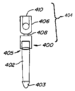
FIG. 12A illustrates an adapter cap device 400 for revising an implanted
arthroplasty device. The adapter
cap device 400 is designed to receive a portion of an implanted spinal
arthroplasty device, such as those illustrated
above. The device 400 is adapted to attach to an elongated shaft or stem 402
which has been positioned within a
vertebral body 12 of a spine. The stem 402 can be, e.g., the previously
implanted shaft of an existing arthroplasty
device (such as that shown in FIG. 7) and a pointed distal end 403. The stem
402 is configured such that it can have
a smooth exterior or an exterior surface treatment (as illustrated). The
exterior treatment would be provided to
facilitate the mating of the stem of the adapter cap device to the body with
which it was mating. The proximal end
403 of device 400 has an adapter cap 404 which has been configured such that
it can be attached to and removed
from the stem 402. The adapter cap 404 is configured to provide an aperture
sized with a diameter that enables a
snug fit around the diameter of the shaft 402. The adapter cap 404 can be
joined to the stem 402 using methods
known in the art, including, but not liniited to, swaging, crimping and/or
bonding. The adapter cap 404 is connected
to a polyaxial housing 406 by a neck 408. The neck 408 connection enables the
polyaxial housing 406 to assume
differing orientations relative to the orientation of the stem 402, thus
accommodating a variety of arthroplasty
devices. The proximal end 410 of the polyaxial housing can be configured to
receive a variety of connectors and
devices depending upon the design of the arthroplasty device to be
accommodated. Alternatively, the existing
proximal end of an implanted device may be cut and removed, and the adapter
cap device 400 may be positioned
over the remaining neck of the device.
Turning now to FIG. 12B, a revision polyaxial device 400 similar to the device
of FIG. 12A is depicted. In
this embodiment, the shaft 402 mates with the adapter cap 414 to provide an
arthroplasty device receiving housing
that is offset to a central axis x of the stem 402. The adapter cap 414 is
similarly configured to the adapter cap 404 of
FIG. 12A in that the cap portion enables the housing 406 to be positioned off
the central axis x of the stem. The
offset adapter cap provides additional flexibility to the design enabling the
adapter cap to mate with a variety of
anatomical surfaces, including resected surfaces or damaged surfaces, along
with a wide variety of arthroplasty
devices. The cap attaches to stem 165.
FIG. 12C illustrates a device 400 implanted into a vertebral body 12 such that
the cap of the device can
function as a pedicle replacement. In this illustration, there is no
preexisting stem or other device, and device 400
therefore includes an integral stem 165. The device 400 can be implanted such
that the device (desirably and
generally) does not intersect the central sagittal axis 411 of the vertebral
body. The housing and cap can be
configured to provide a fixed structure, or can be configured to enable the
housing and cap to be engaged such that
rotation between the two elements is enabled.
FIG. 13A illustrates another embodiment of a device 420 of FIG. 12 suitable
for a stand-alone application,
i.e., without attaching to a portion of an existing implant. In this
embodiment, the stem 422 is configured such that it
incorporates a polyaxial element or housing 424. The housing 424 further
engages a ball 427 attached to a neck 428
that provides further flexibility between the polyaxial element and an
arthroplasty device to which it is mated. When
implanted, as shown in FIG. 13B, the device 420 can be implanted into a
vertebral body 12 such that the device abuts
or intersects the central sagittal axis 410 of the vertebral body. These
devices can be preformed and used for
implantation during the installation of an initial arthroplasty device or can
be used in conjunction with revision
-13-
CA 02576958 2007-02-12
WO 2006/023683 PCT/US2005/029476
surgery to provide additional flexibility and adaptability to the device
performance. In addition, these devices can be
used to attach to commercially available spinal fusion instrumentation,
including generally-available spine rods and
screws.
FIG.14A illustrates an adapter cap implanted in combination with an
arthroplasty device. The arthroplasty
device comprises a body that engages a surface of the vertebra and a stem 165.
The adapter cap of FIG. 13 has been
modified to enable the adapter to communicate with the arthroplasty device.
FIG. 14B illustrates an adapter cap
implanted in combination with another arthroplasty device. In this embodiment,
the adapter cap has again been
modified to enable the cap to mate with the implantable device that restores
the inferior facet. Turning now to
FIG. 14C yet another adapter cap assembly 400 is depicted implanted in
combination with the facet replacement
device of FIG. 8. In this instance, the adapter cap has been modified to
enable the device to receive the threaded bolt
that secures the device.
FiG.15A illustrates a perspective view of component parts of a replaceable
modular stem system for use in
an implantable spinal arthroplasty device. The system 450 includes an
internally and externally threaded auxiliary
sleeve she11452. A threaded female tip 454 fits within an aperture of the
sleeve shel1452. The threaded female tip
454 has a threaded first end 455 and a configured aperture 456 on the opposing
end. A male stem 458 is provided for
communicating with the female threaded tip 454. The male stem 458 has a
configured protrusion 460 (male member)
on one end that mates with the configured aperture 456 (female member) on the
threaded female tip 454. The
configured protrusion 460 is configured to key within the female threaded tip
454 to create a snug fit between the
configured aperture 456 and the configured protrusion 460. The system 450 can
be further held together by use of a
threaded dowel pin 460, that fits within a receiving aperture 462 of the stem
458. Once the system is configured and
in place, the snug fit and keyed configuration of the threaded tip 454 and
configured aperture 456 for receiving the
tip prevents movement of the stem 458 with respect to the sleeve shell 452.
The threaded dowel pin 460, further
prevents movement of the stem 458 with respect to the sleeve shell 452 and the
system overall. If desired, the stems
458 can comprise a set of differing size, length and/or shape stems to
accommodate anatomical and/or surgical
variability, as previously described.
Turning now to FIG. 15B, a side view of replaceable modular stem system 450 is
illustrated. As can be
appreciated by this view, the sleeve shell 452 can be threaded into target
bone 451 using the external threads 455.
Anchoring of the sleeve shell 452 to the bone 451 can be achieved by use of
the threads alone (e.g., for healthy bone)
or the threads in combination with bone cement 464. Additionally, where the
target bone is weak, the site can be
drilled-out, bone cement can be applied, and the stem can then be threaded
into the bone cement. The revision
system depicted in FIGS. 15A-B can be used to revise or replace, for example,
caudal and/or cephalad arms of a
spinal arthroplasty device, such as the devices depicted in FIGS. 6-7. Due to
the modularity of the design, substitutes
for components can easily be employed. As described with respect to FIG. 9,
the sleeve shel1452 can be used to
replace a missing or damaged pedicle, and can be used to assist in
reconstruction.
FIG 16A illustrates a side view of a replaceable modular stem system 470 of an
alternate embodiment to the
system illustrated in FIG. 15. The modular stem system 470 has a sleeve
feature 472 that enables the male stem 474
to fit within the female stem 472. Additionally, an external tie-down or
sleeve 476 can be provided that provides an
additional anchoring feature between the male and female stem. An alternate
anchoring mechanism, in the form of a
set screw 478, is provided to further anchor the two pieces together. The set
screw can have a configured aperture on
its upper surface shaped to mate with a corresponding driver tool, e.g. cross-
headed screwdriver, flat headed screw
driver, and the like. FIG. 16B illustrates the modular system depicted in FIG.
16A, further illustrating the configured
-14-
CA 02576958 2007-02-12
WO 2006/023683 PCT/US2005/029476
aperture 486 on the female stem 472 and the configured male protrusion 480 on
the male stem that provides an anti-
rotation and/or anti-displacement feature(s) between the two pieces when
mated. FIG. 16C illustrates the external
tie-down 476 of FIG. 16A.
FIG. 17 illustrates yet another alternate embodiment of a modular stem system
500 having a modular stem
502 with a female opening 504 for receiving a male connector 506 of a
connecting arm 508. The niating of the
modular stem 502 and the connecting arm 508 is further enhanced by virtue of a
friction and/or compression fit
between the two components. Friction fit can be achieved by use of, for
example, a bushing 510. A suitable bushing
would include a swaging bushing. Systems of this design can be used on a post-
operative revision of a total facet
arthroplasty device. Where the total facet arthroplasty device is revised, the
surgeon may cut the cephalad stem of
the total facet arthroplasty device and remove the caudal bearing. A suitable
cut would be made perpendicular to the
axis of the exposed shaft. Following posterior lumbar interbody fusion (PLIF)
or translaniinar PLIF, a modular rod
extension can be swaged into a dovetail feature, as shown in FIG. 18. FIG. 18
further illustrates the modular stem
system of FIG. 17 connected to a housing 512. The housing can be used to link
elements, such as the cephalad
element to the caudal element, or to connect cephalad arms to a cross-member.
If desired, the caudal cup can be
removed from the caudal stem, to allow access to the intervertebral space for
a PLIF, with the caudal cup or some
other construct replaced back onto the caudal stem once the interbody
procedure has been completed.
As shown in FIG. 19 an implanted arthroplasty device can have a cross-linking
arm 518 installed to further
reinforce the assembly. The cross-linking arm can also further prevent
movement of the arms relative to the cross-
bar member. The cross-linking arm 518 can be implanted to further reinforce
the device or provide additional
control of movement or articulation between the arms, e.g. cephalad arms, and
the cross-member, or can be used to
secure a single loose cephalad arm in a desired position and/or orientation.
The cross-linking arm can be formed of a
single device that connects at either end, using any suitable connection
mechanism or structure. Alternatively, the
cross-linking arm can be comprised of components that mate, to functionally
achieve an arm and provide additional
flexibility with respect to length.
FIG. 20A illustrates a securing device for use in connection with an
arthroplasty device to revise and/or
modify, control, or limit motion of the arthroplasty device. The securing
device has a body 520 with a distal surface
521 having pair of prongs 522, 522'. When installed, the prongs 522, 522' form
a base and are positioned below the
crossbar member and the indenture 524 of the securing device engages the
anchors on three sides. When used with a
device of FIG. 7, the prongs can be positioned below the caudal cup which
receives an end of the crossbar member,
while the top sits above the crossbar end (110, 115) to secure the end in
place within the caudal cup 150.
The prongs 522, 522' engage a wa11526 of the securing device on one side. The
wa11526 mates with a top
or roof 528 that fits above the cross-bar member. The top 528 has an aperture
529. The aperture 529 can function as
a detent, catch or plunger to snap fit over the ball end 110 of the crossbar
member in an arthroplasty device.
Alternatively, the securing device can be a securing mechanism, such as a set
screw 530. FIG. 20B is a top view of
the securing device 520. From this perspective, it is apparent that the top
528 can be positioned off a central axis of
the device to the two prongs 522, 522', thus also potentially positioning the
aperture 529 off the central axis as well.
FIG. 20C is a side view of the securing device, illustrating the angled
configurations of the sides 531, 531' back wall
526. The angled configuration positions the top 528, which can have a smaller
dimension in at least one direction
(e.g., length or width) than the length or width formed by the prongs and the
wall. FIG. 20D is a bottom view of the
securing device 520. FIG. 20E is a cross-sectional view of the securing device
taken through an axis parallel to the
prongs 522, 522'.
-15-
CA 02576958 2007-02-12
WO 2006/023683 PCT/US2005/029476
FIG. 21A illustrates a side view of the securing device of FIG. 21 in
combination with a portion of an
arthroplasty device, such as the arthroplasty device of FIG. 7. The prongs
522, 522' sit below the caudal cup 150,
holding the caudal cup in a fixed position. The top 528 of the securing device
520 sits above an end of the cross-
member 110, which fits within the caudal cup 150. An anchoring device 530 (see
FIG. 20A) can be fed through the
aperture to engage the end of the cross-member and hold it in position within
the caudal cup 150. As illustrated, the
caudal cup 150 is tilted t toward an axial plane 52, enabling the caudal cup
to secure the cross-member at a location.
Adjustment of the position of the caudal cup relative to the cross-member end
can affect the position of the device.
FiG. 21B illustrates a perspective view of the securing device in combination
with a portion of the arthroplasty
device. From this perspective, a set screw 530 located within the aperture 529
on the top of the securing device can
be seen. FIG. 21C is a perspective view from a partially anterior view of the
securing device again in combination
with a portion of the arthroplasty device. FIG. 21D is a top view of the
securing device 520 with a portion of the
arthroplasty device. As evident from this perspective, the caudal cup extends
on one side past the prong 522'. The set
screw 530 is positioned off-center relative to the length oflthe securing
device, but the top of the securing device is
positioned over the end of the cross-member. FIG. 21E is a bottom view of the
securing device engaging an
arthroplasty device. From this view, it is illustrated that the prongs 522,
522' are seated beneath, for example, the
caudal cup of the arthroplasty device.
Thus, the implanted arthroplasty device can be revised to incorporate locks or
"fusion caps" that desirably
convert the device from an articulating joint replacement construct to a non-
articulating (or controlled and/or limited
articulation) spinal fusion construct. In this embodiment, the fusion cap can
be installed on or into the caudal cups to
desirably immobilize the cephalad bearings within the cups. In various
embodiments, the fusion caps could
imrnobilize the cephalad bearings by direct compression or contact, through
use of a set screw or other device to
secure the cephalad bearing relative to the cup, or the fusion cap could
contain or cover an encapsulating material,
such as bone cement, which could fill the caudal cup and immobilize the
cephalad bearing. Various techniques
could be used in conjunction with the installation of such fusion caps, and
the cap could be installed prior to, during,
or after the completion of a concurrent spinal fusion procedure, including the
removal of intervertebral disc material,
installation of fusion cages, and/or introduction of material (such as bone
graft material) that desirably promotes
spinal fusion. Alternative embodiments could incorporate bearings of different
shapes or sizes (not shown),
including square or non-spherical bearings and/or bearings shaped to that fit
snugly into and accommodate most or
all of the interior of the caudal cup (not shown), that can be secured within
the cup in a similar manner.
Turning now to FIG. 22A, a perspective view of an implanted arthroplasty
device 600 with the securing
device of FIG. 21 is illustrated. The arthroplasty device 600 features a pair
of caudal cups 150 engaging a cross-
member 110. The cephalad arms have been removed, but it has been determined
desirable to keep the caudal cups
and cross-bar in place. The use of the securing device enables the caudal cup
and crossbar member to be retained in
position even without one or more of the cephalad arms to anchor the cross-
member. Additionally, as will be
appreciated by those of skill in the art, one of the two cephalad arms could
be removed with the use of one or two of
the securing devices to provide a three-point secured device (i.e., rigidly
connecting two caudal cups to a single
cephalad arm). The securing device engages the caudal cup and an end of the
cross-member in the manner described
above. FIG. 22B is a perspective view of another implanted arthroplasty device
602 having a pair of caudal cups 150,
150' engaging a cross-member 110 and a pair of cephalad arms 120, 120'
extending vertically toward the adjacent
vertebra 12 along with the securing device of FIG. 21. FIG. 22C is a
perspective view of yet another implanted
arthroplasty device 604 with the securing device of FIG. 21.
-16-
CA 02576958 2007-02-12
WO 2006/023683 PCT/US2005/029476
Additional modifications of the caudal cup of an arthroplasty device are also
possible in order to improve
the operation and reliability of the arthroplasty device through the range of
spinal motion. Further, these
modifications can change the operation of the device from one enabling a full
range of motion, to a device that
enables less than a full range of motion, or to a device that restricts range
of motion (this "restriction" could extend
from allowing full motion to allowing partial or controlled motion to allowing
no motion - thus functionally
achieving some of all of the effects of a fusion device). One such
modification is illustrated in FIG. 23. Caudal cup
150' is a modified version of the caudal cup 150 shown in FIG. 7. The caudal
cup 150' includes an upper crossbar
end retainer 702 and a lower crossbar end retainer 704. The upper and lower
crossbar end retainers 702, 704 may
optionally be provided to reduce the likelihood that the crossbar ends 110,
115 will slide out of contact with or leave
an acceptable area adjacent the caudal cup surface 155 (dislocate). In a
similar manner, the posterior surface of the
caudal cup could also be closed (not shown), thereby capturing and holding the
crossbar ends 110, 115 and limiting
and/or preventing posterior movement of the crossbar relative to the caudal
cups. In this alternate embodiment, the
caudal cups could also comprise a "clamshell" design with the lower portion
(as shown in FIG. 23) and a mating
shape (not shown) that clamps, bolts, clips, or bonds to the lower portion,
substantially closing the posterior side of
the cup.
FIG. 24A illustrates yet another alternate design of a caudal cup 150
incorporating a flange 712 which
creates a pocket 714 to contain and/or secure a cephalad bearing element of an
arthroplasty device.(such as the
device shown in FIG. 6) that can be positioned within the pocket when the
arthroplasty device is assembled. When
this design of caudal cup 150 is deployed in an arthroplasty device, it
secures the bearing element 115 (as shown in
FIG. 23) when the arthroplasty device is articulated to one or more extreme
limits of its range of motion. Thus, for
example, when the cephalad and caudal elements are compressed together (such
as during extension of the spine),
the cephalad bearing element (115 in FIG. 23) will slide along the caudal
bearing element in the cephalad direction,
coming to rest in the pocket 714 formed by the interior surface 715 of the
caudal cup 150. When the bearing (not
shown) is positioned within the pocket 714 any increased compressive force
acting on the device will desirably seat
the bearing even further into the pocket 714, reducing and/or eliminating any
opportunity for the bearing to slide out
of the cup and potentially dislocate the device. If desired, a similar flange
and pocket (not shown) can be formed on
the opposing (cephalad) side of the caudal cup 150, to capture the cephalad
bearing and prevent dislocation of the
bearing surface from the cephalad cut during flexion of the device. Thus, the
flange can provide a hard stop for the
bearing surface during flexion. If desired, this alternative caudal cup design
(incorporating the flange 712) could be
implanted in patients prone to dislocation (during the initial facet
replacement procedure) or it could be implanted in
a subsequent surgical procedure to replace a non-flanged caudal cup after the
patient has dislocated the facet joint
replacement incorporating a non-flanged cup design. Similarly, the various
other embodiments disclosed herein
could be used to replace and/or retrofit components already implanted within a
patient, or could be used
prophylactically during the initial implantation surgery to ensure against
failure of the implant.
In alternative embodiments, such as that shown in FiG. 24B, additional
crossbar motion can be
accommodated by altering the caudal cup width (wcõP) or adjusting the distance
between the medial edge 721 and the
lateral edge 723 in some embodiments. If desired, the upper edge 720 can be
configured to curve over the top to
enclose (either partially or fully) the upper portion of the cup 150. In other
embodiments, the radius of the curve that
transitions between the lateral edge 723 and the upper edge 720 and the radius
of the curve that transitions between
the lateral edge 723 and the lower edge 725 may also be adjusted to
accommodate the various shapes of the crossbar
end 115 and outer surface. In additional alternative embodiments, the medial
edge 721 and lateral edge 723 can be
-17-
CA 02576958 2007-02-12
WO 2006/023683 PCT/US2005/029476
configured such that the edges are nonparallel, with respect to each other. In
other embodiments, the medial edge
721 and the lateral edge 723 could have an arcuate shape, or the cup 150 could
be completely enclosed with a
flexible and/or rigid cover or cap. In other embodiments, the medial edge 721
could have a raised lip or ridge (not
shown) which would desirably assist in retaining the cephalad bearing within
the caudal cup 150. Such
arrangements could help prevent dislocation of the construct and/or allow for
spontaneous and/or controlled
relocation of the bearing surface (operatively, minimally invasively or non-
invasively, including non-operative
manipulation of the patient's spine through chiropractic procedures, etc.).
In one alternate embodiment, once the cephalad and caudal components of the
device has been secured to
the targeted vertebral bodies, one or more elastic compression devices or
"bands" 740 could be secured about the
caudal cups and bearing elements (see FIG. 24c), tothe vertebral bodies
themselves, between other parts of the
cephalad or caudal arms, or any combination thereof. This configuration could
be especially useful if a device
dislocates one or more times. Properly positioned and/or tensioned, these
"bands" would tend to keep the bearing
surfaces and caudal cups in contact and/or close proximity, even under extreme
and/or unusual loading conditions,
and thus reduce and/or eliminate the opportunity for the bearing elements to
dislocate. Moreover, in the event that
dislocation of the implant did occur, the bands could prevent and/or liniit
motion of the dislocated joint (by holding
the bearing surfaces and caudal cups together), and thus reduce or eliminate
damage to other tissues (such as the
spinal cord, various other nerves and/or circulatory/connective tissues)
resulting from the dislocation. In fact, the
compression of the bands might make it possible to eventually "reduce" the
dislocation and/or repair the dislocated
device through external manipulation and/or minimally-invasive surgery. If
desired, one or more "bands" could be
secured between the articulating surfaces of the device, or between the
various arms, cups, stems and/or cross-arms
of the construct elements, with varying results. In one embodiment, such a
band could be looped around the base of
the caudal cup, and around the corresponding cross-arm, in a figure-8 shape.
Properly positioned and tensioned, this
arrangement would allow the cup and cephalad bearing to articulate without
allowing the band to slip off (either or
both) the cup and cross-arm. Depending upon the length and size of the band,
and the tension therein, the band
could positioned and tighten to reduce and/or ultimately prevent any
significant articulation of one or both sides of
the facet joint replacement device. If desired, the band could be tightened
and/or loosened in a minimally-invasive
manner, during the implantation procedure and/or during a subsequent
procedure.
In another alternate embodiment, the compression device could comprise an
elastic or pliable material,
which may or may not be surrounded by a non-elastic housing, whereby the
elastic material allows various
movement of the bearing surfaces (with resistance commensurate to the
flexibility of the material, as well as
flexibility allowed by the coupling to the device components), but the
optional non-elastic housing acts as an
ultimate "stop" to movement of the bearing surfaces relative to the caudal
cup. Such embodiments could include
one or more "encapsulated" bearing surfaces, such as shown in FIGS. 24C-D,
which show two caudal cup and
cephalad bearing pairs (of a facet replacement device), each pair surrounded
by a flexible skin or "jacket" 740 which
permits relative movement between the cup and bearing, but which desirably
encapsulates or isolates the cup and
bearing pair from the surrounding environment (totally or partially or some
combination thereof). In practice, the
jacket 740 can serve many functions, including (but not limited to) (1) as a
shock absorber or brake to slow, control,
modify and/or limit movement of the bearing/cup complex throughout and/or at
the extreme ranges of motion, (2) as
a stop or limiter to reduce and/or prevent complete or partial dislocation of
the joint, (3) as a barrier to prevent
surrounding tissues from invading the bearing surfaces and/or being "pinched"
or damaged between moving
surfaces, and (4) as a barrier or "filter" to prevent "bearing wear
particulate," or other bearing by-products, from
-18-
CA 02576958 2007-02-12
WO 2006/023683 PCT/US2005/029476
reaching and impacting surrounding tissues (or to contain fluids or other
materials including, but not limited to, joint
lubricating fluids, antibiotics and/or fluids that provide a biological marker
and/or indicator - including bioreactive
materials - upon rupture or compromise of the barrier). In a similar manner,
the jacket 740 could encompass the
entire bearing construct, with only the cephalad and caudal stems (and
possibly the crossbar, depending upon
whether the jacket encompasses one or both bearing constructs) protruding
through the jacket and extending into the
vertebral bodies. Depending upon the type of polymer (or other material) used,
as well as the physical properties
and orientation of the polymer, the jacket 740 could be designed to control
the motion of the device in a desired
manner, and could also control the movement of the device to more accurately
replicate the natural motion of the
spinal segment. For instance, a polymer jacket could be designed to allow a
greater degree of freedom in
flexion/extension, but limit (to some extent) the degree of lateral bending or
torsion of the same segment, by proper
choice and orientation of the polymer or other material. In one alternative
embodiment, the band could comprise a
flexible, polymeric (including, but not limited to, biocompatible polymeric)
material.
In various alternative embodiments, the physical properties of the materials
used could be selected based on
an ability to alter over time or in response to one or more biological,
environmental, temperature and/or externally
induced factors, altering the properties of the material (i.e., polymers,
ceramics, metals - Nitinol - etc.). For
example, the material could comprise a material that hardens over time (or in
the presence of body fluids, proteins,
or body heat, etc.), which initially allows the components to freely
articulate at the time of implantation (and thus
minimize the stresses experienced by the anchoring components), but which
hardens and subsequently resists
movement to a greater degree once the component anchoring has solidified or
bonded to the surrounding bone.
Biodegradable materials can be used in embodiments to adapt an implanted
device to achieve a temporary fixation of
the device. By preventing movement for a period of time, healing can be
facilitated, among other things. Once a
suitable amount of time has passed and the biodegradable material (or other
types of materials) degrades (or
otherwise alters its material properties in some manner), the device will then
return to its initial state of
biomechanical movement. Where the material alteration is induced by externally
induced factors (such as directed
radiation, rf and/or sonic energy), the external factor could desirably alter
the physical properties of the material(s) in
a partially or completely reversible manner (depending upon the type,
duration, frequency and/or amplitude of the
induced factor), allowing for controlled alteration of the material properties
in a minimally-invasive and/or
completely non-invasive manner.
Similarly, the "band" could comprise an elastic, non-elastic or rigid
material, such as stainless steel cable,
which desirably prevents relative motion of the device components beyond a
certain pre-defined maximum
extension, flexion, rotation and/or torsion. In various embodiments, the band
could alternatively be installed to limit
motion of the device to prevent dislocation, or to minimize or control the
articulation of the device to some degree
(such as to protect a disc replacement device against unwanted motion in one
or more directions, protect an adjacent
fused level against unwanted stresses, or to protect various tissues from
experiencing stresses and/or damage). If
desired, the cable could be tightened or loosened post-surgery, in a minimally-
invasive manner, to alter performance
of the device.
Where the revision involves a spinal level incorporating an artificial disc
replacement, the revision
instrumentation could include wedges or "shims" that could be used to
inunobilize the artificial disc (thereby
augmenting the motion modification provided by the revision component) and/or
further increase the likelihood of
achieving a successful fusion in combination with the revision constructs
described herein.
-19-
CA 02576958 2007-02-12
WO 2006/023683 PCT/US2005/029476
FiG. 25 illustrates a method for altering the biomechanics of an implanted
spinal arthroplasty device, or
revising an implanted arthroplasty device. An incision may be created in a
selected location to access the implanted
arthroplasty device 800. However, as will be appreciated by those of skill in
the art, scenarios can arise wherein a
surgeon is implanting a spinal arthroplasty device and determines that the
condition of the patient warrants revising
the biomechanics of the device during the initial implantation. More commonly,
however, revision will be performed
at a time subsequent to implantation of the spinal arthroplasty device, thus
requiring a second, or subsequent,
surgical intervention.
Once the incision is made the implanted arthroplasty device is accessed 802.
Unless, the biomechanics have
been assessed prior to surgical access and a device has been preselected, the
surgeon can then select a revision
device 804, e.g. from a kit, that is adapted to alter the biomechanics or
revise the arthroplasty device. Whether
selected in advance of surgery, or during the surgical procedure, the surgeon
next inserts the revision device 806. As
discussed above, the revision device can be one that alters the biomechanics,
whether temporarily or permanently, or
otherwise revises the implanted arthroplasty device. The step of selecting a
revision device and implanting a revision
device can be repeated as often as required to achieve the desired result.
Once the surgeon is satisfied that the desired
result is achieved, the incision is closed 808.
While preferred embodiments of the present invention have been shown and
described herein, it will be
obvious to those skilled in the art that such embodiments are provided by way
of example only. Numerous
variations, changes, and substitutions will now occur to those skilled in the
art without departing from the invention.
It should be understood that various alternatives to the embodiments of the
invention described herein may be
employed in practicing the invention. It is intended that the following claims
define the scope of the invention and
that methods and structures within the scope of these claims and their
equivalents be covered thereby.
-20-