Note: Descriptions are shown in the official language in which they were submitted.
CA 02577097 2007-02-13
WO 2006/026105 PCT/US2005/028515
HYBRID LESION FORMATION APPARATUS, SYSTEMS
AND METHODS
BACKGROUND OF THE INVENTIONS
1. Field of Inventions
The present inventions relate generally to devices for performing
therapeutic operations on body tissue.
2. Description of the Related Art
There are many instances where electrophysiology devices are used to
form therapeutic lesions in tissue. Therapeutic lesions are frequently formed
to treat conditions in the heart, prostate, liver, brain, gall bladder,
uterus,
breasts, lungs and other solid organs. Electromagnetic radio frequency ("RF")
may, for example, be used to heat and eventually kill (i.e. "ablate") tissue
to
form a lesion. During the ablation of soft tissue (i.e. tissue other than
blood,
bone and connective tissue), tissue coagulation occurs and it is the
coagulation that kills the tissue. Thus, references to the ablation of soft
tissue
are necessarily references to soft tissue coagulation. "Tissue coagulation" is
the process of cross-linking proteins in tissue to cause the tissue to jell.
In soft
tissue, it is the fluid within the tissue cell membranes that jells to kill
the cells,
thereby killing the tissue.
The tissue coagulation energy is typically supplied by an
electrosurgical unit ("ESU") during the therapeutic procedure. More
specifically, after an electrophysiology catheter, surgical probe or clamp has
been connected to the ESU, and the electrodes or other energy transmission
elements on the catheter, surgical probe or clamp have been positioned
adjacent to the target tissue, energy from the ESU is transmitted through the
energy transmission elements to the tissue to from a lesion. The amount of
power required to coagulate tissue ranges from 5 to 150 W.
Some electrophysiology procedures require the use of more than one
electrophysiology device. One example of such a procedure involves the
formation of therapeutic lesions to the treat cardiac conditions such as
atrial
fibrillation. Here, a clamp may be used to create a first transmural
epicardial
lesion around the right pulmonary vein pair and a second transmural
epicardial lesion around the left pulmonary vein pair. Thereafter, if needed,
a
CA 02577097 2007-02-13
WO 2006/026105 PCT/US2005/028515
surgical probe may be used to create a linear transmural epicardial lesion
between the right and left pulmonary vein pairs. A linear transmural lesion
that
extends from the lesion between the right and left pulmonary vein pairs to the
left atrial appendage may also be created.
The present inventors have determined that conventional lesion
formation devices are susceptible to improvement. For example, the present
inventors have determined that the aforementioned procedure is inconvenient
because it requires the surgical staff to disconnect the clamp from the ESU
and connect the surgical probe to the ESU during the procedure. The
inconvenience is compounded in those instances where the ESU resets and
performs a diagnostic procedure each time a device is connected thereto. The
present inventors have also determined that there may be more efficient and
cost effective ways, in terms of materials, manufacturing, sterilization,
shipping, etc., to provide physicians with the capabilities of two separate
devices, such as the aforementioned separate clamp and surgical probe.
SUMMARY OF THE INVENTIONS
An apparatus in accordance with one invention herein includes a probe
component including at least one energy transmission device and an electrical
connector operably connected to the at least one energy transmission device
and a clamp component including at least one energy transmission device
operably connected to the probe component electrical connector.
A lesion formation apparatus in accordance with one invention herein
includes a tissue coagulation probe including an energy transmission device
carried, a bipolar tissue coagulation device including first and second energy
transmission devices, a first connector that facilitates connection of the
energy
transmission device on the tissue coagulation probe and the first energy
transmission device on the bipolar tissue coagulation device to a power output
port, and a second connector that connects the second energy transmission
device on the bipolar tissue coagulation device to a power return port.
A system in accordance with one invention herein includes a source of
tissue coagulation energy and a lesion formation apparatus including a probe
component and a clamp component.
2
CA 02577097 2007-02-13
WO 2006/026105 PCT/US2005/028515
A method in accordance with one invention herein includes the step of
simultaneously connecting a tissue coagulation probe and a clamp-based
tissue coagulation device to the same power output port on a source of tissue
coagulation energy.
BRIEF DESCRIPTION OF THE DRAWINGS
Detailed description of preferred embodiments of the inventions will be
made with reference to the accompanying drawings.
Figure 1 is a plan view of a hybrid lesion formation apparatus in
accordance with a preferred embodiment of a present invention. -
Figure 2 is a section view taken along line 2-2 in Figure 1.
Figure 3 is a section view taken along line 3-3 in Figure 1.
Figure 4 is an end view of the handle illustrated in Figure 1.
Figure 5 is a plan view of a clamp in accordance with a preferred
embodiment of a present invention.
Figure 6 is a section view taken along line 6-6 in Figure 5.
Figure 7 is a top view of a portion of the clamp illustrated in Figure 5.
Figure 8 is a plan view of a clamp component in accordance with a
preferred embodiment of a present invention.
Figure 9 is a side, partial section view of a portion of the clamp
component illustrated in Figure 8.
Figure 10 is a side, partial section view of a portion of the clamp
component illustrated in Figure 8.
Figure 11 is a section view taken along line 11-11 in Figure 9.
Figure 12 is a section view taken along line 12-12 in Figure 10.
Figure 13 is a perspective view of a surgical system in accordance with
a preferred embodiment of a present invention.
Figure 14 is a plan view of a hybrid lesion formation apparatus in
accordance with a preferred embodiment of a present invention.
3
CA 02577097 2007-02-13
WO 2006/026105 PCT/US2005/028515
DETAILED DESCRIPTION OF THE PREFERRED EMBODIMENTS
The following is a detailed description of the best presently known modes
of carrying out the inventions. This description is not to be taken in a
limiting
sense, but is made merely for the purpose of illustrating the general
principles of
the inventions.
The detailed description of the preferred embodiments is organized as
follows:
1. Introduction
II. Exemplary Hybrid Lesion Formation Apparatus
III. Exemplary Systems
The section titles and overall organization of the present detailed
description are
for the purpose of convenience only and are not intended to limit the present
inventions.
1. Introduction
This specification discloses a number of structures, mainly in the
context of cardiac treatment, because the structures are well suited for use
with myocardial tissue. Nevertheless, it should be appreciated that the
structures are applicable for use in therapies involving other types of soft
tissue. For example, various aspects of the present inventions have
applications in procedures concerning other regions of the body such as the
prostate, liver, brain, gall bladder, uterus, breasts, lungs, and other solid
organs.
II. Exemplary Hybrid Lesion Formation Apparatus
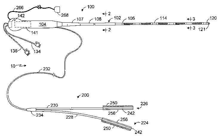
A hybrid lesion formation apparatus in accordance with one
embodiment of a present invention is generally represented by reference
numeral 10 in Figure 1. The exemplary embodiment includes a surgical probe
component 100 and a clamp component 200. The clamp component 200 in
the exemplary embodiment is adapted to be removably secured to a clamp so
as to convert a conventional clamp into a electrophysiology device that may
be used to form lesions in the manner discussed in greater detail below.
Alternatively, in other implementations, the clamp component may include the
clamp itself. The surgical probe component 100 and clamp component 200
preferably share a common electrical connector which may be used to
4
CA 02577097 2007-02-13
WO 2006/026105 PCT/US2005/028515
connect the hybrid lesion formation apparatus 10 to an electrosurgical unit
("ESU") in the manner described below with reference to Figure 13.
There are a variety of advantages associated with such a device. By
way of example, but not limitation, providing a surgical probe component 100
and a clamp component 200 in a single device facilitates the use of a single
handle (and associated electrical connectors). A conventional surgical system
including a surgical probe and a clamp would have two handles. In addition to
cost savings, the use of a single handle (and associated electrical
connectors)
allows the physician to avoid the inconveniences associated with
disconnecting one device from an ESU and connecting another during a
surgical procedure. The sterilization, packaging and shipment of the present
hybrid lesion formation apparatus may also be accomplished in a manner that
is more efficient than the sterilization, packaging and shipment of separate
devices.
Referring to Figures 1-4, the surgical probe component 100 in the
exemplary implementation includes a relatively short shaft 102, a handle 104
that is secured to the shaft, and one or more electrodes 106 or other energy
transmission devices on the distal portion of the shaft. A strain relief
element
107 may also be provided. The shaft 102 is preferably, although not
necessarily, about 13 cm to 51 cm in length, and most preferably about 20 cm
to 30 cm in length. The shaft 102 is also preferably relatively stiff. In
other
words, the shaft 102 is rigid, malleable, or somewhat flexible. A rigid shaft
cannot be bent. A malleable shaft is a shaft that can be readily bent by the
physician to a desired shape, without springing back when released, so that it
will remain in that shape during the surgical procedure. Thus, the stiffness
of a
malleable shaft must be low enough to allow the shaft to be bent, but high
enough to resist bending when the forces associated with a surgical
procedure are applied to the shaft. A somewhat flexible shaft will bend and
spring back when released. However, the force required to bend the shaft
must be substantial.
In the exemplary implementation illustrated in Figures 1-4, the shaft
102 consists of a proximal portion 108, including a malleable hypotube 110
and a non-conductive outer polymer coating 112, and distal portion 114,
5
CA 02577097 2007-02-13
WO 2006/026105 PCT/US2005/028515
including a malleable mandrel 116 and a multi-lumen electrically non-
conductive outer structure 118. The proximal portion 108 will typically be
about 15 to 40 cm in length, while the distal portion will typically be about
6 to
15 cm in length. The proximal end of the malleable mandrel 116 is secured to
the inner surface of the distal end of the hypotube 110 by, for example,
soldering, spot welding or adhesives. Mechanical methods, such as crimping
and mechanical fasteners, may also be employed. The distal end of the
malleable mandrel 116 is secured to a tip member 120. The exemplary tip
member 120 is provided with a suture aperture 121 (or a suture groove). If
desired, physicians may pass a suture through the aperture 121 (or around a
suture groove) and use the suture to pull the shaft 102 around a body
structure.
The handle 104 is configured to be gripped by the physician and used
to press the shaft distal portion 114 and electrodes 106 against tissue. To
that
end, the exemplary handle 104 is also about 7 to 18 cm in length and about 2
to 5 cm around its perimeter (measured perpendicularly to the longitudinal
axis), which is suitable for gripping by the physician.
The exemplary surgical probe component 100 is a fluid cooled surgical
probe and, as illustrated in Figure 3, the electrically non-conductive outer
structure 118 includes fluid inlet and outlet lumens 122 and 124, power and
signal wire lumens 126 and 128, a central lumen 130 for the mandrel 116. To
that end, the tip member 120 includes a connection lumen (not shown) that
connects the inlet lumen 122 to the outlet lumen 124, as well as a pair of
plugs (not shown) to seal the power and signal wire lumens 126 and 128.
Heat from the electrodes 106 is transferred through the outer structure 118 to
fluid that is flowing through the inlet and outlet lumens 122 and 124.
Accordingly, in addition to being electrically non-conductive, the material
used
to form the outer structure 118 should be relatively high in thermal
conductivity. As used herein, "relatively high" thermal conductivity is at
least
about 1 W/m=K and preferably ranges from about 1 to about 10 W/m=K.
Suitable electrically non-conductive, thermally conductive thermoplastics for
the outer structure 118 include flexible thermoplastic polymer materials, such
as nylon or polyurethane, which are filled with a filler that promotes heat
6
CA 02577097 2007-02-13
WO 2006/026105 PCT/US2005/028515
transfer. Suitable fillers include graphite, aluminum, tungsten and ceramic
powders. Another suitable filler is Carborundum CarboThermTM boron nitride
powder manufactured by Saint-Gobain in Cavaillon, France.
In addition to the aforementioned fillers, heat transfer may be promoted
by minimizing the thickness of the electrically non-conductive material
between the lumens 122 and 124 and the electrodes 106 and by maximizing
the cross-sectional area of the inlet and outlet lumens. With respect to the
outer structure 118 illustrated in Figure 3, for example, in an implementation
where the outer diameter of the outer structure is about 8 French (2.66 mm),
the thickness of the outer wall 132 between the electrodes 106 and the inlet
and outlet lumens 122 and 124 will be about 0.08 mm to about 0.36 mm. It
should be noted that when the outer wall thickness is about 0.02 mm or less,
materials with less than "relatively high" thermal conductivities, such as
Pebax material and polyurethane, may also be used for the outer structure
118.
Suitable materials for the malleable hypotube 110 include annealed
stainless steel, while the suitable material for the mandrel 116 includes
annealed stainless steel and beryllium copper.
As illustrated for example in Figures 1-4, fluid may be supplied to the
surgical probe component 100 by way of an infusion tube 134, which is
connected to the inlet lumen 122. The infusion tube 134 extends through an
aperture 135 in the handle 104 and is provided with stop-cock, which may be
connected to a fluid supply and control apparatus 300 in the manner
described below with reference to Figure 13. Similarly, a ventilation tube 136
is connected to the outlet lumen 124 and extends through an aperture 137 in
the handle 104. The ventilation tube 136 also includes a stopcock that may be
connected to the fluid supply and control apparatus.
The electrodes 106 in the exemplary probe component 100 illustrated
in Figures 1-4 are electrically coupled to individual power wires 138 that
pass
from the power wire lumen 126, and through a power wire tube 140, to an
electrical connector 141 that is associated with a slot 142 in the handle 104.
Suitable electrical connectors include PC boards, edge card connectors,
subminiature D connectors, ribbon cable connectors, and pin and socket
7
CA 02577097 2007-02-13
WO 2006/026105 PCT/US2005/028515
connectors. A plurality of temperature sensors 144, such as thermocouples or
thermistors, may be located on, under, abutting the longitudinal end edges of,
or in between, the electrodes 106. A reference thermocouple (not shown) may
also be provided. In the exemplary implementation, temperature sensors 144
are located at both longitudinal ends of each electrode 106. The temperature
sensors 144 are connected to the electrical connector 141 by signal wires
146, which pass through the signal wire lumen 128 and a signal wire tube
148. The temperature sensors 144 are also located within a linear channel
150 that is formed in the non-conductive outer structure 118. The linear
channel 150 insures that the temperature sensors will all face in the same
direction (e.g. facing tissue) and be arranged in linear fashion.
The number of electrodes carried on the shaft distal portion 114 will
typically depend upon the number of power connections available on the ESU
and common electrical connector 141 (e.g. a PC board) as well as the number
and purpose of the electrodes carried by the clamp component 200. In the
exemplary implementation, the clamp component 200 includes two electrodes
that are used to transmit energy and one that is used to return energy when
operating in a bipolar mode, as is discussed below with reference to Figures
8-12. In those instances where the ESU and common electrical connector 141
are configured for seven electrodes and two temperature sensors for each
transmitting electrode, the probe component 100 will include five spaced
electrodes 106.
The spaced electrodes 106 are preferably in the form of wound, spiral
closed coils. The coils are made of electrically conducting material, like
copper alloy, platinum, or stainless steel, or compositions such as drawn-
filled
tubing (e.g. a copper core with a platinum jacket). The electrically
conducting
material of the coils can be further coated with platinum-iridium or gold to
improve its conduction properties and biocompatibility. Preferred coil
electrodes are disclosed in U.S. Patent No. 5,797,905 and 6,245,068.
Alternatively, the electrodes 106 may be in the form of solid rings of
conductive material, like platinum, or can comprise a conductive material,
like
platinum-iridium or gold, coated upon the device using conventional coating
techniques or an ion beam assisted deposition (IBAD) process. For better
8
CA 02577097 2007-02-13
WO 2006/026105 PCT/US2005/028515
adherence, an undercoating of nickel, silver or titanium can be applied. The
electrodes can also be in the form of helical ribbons. The electrodes can also
be formed with a conductive ink compound that is pad printed onto a non-
conductive tubular body. A preferred conductive ink compound is a silver-
based flexible adhesive conductive ink (polyurethane binder), however other
metal-based adhesive conductive inks such as platinum-based, gold-based,
copper-based, etc., may also be used to form electrodes. Such inks are more
flexible than epoxy-based inks. Open coil electrodes may also be employed.
Still other types of electrodes are formed from electroless plated copper on a
polyimide film or tubular substrate. Gold, nickel or silver should be plated
over
the copper for electrochemical stability and improved biocompatibility. The
plating can be applied in continuous form (up to about 1-2 cm in length at
most) or can be applied in a pattern that is designed to improve current
density distributions and/or electrode flexing characteristics. Temperature
sensors (e.g. thermocouples) may be incorporated into the electrode structure
by placing the temperature sensors in a channel in the polyimide film or
tubular substrate and then plating over them.
The exemplary flexible electrodes 106 are preferably about 4 mm to
about 20 mm in length. In the preferred embodiments, the electrodes are 12.5
mm in length with 1 mm to 3 mm spacing, which will result in the creation of
continuous lesion patterns in tissue when coagulation energy is applied
simultaneously to adjacent electrodes. For rigid electrodes, the length of the
each electrode can vary from about 2 mm to about 10 mm. Using multiple
rigid electrodes longer than about 10 mm each adversely effects the overall
flexibility of the device, while electrodes having lengths of less than about
2
mm do not consistently form the desired continuous lesion patterns.
Additional details concerning fluid cooled surgical probes similar to that
described above are presented in U.S. Patent App. Pub. No. 2003/0078644,
which is entitled "Apparatus for Supporting Diagnostic and Therapeutic
Elements in Contact With Tissue Including Dual Lumen Cooling Device."
Although the exemplary surgical probe component 100 is an internally
cooled, fluid cooled surgical probe, the present inventions are not limited to
such probes. Other exemplary surgical probes include, for example, externally
9
CA 02577097 2007-02-13
WO 2006/026105 PCT/US2005/028515
cooled, fluid cooled surgical probes such as those illustrated in U.S. Patent
App. Pub. No. 2003/0014048, which is entitled "Fluid Cooled Apparatus for
Supporting Diagnostic and Therapeutic Elements in Contact with Tissue" and
non-cooled surgical probes such as those illustrated in U.S. Patent No.
6,645,200. The exemplary surgical probe component 100 may also be
replaced with a catheter component in those instances where percutaneous
access (e.g. access through the femoral vein to a chamber within the heart) is
desired. Suitable catheters are disclosed in U.S. Patent Nos. 6,013,052,
6,203,525, 6,214,002 and 6,241,754.
Turning to the clamp component, the exemplary clamp component 200
illustrated in Figure 1 is configured such that it may be removably secured to
a
clamp. As used herein, the term "clamp" includes, but is not limited to,
clamps,
clips, forceps, hemostats, and any other surgical device that includes a pair
of
opposable clamp members that hold tissue, at least one of which is movable
relative to the other. In some instances, the clamp members are connected to
a scissors-like arrangement including a pair of handle supporting arms that
are pivotably connected to one another. The clamp members are secured to
one end of the arms and the handles are secured to the other end. Certain
clamps that are particularly useful in minimally invasive procedures also
include a pair of handles and a pair of clamp members. Here, however, the
clamp members and handles are not mounted on the opposite ends of the
same arm. Instead, the handles are carried by one end of an elongate
housing and the clamp members are carried by the other. A suitable
mechanical linkage located within the housing causes the clamp members to
move relative to one another in response to movement of the handles. The
clamp members may be linear or have a predefined curvature that is
optimized for a particular surgical procedure or portion thereof. The clamp
members may also be rigid or malleable.
One example of a clamp to which the clamp component 200 may be
secured is generally represented by reference numeral 202 in Figures 5-7.
The clamp 202 includes a pair of rigid arms 204 and 206 that are pivotably
connected to one another by a pin 208. The proximal ends of the arms 204
and 206 are respectively connected to a pair of handle members 210 and
CA 02577097 2007-02-13
WO 2006/026105 PCT/US2005/028515
212, while the distal ends are respectively connected to a pair of clamp
members 214 and 216. The clamp members 214 and 216 may be rigid or
malleable and, if rigid, may be linear or have a pre-shaped curvature. A
locking device 218 locks the clamp in the closed orientation, and prevents the
clamp members 214 and 216 from coming any closer to one another than is
illustrated in Figure 5, thereby defining a predetermined spacing between the
clamp members. The clamp 202 is also configured for use with a pair of soft,
deformable inserts (not shown) that may be removably carried by the clamp
members 214 and 216 and allow the clamp to firmly grip a bodily structure
without damaging the structure. To that end, the clamp members 214 and 216
each include a slot 220 (Figures 6 and 7) that is provided with a sloped inlet
area 222 and the inserts include mating structures that are removably friction
fit within the slots. The exemplary clamp component 200 may be mounted on
the clamp members in place of the inserts.
With respect to clamp component itself, the clamp component 200 in
the exemplary hybrid lesion formation apparatus 10 illustrated in Figure 1
includes a first energy transmission device 224 that may be connected to one
of the clamp members 214 and 216 (Figures 5 and 13) and a second energy
transmission device 226 that may be connected to the other. The energy
transmission devices 224 and 226 are respectively carried on support
structures 228 and 230, which are connected to a cable 232 by a molded
junction 234. The cable 232 enters the handle 104 and, preferably, enters the
handle just proximally of the strain relief element 107.
Although clamp components in accordance with the present invention
may be operated in bipolar and unipolar modes, the exemplary clamp
component 200 is configured so as to be especially useful in a bipolar mode
wherein the first energy transmission device 224 will transmit energy through
tissue to the second energy transmission device 226. To that end, and as
illustrated for example in Figures 8-12, the first energy transmission device
224 includes a pair of electrodes 236 and 238 that may be independently
controlled, while the second energy transmission device 226 includes a single
electrode 240. Such an arrangement provides for higher fidelity control of the
11
CA 02577097 2007-02-13
WO 2006/026105 PCT/US2005/028515
overall region that is transmitting energy and a gap free, constant potential
region on the return side.
The first and second energy transmission devices 224 and 226 in the
illustrated embodiment illustrated in Figures 8-12 are also provided with
respective mounting devices 242 that may be used to mount the clamp
component 200 in general, and the energy transmission devices in particular,
on the clamp 202. Additionally, although the configuration of the clamp
component 200 may vary from application to application to suit particular
situations, the exemplary clamp component is configured such that the
electrodes 236 and 238 will be parallel to, and relatively close to one
another
(i.e. a spacing of about 1-10 mm), the electrode 240 when the clamp 202 is in
the closed orientation. Such an arrangement will allow the clamp component
200 to grip a bodily structure without cutting through the structure.
Referring more specifically to Figures 9 and 11, each mounting device
242 includes a base member 246 that has a groove 248 which is configured
to receive the support structure 228 and electrodes 236 and 238 (or support
structure 230 and electrode 240). About 20% of the electrode surface (i.e.
about 75 of the 360 circumference) is exposed in the illustrated
embodiment. Adhesive may be used to hold the support structures and
electrodes in place. The mounting device also includes a connector 250 that
is configured to removably mate with the clamp slot 220 (Figures 6 and 7).
The exemplary connector 250 is provided with a relatively thin portion 252 and
a relatively wide portion 254, which may consist of a plurality of spaced
members (as shown) or an elongate unitary structure, in order to correspond
to the shape of the slot 220.
The exemplary energy transmission devices 224 and 226 may also
include a wettable fluid retention element 256 that is saturated with ionic
fluid
(such as saline) prior to use. Suitable materials for the fluid retention
elements
256 include biocompatible fabrics commonly used for vascular patches (such
as woven Dacron ), open cell foam materials, hydrogels, nanoporous balloon
materials (with very slow fluid delivery to the surface), and hydrophilic
nanoporous materials. The effective electrical resistivity of the fluid
retention
element 256 when wetted with 0.9% saline (normal saline) should range from
12
CA 02577097 2007-02-13
WO 2006/026105 PCT/US2005/028515
about 1 0-cm to about 2000 SZ-cm. A preferred resistivity for epicardial and
endocardial procedures is about 1000 92-cm. Alternatively, one or both of the
fluid retention elements may be removed so that the electrodes contact the
tissue directly.
The electrodes 236 and 238 in the exemplary clamp component
illustrated in Figures 8-12 are connected to power wires 258, while the
electrode 240 is connected to a power wire 260. The power wires 258 and
260 extend through the support structures 228 and 230, respectively, as well
as the cable 232, and into the handle 104. The power wires 258 are
connected to the electrical connector 141 (Figures 1 and 4) that is associated
with the slot 142 in the handle 104. As such, the electrodes 236 and 238 and
associated power wires 258 from the clamp component 200 are connected to
the same electrical connector as the power wires 138 from the probe
component 100. Conversely, the power wire 260 extends through a cable 266
(Figure 1), which enters the proximal end of the handle 104 through an
aperture 267, to a connector 268 so that the electrode 240 may be connected
to one of the power return ports 340 on the ESU 322 (Figure 13).
A plurality of temperature sensors 262 (Figure 11), such as
thermocouples or thermistors, may be located on, under, abutting the
longitudinal end edges of, or in between, the electrodes 236 and 238. A
reference thermocouple (not shown) may also be provided. In the exemplary
implementation, temperature sensors 262 are located at both longitudinal
ends of each of the electrodes 236 and 238. The temperature sensors 262
are connected to the electrical connector 141 by signal wires 264, which pass
through the support structure 228 and cable 232. In other words, the signal
wires 264 from the clamp component 200 are connected to the same
electrical connector 141 (a PC board in the exemplary embodiment) as the
signal wires 146 from the probe component 100. The temperature sensors
262 are also located within a linear channel 263 that is formed in the support
structure 228. The linear channel insures that the temperature sensors will
all
face in the same direction (e.g. facing tissue) and be arranged in linear
fashion.
13
CA 02577097 2007-02-13
WO 2006/026105 PCT/US2005/028515
With respect to dimensions and materials, the support structures 228
and 230 are flexible tubular structures which have an outer diameter that is,
depending on the diameter of the electrodes 236, 238 and 240, typically
between about 1.5 mm and about 3 mm. The support structures 228 and 230
in the illustrated embodiment, which are intended for use in cardiovascular
applications, have an outer diameter of about 2 mm. Suitable support
structure materials include, for example, flexible biocompatible thermoplastic
tubing such as unbraided Pebax material, polyethylene, or polyurethane
tubing.
The mounting devices 242 are preferably formed from polyurethane.
The length of the mounting devices 242 will vary according to the intended
application. In the area of cardiovascular treatments, it is anticipated that
suitable lengths will range from, but are not limited to, about 4 cm to about
10
cm. In the exemplary implementation, the base members 242 are about 6 cm
in length.
A variety of other suitable clamp based energy transmission devices
that may be incorporated into hybrid lesion formation apparatus in accordance
with the present inventions are illustrated in U.S. Patent App. Pub. No.
2003/0158547, which is entitled "Apparatus for Converting a Clamp Into an
Electrophysiology Device."
III. Exemplary Systems
A tissue coagulation system 1000 in accordance with one embodiment
of a present invention is illustrated in Figure 13. The exemplary system 1000
includes the hybrid lesion formation apparatus 10, a fluid supply and control
apparatus 300 and a power supply and control apparatus 320. In addition, the
clamp component 200 is mounted on the clamp 202 to form a clamp-based
tissue coagulation device.
The fluid supply and control apparatus 300, which may be used to
supply cooling fluid to the surgical probe component 100, includes housing
302, a fluid outlet port 304, and a fluid inlet port 306. The fluid outlet
port 304
may be coupled to the stopcock or other connector associated with the
infusion tube 134 (and, therefore, to the inlet lumen 122) by a connector tube
308, while the fluid inlet port 306 may be coupled to the stopcock or other
14
CA 02577097 2007-02-13
WO 2006/026105 PCT/US2005/028515
connector associated with the ventilation tube 136 (and, therefore, to the
outlet lumen 124) by a connector tube 310. An infusion pump capable of
variable flow rates is one example of a suitable fluid supply and control
apparatus.
The cooling fluid is not limited to any particular fluid. Preferably,
however, the fluid will be a low or electrically non-conductive fluid such as
sterile water or 0.9% saline solution in those instances where the fluid will
not
be used to transmit current to tissue. A suitable fluid inlet temperature is
about
0 to 25 C and the fluid supply and control apparatus 300 may be provided
with a suitable cooling system, if desired, to bring the temperature of the
fluid
down to the desired level. In a five electrode embodiment where 150 W is
being supplied to the electrodes 106, for example, a suitable constant fluid
flow rate is about 5 mI/min to about 20 ml/min.
The power supply and control apparatus 320 includes an
electrosurgical unit ("ESU") 322 that supplies and controls RF power. A
suitable ESU is the Model 4810A ESU sold by Boston Scientific Corporation
of Natick, Massachusetts, which is capable of supplying and controlling power
on an electrode-by-electrode basis. This is sometimes referred to as "multi-
channel control." Typically, power will be controlled as a function of the
temperature at each electrode in order to insure that tissue is coagulated
without over-heating and causing coagulum and charring. With respect to
temperature sensing, temperature at the electrodes 106 on the surgical probe
component 100, as well as the electrodes 236 and 238 on the clamp
component 200, is measured by the aforementioned temperatures sensors
144 and 262. Alternatively, in those instances where temperature sensors are
not employed, the respective temperatures at each electrode 106, 236 and
238 may be determined by measuring impedance at each electrode.
The power and signal wires 138, 146, 258 and 264 should be
connected to the electrical connector 141 in such a manner that the physician
will know in advance which of the ESU control channels correspond to the five
electrodes 106 on the probe component 100 and which of the ESU control
channels correspond the electrodes 236 and 238 on the clamp component
200. In one exemplary configuration, control channels 1 and 2 may be used
CA 02577097 2007-02-13
WO 2006/026105 PCT/US2005/028515
for the clamp component electrodes 236 and 238 and control channels 3-7
may be used for the five probe component electrodes 106.
The ESU 322 transmits energy to the electrodes 106, 236 and 238 by
way of a cable 324. The cable 324 includes a connector 326, which may be
connected to the electrical connector 141 in the probe handle 104, and a
connector 328, which may be connected to a power output port 330 on the
ESU 322.
Tissue coagulation energy emitted by the electrodes 106 is returned to
the ESU 322 through an indifferent electrode 334 that is externally attached
to
the skin of the patient with a patch, or one or more electrodes (not shown)
that
are positioned in the blood pool, and a cable 336. The cable 336 includes a
connector 338 that may be connected to one of the power return ports 340 on
the ESU 322. Similarly, tissue coagulation energy emitted by the electrode
236 and 238 on the energy transmission device 224 is returned to the ESU
322 by way of the electrode 240 on the energy transmission device 226, the
power wires 260 and the cable 266. The cable 326 is connected to the other
ESU power return port 340 by the connector 268. Preferably, the ESU power
output port 330 and corresponding connector 328 have different
configurations than the power return port 340 and corresponding connectors
268 and 338 in order to prevent improper connections.
The exemplary tissue coagulation system 1000 illustrated in Figure 13
may be used to form a variety of lesions in a variety of anatomical
structures.
By way of example, but not limitation, the tissue coagulation system 1000 may
be used in the following manner to form lesions in myocardial tissue to cure
atrial fibrillation.
After the clamp component 200 has been secured to the clamp 202
and the hybrid lesion formation apparatus 10 has been connected to the ESU
322 by the connectors 328 and 368, the clamp 202 may be used to position
the clamp component energy transmission devices 224 and 226 on left atrial
tissue adjacent to opposite sides of the right pulmonary vein pair. The clamp
members 214 and 216 may then be brought into a completely closed
orientation or, depending on the tissue structure, a slightly open orientation
so
long as the pulmonary veins are firmly held. The ESU 322 is used to supply
16
CA 02577097 2007-02-13
WO 2006/026105 PCT/US2005/028515
coagulation energy to the electrodes 236 and 238, and energy is returned to
the ESU by way of the electrode 240. Energy will be continued to be supplied
in a controlled manner based on the temperatures monitored by the
temperature sensors 262 until a transmural epicardial lesion around the right
pulmonary vein pair is formed. This process is then repeated on the left
pulmonary vein pair. It should be noted, however, that individual lesions may
be formed around each of the pulmonary veins instead of around the
pulmonary vein pairs. The clamp component 200 and clamp 202 may then be
placed on the sterile drape covering the patient, where it can remain until
the
ablation procedure is completed.
The surgical probe component 100 of the hybrid lesion formation
apparatus 10 may then be used, if necessary, to touch up the lesions formed
by the clamp component 200. As noted above, this may be accomplished
without disconnecting the clamp component 200 from the ESU 322 and then
connecting surgical probe component 100 to the ESU because both
components share the electrical connector 141 in the handle 104. Tissue
coagulation energy from the ESU 322 will be supplied to one, some or all of
the electrodes 106 and returned to the ESU by way of the indifferent electrode
334. The surgical probe component 100 may also be used to create a linear
transmural epicardial lesion between the right and left pulmonary vein pairs
and/or a linear transmural lesion that extends from the lesion between the
right and left pulmonary vein pairs to the left atrial appendage.
Although the inventions disclosed herein have been described in terms
of the preferred embodiments above, numerous modifications and/or
additions to the above-described preferred embodiments would be readily
apparent to one skilled in the art.
By way of example, but not limitation, the electrical connector 141 may
be located at the end of a cable that extends outwardly from the handle,
instead of within the handle, so that the cable 324 may be eliminated.
Turning to Figure 14, the clamp component 200a in the exemplary
hybrid lesion formation apparatus 10a, which is otherwise identical to the
hybrid lesion formation apparatus 10, is provided with tissue stimulation (or
"pacing") electrodes 239 and 241 on the energy transmission devices 224a
17
CA 02577097 2007-02-13
WO 2006/026105 PCT/US2005/028515
and 226a. The tissue stimulation electrodes 239 and 241 are carried on the
ends of the support structures 228 and 230. The tissue stimulation electrodes
239 and 241 are also connected to signal lines 243 and 245, which extend
through the support structures 228 and 230 and cable 232, as well as through
the proximal end of the handle 104, to connectors 247 and 249. This allows
the tissue stimulation electrodes 239 and 241 to be connected to a
conventional pacing apparatus, such as the Medtronic Model Nos. 5330 and
5388 external pulse generators, or to an ECG machine that is capable of
monitoring and recording electrical impulses.
The tissue stimulation electrodes 239 and 241 may then be used to
supply a bipolar pacing pulse (e.g. about 20 mA) on the side opposite the left
atrium of a lesion formed with the hybrid lesion formation apparatus 10a. The
physician can determine whether or not a therapeutic lesion (or "complete
block") has been formed by observing the left atrium. If the pacing pulse is
able to cross the lesion, the heart will beat faster (e.g. 120 beats/minute).
This
may be determined by observation or by use of an ECG machine that is
monitoring the heart. Here, additional coagulation will be required to
complete
the lesion. The failure to stimulate the heart from the side of the lesion
opposite the left atrium is, on the other hand, indicative of the formation of
a
therapeutic lesion. Nevertheless, because muscle bundles are not always
connected near the pulmonary veins, it is preferable that the stimulation
energy be applied to a number of tissue areas on the side of the lesion
opposite the left atrium to reduce the possibility of false negatives.
Alternatively, the tissue stimulation electrodes 239 and 241 may be used to
monitor tissue within the region that was intended to be isolated. In the
context of pulmonary vein isolation, for example, the tissue stimulation
electrodes 239 and 241 may be placed in contact with viable tissue on the
pulmonary vein side of the lesion.
Additional information concerning tissue stimulation electrodes, as well
as the manner in which they may be employed in conjunction with a clamp
based device, is provided in U.S. Patent App. Pub. No. 2005/0119653 Al,
which is entitled "Surgical Methods And Apparatus For Forming Lesions In
Tissue And Confirming Whether A Therapeutic Lesion Has Been Formed."
18
CA 02577097 2007-02-13
WO 2006/026105 PCT/US2005/028515
It is intended that the scope of the present inventions extend to all such
modifications and/or additions and that the scope of the present inventions is
limited solely by the claims set forth below.
19