Note: Descriptions are shown in the official language in which they were submitted.
CA 02577388 2007-02-16
WO 2006/023867 PCT/US2005/029826
NOVEL ELECTRODE ASSEMBLY FOR MEDICAL ELECTRICAL LEADS
The present invention pertains to medical electrical systems and more
particularly to electrode assemblies.
Cardiac stimulation systems commonly include a pulse-generating device, such
as a pacemaker or implantable cardioverter/defibrillator that is electrically
connected to
the heart by at least one medical electrical electrode. A medical electrical
electrode
delivers electrical pulses emitted by the device to the heart and may also
sense cardiac
signals so the device may monitor the electrical activity of the heart. These
electrical
pulses are typically conducted between the device and electrodes via elongate
conductors extending within one or more leads.
In recent years, with the development of cardiac resynchronization therapy,
pacing of the left ventricle has been achieved by implanting transvenous lead
electrodes
in vessels of the coronary venous system of the heart in order to stimulate an
epicardial
surface of the left ventricle. Thus there is a need for electrode assemblies
that are suited
for delivery to, and function within in a vessel environment.
The following drawings are illustrative of particular embodiments of the
invention and therefore do not limit its scope, but are presented to assist in
providing a
proper understanding of the invention. The drawings are not to scale (unless
so stated)
and are intended for use in conjunction with the explanations in the following
detailed
description. The present invention will hereinafter be described in
conjunction with the
appended drawings, wherein like numerals denote like elements, and:
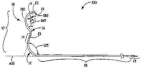
Figure IA is a plan view of a medical electrical lead according to one
embodiment of the present invention;
Figure 1B is a schematic of the lead of Figure IA implanted in a coronary
venous system from an anterior perspective;
Figure 1 C is an enlarged view of a distal portion of the lead shown in Figure
lA
implanted within a coronary vein;
CA 02577388 2007-02-16
WO 2006/023867 PCT/US2005/029826
-2-
Figure 2 is an enlarged detailed plan view of a lead electrode assembly
according to one embodiment of the present invention; and
Figure 3 is an enlarged detailed section view of another lead electrode
assembly
according to another embodiment of the present invention.
The following detailed description is exemplary in nature and is not intended
to
limit the scope, applicability, or configuration of the invention in any way.
Rather, the
following description provides a practical illustration for implementing
exemplary
embodiments of the invention.
Figure lA is a plan view of a medical electrical lead 100 according to one
embodiment of the present invention. Figure IA illustrates lead 100 including
an
approximately straight proximal lead body portion 15, which is terminated at a
proximal end by a lead connector 13, and a pre-formed distal lead body portion
17
extending distally from proximal portion 15. Figure 1A further illustrates
distal lead
body portion 17 including a first arcuate segment 12 bending in a first
direction, an
approximately straight segment 14 extending from first arcuate segment 12, a
second
arcuate segment 16 extending from straight segment 14 and bending in a second,
generally distal, direction, a third arcuate segment 18 bending in a third,
generally
proximal, direction, and a distal tip segment 19 extending from the third
arcuate
segment 18. According to the illustrated embodiment of the present invention,
lead
100 further includes a first electrode E1 coupled to approximately straight
segment 14
and second electrode coupled to distal tip segment 19; the position of pre-
formed
curves of arcuate segments of distal portion 17 with respect to electrodes El
and E2
provide for epicardial contact of electrodes E1 and E2 when implanted in a
coronary
vessel, as will be further described below.
Figure IA further illustrates angles 125, 165 and 185 of arcs included in
arcuate
segments 12, 16 and 18, respectively; according to some embodiments of the
present
invention, dimensions of the arcs are as indicated in Table 1.
CA 02577388 2007-02-16
WO 2006/023867 PCT/US2005/029826
-3-
Table 1: Arc Dimensions
Arcuate Segment Arc radius (inch) range Arc angle range
12 -0.2 - -0.3 Angle 125: -45 - -90
16 -0.2 - -0.4 Angle 165: -10 - -40
18 -0.1 - -0.4 Angle 185: -60 - -100
Furthermore, a length of straight segment 14, according to some embodiments,
is from approximately 0.2 to approximately 0.7 inch and a length of distal tip
segment
19 is from approximately 0.05 inch to approximately 0.2 inch. According to one
embodiment electrode E2 terminates distal tip segment 19, which may or may not
extend proximally from electrode; according to another embodiment a portion of
distal
tip segment 19 extends distally from electrode E2 as illustrated by dashed
lines in
Figure 1 and this extension may or may not be curved. Distal lead body portion
17 is
alternately described as being canted, bending at angle 125 with respect to a
longitudinal axis A15 of proximal portion 15 and including a hump-like
segment,
corresponding to segment 18, extending from approximately straight segment 14
and
having a distal apex 180. According to one embodiment of the present
invention, the
arc of segment 18 has a chord length of approximately 0.4 inch to
approximately 0.7
inch and distal apex 180 of segment 18 has a height H of approximately 0.1
inch to
approximately 0.3 inch.
General construction details concerning lead 100, for example of arrangement
of conductors and insulation, coupling of electrodes to conductors, and
assembly of
connector 13, are well known to those skilled in the art. Conductors coupling
electrodes E1 and E2 to connector contacts of connector 13 may be side-by-side
cables
or coaxial coils, either of which may be formed of wires made from MP35N
alloy; and
insulation formed about conductors for electrical isolation inay formed of
polyurethane,
fluoropolymers, silicone, polyimide or any combination thereof. Methods for
pre-
forming distal portion 17 include pre-forming of conductors extending therein
and/or
sheaths extending about the conductors; according to one method one or more
sheaths
extending between proximal.lead body portion 15 and distal tip segment 17 are
formed
of polyurethane, which is heat set into the preformed curve; such a method is
further
described in U.S. 5,999,858, which is incorporated herein by reference.
CA 02577388 2007-02-16
WO 2006/023867 PCT/US2005/029826
-4-
Figure 1B is a schematic of lead 100 implanted in a coronary venous system
193, and Figure 1C is an enlarged view of distal lead body portion 17 therein.
Figure
1B illustrates lead 100 having been passed through a coronary sinus 191 into
coronary
vasculature 193 such that electrodes El and E2 are positioned for left
ventricular
pacing. According to some embodiments of the present invention both electrodes
El
and E2 are designed for pacing stimulation so that one of the two electrodes
may be
selected for ventricular pacing based on a preferred implant position; as
illustrated in
Figure 1C, the pre-formed curvature of distal lead body portion 17 assures
that both
electrodes El and E2 contact a left ventricular epicardial surface 175.
Electrodes El
and E2 may each have a surface area ranging between approximately 2 square
millimeters and approximately 10 square millimeters and may be formed from any
suitable material known to those skilled in the art, for example platinum-
iridium and
titanium. Dashed lines in Figure 1C show an alternate distal lead body portion
wherein
a pre-formed hump (i.e. segment 18, Figure 1 A) is not included in order to
illustrate a
need for the hump when two electrodes are included in the distal lead body
portion.
Figure 1C also shows how canted distal portion 17 serves to force electrode E2
into
contact with epicardial surface 175.
Figure 1C further illustrates that pre-formed segments 12, 16 and 18 (Figure
IA) of distal portion 17 are flexible to bend in compliance with external
forces such as
that applied by the vessel walls of coronary vasculature 193. These segments
may also
be bent in compliance with an internal force applied by a stylet inserted
within a lumen
of lead 100.
Figure 2 is an enlarged detailed plan view of a lead electrode assembly,
corresponding to first electrode El illustrated in Figures IA-C, according to
one
embodiment of the present invention. Figure 2 illustrates approximately
straight
segment 14 of distal lead body portion 17 extending away from electrode E1
toward
segment 12(Figure IA); El may be positioned along segment 14 such that segment
14
further extends in an opposite direction from electrode E1, or such that
electrode El is
in close proximity or adjacent to second arcuate segment 16 (thus segment
14/16
indicated in Figure 2). Figure 2 further illustrates electrode El including a
central
portion having a maximum diameter D2 that is greater than diameters D1 and D1'
of
segments 14 and 14/16, respectively, while either end of electrode El is
approximately
CA 02577388 2007-02-16
WO 2006/023867 PCT/US2005/029826
-5-
flush with diameters D1 and D1'. According to some embodiments of the present
invention, a ratio of diameter D2 to diameters D1 and D1' is from
approximately 1.1 to
approximately 1.6. It is likely that an active outer surface of electrode E1
in proximity
to D2 will make best contact with epicardial tissue, for example epicardial
surface 175
illustrated in Figure 1C.
According to the illustrated embodiment the active outer surface of electrode
El
has a generally arcuate profile and includes a recess 21, approximately
aligned with a
longitudinal center of electrode E 1 and in which a therapeutic or bioactive
agent 22 is
held, agent 22 being adapted to disperse out from recess 21 upon implantation
of
electrode E1. According to an alternate embodiment, a recess holding an agent
is offset
from the longitudinal center of E1, as illustrated in Figure 2 with dashed
lines in
proximity to segment 14. Although Figure 1 illustrates recess extending about
a
circumference of electrode E 1, alternate embodiments of the present invention
include
recesses, of a generally macroscopic scale, which are discrete in nature and
of various
orientations. Other dashed lines in Figure 2 illustrate alternate profiles of
agent 22
including arcuate and flat profiles which may be either protruding, flush or
recessed
with respect to adjacent outer surface of electrode El. According to one set
of
embodiments of the present invention, agent 22 is embedded in a polymer
matrix, and,
according to a particular embodiment, agent 22 is an anti-inflammatory agent
such as a
steroid, for example dexamethasone sodium phosphate, dexamethasone acetate, or
beclomethasone diproprionate, embedded in a polyurethane or silicone matrix
such that
the steroid may elute from the matrix to prevent inflammation at the electrode
contact
site. Methods for forming such compounds for application in embodiments of the
present invention are well known to those skilled in the art. According to
another set of
embodiments, a surface of recess 21 includes a microstructure in which agent
22 is
embedded, for example a platinized surface in which beclomethasone is
embedded.
Figure 3 is an enlarged detailed section view of another lead electrode
assembly, corresponding to second electrode E2 illustrated in Figures lA-C,
according
to another embodiment of the present invention. Figure 3 illustrates lead 100
including
a lumen 30 formed by a conductor coil 31 and a core 33 to which conductor coil
31 and
electrode E2 are coupled; lumen 30 is terminated at a distal end of distal tip
segment 19
with a resilient element 34 mounted upon core 33 and adjacent to electrode E2.
CA 02577388 2007-02-16
WO 2006/023867 PCT/US2005/029826
-6-
According to the illustrated embodiment, element 34 is generally cup shaped
and
includes an outer surface 302, which forms a portion of an external surface 32
of distal
tip segment 19 of distal lead body portion 17 (Figure IA), and an inner
surface 300
adapted both to seal off lumen 30 and to spread apart to allow passage of an
elongate
member, for example a guide wire, by nature of the resiliency of element 34.
U.S.
patent 6,192,280 describes in part the assembly illustrated in Figure 3 and is
incorporated herein in its entirety. According to some embodiments of the
present
invention, element 34 further includes a therapeutic or bioactive agent
embedded
therein which is adapted to disperse out from outer surface 302 upon
implantation of
lead 100. According to one embodiment, the agent is an anti-inflammatory agent
such
as a steroid, for example dexamethasone sodium phosphate, dexamethasone
acetate, or
beclomethasone diproprionate, and element 34 is formed by transfer molding a
blend of
the steroid (10%-50% by weight) and a silicone rubber, according to methods
known to
those skilled in the art of silicone molding.
In the foregoing detailed description, the invention has been described with
reference to specific embodiments. However, it may be appreciated that various
modifications and changes can be made without departing from the scope of the
invention as set forth in the appended claims. For example, the inventive
electrode
assemblies described herein are not limited to the lead bodyembodiments
described
herein and may be incorporated in many types of medical electrical systems.
Furthermore, although embodiments of the present invention have been described
herein in the context of cardiac pacing from the coronary venous vasculature,
the scope
of the present invention is not limited to this particular application and
embodiments of
the present invention may be applied to other bodily environments.
