Note: Descriptions are shown in the official language in which they were submitted.
CA 02577819 2007-02-20
WO 2006/029052 PCT/US2005/031469
-1-
METHODS FOR ASSESSING ATHEROSCLEROSIS
PRIORITY CLAIM
This application claims the benefit of U.S. Provisional Application No.
60/607,03 1, filed September 3, 2004 and U.S. Provisional Application No.
60/618,275, filed October 12, 2004, both of which are incorporated by
reference
herein in their entirety.
FIELD
This relates to the field of vascular disease such as atherosclerosis, more
specifically to methods for detecting atherosclerosis using markers expressed
in
peripheral blood or secreted into the serum.
BACKGROUND
Cardiovascular disease is a major health risk throughout the industrialized
world. Atherosclerosis, the most prevalent of cardiovascular diseases, is the
principal cause of heart attack, stroke and gangrene of the extremities. It is
also the
principal cause of death in the United States.
Atherosclerosis is a complex disease involving many cell types and
molecular factors (for review, see Ross, Nature 362:801-809, 1993). The
process is
believed to occur as a response to insults to the endothelial cell layer that
lines the
wall of the artery. The process includes the formation of fibrofatty and
fibrous
lesions or plaques, preceded and accompanied by inflammation. The advanced
lesions of atherosclerosis may occlude an artery, and result from an excessive
inflammatory-fibroproliferative response to numerous different forms of
insult. For
example, shear stresses are thought to be responsible for the frequent
occurrence of
atherosclerotic plaques in regions of the circulatory system where turbulent
blood
flow occurs, such as branch points and irregular structures.
The first event that is observed in the formation of an atherosclerotic plaque
occurs when blood-borne monocytes adhere to the vascular endothelial layer and
transmigrate through to the sub-endothelial space. Adjacent endothelial cells
at the
same time produce oxidized low density lipoprotein (LDL). These oxidized LDLs
CA 02577819 2007-02-20
WO 2006/029052 PCT/US2005/031469
-2-
are then taken up in large amounts by the monocytes through scavenger
receptors
expressed on their surfaces. The lipid-filled monocytes are termed "foam
cells," and
are the major constituent of the fatty streak. Interactions between foam cells
and the
endothelial and SMCs which surround them can eventually lead to smooth muscle
cell proliferation and migration, and the formation- of a fibrous plaque. Such
plaques
occlude the blood vessel concerned and restrict the flow of blood, resulting
in
ischemia.
Ischemia is characterized by a lack of oxygen supply in tissues of organs due
to 'inadequate perfusion. The most common cause of ischemia in the heart is
atherosclerotic disease of epicardial coronary arteries. By reducing the lumen
of
these vessels, atherosclerosis causes an absolute decrease in myocardial
perfusion in
the basal state or limits appropriate increases in perfusion when the demand
for flow
is augmented.
The principal surgical approaches to the treatment of ischemic
atherosclerosis are bypass grafting, endarterectomy and percutaneous
translumenal
angioplasty (PCTA). The latter approach often fails due to restenosis, in
which the
occlusions recur and often become even worse. This is estimated to occur at an
extraordinarily high (30-50%) rate. It appears that much of the restenosis is
due to
further inflammation, smooth muscle accumulation and thrombosis. There remains
a need for methods to diagnose and/or treat atherosclerosis. Most current
methods
involve evaluation of the arteries themselves or vascular function.
SUIVIMARY
It is disclosed herein that FOS and DUSP1 expression is increased in
mononuclear cells, such as in peripheral blood monocytes, in subjects with
atherosclerosis. It is also disclosed that following an effective treatinent
for
atherosclerosis, FOS and DUSP1 is decreased in peripheral blood monocytes,
serum
and/or plasma.
In one embodiment, a non-invasive method for the diagnosis of
atherosclerosis, or for determining the risk for the development or
progression of
atherosclerosis, is provided. In one example, the method includes assaying the
expression of FOS, DUSP1, or both FOS and DUSPl in monocytes from the
CA 02577819 2007-02-20
WO 2006/029052 PCT/US2005/031469
-3-
subject, wherein an increase in the expression of FOS, DUSP1, or both FOS and
DUSP1 in monocytes in the sample as compared to a control indicates that the
subject has atherosclerosis. In one example, the monocytes are in a peripheral
blood
sample. In another example, FOS and or DUSP1 are assessed in a serum or plasma
sample from the subject.
In another embodiment, a method is disclosed for determining if a
pharmaceutical agent is effective for treatment of atherosclerosis in a
subject. The
method includes assaying the expression of FOS, DUSP1, or both FOS and DUSP1
in a monocytes treated with the pharmaceutical agent, wherein a decrease in
the
expression of FOS, DUSP1, or both FOS and DUSP1 in monocytes in the sample as
compared to a control indicates that the pharmaceutical agent is effective for
the
treatment of atherosclerosis. In one example, a peripheral blood sample is
utilized
that includes monocytes. In another example, a monocyte cell line is utilized.
The foregoing and other features and advantages will become more apparent
from the following detailed description of several embodiments, which proceeds
with reference to the accompanying figures.
BRIEF DESCRIPTION OF THE FIGURES
FIGS. 1A-1E are digital images and graphs showing mononuclear cell
mRNA expression levels of the candidate genes identified by SAGE in normal
control subjects and carotid endarterectomy patients. FIG. lA shows normalized
fold-change expression levels of the candidate genes are color-coded (red,
induced;
green, repressed). The subjects are ordered by the average expression values
of the
six genes (AVG). The three groups are composed of: A, younger controls Al and
A2; Controls, normal subjects C1-C19; and Patients, carotid endarterectomy
patients
P? -P25. FIG.1B is a bar graph showing relative expression levels of the top
two
candidate genes, FOS and DUSPl, and plasma hsCRP levels in Control (n=19)
versus Patient (n=25). Values shown as mean SE. P values for difference
between
control and patient were calculated using Student's t-test. FIG.1 C is a bar
graph of
controls and patients ordered by the relative level of FOS expression within
each
group. Diamonds indicate history of coronary revascularization either by
angioplasty or coronary artery bypass graft surgery (Revasc.); squares,
current HMG
CA 02577819 2007-02-20
WO 2006/029052 PCT/US2005/031469
-4-
CoA reductase inhibitor treatment (Statin); circles, current aspirin treatment
(ASA).
All RT-PCR measurements done in duplicates and repeated at least two times.
FIG.
1D is a line graph of receiver operating characteristic curves for the utility
of FOS
(solid circle and line) and hsCRP (square and dashed line) at identifying
coronary
revascularization patients. FIG.1E is a bar graph of controls and patients
ordered
by the relative level of DUSP1 expression within each group. The patient (P)
and
control (C) numbers correspond to the numbering in FIG. 1C, thus the clinical
information denoted by Diamonds, Squares and Circles for (Revasc.), (Statin)
and
(ASA), is maintained in this pannel. There is a high correlation between FOS
and
DUSPl expression levels between controls and patients.
FIGS. 2A-2D are digital images and graphs showing expression of FOS in
human carotid plaque macrophages and in activated human monocytic cells and
ApoE KO mouse splenocytes. FIG. 2A is a digital image of fresh frozen sections
of
human carotid artery plaques stained with hematoxylin and eosin (H&E),
negative
control immunoglobulin (Control Ig) and antibodies against CD14 or FOS. CD14+
staining of macrophages colocalizes with FOS immunoreactivity (25x
magnification). Note that the CD 14 staining gives a more diffuse appearance
consistent with cell surface plasma membrance staining while the FOS pattern
is
more punctate consistent with nuclear localization. For the digital image
shown as
FIG. 2B, from four patients, the corresponding mononuclear cell (MNC),
circulating
monocyte (Mono) and carotid plaque purified macrophage (Mac) preparations were
used for quantitative RT-PCR. The normalized expression levels shown were
obtained as described in FIG. lA. Note the progressively higher pattern of
candidate
gene expression associated with increasing concentration of monocytes and
activation into macrophages. FIG. 2C is a digital image wherein five different
human monocytic cell lines were stimulated with 20 nM PMA for the indicated
times (h) and RT-PCR performed as described above. FIG. 2D is a bar graph
showing the difference in relative expression of FOS mRNA in splenocytes from
ApoE gene knockout (KO, n=11) and wild-type (WT, n=14) mice. Values
expressed as mean+SE, P=0.04.
CA 02577819 2007-02-20
WO 2006/029052 PCT/US2005/031469
-5-
FIGS. 3A-3B are digital images and graphs showing the functional effects of
statin and FOS siRNA inhibition on monocyte activation by PMA. FIG. 3A is a
bar
graph and a digital image wherein THP1 cells were pretreated with simvastatin
and/or mevalonate for 20 hours prior to stimulation with 2 nM PMA. Cell
adhesion
was determined 4 hours after PMA stimulation; cumulative MCP-1 release into
medium was assayed 24 hours after PMA stimulation. Western blot shows FOS
protein levels after 4 hours of PMA stimulation for the indicated conditions.
P
values for the difference in cell adhesion and MCP-1 release after statin
treatment
were 0.004 and 0.04, respectively. FIG. 3B is a bar graph and a digital image
wherein THP1 cells were stimulated with 2 nM PMA 30 minues after siRNA
transfection for 4 hours. Control (-) cells were mock transfected without
siRNA as a
transfection control. The difference between the nonspecific sequence (NS) and
FOS target sequence (FOS) siRNAs were significant, P=0.006. Data shown are
representative of experiments repeated at least three times in duplicates or
triplicates.
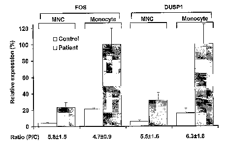
FIG. 4 is a bar graph of FOS and DUSP1 fold change ratios in patients
compared to controls (Ratio (P/C)) are preserved whether whole mononuclear
cells
(MNC) or purified monocytes (Monocyte) are used for RT-PCR. Values shown as
mean SE, n=6, for patients and controls.
FIGS. 5A-5B are a set of plots and digital images showing the confirmation
of monocyte and macrophage purity. FIG. 5A is a set of plots from flow
cytometry
showing the relative distribution profiles of CD 14- (negative) and CD14+
(positive
with anti-CD14 antibody conjugated to fluorescein isothiocyanate (FITC)) cells
in
the mononuclear cell (MNC), purified monocyte (Mono) and monocyte-depleted
(Non-mono) fractions. FIG. 5B is a set of digital images of RT-PCR of
undiluted
(1) and one-tenth diluted (0.1) cDNA from different fractions of blood and
carotid
plaque purification. Cell markers: control genes, glyceraldehyde-3-phosphate
dehydrogenase (GAPD), and translation initiation factor (TIF); monocyte, CD14;
macrophage, macrophage mannose receptor (CD206); lymphocyte, CD3; platelet,
glycoprotein IIb (GPIlb). NTC, no template control; RT-, no reverse
transcriptase;
SN, plaque suspension cells after CD14+ macrophage depletion.
CA 02577819 2007-02-20
WO 2006/029052 PCT/US2005/031469
-6-
FIG. 6 is a digital image of FOS protein expression in plasma. It shows a
Western blot using anti-FOS antibody on equal amounts of four controls and
four
patients' plasma protein (50 micrograms). As positive control for FOS protein,
THP1 cells were stimulated with PMA (C+). The two lower panels for controls
and
patients show the same corresponding samples re-run on opposite sides of the
gel to
control for potential differences in transfer efficiency of proteins in
different areas of
the gel.
SEQUENCE LISTING
The nucleic and amino acid sequences listed in the accompanying sequence
listing are shown using standard letter abbreviations for nucleotide bases,
and three
letter code for amino acids, as defined in 37 C.F.R. 1.822. Only one strand of
each
nucleic acid sequence is shown, but the complementary strand is understood as
included by any reference to the displayed strand. In the accompanying
sequence
listing:
SEQ ID NOs: 1-2 are the nucleic acid sequence of a human GAPD forward
and a reverse primer, respectively.
SEQ ID NOs: 3-4 are the nucleic acid sequence of a human TIF forward and
a reverse primer, respectively.
SEQ ID NOs: 5-6 are the nucleic acid sequence of a human FOS forward and
a reverse primer, respectively.
SEQ ID NOs: 7-8 are the nucleic acid sequence of a human DUSP 1 forward
and a reverse primer, respectively.
SEQ ID NOs: 9-10 are the nucleic acid sequence of a human NFKB1A
forward and a reverse primer, respectively.
SEQ ID NOs: 11-12 are the nucleic acid sequence of a human ID2 forward
and a reverse primer, respectively.
SEQ ID NOs: 13-14 are the nucleic acid sequence of a human PER1
forward and a reverse primer, respectively.
SEQ ID NOs: 15-16 are the nucleic acid sequence of a human SAP30
forward and a reverse primer, respectively.
CA 02577819 2007-02-20
WO 2006/029052 PCT/US2005/031469
-7-
SEQ ID NOs: 17-18 are the nucleic acid sequence of a human CD14 forward
and a reverse primer, respectively.
SEQ ID NOs: 19-20 are the nucleic acid sequence of a human CD206
forward and a reverse primer, respectively.
SEQ ID NOs: 21-22 are the nucleic acid sequence of a human CD3 forward
and a reverse primer, respectively.
SEQ ID NOs: 23-34 are the nucleic acid sequence of a human GP11b
forward and a reverse primer, respectively.
SEQ ID NOs: 25-26 are the nucleic acid sequence of a mouse TIF forward
and a reverse primer, respectively.
SEQ ID NOs: 27-28 are the nucleic acid sequence of a mouse FOS forward
and a reverse primer, respectively.
SEQ ID NOs: 29-30 are the nucleic acid sequence of a mouse DUSP1
forward and a reverse primer, respectively.
SEQ ID NOs: 31-34 are FOS siRNA target nucleic acid sequences.
SEQ ID NO: 35 is the nucleic acid sequence of the CD14 SAGE tag
sequence.
SEQ ID NO: 36 is the nucleic acid sequence of the CD163 SAGE tag
sequence.
SEQ ID NO: 37 is the nucleic acid sequence of the CD3E SAGE tag
sequence.
SEQ ID NO: 38 is the nucleic acid sequence of the CD79A SAGE tag
sequence.
SEQ ID NO: 39 is the nucleic acid sequence of the CD99 SAGE tag
sequence.
SEQ ID NO: 40 is the nucleic acid sequence of the FOS SAGE tag sequence.
SEQ ID NO: 41 is the nucleic acid sequence of the dual specificity
phosphatase 1 (DUSP1) tag sequence.
SEQ ID NO: 42 is the nucleic acid sequence of the NF kappa gene in B-cell
inhibitor (NFKBIA) SAGE tag sequence.
SEQ ID NO: 43 is the nucleic acid sequence of the inhibitor of DNA 2(ID2)
SAGE tag sequence.
CA 02577819 2007-02-20
WO 2006/029052 PCT/US2005/031469
-8-
SEQ ID NO: 44 is the nucleic acid sequence of the period homolog 1(PERl)
SAGE tag sequence.
SEQ ID NO: 45 is the nucleic acid sequence of the sin3-associated
polypeptide, 30 kDa (SAP30) SAGE tag sequence.
DETAILED DESCRIPTION
1. Abbreviations
AVG: average
BMI: body mass index
CEA: Carotid endarterectomy
DUSP1: dual specificity phosphatase 1
FITC: fluorescein isothiocyanate
FOS: Biskis-Jinkins osteosarcoma
GADP: glyceraldehyde-3-phosphate dehydrogenase
GPIIb: glycoprotein IIb
hsCRP: high sensitivity C-reactive protein
ID 1: inhibitor of DNA binding 2
kDa: kilodaltons
KO: knock-out
MAPK: mitogen activated protein kinase
MCP-1: monocyte chemoattractant protein 1
MNC: mononuclear cells
NTC: no template control
PCR: polymerase chain reaction
PERl : period homolog 1
PMA: phorbo 12-myristate 13-acetate
ROC: receive operating characteristic
RT: reverse transcriptase
SAP30: sin-3 associated polypeptide, 30 kDa
SAGE: serial analysis of gene expression
SE: standard error
CA 02577819 2007-02-20
WO 2006/029052 PCT/US2005/031469
-9-
siRNA: small inhibitory RNA
SN: plaque suspension cells after CD14+ macrophage depletion
TIF: translation initiation factor
WT: wild-type
II. Terms
Unless otherwise noted, technical terms are used according to conventional
usage. Definitions of common terms in molecular biology may be found in
Benjamin Lewin, Genes V, published by Oxford University Press, 1994 (ISBN 0-19-
854287-9); Kendrew et al. (eds.), The Encyclopedia ofMolecularBiology,
published
by Blackwell Science Ltd., 1994 (ISBN 0-632-02182-9); and Robert A. Meyers
(ed.), Molecular Biology and Biotechnology: a Comprehensive Desk Reference,
published by VCH Publishers, Inc., 1995 (ISBN 1-56081-569-8).
In order to facilitate review of the various embodiments of this disclosure,
the following explanations of specific terms are provided:
Alter: A change in an effective amount of a substance of interest, such as a
polynucleotide or polypeptide. The amount of the substance can changed by a
difference in the amount of the substance produced, by a difference in the
amount of
the substance that has a desired function, or by a difference in the
activation of the
substance. The change can be an increase or a decrease. The alteration can be
in
vivo or in vitro.
In several embodiments, altering an amount of a polypeptide or
polynucleotide is at least about a 50%, 60%, 70%, 80%, 90%, 95%, 96%, 97%,
98%, 99%, or 100% increase or decrease in the effective amount (level) of a
substance. In specific example, an increase of a polypeptide or polynucleotide
is at
least about a 50%, 60%, 70%, 80%, 90%, 95%, 96%, 97%, 98%, 99%, or 100%
increase in FOS and/or DUSP1 polypeptide or polynucleotide as compared to a
control, a statistical normal, or a standard value chosen for specific study.
In another
specific example, an decrease of a polypeptide or polynucleotide, such as
following
the initiation of a therapeutic protocol, is at least about a 50%, 60%, 70%,
80%,
90%, 95%, 96%, 97%, 98%, 99%, or 100% decrease in FOS and/or DUSP1
CA 02577819 2007-02-20
WO 2006/029052 PCT/US2005/031469
-10-
polypeptide or polynucleotide as compared to a control, a statistical normal,
or a
standard value chosen for specific study.
Atherosclerosis: The progressive narrowing and hardening of a blood vessel
over time. Atherosclerosis is a common form of ateriosclerosis in which
deposits of
yellowish plaques (atheromas) containing cholesterol, lipoid material and
lipophages
are formed within the intima and inner media of large and medium-sized
arteries.
Treatment of atherosclerosis includes reversing or slowing the progression of
atherosclerosis, for example as measured by the presence of atherosclerotic
lesions
and/or functional signs of the disease, such as improvement in cardiovascular
function as measured by signs (such as peripheral capillary refill), symptoms
(such
as chest pain and intermittent claudication), or laboratory evidence (such as
that
obtained by EKG, angiography, or other imaging techniques). "Assessing
atherosclerosis" indicates determining if a subject of interest has
atherosclerosis,
determining the prognosis of the subject of interst, and/or determining if a
therapeutic regimen administered to the subject is effective in treating the
subject.
Binding or stable binding: An association between two substances or
molecules, such as the hybridization of one nucleic acid molecule to another
(or
itself), the association of an antibody with a peptide, or the association of
a protein
with another protein or nucleic acid molecule. An oligonucleotide molecule
binds or
stably binds to a target nucleic acid molecule if a sufficient amount of the
oligonucleotide molecule forms base pairs or is hybridized to its target
nucleic acid
molecule, to permit detection of that binding.
Binding can be detected by any procedure known to one skilled in the art,
such as by physical or functional properties of the formed complexes, such as
a
target:oligonucleotide complex or a target:antibody complex. For example,
binding
can be detected functionally by determining whether binding has an observable
effect upon a biosynthetic process such as expression of a gene, DNA
replication,
transcription, translation, and the like.
Physical methods of detecting the binding of complementary strands of
nucleic acid molecules, include but are not limited to, such methods as DNase
I or
chemical footprinting, gel shift and affinity cleavage assays, Northern
blotting, dot
blotting and light absorption detection procedures. For example, one method
CA 02577819 2007-02-20
WO 2006/029052 PCT/US2005/031469
-11-
involves observing a change in light absorption of a solution containing an
oligonucleotide (or an analog) and a target nucleic acid at 220 to 300 nm as
the
temperature is slowly increased. If the oligonucleotide or analog has bound to
its
target, there is a sudden increase in absorption at a characteristic
temperature as the
oligonucleotide (or analog) and target disassociate from each other, or melt.
In
another example, the method involves detecting a signal, such as a detectable
label,
present on one or both nucleic acid molecules (or antibody or protein as
appropriate).
In one example, the binding between an oligomer and its target nucleic acid
is characterized by the temperature (Tm) at which 50% of the oligomer is
melted
from its target. A higher (Tm) means a stronger or more stable complex
relative to a
complex with a lower (Tm).
Blood vessel: The vessels through which blood circulates. In general, blood
vessels are elastic tubular channels that are lined with endothelium. Blood
vessels
include the arteries, veins and capillaries. Specific, non-limiting examples
of a
blood vessel include a vena cava, a thoracic aorta, a saphanous vein, a
mammary
artery, the brachial artery and a capillary. In another embodiment, a blood
vessel
includes the smaller arteries and veins. In yet another embodiment, a blood
vessel is
a capillary of the microvascular circulation.
Buffy coat: A thin yellow or white layer of leukocytes that appears on top
of a mass of packed red cells when whole blood is centrifuged.
Cardiovascular: Pertaining to the heart and/or blood vessels.
Cardiovascular risk: The likelihood of the development of disorders
related to the cardiovascular system, such as, but not limited to, myocardial
ischemia
and infarction, intermittent claudication, bowel ischemia, retinal ischemia,
transient
ischemic attacks, ischemic strokes, and other conditions associated with
cardiovascular dysfunction. In a specific non-limiting example, the disorder
is
myocaridal ischemia or infarction.
Cholesterol lowering agent: An agent, such as a pharmaceutical, vitamin,
or small molecule, that lowers the level of cholesterol in a subject. One of
skill in
the art can readily identify assays, such as blood screening, to determine the
effect of
cholesterol. Agents include, but are not limited to, niacin, the statins
(e.g., ZocorTM,
CA 02577819 2007-02-20
WO 2006/029052 PCT/US2005/031469
-12-
LipitorTM, PravacolTM, LescorTM, MevacorTM), binding resins (e.g.,
QuestranTM), and
fibrates (e.g. LopidTM, Lipidil MicroT"')
DUSPI: Dual specificity phosphatase 1, which is known to be induced by
oxidative stress and heat shock. DUSP1 has also been called CL100, MVHl, MKP-
1 and DTPN10. Exemplary human DUSP 1 amino acid and nucleic acid sequence
can be found at GenBank Accession No.U01669 (June 11, 1994) and X68277 (April
18, 2005), and Swiss-Prot No. P28562 (February 23, 2996), which are
incorporated
herein by reference. In humans, the DUSP1 gene is encoded on chromosome 5.
DUSPl is a dual specification phosphatase that dephosphorylates MAP kinase ERK
at Tyr-185. Orthogs from chimpanzee, rat, mouse, and zebrafish have been
identified (see GeneCard for DUSPl, GC05M1721127, which is available on the
internet at the Weizmann Institute of Science Website).
FOS: An oncogene, Finkel-Biskis-Jinkins osteosarcoma (FOS) gene. FOS
was identified in a mouse osteosarcoma, encoding a transcription factor. The
product of this oncogene works with the product of another oncogene, the jun
oncogene, to abnormally change the rate of transcription of certain other
genes.
c-FOS is the cellular homolog of the viral v-FOS oncogene found in FBJ (Finkel-
Biskis-Jinkins) and FBR murine osteosarcoma viruses (MSV). The human FOS
gene maps to chromosome 14q21-q31. FOS has been identified as TIS28, a gene
inducible in several cell types by Phorbol esters. Exemplary amino acid and
nucleic
acid sequence for the murine and human FOS are shown in GenBank Accession No.
BC029814 (June 30, 2004) and V01512 (November 21, 2004), respectively, and is
shown as Swiss-Prot No. P0110 (July 1, 1986), which are incorporated herein by
reference.
Without being bound by theory, c-FOS is thought to have an important role
in signal transduction, cell proliferation and differentiation. It is a
nuclear protein
which, in combination with other transcription factors (for example: c jun )
acts as a
trans-activating regulator of gene expression. Orthogs from chimpanzee, rat,
mouse,
and zebrafish have been identified (see GeneCard for FOS, GC14P074815, which
is
available on the internet at the Weizmann Institute of Science website).
Framingham Risk Score: A risk factor score that is used for predicting
future risk of coronary artery disease in individuals free of disease, based
on the
CA 02577819 2007-02-20
WO 2006/029052 PCT/US2005/031469
-13-
measurement of risk factors including age, gender, systolic blood pressure,
cigarette
smoking, glucose intolerance, left ventricular hypertrophy, as well as total
cholesterol, low density lipoprotein (LDL) and high density lipoprotein (HDL)
levels (Wilson et al., Am JCardiol 59:91G-94G, 1987).
Leukocyte: Cells in the blood, also termed "white cells," that are involved
in defending the body against infective organisms and foreign substances.
Leukocytes are produced in the bone marrow. There are five main types of white
blood cells, subdivided between two main groups: polymorphonuclear leukocytes
(neutrophils, eosinophils, basophils) and mononuclear leukocytes (monocytes
and
lymphocytes). When an infection is present, the production of leukocytes
increases.
Lymphocytes: A type of white blood cell that is involved in the immune
defenses of the body. There are two main types of lymphocytes: B cell and T
cells.
Microarray: An "array" is an arrangement of molecules, such as biological
macromolecules (such as peptides or nucleic acid molecules) or biological
samples
(such as tissue sections), in addressable locations on or in a substrate. A
"microarray" is an array that is miniaturized so as to require or be aided by
microscopic examination for evaluation or analysis. Arrays including
biological
materials are sometimes called DNA chips or biochips. Generally, DNA is either
spotted, using pins or an ink j et printer, or synthesized directly on the
array using
PCR or photolithography. The DNA may be either double-stranded copies of
transcripts or shorter single-stranded oligonucleotides. In one embodiment,
for
microarray analysis, RNA is first extracted from a sample; the RNA can be
amplified prior to analysis. Subsequently, the RNA itself, complementary DNA,
or
amplified RNA is labeled. The labeled nucleic acid is hybridized,
competitively or
noncompetitively, to the microarray. Complementary sequences remain bound to
the array and unbound sequences are washed off. Expressed genes are identified
by
the position ofbound probes on the array. Microarrays are available from a
number
of commercial sources, or can be produced in individual laboratories. In
addition,
computer software that can be used to analyze the microarray data is available
commercially from a number of sources and on the internet (see the dchip
website,
or the tigr website, for examples).
CA 02577819 2007-02-20
WO 2006/029052 PCT/US2005/031469
-14-
Hybridization: To form base pairs between complementary regions of two
strands of DNA, RNA, or between DNA and RNA, thereby forming a duplex
molecule. Hybridization conditions resulting in particular degrees of
stringency will
vary depending upon the nature of the hybridization method and the composition
and length of the hybridizing nucleic acid sequences. Generally, the
temperature of
hybridization and the ionic strength (such as the Na+ concentration) of the
hybridization buffer will determine the stringency of hybridization.
Calculations
regarding hybridization conditions for attaining particular degrees of
stringency are
discussed in Sambrook et al., (1989) Molecular Cloning, second edition, Cold
Spring Harbor Laboratory, Plainview, NY (chapters 9 and 11). An exemplary non-
limiting set of very high stringency conditions (detects sequences that share
90%
identity) include hybridization in 5x SSC at 65 C for 16 hours, washing twice
in 2x
SSC at room temperature (RT) for 15 minutes each, and washing twice in 0.5x
SSC
at 65 C for 20 minutes each. An exemplarynon-limiting set of high stringency
conditions (detects sequences that share 80% identity or greater) include
hybridization in 5x-6x SSC at 65 C-70 C for 16-20 hours, washing twice in 2x
SSC
at RT for 5-20 minutes each, and washing twice in lx SSC at 55 C-70 C for 30
minutes each.
Label: An agent capable of detection, for example by ELISA,
spectrophotometry, flow cytometry, or microscopy. For example, a label can be
attached to a nucleic acid molecule or protein, thereby permitting detection
of the
nucleic acid molecule or protein. Examples of labels include, but are not
limited to,
radioactive isotopes, enzyme substrates, co-factors, ligands,
chemilurninescent
agents, fluorophores, haptens, enzymes, and combinations thereof. Methods for
labeling and guidance in the choice of labels appropriate for various purposes
are
discussed for example in Sambrook et al. (Molecular Cloning: A Laboratory
Manual, Cold Spring Harbor, New York, 1989) and Ausubel et al. (In Current
Protocols in Molecular Biology, John Wiley & Sons, New York, 1998).
Monocyte: A relatively large mononuclear leukocyte (16-22 ~Lm in
diameter). Monocytes normally constitute 3-7% of the leukocytes of the
circulating
blood, and are normally found in lymph nodes, spleen, bone marrow and loose
connective tissue. When treated with histological dyes, monocytes manifest an
CA 02577819 2007-02-20
WO 2006/029052 PCT/US2005/031469
-15-
abundant pale blue or blue-gray cytoplasm that contains numerous, fine, dust-
like,
red-blue granules; vacuoles are frequently present; the nucleus is usually
indented,
or slightly folded, and has a stringy chromatin structure that seems more
condensed
where the delicate strands are in contact. Generally, monocytes have an ovoid
or
kidney-shaped nucleus, containing lacy, linear chromatin, and abundant gray-
blue
cytoplasm filled with fine reddish and azurophilic granules. Circulating
monocytes
in blood differentiate into macrophages when they migrate into tissues.
Polynucleotide: A linear nucleotide sequence, including sequences of greater
than 100 nucleotide bases in length.
Polypeptide: Any chain of amino acids, regardless of length or post-
translational modification (e.g., glycosylation or phosphorylation).
Purified or Isolated: The term "purified" or "isolated" does not require
absolute purity; rather, it is intended as a relative term. A purified nucleic
acid or
protein is isolated or purified away from other biological components in the
cell of
the organism in which the component naturally occurs, i.e., other chromosomal
and
extrachromosomal DNA and RNA, and proteins Nucleic acids, peptides and
proteins
which have been "isolated" thus include nucleic acids and proteins purified by
standard purification methods. The term also embraces nucleic acids, peptides
and
proteins prepared by recombinant expression in a host cell as well as
chemically
synthesized nucleic acids.
Thus, for example, a purified cell preparation is one in which the cell,
protein
or nucleic acid referred to is more pure than the cell in its natural
environment
within a tissue. In one embodiment, a "substantially purified" population of a
specific cell type is a composition of cells that includes less than about
20%, less
than about 15%, or less than about 10% of cells of a different phenotype.
Thus, a
substantially purified population of cells includes greater than 80%, greater
than
85%, or greater than 90% of the cells of interest. In another embodiment, a
process
that produces a purified population of cells is a process that produces a
population of
cells so that more than 50% of the resulting population is the cell type of
interest.
Statin: Any of a class of lipid-lowering drugs that reduce serum cholesterol
levels by inhibiting a key enzyme involved in the biosynthesis of cholesterol.
Example statins include atorvastatin (Lipitor ), fluvastatin (Lescol(D),
lovastatin
CA 02577819 2007-02-20
WO 2006/029052 PCT/US2005/031469
-16-
(Mevacor , Altocor , not marketed in the UK), pravastatin (Pravachol ,
Selektine , Lipostat ), rosuvastatin (Crestor ), simvastatin (Zocor ). There
are
two groups of statins: (1) Fermentation-derived: lovastatin, simvastatin and
pravastatin, and (2) Synthetic statins: fluvastatin, atorvastatin,
cerivastatin and
rosuvastatin. Generally, statins act by competitively inhibiting 3-hydroxy-3-
methylglutaryl coenzyme A (HMG CoA) reductase, an enzyme of the HMG-CoA
reductase pathway, the body's metabolic pathway for the synthesis of
cholesterol.
The structure of one exemplary statin, Lovastatin, is shown below.
mo
CH,4
cH3
Subject: Any subject that has a vascular system and has hematopoietic cells.
In one embodiment, the subject is a non-human mammalian subject, such as a
monkey, mouse, rat, rabbit, pig, goat, sheep or cow. In another embodiment,
the
subject is a human subject.
Therapeutically effective amount: An amount of a pharmaceutical
preparation that alone, or together with a pharmaceutically acceptable carrier
or one
or more additional therapeutic agents, induces the desired response. A
therapeutic
agent, such as an anticoagulant, is administered in therapeutically effective
amounts.
Effective amounts a therapeutic agent can be determined in many different
ways, such as assaying for a reduction in atherosclerotic disease or
improvement of
physiological condition of a subject having vascular disease. Effective
amounts
also can be determined through various in vitro, in vivo or in situ assays.
Therapeutic agents can be administered in a single dose, or in several doses,
for example daily, during a course of treatment. However, the effective amount
of
can be dependent on the source applied, the subject being treated, the
severity and
type of the condition being treated, and the manner of administration.
In one example, it is an amount sufficient to partially or completely
alleviate
symptoms of vascular disease within a subject. Treatment can involve only
slowing
CA 02577819 2007-02-20
WO 2006/029052 PCT/US2005/031469
-17-
the progression of the vascular disease temporarily, but can also include
halting or
reversing the progression of the vascular disease permanently. For example, a
pharmaceutical preparation can decrease one or more symptoms of vascular
disease,
for example decrease a symptom by at least 20%, at least 50%, at least 70%, at
least
90%, at least 98%, or even at least 100%, as compared to an amount in the
absence
of the pharmaceutical preparation.
Treating a disease: "Treatment" refers to a therapeutic intervention that
ameliorates a sign or symptom of a disease or pathological condition, such a
sign,
parameter or symptom of vascular disease (for example, atherosclerosis).
Treatment can also induce remission or cure of a condition, such as vascular
disease. In particular examples, treatment includes preventing a disease, for
example by inhibiting the full development of a disease, such as preventing
development of vascular disease. Prevention of a disease does not require a
total
absence of vascular disease. For example, a decrease of at least 50% can be
sufficient.
Vascular function: The function of the blood vessels. Decreased vascular
function is associated with atherosclerosis, myocardial infarction,
intermittent
claudication, bowel ischemia, retinal ischemia, transient ischemic attacks
(TIAs),
ischemic strokes, restenosis after angioplasty, transplant atherosclerosis,
unstable
angina, sudden death and alterations in blood pressure.
Vascular function assessment: An assay that measures the function of the
vascular system. Assays include measurement of a parameter of the blood,
assays of
arterial hyperplasia, vascular contractility measurements, brachial reactivity
measurements, and morphometric measurements. Similarly, an endothelial cell
assessment is a test that measures a function or parameter of an endothelial
cell.
"Decreased vascular function" indicates a decrease in any function of the
blood
vessels, as compared to a standard value or a control sample. Thus, in one
example,
decreased vascular function is a decrease in a vascular contractility, as
compared to
a known value for normal vascular contractility. In another example, decreased
vascular function is the lower contractility of a blood vessel as compared to
the
contractility of a vessel known to not be affected by a disease or a disorder.
In a
further example, decreased vascular function is a lower vascular contractility
as
CA 02577819 2007-02-20
WO 2006/029052 PCT/US2005/031469
-18-
compared to the contractility of a vessel from the same subject at an earlier
time
point. "Cardiovascular risk" is the probability that a subject has or will
develop a
vascular disease in the future.
Vascular tissue: Tissue consisting of, or containing, vessels as an essential
part of a structure. Vascular tissue operates by means of, or is made up of an
arrangement of, vessels. Vascular tissue includes the arteries, veins,
capillaries,
lacteals, microvasculature, etc. In one embodiment, vascular tissue includes a
highly
vascularized organ (e.g. the lung). In another embodiment, vascular tissue is
a blood
vessel, or a portion thereof. Cells isolated from a vascular tissue are a
population of
cells isolated from the remaining components of the tissue.
Assessment of Vascular Function
A method of assessing vascular function in a subject is disclosed herein.
Specifically, the method is of use in assessing (for example, determining the
diagnosis or prognosis of) atherosclerosis. In several embodiments, the method
includes assaying expression of FOS mRNA or the presence of FOS polypeptide.
In
additional embodiments, the method includes assaying expression of DUSPl inRNA
or the presence of DUSP1 polypeptide. The method can include monitoring FOS
and/or DUSP1 in blood, serum or plasma.
The method can be used, for example, to predict future cardiovascular risk.
Specifically, the method can be used to predict risk for myocardial
infarction,
intermittent claudication, bowel ischemia, retinal ischemia, transient
ischemic
attacks (TIAs), ischemic strokes, restenosis after angioplasty, transplant
atherosclerosis, unstable angina, sudden death, and other conditions
associated with
cardiovascular dysfunction. In one specific, non-limiting example, the
assessment of
FOS or DUSP1 is of use in predicting cardiovascular risk for myocardial
ischemia
and/or infarction. Cardiovascular risk indicates the potential for a future
cardiovascular event, such as myocardial infarction, intermittent
claudication, bowel
ischemia, retinal ischemia, transient ischemic attacks (TIAs), ischemic
strokes,
restenosis after angioplasty, transplant atherosclerosis, unstable angina,
sudden
death, and other conditions associated with cardiovascular dysfunction.
Factors
involved in cardiovascular risk include, but are not limited to, serum
cholesterol,
CA 02577819 2007-02-20
WO 2006/029052 PCT/US2005/031469
-19-
hypertension, diabetes, sex and age. The method can also be used to assess the
severity of a disease, such as atherosclerosis.
Methods are provided herein for evaluating vascular risk, for example for
determining whether a subject, such as an otherwise healthy subject, or a
subject
suspected or at risk of having vascular disease, has vascular disease or will
likely
develop vascular disease in the future. In particular examples, the method can
determine with a reasonable amount of sensitivity and specificity whether a
subject
has or will likely develop a vascular disease in the future. In some examples,
isolated or purified PBMCs, serum, blood or plasma obtained from the subject
are
used to predict the subject's risk of vascular disease. In one example, the
subject is
apparently healthy, such as a subject who does not exhibit symptoms of
vascular
disease (for example has not previously had an acute adverse vascular event
such as
a myocardial infarction or a stroke). In some examples, a healthy subject is
one that
if examined by a medical professional, would be characterized as healthy and
free of
symptoms of vascular disease. In another example, the subject is suspected of
having a vascular disease, or is suspected of being at risk of developing a
vascular
disease in the future. For example, such a subject may have elevated
cholesterol or
tri-glyceride levels, elevated C-reactive protein levels, or high blood
pressure.
In a specific, non-limiting example, the expression of FOS and/or DUSP1 in
monocytes is used to non-invasively diagnose atherosclerosis. For example,
expression of FOS and/or DUSP 1 can be used to assess the severity and/or the
progression of the disease. In one embodiment, the expression of FOS and/or
DUSP1 in monocytes is assessed. The monocytes can be in an atherosclerotic
lesion
or can be circulating monocytes in the peripheral blood. In additional
embodiments,
the amount of FOS into the plasma or serum is assessed. Thus, in several
examples,
the method includes measuring the expression of FOS and/or DUSP1 in the
peripheral blood, plasma, serum, or in peripheral blood mononuclear cells, to
determine the risk for developing a cardiovascular condition such as, but not
limited
to, atherosclerosis. Such assessments can assist in determining whether to
initiate
therapy, for example, with lifestyle (including dietary) intervention or
pharmacologic (drug) therapy.
CA 02577819 2007-02-20
WO 2006/029052 PCT/US2005/031469
-20-
The methods disclosed herein include assaying the expression of FOS,
DUSPl, or both FOS and DUSPl. An increase in the expression of FOS and/or
DUSP1 in a sample including monocytes as compared to a control sample
indicates
decreased vascular function, for example, increased future cardiovascular risk
or
development of atherosclerosis. In one specific, non-limiting example, an
assessment of the risk of a subject to develop vascular disease, or an
assessment of
vascular function is made by evaluating the expression of FOS and/or DUSP 1 in
peripheral blood mononuclear cells (PBMC).
In a further specific, non-limiting example the expression of FOS and/or
DUSP 1 are used to assess the efficacy of a therapeutic protocol. The
treatment
protocol can include any therapy for atherosclerosis designed to reverse or
slow the
progression of atherosclerosis, including but not limited to treatment with
statins,
niacin or other cholesterol-lowering agents, anti-inflammatory agents, or any
other
pharmaceutical compound. In this embodiment, a sample including monocytes,
and/or a sample of blood, serum or plasma, can be taken from a subject prior
to
initiation of therapy. After therapy is initiated, an additional sample
including
monocytes, and/or a sample of blood, serum or plasma, is taken from the
subject. A
decrease in the amount of FOS and/or DUSPl indicates that the therapy is
efficacious. In addition, the subject can be monitored over time to evaluate
the
continued effectiveness of the therapeutic protocol. The effect of different
dosages
can also be evaluated, by comparing the expression of FOS and/or DUSP 1 in a
sample from the subject receiving a first dose to the expression of FOS and/or
DUSP1 in a sample from the subject receiving a second (different) dose.
A variety of methods can be employed to detect FOS and/or DUSP 1
expression in monocytes in an atherosclerotic lesion or in the peripheral
blood,
serum, or plasma. These methods include the use of nucleic acid probes,
antibodies
or other analytical techniques such as mass spectrometry to detect FOS and/or
DUSP1 expression. The expression of FOS and/or DUSP 1 is assessed in
monocytes, such as monocytes in an atherosclerotic lesion or peripheral blood
monocytes, or in a blood, peripheral blood, or serum sample. In one specific,
non-
limiting example, the method specifically excludes detection of FOS and/or
DUSP 1
in vascular smooth muscle, such that the expression of FOS and/or DUSP 1 is
CA 02577819 2007-02-20
WO 2006/029052 PCT/US2005/031469
-21-
evaluated in monocytes only (or in the blood, plasma or serum only). Thus, in
one
embodiment, the assay system is designed to distinguish expression of FOS
and/or
DUSP 1 in monocytes. Thus, in one embodiment, the expression of FOS and/or
DUSPl is not evaluated in the vascular tissue, such as in vascular smooth
muscle.
In another embodiment, the assay is designed to detect the release into plasma
from
the expression of FOS and DUSP1 in vascular tissue. In several examples, the
assay
can be performed in isolated peripheral blood monocytes (PBMC), plasma, blood
or
serum.
Detection of FOS and D USPI Nucleic Acids
In one embodiment, nucleic acid based methods are utilized. These methods
include serial analysis of gene expression (SAGE techniques), RT-PCR,
quantitative
PCR, real time PCR, Northern blot, dot blots, microarrays, amongst others.
Generally, with regard to nucleic acids, any method can be utilized provided
it can
detect the expression of target gene mRNA (FOS and/or DUSP1) as compared to a
control. One of skill in the art can readily identify an appropriate control,
such as a
sample from a subject known not to have a disorder (a negative control), a
sample
from a subject known to have a disorder (a positive control), or a known
amount of
nucleic acid encoding FOS and/or DUSPl (a standard or a normal level found in
a
healthy subject). Statistically normal levels can be determined for example,
from a
subject with known not be have atherosclerosis, and to at low risk for a
cardiac
event. In one non-limiting example, normal levels can be assessed by measuring
FOS and/or DUSP1 in the blood, serum, or plasma of young adults, who do not
smoke or drink, exercise regularly, have no known history of cardiac events,
and no
familial history of heart disease.
The methods described herein may be performed, for example, by utilizing
pre-packaged diagnostic kits comprising at least one specific nucleic acid
probe,
which may be conveniently used, such as in clinical settings, to diagnose
patients
exhibiting cardiovascular disease symptoms or at risk for developing
cardiovascular
disease. In one embodiment, this assay is performed in a medical laboratory on
a
sample of peripheral blood, cells isolated from the peripheral blood, serum or
plasma.
CA 02577819 2007-02-20
WO 2006/029052 PCT/US2005/031469
-22-
The diagnostic procedures can be performed "in situ" directly upon blood
smears (fixed and/or frozen), or on tissue biopsies, such that no nucleic acid
purification is necessary. DNA or RNA from a sample can be isolated using
procedures which are well known to those in the art.
Nucleic acid reagents that are specific to the nucleic acid of interest,
namely
the nucleic acid encoding FOS or DUSPl, can be readily generated given the
sequences of these genes for use as probes and/or primers for such in situ
procedures
(see, for example, Nuovo, G. J., 1992, PCR in situ hybridization: protocols
and
applications, Raven Press, NY).
A differential display procedure can be utilized based on Northern analysis
and/or RT-PCR. An exemplary method is disclosed in the examples section below.
In one embodiment, the methods disclosed herein include the use of an ordered
array
of nucleic acids representing thousands of genes on a solid support. mRNA from
the
cells of interest are used to create a labeled, first strand cDNA probe that
is then
hybridized to the microarray. In one embodiment, two mRNA samples are directly
compared to the same microarray by incorporating different labels into the
cDNA
probes derived from the samples. The extent of hybridization of the probes to
each
nucleic acid sequence on the microarray is then quantitated and the ratio of
the pixel
intensities for each label is used as a measure of the relative mRNA
expression in
the two samples. In one embodiment, the array is an array of nucleic acids
expressed by the immune system or the cardiovascular system.
In one example, a lymphochip is utilized, which includes nucleic acid
sequences derived from high-throughput sequencing of cDNA clones from
libraries
of human immune cells. The array can incorporate, for example, thousands of
clones from a library prepared from the immune system or the cardiovascular
system. The array can also include genes of known structure and function based
on
their established role in immune cell differentiation, response and disorders.
These
types of arrays are well known in the art (see, for example, Staudt, Trends
Inamunol.
22:35-40, 2001; Staudt and Brown, Ann. Rev. Immunol. 18:829-859, 2000;
Alizadeh
et al., Nature 403:503-511, 2000; Alizadeh et al., Cold Spring Harbor Synap.
Quant.
Biol. 64:71-78, 1999; U.S. Patent Application No. 20030203416A1, all of which
are
incorporated herein by reference).
CA 02577819 2007-02-20
WO 2006/029052 PCT/US2005/031469
-23-
The array can be a high density array, such that the array includes greater
than about 100, greater than about 1000, greater than about 16,000 and most
greater
than about 65,000 or 250,000 or even greater than about 1,000,000 different
oligonucleotide probes. The oligonucleotide probes generally range from about
5 to
about 50 nucleotides, such as about 10 to about 40 nucleotides in length or
from
about 15 to about 40 nucleotides in length.
The location and sequence of each different oligonucleotide probe sequence
in the array is known. Moreover, in a high density array, the large number of
different probes occupies a relatively small area so that there is a probe
density of
greater than about 60 different oligonucleotide probes per cm2, such as
greater than
about 100, greater than about 600, greater than about 1000, greater than about
5,000,
greater than about 10,000, greater than about 40,000, greater than about
100,000, or
greater than about 400,000 different oligonucleotide probes per cm2. The small
surface area of the array (such as less than about 10 cm2, less than about 5
cm2, less
than about 2 cm2) permits extremely uniform hybridization conditions
(temperature
regulation, salt content, etc.) while the extremely large number of probes
allows
parallel processing of hybridizations.
Generally, the methods of monitoring gene expression using array
technology involve (1) providing a pool of target nucleic acids comprising RNA
transcript(s) of one or more target gene(s), or nucleic acids derived from the
RNA
transcript(s); (2) hybridizing the nucleic acid sample to an array of probes
(including
control probes), that can be a high density array; and (3) detecting the
hybridized
nucleic acids and calculating a relative expression (transcription) level. In
the
present application, the expression of FOS and/or DUSPl is evaluated.
In order to measure the transcription level of a gene or genes, it is
desirable
to provide a nucleic acid sanzple comprising inRNA transcript(s) of the gene
or
genes, or nucleic acids derived from the mRNA transcript(s). As used herein, a
nucleic acid derived from an mRNA transcript refers to a nucleic acid for
whose
synthesis the mRNA transcript or a subsequence thereof has ultimately served
as a
template, such as a cDNA ("first strand" transcribed from the mRNA). Thus, a
cDNA reverse transcribed from an mRNA, an RNA transcribed from that cDNA, a
DNA amplified from the cDNA, an RNA transcribed from the amplified DNA, etc.,
CA 02577819 2007-02-20
WO 2006/029052 PCT/US2005/031469
-24-
are all derived from the mRNA transcript. Detection of such products is
indicative
of the presence and/or abundance of the original transcript in a sample. Thus,
suitable samples include, but are not limited to, mRNA transcripts of the gene
or
genes, cDNA reverse transcribed from the mRNA, cRNA transcribed from the
cDNA, and the like.
Generally, the transcription level (and thereby expression) of one or more
genes in a sample is quantified, so that the nucleic acid sample is one in
which the
concentration of the mRNA transcript(s) of the gene or genes, or the
concentration
of the nucleic acids derived from the mRNA transcript(s), is proportional to
the
transcription level (and therefore expression level) of that gene. The
hybridization
signal intensity should also be proportional to the amount of hybridized
nucleic acid.
Generally, the proportionality is relatively strict (for example, a doubling
in
transcription rate results in approximately a doubling in mRNA transcript in
the
sample nucleic acid pool and a doubling in hybridization signal), one of skill
will
appreciate that the proportionality can be more relaxed and even non-linear.
Thus,
for example, an assay where a 5 fold difference in concentration of the target
mRNA
results in a 3 to 6 fold difference in hybridization intensity can be
sufficient. Where
more precise quantification is required, controls can be run to correct for
variations
introduced in sample preparation and hybridization as described herein. In
addition,
serial dilutions of "standard" target mRNAs can be used to prepare calibration
curves according to methods well known to those of skill in the art. Of
course,
where simple detection of the presence or absence of a transcript (such as FOS
and/or DUSP1) is desired, controls or calibrations may not be required.
In one embodiment, a nucleic acid sample is utilized, such as the total
mRNA isolated from a biological sample. The biological sample can be from any
biological tissue or fluid from the subject of interest, such as a subject who
is
suspected of having cardiovascular disease. Such samples include, but are not
limited to, blood, blood cells (such as white blood cells) or tissue biopsies
including
vascular tissue. However, the sample could also be peritoneal fluid, and
pleural
fluid, cerebral spinal fluid, or cells separated from a sample.
Nucleic acids (such as mRNA) can be isolated from the sample according to
any of a number of methods well known to those of skill in the art. Methods of
CA 02577819 2007-02-20
WO 2006/029052 PCT/US2005/031469
-25-
isolating total mRNA are well known to those of skill in the art. For example,
methods of isolation and purification of nucleic acids are described in detail
in
Chapter 3 of Laboratory Techniques in Biochemistry and Molecular Biology:
Hybridization With Nucleic Acid Probes, Part I. Theory and Nucleic Acid
Preparation, P. Tijssen, ed. Elsevier, N.Y. (1993) and Chapter 3 of Laboratory
Techniques in Biochemistry and Molecular Biology: Hybridization With Nucleic
Acid Probes, Part I. Theory and Nucleic Acid Preparation, P. Tijssen, ed.
Elsevier,
N.Y. (1993). In one example, the total nucleic acid is isolated from a given
sample
using, for example, an acid guanidinium-phenol-chloroform extraction method,
and
polyA+ mRNA is isolated by oligo dT column chromatography or by using (dT)n
magnetic beads (see, for example, Sambrook et al, Molecular Cloning: A
Laboratory Manual (2nd ed.), Vols. 1-3, Cold Spring Harbor Laboratory, (1989),
or
Current Protocols in Molecular Biology, F. Ausubel et al., ed. Greene
Publishing
and Wiley-Interscience, N.Y. (1987)). In another example, oligo-dT magnetic
beads may be used to purify mRNA (Dynal Biotech Inc., Brown Deer, WI).
The nucleic acid sample can be amplified prior to hybridization. If a
quantitative result is desired, a method is utilized that maintains or
controls for the
relative frequencies of the amplified nucleic acids. Methods of "quantitative"
amplification are well known to those of skill in the art. For example,
quantitative
PCR involves simultaneously co-amplifying a known quantity of a control
sequence
using the same primers. This provides an internal standard that can be used to
calibrate the PCR reaction. The array can then include probes specific to the
internal
standard for quantification of the amplified nucleic acid.
Suitable amplification methods include, but are not limited to, polymerase
chain reaction (PCR) (see Innis et al., PCR Protocols, A guide to Methods and
Application, Academic Press, Inc. San Diego, 1990), ligase chain reaction
(LCR)
(see Wu and Wallace, Genomics 4:560, 1989; Landegren et al., Science 241:1077,
1988; and Barringer, et al., Gene 89:117, 1990), transcription amplification
(Kwoh
et al., Proc. Natl. Acad. Sci. U.S.A. 86:1173, 1989), and self-sustained
sequence
replication (Guatelli et al., Proc. Nat. Acad. Sci. U.S.A. 87:1874, 1990). In
one
embodiment, the sample mRNA is reverse transcribed with a reverse
transcriptase
and a primer consisting of oligo dT and a sequence encoding the phage T7
promoter
CA 02577819 2007-02-20
WO 2006/029052 PCT/US2005/031469
-26-
to provide single stranded DNA template (termed "first strand"). The second
DNA
strand is polymerized using a DNA polymerase. After synthesis of double-
stranded
cDNA, T7 RNA polymerase is added and RNA is transcribed from the cDNA
template. Successive rounds of transcription from each single cDNA template
S results in amplified RNA.
Methods of in vitro polymerization are well known to those of skill in the art
(see, for example, Sambrook, supra; Van Gelder et al., Proc. Natl. Acad. Sci.
U.S.A.
87:1663-1667, 1990). The direct transcription method provides an antisense
(aRNA) pool. Where antisense RNA is used as the target nucleic acid, the
oligonucleotide probes provided in the array are chosen to be complementary to
subsequences of the antisense nucleic acids. Conversely, where the target
nucleic
acid pool is a pool of sense nucleic acids, the oligonucleotide probes are
selected to
be complementary to subsequences of the sense nucleic acids. Finally, where
the
nucleic acid pool is double stranded, the probes may be of either sense as the
target
nucleic acids include both sense and antisense strands.
The protocols include methods of generating pools of either sense or
antisense nucleic acids. Indeed, one approach can be used to generate either
sense
or antisense nucleic acids as desired. For example, the cDNA can be
directionally
cloned into a vector (for example Stratagene's pBluscript II KS (+) phagemid)
such
that it is flanked by the T3 and T7 promoters. In vitro transcription with the
T3
polymerase will produce RNA of one sense (the sense depending on the
orientation
of the insert), while in vitro transcription with the T7 polymerase will
produce RNA
having the opposite sense. Other suitable cloning systems include phage lambda
vectors designed for Cre-loxP plasmid subcloning (see, for example, Palazzolo
et al.,
Gene 88:25-36, 1990).
In one embodiment, the nucleic acid from the tissue, peripheral blood, or
other sample can be immobilized, for example, to a solid support such as a
membrane, including nylon membranes or nitrocellulose, or a plastic surface
such as
that on a microtitre plate or polystyrene beads. Labeled nucleic acid probes
that
specifically bind FOS and/or DUSP1 are bound to the immobilized sample. The
labels include radiolabels, enzymatic labels, and binding reagehts (such as
avidin or
CA 02577819 2007-02-20
WO 2006/029052 PCT/US2005/031469
-27-
biotin). Detection of the annealed, labeled nucleic acid reagents is
accomplished
using standard techniques well known to those in the art.
In one embodiment, the hybridized nucleic acids are detected by detecting
one or more labels attached to the sample nucleic acids. The labels can be
incorporated by any of a number of methods. In one example, the label is
simultaneously incorporated during the amplification step in the preparation
of the
sample nucleic acids. Thus, for example, polymerase chain reaction (PCR) with
labeled primers or labeled nucleotides will provide a labeled amplification
product.
In one embodiment, transcription amplification, as described above, using a
labeled
nucleotide (such as fluorescein-labeled UTP and/or CTP) incorporates a label
into
the transcribed nucleic acids.
Alternatively, a label may be added directly to the original nucleic acid
sample (such as mRNA, polyA inRNA, cDNA, etc.) or to the amplification product
after the amplification is completed. Means of attaching labels to nucleic
acids are
well known to those of skill in the art and include, for example, nick
translation or
end-labeling (e.g. with a labeled RNA) by kinasing of the nucleic acid and
subsequent attachment (ligation) of a nucleic acid linker joining the sample
nucleic
acid to a label (e.g., a fluorophore).
Detectable labels suitable for use include any composition detectable by
spectroscopic, photochemical, biochemical, immunochemical, electrical, optical
or
chemical means. Useful labels include biotin for staining with labeled
streptavidin
conjugate, magnetic beads (for example DYNABEADSTM), fluorescent dyes (for
example, fluorescein, Texas red, rhodamine, green fluorescent protein, and the
like),
radiolabels (for example, 3 H,12s I, 35 S,14 C, or 32 P), enzymes (for
example,
horseradish peroxidase, alkaline phosphatase and others commonly used in an
ELISA), and colorimetric labels such as colloidal gold or colored glass or
plastic
(for example, polystyrene, polypropylene, latex, etc.) beads. Patents teaching
the use
of such labels include U.S. Patent No. 3,817,837; U.S. Patent No. 3,850,752;
U.S.
Patent No. 3,939,350; U.S. Patent No. 3,996,345; U.S. Patent No. 4,277,437;
U.S.
Patent No. 4,275,149; and U.S. Patent No. 4,366,241.
Means of detecting such labels are also well known. Thus, for example,
radiolabels may be detected using photographic film or scintillation counters,
CA 02577819 2007-02-20
WO 2006/029052 PCT/US2005/031469
-28-
fluorescent markers may be detected using a photodetector to detect emitted
light.
Enzymatic labels are typically detected by providing the enzyme with a
substrate
and detecting the reaction product produced by the action of the enzyme on the
substrate, and colorimetric labels are detected by simply visualizing the
colored
label.
The label may be added to the target (sample) nucleic acid(s) prior to, or
after, the hybridization. So-called "direct labels" are detectable labels that
are
directly attached to or incorporated into the target (sample) nucleic acid
prior to
hybridization. In contrast, so-called "indirect labels" are joined to the
hybrid duplex
after hybridization. Often, the indirect label is attached to a binding moiety
that has
been attached to the target nucleic acid prior to the hybridization. Thus, for
example,
the target nucleic acid may be biotinylated before the hybridization. After
hybridization, an avidin-conjugated fluorophore will bind the biotin bearing
hybrid
duplexes providing a label that is easily detected (see Laboratory Techniques
in
Biochemistry and Molecular Biology, Vol. 24: HybYidization With Nucleic Acid
Probes, P. Tijssen, ed. Elsevier, N.Y., 1993).
Nucleic acid hybridization simply involves providing a denatured probe and
target nucleic acid under conditions where the probe and its complementary
target
can form stable hybrid duplexes through complementary base pairing. The
nucleic
acids that do not form hybrid duplexes are then washed away leaving the
hybridized
nucleic acids to be detected, typically through detection of an attached
detectable
label. It is generally recognized that nucleic acids are denatured by
increasing the
temperature or decreasing the salt concentration of the buffer containing the
nucleic
acids. Under low stringency conditions (e.g., low temperature and/or high
salt)
hybrid duplexes (e.g., DNA:DNA, RNA:RNA, or RNA:DNA) will form even where
the annealed sequences are not perfectly complementary. Thus, specificity of
hybridization is reduced at lower stringency. Conversely, at higher stringency
(e.g.,
higher temperature or lower salt) successful hybridization requires fewer
mismatches.
One of skill in the art will appreciate that hybridization conditions can be
designed to provide different degrees of stringency. In a one embodiment,
hybridization is performed at low stringency in this case in 6xSSPE-T at 37 C
CA 02577819 2007-02-20
WO 2006/029052 PCT/US2005/031469
-29-
(0.005% Triton X-100) to ensure hybridization and then subsequent washes are
perfonned at higher stringency (e.g., 1 XSSPE-T at 37 C) to eliminate
mismatched
hybrid duplexes. Successive washes may be performed at increasingly higher
stringency (e.g., down to as low as 0.25xSSPE-T at 37 C to 50 C) until a
desired
level of hybridization specificity is obtained. Stringency can also be
increased by
addition of agents such as formamide. Hybridization specificity may be
evaluated
by comparison of hybridization to the test probes with hybridization to the
various
controls that can be present (e.g., expression level control, normalization
control,
mismatch controls, etc.).
In general, there is a tradeoff between hybridization specificity (stringency)
and signal intensity. Thus, in one embodiment, the wash is performed at the
highest
stringency that produces consistent results and that provides a signal
intensity
greater than approximately 10% of the background intensity. Thus, the
hybridized
array may be washed at successively higher stringency solutions and read
between
each wash. Analysis of the data sets thus produced will reveal a wash
stringency
above which the hybridization pattern is not appreciably altered and which
provides
adequate signal for the particular oligonucleotide probes of interest. These
steps
have been standardized for commercially available array systems.
Methods for evaluating the hybridization results vary with the nature of the
specific probe nucleic acids used as well as the controls provided. In one
embodiment, simple quantification of the fluorescence intensity for each probe
is
determined. This is accomplished simply by measuring probe signal strength at
each
location (representing a different probe) on the array (for example, where the
label is
a fluorescent label, detection of the amount of florescence (intensity)
produced by a
fixed excitation illumination at each location on the array). Comparison of
the
absolute intensities of an array hybridized to nucleic acids from a "test"
sample
(such as from a patient treated with a therapeutic protocol) with intensities
produced
by a "control" sample (such as from the same patient prior to treatment with
the
therapeutic protocol) provides a measure of the relative expression of the
nucleic
acids that hybridize to each of the probes.
Changes in expression detected by these methods for instance can be
different for different therapies, and may include increases or decreases in
the level
CA 02577819 2007-02-20
WO 2006/029052 PCT/US2005/031469
-30-
(amount) or functional activity of such nucleic acids, their expression or
translation
into protein, or in their localization or stability. An increase or a decrease
can be, for
example, about a 1-fold, 2-fold, 3-fold, 4-fold, 5-fold, change (increase or
decrease)
in the expression of a particular nucleic acid, such as a nucleic acid
encoding FOS
and/or DUSP 1.
Certain of the encompassed methods involve measuring an amount of the
molecule in a sample that includes monocytes (such as a serum, blood or tissue
sample) derived or taken from the subject, in which a difference (an increase
or a
decrease) in the level of the molecule relative to that present in a sample
derived or
taken from the subject at an earlier time, is diagnostic for atherosclerosis
or
prognostic for the usefulness of the specific therapeutic protocol. Certain of
the
encompassed methods involve measuring an amount of a molecule in a sample
derived or taken from the subject, compared to the level of the molecule
relative to
that present in a control sample, such as a subject that correctly responds,
or does not
respond, to the therapeutic protocol of interest. Although this can be
accomplished
using nucleic acid arrays, it does not require the use of such a nucleic acid
array.
Alterations, including increases or decreases in the expression of nucleic
acid
molecules can be detected using, for instance, in vitro nucleic acid
amplification
and/or nucleic acid hybridization. The results of such detection methods can
be
quantified, for instance by determining the amount of hybridization or the
amount of
amplification.
Detection of FOS and D USPI Polypeptides
In several embodiment, an amount of FOS and/or DUSP1 polypeptides are
measured. This can be accomplished using immunoassays or using spectrometric
methods. The expression of FOS and/or DUSP1 can be prepared to a control. One
of skill in the art can readily identify an appropriate control, such as a
sample from a
subject known not to have a disorder (a negative control), a sample from a
subject
known to have a disorder (a positive control), or a known amount of FOS and/or
DUSP1 polypeptide (a standard or a normal level found in a healthy subject).
Statistically normal levels of FOS and/or DUSP 1 polypeptide can be determined
for
example, from a subject with known not be have atherosclerosis, and to at low
risk
CA 02577819 2007-02-20
WO 2006/029052 PCT/US2005/031469
-31-
for a cardiac event. In one non-limiting example, normal levels can be
assessed by
measuring FOS and/or DUSPl in the blood, serum, or plasma of young adults, who
do not smoke or drink, exercise regularly, have no known history of cardiac
events,
and no familial history of heart disease.
Both monoclonal and polyclonal antibodies, and fragments thereof, can also
be utilized to detect and quantify the expression of FOS and/or DUSP1. This
can be
accomplished, for example, by immunohistochemistry, immunoassay (such as
enzyme-linked immunosorbent assay (ELISA) or radioimmunoassay (RIA)),
Western blotting, flow cytometric or fluorimetric detection. The antibodies
(or
fragments thereof) can be employed histologically, as in immunofluorescence or
immunoelectron microscopy, for in situ detection of FOS and/or DUSPl. In situ
detection includes contacting a histological specimen from a subject with
labeled
antibody, and detecting binding of the antibody to monocytes within the
sample. A
wide variety of histological methods (such as staining procedures) can be
modified
in order to achieve such in situ detection.
Generally, immunoassays for FOS and DUSP1 typically include incubating a
biological sample including monocytes, such as a biological fluid, a tissue
extract, or
freshly harvested cells, in the presence of antibody, and detecting the bound
antibody by any of a number of techniques well known in the art. The
biological
sample can be blood, serum or plasma. The biological sample can also be
isolated
monocytes. The biological sample can be brought in contact with and
immobilized
onto a solid phase support or carrier such as nitrocellulose, or other solid
support
which is capable of immobilizing cells, cell particles or soluble proteins.
The
support may then be washed with suitable buffers followed by treatment with
the
antibody that binds FOS and or the antibody that binds DUSP 1. The solid phase
support can then be washed with the buffer a second time to remove unbound
antibody. If the antibody is directly labeled, the amount of bound label on
solid
support can then be detected by conventional means. If the antibody is
unlabeled, a
labeled second antibody, which detects that antibody that specifically binds
FOS
and/or the antibody can be used.
By "solid phase support or carrier" is intended any support capable of
binding an antigen or an antibody. Well-known supports or carriers include
glass,
CA 02577819 2007-02-20
WO 2006/029052 PCT/US2005/031469
-32-
polystyrene, polypropylene, polyethylene, dextran, nylon, amylases, natural
and
modified celluloses, polyacrylamides, gabbros and magnetite. The nature of the
carrier can be either soluble to some extent or insoluble for the purposes of
the
present disclosure. The support material may have virtually any possible
structural
configuration so long as the coupled molecule is capable of binding to an
antigen or
antibody. Thus, the support configuration may be spherical, as in a bead, or
cylindrical, as in the inside surface of a test tube, or the external surface
of a rod.
Alternatively, the surface may be flat such as a sheet or test strip.
In one embodiment, proteins are isolated from a sample including
monocytes, such as a peripheral blood sample. In other embodiments, proteins
are
isolated from serum or plasma. In one embodiment, an enzyme linked
immunosorbent assay (ELISA) is utilized to detect the protein (Voller, "The
Enzyme
Linked Immunosorbent Assay (ELISA)," Diagnostic Horizons 2:1-7, 1978,
Microbiological Associates Quarterly Publication, Walkersville, Md.; Voller et
al.,
J. Clin. Pathol. 31:507-520, 1978; Butler, Meth. Enzymol. 73:482-523, 1981;
Maggio, (ed.) Enzyme Iminunoassay, CRC Press, Boca Raton, Fla., 1980;
Ishikawa,
et al., (eds.) Enzyme Immunoassay, Kgaku Shoin, Tokyo, 1981). In this method,
an
enzyme which is bound to the antibody will react with an appropriate
substrate,
preferably a chromogenic substrate, in such a manner as to produce a chemical
moiety which can be detected, for example, by spectrophotometric, fluorimetric
or
by visual means. Enzymes which can be used to detectably label the antibody
include, but are not limited to, malate dehydrogenase, staphylococcal
nuclease,
delta-5-steroid isomerase, yeast alcohol dehydrogenase, alpha-
glycerophosphate,
dehydrogenase, triose phosphate isomerase, horseradish peroxidase, alkaline
phosphatase, asparaginase, glucose oxidase, beta-galactosidase, ribonuclease,
urease, catalase, glucose-6-phosphate dehydrogenase, glucoamylase and
acetylcholinesterase. The detection can be accomplished by colorimetric
methods
which employ a chromogenic substrate for the enzyme. Detection can also be
accomplished by visual comparison of the extent of enzymatic reaction of a
substrate in comparison with similarly prepared standards.
However, detection can also be accomplished using any of a variety of other
immunoassays. For example, by radioactively labeling the antibodies or
antibody
CA 02577819 2007-02-20
WO 2006/029052 PCT/US2005/031469
-33-
fragments, it is possible to detect fingerprint gene wild-type or mutant
peptides
through the use of a radioimmunoassay (RIA) (see, for example, Weintraub, B.,
Principles of Radioimrnunoassays, Seventh Training Course on Radioligand Assay
Techniques, The Endocrine Society, March, 1986, which is incorporated by
reference herein). In another example, a sensitive and specific tandem
immunoradiometric assay may be used (see Shen and Tai, J. Biol. Chem., 261:25,
11585-11591, 1986). The radioactive isotope can be detected by such means as
the
use of a gamma counter or a scintillation counter or by autoradiography.
It is also possible to label the antibody with a fluorescent compound. When
the fluorescently labeled antibody is exposed to light of the proper
wavelength, its
presence can then be detected due to fluorescence. Among the most commonly
used
fluorescent labeling compounds are fluorescein isothiocyanate, rhodamine,
phycoerythrin, phycocyanin, allophycocyanin, o-phthaldehyde and fluorescamine.
The antibody can also be detectably labeled using fluorescence emitting metals
such
, as 152 Eu, or others of the lanthanide series. These metals can be attached
to the
antibody using such metal chelating groups as diethylenetriaminepentacetic
acid
(DTPA) or ethylenediaminetetraacetic acid (EDTA). The antibody also can be
detectably labeled by coupling it to a chemiluminescent compound. The presence
of
the chemiluminescent-tagged antibody is then determined by detecting the
presence
of luminescence that arises during the course of a chemical reaction. Examples
of
particularly useful chemiluminescent labeling compounds are luminol,
isoluminol,
theromatic acridinium ester, imidazole, acridinium salt and oxalate ester.
Likewise,
a bioluminescent compound can be used to label the antibody of the present
invention. Bioluminescence is a type of chemiluminescence found in biological
systems in which a catalytic protein increases the efficiency of the
chemiluminescent
reaction. The presence of a bioluminescent protein is determined by detecting
the
presence of luminescence. Important bioluminescent compounds for purposes of
labeling are luciferin, luciferase and aequorin.
Any method known to those of skill in the art can be used to detect and
quantify FOS and/or DUSP1 protein. Thus, in additional embodiments, a
spectrometric method is utilized. Spectrometric methods include mass
spectrometry,
nuclear magnetic resonance spectrometry, and combinations thereof. In one
CA 02577819 2007-02-20
WO 2006/029052 PCT/US2005/031469
-34-
example, mass spectrometry is used to detect the presence of FOS and/or DUSP1
protein in a biological sample, such as a blood sample, a serum sample, or a
plasma
sample (see for example, Stemmann, et al., Cell Dec 14;107(6):715-26, 2001;
Zhukov et al., "From Isolation to Identification: Using Surface Plasmon
Resonance-
Mass Spectrometry in Proteomics, PharmaGenomics, March/April 2002, available
on the PharmaGenomics website on the internet).
Screening for Agents of Use in the Treatment of Atherosclerosis
A method is provided herein for selecting an agent that is of use in the
treatment of atherosclerosis. The method includes contacting monocytes with
the
test compound of interest, and evaluating the expression of FOS, the
expression of
DUSP 1, or the expression of both FOS and DUSP 1.
The monocytes can be in vitro. In one embodiment, the monocytes can be
cells from a monocyte cell line, including human and non-human cells. Specific
examples of monocyte cell lines are THP-1, U937, HL-60, K562, MonoMac6,
J774A.1, RAW 264.7, and LADMAC. In another embodiment, the monocytes can
also be peripheral blood monocytes from a subject. In one embodiment,
peripheral
blood monocytes are isolated from the other blood components.
The monocytes can also be in vivo. In one example, a therapeutically
effective amount of a pharmaceutical agent of interest is administered to a
subject.
A sample including monocytes is taken from the subject, and the expression of
FOS,
DUSP1, or both FOS and DUSP1 is assessed. For example, the sample can be
peripheral blood.
The expression of FOS, DUSPl, or both FOS and DUSP1, can be compared
to a control. One of skill in the art can readily identify an appropriate
control, such
as a sample from a subject known not to have a disorder (a negative control),
a
sample from a subject known to have a disorder (a positive control), or a
known
amount of nucleic acid encoding FOS and/or DUSPl (a standard or a normal level
found in a healthy subject). Statistically normal levels can be determined for
example, from a subject with known not be have atherosclerosis, and to at low
risk
for a cardiac event. In one non-limiting example, normal levels can be
assessed by
measuring FOS and/or DUSP 1 in the blood, serum, or plasma of young adults,
who
CA 02577819 2007-02-20
WO 2006/029052 PCT/US2005/031469
-35-
do not smoke or drink, exercise regularly, have no known history of cardiac
events,
and no familial history of heart disease. Suitable controls also include a
standard
value, the level of FOS and/or DUSP1 in monocytes not contacted with the
agent,
and the level of FOS and/or DUSP1 is a sample from a subject not administered
the
test agent or administered only the carrier for the test agent, such as a
buffer
The test agent can be any compound of interest, including chemical
compounds, small molecules, polypeptides or other biological agents (for
example
antibodies or cytokines). In several examples, a panel of potential agents is
screened, such as a panel of cytokines, pharmaceutical agents (such as
statins) or
growth factors is screened.
Methods for preparing a combinatorial library of molecules that can be tested
for a desired activity are well known in the art and include, for example,
methods of
making a phage display library of peptides, which can be constrained peptides
(see,
for example, U.S. Patent No. 5,622,699; U.S. Patent No. 5,206,347; Scott and
Smith,
Science 249:386-390, 1992; Markland et al., Gene 109:13-19, 1991), a peptide
library (U.S. Patent No. 5,264,563); a peptidomimetic library (Blondelle et
al.,
Trends Anal Chem. 14:83-92, 1995); a nucleic acid library (O'Connell et al.,
Proc.
Natl Acad. Sci. U.S.A. 93:5883-5887, 1996; Tuerk and Gold, Science 249:505-
510,
1990; Gold et al., Ann. Rev. Biochem. 64:763-797, 1995); an oligosaccharide
library
(York et al., Carb. Res. 285:99-128, 1996; Liang et al., Science 274:1520-
1522,
1996; Ding et al., Adv. Expt. Med. Biol. 376:261-269, 1995); a lipoprotein
library
(de Kruif et al., FEBSLett. 399:232-236, 1996); a glycoprotein or glycolipid
library
(Karaoglu et al., J Cell Biol. 130:567-577, 1995); or a chemical library
containing,
for example, drugs or other pharmaceutical agents (Gordon et al., JMed.
Chem. 37:1385-1401, 1994; Ecker and Crooke, BioTechnology 13:351-360, 1995).
Polynucleotides can be particularly useful as agents that can alter a function
of ES
cells because nucleic acid molecules having binding specificity for cellular
targets,
including cellular polypeptides, exist naturally, and because synthetic
molecules
having such specificity can be readily prepared and identified (see, for
example,
U.S. Patent No. 5,750,342).
In one embodiment, for a high throughput format, monocytes can be
introduced into wells of a multiwell plate or of a glass slide or microchip,
and can be
CA 02577819 2007-02-20
WO 2006/029052 PCT/US2005/031469
-36-
contacted with the test agent. Generally, the cells are organized in an array,
particularly an addressable array, such that robotics conveniently can be used
for
manipulating the cells and solutions and for monitoring the monocytes,
particularly
with respect to the function being examined. An advantage of using a high
throughput format is that a number of test agents can be examined in parallel,
and, if
desired, control reactions also can be run under identical conditions as the
test
conditions. As such, the methods disclosed herein provide a means to screen
one, a
few, or a large number of test agents in order to identify an agent that can
alter a
function of monocytes, for example, an agent that alters FOS expression, DUSP
1
expression or both. In one embodiment, an agent is identified that decreases
FOS
expression, DUSP1 expression, or both, as compared to a control. The decrease
can
be, for example, at least about 30%, such as at least about 50%, sucli as at
least
about 55%, at least about 70%, at least about 75%, at least about 80%, at
least about
85% or at least about 90%. The control can be a standard value, a cell not
contacted
with the agent, a cell contacted with an agent known to affect the expression
of FOS,
DUSP 1, or both, or a cell contacted with a pharmaceutical carrier, or a cell
contacted
with an agent known not to affect the expression of FOS, DUSP 1, or both.
The cells are contacted with test compounds sufficient for the compound to
interact with the cell. When the compound binds a discrete receptor, the cells
are
contacted for a sufficient time for the agent to bind its receptor. In some
embodiments, the cells are incubated with the test compound for an amount of
time
sufficient to affect phosphorylatiori of a substrate. In some embodiments,
cells are
treated in vitro with test compounds at 37 C in a 5% CO2 humidified
atmosphere.
Following treatment with test compounds, cells are washed with Caa+ and M g2+
free PBS and total protein is extracted as described (Haldar et al., Cell
Death Diff.
1:109-115, 1994; Haldar et al., Nature 342:195-198, 1989; Haldar et al.,
Cancer
Res. 54:2095-2097, 1994). In additional embodiments, serial dilutions of test
compound are used.
Methods of Treatment of Atherosclerosis
Methods are disclosed herein for improving vascular function in a subject.
The methods include administering to the subject a therapeutically effective
amount
CA 02577819 2007-02-20
WO 2006/029052 PCT/US2005/031469
-37-
of an agent identified using the methods disclosed herein to treat a disorder
in a
subject. In one embodiment, the subject has atherosclerosis. In other
embodiments,
the subject has had a myocardial infarction, or has intermittent claudication,
bowel
ischemia, retinal ischemia, transient ischemic attacks (TIAs), ischemic
strokes,
restenosis after angioplasty, transplant atherosclerosis, unstable angina, or
another
condition associated with cardiovascular dysfunction.
An agent is identified using the methods disclosed herein, and a
therapeutically effective dose is determined by various methods, including
generating an empirical dose-response curve, predicting potency and efficacy
of
using modeling, and other methods used in the biological sciences. In general,
a
therapeutically effective amount of the agent is an amount sufficient to
prevent,
treat, reduce, eliminate and/or ameliorate a symptom and/or the underlying
causes of
the disease or disorder being treated, such as any condition associated with
cardiovascular dysfunction. In one embodiment, a therapeutically effective
amount
is an amount sufficient to treat atherosclerosis, or to lower cholesterol. The
therapeutically effective amount will be dependent on the subject being
treated (e.g.
the species or size of the subject), the type of cardiovascular dysfunction
suffered by
the subject, and the location of administration of the agent (e:g.
intravenously,
locally, etc). One or multiple doses can be administered. Administration can
be
systemic or local, and can be by any route, such as intramuscular,
subcutaneous,
intravascular, intraperitoneal, intranasal, or oral administration.
Administration can
be by injection. Specific, non-limiting examples of administration by
injection
include administration by subcutaneous injection, intramuscular injection, or
intravenous injection. If administration is intravenous, an injectible liquid
suspension of endothelial progenitor cells can be prepared and administered by
a
continuous drip or as a bolus. The therapeutically effective amount can be
administered in conjunction with another agent, such as a statin or an agent
that
affects monocyte function.
Unless otherwise explained, all technical and scientific terms used herein
have the same meaning as commonly understood by one of ordinary skill in the
art
to which this disclosure belongs. The singular terms "a," "an," and "the"
include
plural referents unless context clearly indicates otherwise. Similarly, the
word "or"
CA 02577819 2007-02-20
WO 2006/029052 PCT/US2005/031469
-38-
is intended to include "and" unless the context clearly indicates otherwise.
It is
further to be understood that all base sizes or amino acid sizes, and all
molecular
weight or molecular mass values, given for nucleic acids or polypeptides are
approximate, and are provided for description. Although methods and materials
similar or equivalent to those described herein can be used in the practice or
testing
of this disclosure, suitable methods and materials are described below. The
term
"comprises" means "includes." All publications, patent applications, patents,
and
other references mentioned herein are incorporated by reference in their
entirety. In
case of conflict, the present specification, including explanations of terms,
will
control. In addition, the materials, methods, and examples are illustrative
only and
not intended to be limiting.
The disclosure is illustrated by the following non-limiting Examples.
EXAMPLES
The importance of inflammation in atherosclerosis has become well
established as evidenced by the clinical use of inflammatory markers such as
high-
sensitivity C-reactive protein (hsCRP) for cardiac risk stratification (Libby
et al.,
Circulation 105(9):1135-1143, 2002; Ross, NEngl JMed 340(2):115-126, 1999).
With increasing lifespan and prevalence of cardiac risk factors such as
obesity and
the metabolic syndrome, the discovery of new biomarkers and therapeutic
targets
can help improve the management of this disease commonly associated with aging
and insulin resistance.
Cardiovascular investigators have been limited by a number of factors such
as difficulty in obtaining diseased tissue, functional complexity of the
system, and
lack of in vitro hunzan disease models. The variety of blood cells which
circulate
throughout the body present an ideal tissue for atherosclerosis studies for
four
reasons: 1) they are easily accessible and include inflammatory cells such as
monocytes which are critical elements in the atherosclerotic process; 2)
circulating
blood cells are in intermittent intimate contact with the diseased
endovascular lumen
and as such may serve as reporters; 3) blood cells have defined cell surface
markers
facilitating their purification to homogeneity; 4) there are immortalized
human
CA 02577819 2007-02-20
WO 2006/029052 PCT/US2005/031469
-39-
monocytic cell lines, which retain differentiated phenotypes, and can thus
support in
vitro studies.
To identify disease markers and genes involved in pathogenesis, gene
expression was quantified in circulating monocytes from patients with
atherosclerosis and compared the results to those of normal subjects using the
serial
analysis of gene expression (SAGE) technique (Polyak et al., J Clin Oncol
19(11):2948-2958, 2001; Saha et al., Nat Biotechnol 20(5):508-512, 2002;
Patino et
al., Circ Res 91(7):565-569, 2002, which are all incorporated by reference
herein).
The analyses presented herein revealed higher levels of various stress
response and
inflammatory gene transcripts in the monocytes of patients compared to normal
controls, and one in particular, FOS, was strongly expressed in the
circulating
monocytes of patients. In comparison to plasma hsCRP, elevated FOS transcript
levels were more significantly associated with patients who had severe
atherosclerosis that required coronary revascularization. FOS was initially
identified
as the transforming activity of a murine osteosarcoma virus, and the human
homolog
of this viral oncogene has subsequently been well characterized (Finkel et
al.,
Scietace 151(711):698-701, 1966; Ransone et al., Annu Rev Cell Biol 6:539-557,
1990). Though FOS has been studied in myeloid cell differentiation and
activation,
its role in monocytes and atherosclerosis is demonistrated herein with
complementary clinical and basic experimental data showing that FOS is a
marker
and mediator of atherosclerosis.
Example 1
Materials and Methods
The following material and methods were used in the experiments disclosed
herein:
Human subjects: All patients and normal volunteers were recruited after
informed consent. The patients were selected from those scheduled to undergo
carotid endarterectomy for atherosclerotic disease according to standard
surgical
guidelines. The normal control subjects were screened to ensure absence of
significant atherosclerosis based on history and physical examination,
electrocardiogram, echocardiogram, exercise stress testing and carotid artery
CA 02577819 2007-02-20
WO 2006/029052 PCT/US2005/031469
-40-
ultrasonogram with intima-media thickness (IIVIT) measurements. The exclusions
criteria for all subjects were: history of chronic infections, vasculitis or
any other
inflammatory disease, neoplastic disease, immunosuppressive therapy and
chemotherapy.
Blood purification: Blood samples were collected from controls and from
patients intraoperatively and processed within 1 hour of collection as
previously
described (Holodniy et al., JClin Microbiol 33(6):1562-1566, 1995). Blood
samples were collected into Vacutainer CPT tubes (Becton Dickinson, Franklin
Lakes, NJ) containing sodium citrate and Ficoll Hypaque gradient with a gel
barrier
that allowed a one-step isolation of mononuclear cells (MNC) then subsequently
processed at 4 C. MNCs were resuspended in RNA Lysis/Binding buffer (Dynal
Biotech Inc. Brown Deer, WI) for RT-PCR as described below. Monocytes were
obtained by double column purification using CD 14 MicroBeads and Fc Blocking
reagent according to protocol (Miltenyi Biotec, Auburn, CA). Cell counts and
viability were determined by Trypan Blue exclusion (>95%) and purity
determined
by flow-cytometry (>95% CD14+) and RT-PCR (FIGS. 4A-B).
Macrophage purification: Within one hour of surgical resection, human
carotid artery plaques were processed as described with the following
modifications
(St. Croix et al., Science 289(5482):1197-1202, 2000; Liu-Wu et al., Cytametry
29(2):155-164, 1997). The tissue was rinsed, cut into fine 0.5 mm cubes and
digested in Hank's Balanced Salt Solution (HBSS, HEPES 4.8 mg/ml) containing
collagenase type IV (450 units/ml), DNase I(500 units/ml) and trypsin
inhibitor
(Img/ml) (Worthington Bichemical Co., Lakewood, NJ) for 30 minutes to 1 hour
at
37 C. The resulting cell suspension was sequentially filtered through 600 to
40 m
nylon filters (Spectrum Laboratories, Inc., Rancho Dominguez, CA) and
macrophages isolated using CD14 Microbeads as described for the monocytes.
Cell
viability was greater than 95% by Trypan Blue exclusion. Macrophage purity was
determined by CD14+ immunoreactivity (>90%) and by RT-PCR (FIG. 4B).
Mice and splenocytes: C57BL/6J ApoE gene knockout mice (at least 10
generation backcrossed, Jackson Laboratory, Bar Harbor, ME) were maintained on
normal chow (4.5% fat) per animal care guidelines. At age 17-21 week mice were
sacrificed and their spleens placed in ice-cold RPMI media, gently ground and
CA 02577819 2007-02-20
WO 2006/029052 PCT/US2005/031469
-41-
filtered through a 40 gm filter and erythrocytes lysed in cold ACK buffer (Bio-
Whittaker, Walkersville, MD). Purified splenocytes (25-50 X 106 cells per
animal)
with greater than 80% viability were resuspended in RNA lysis buffer.
Cell lines and tissue culture: All human monocytic cell lines were obtained
from the American Type Culture Collection (Manassas, VA) and maintained per
protocol. The MonoMac6 cell line has been described (Ziegler-Heitbrock et al.,
Int
JCancer 41(3):456-461, 1988).
SAGE: SAGE libraries were made according to the LongSAGE protocol
(Saha et al., Nat Biotechnol 20(5):508-512, 2002). A SpectruMedix 192-
capillary
automated sequencer (SpectruMedix, State College, PA) was used for sequencing
50,000 to 100,000 tags per library. SAGE tags were counted using the SAGE2000
software (see the sagenet website, available online) normalized to 100,000
tags per
library and identified using the Unigene/SAGEmap database (Lash et al., Genome
Res 10(7):1051-1060, 2000). Tags matching a single Unigene cluster were summed
and fold-change/total tag queries were performed using Microsoft Access.
Quantitative real-time RT-PCR: mRNA from lystates (105 cells) were
purified by binding to poly(dT) magnetic beads (Dynal Biotech Inc. Brown Deer,
WI) and reverse transcribed using Superscript II (Invitrogen, Carlsbad, CA).
All
primer sequences for the various genes are provided in Table 1. Standard
quantitative RT-PCR was performed in duplicates at least two to three times
using
SYBR Green (Molecular Probes, Eugene, OR) and TaqMan protocols on the
7900HT Sequence Detection System (Applied Biosystems, Inc., Foster City, CA)
(Cerutti et al., J Clin Invest 113(8):1234-1242, 2004). RT-PCR data were
normalized by
measuring average cycle threshold (Ct) ratios between candidate genes and two
different control genes, eukaryotic translation initiation factor (EIF3S5 or
TIF) and
GAPD. The formula 2"Canaiaace>/2Cc(Controi) was used to calculate normalized
ratios.
Color-coded normalized fold changes were generated from log transformed
control-
normalized ratios (nomlalized Ct ratio divided by the average Ct ratio of all
control
samples) using Cluster v2.2 and Treeview Software (available online through
the
Rana/Eisen Software webiste, maintained by the U.S. government (Cerutti et
al., J
Clin Invest 113(8):1234-1242, 2004).
CA 02577819 2007-02-20
WO 2006/029052 PCT/US2005/031469
-42-
The primer sequences are listed below.
Table 1
RT-PCR primer sequences
Gene Forward (5' - 3') Reverse (5' - 3')
Human
CATCTCTGCCCCCTCTGCT ACGCCTGCTTCACCACCTT
GAPD
(SEQ ID NO: 1) (SEQ ID NO: 2)
GACACAAGTCTCCAGAACGGC TGGTCTCAAAGTCATCGGGAA
TIF
(SEQ ID NO: 3) (SEQ ID NO: 4)
GGAGGACCTTATCTGTGCGTGA GAACACACTATTGCCAGGAACACA
FOS
(SEQ ID NO: 5) (SEQ ID NO: 6)
GGAGGACAACCACAAGGCAGA TGTGTCGTCGGGAATAATACTGGT
DUSP1
(SEQ ID NO: 7) (SEQ ID NO: 8)
TACGAGCAGATGGTCAAGGAGC TTCAGGATGGAGTGGAGGTGC
NFKBIA
(SEQ ID NO: 9) (SEQ ID NO: 10)
CCCAGAACAAGAAGGTGAGCAA CAAGTAAGAGAACACCCTGGGAAG
ID2
(SEQ ID NO: 11) (SEQ ID NO: 12)
TCCAGTCCAGCCTTACCTACAGC CCAACCCTCAAGAGTCAGATTCAG
PER1
(SEQ ID NO: 13) (SEQ ID NO: 14)
GCATCTCCCAGAAGAAGGTGAAG TAAGTCCTGGTCTGGTTGGTAGC
SAP30
(SEQ ID NO: 15) (SEQ ID NO: 16)
TCCGAAGCCTTCCAGTGTGT ACAGAGAGCCGCCATCAGTC
CD 14
(SEQ ID NO: 17) (SEQ ID NO: 18)
TGGTTTCCATTGAAAGTGCTGC TTCCTGGGCTTGACTGACTGTTA
CD206
(SEQ ID NO: 19) (SEQ ID NO: 20
TTCCCAACCCAGACTATGAGC AAGGAGGGAACTGAACGGAG
CD3
(SEQ ID NO: 21) (SEQ ID NO: 22)
ACAGATCTTCCTGCCAGAGC CACCCACCAGATTGGAATGGC
GPIlb
(SEQ ID NO: 23) (SEQ ID NO: 24
Mouse
CTGAGGATGTGCTGTCTGGGAA CCTTTGCCTCCACTTCGGTC
TIF
(SEQ ID NO: 25) (SEQ ID NO: 26)
TGGAGCCAGTCAAGAGCATCA GGTAGGTGAAGACAAAGGAAGACG
FOS
(SEQ ID NO: 27) (SEQ ID NO: 28)
TTTGAGTTTGTGAAGCAGAGGCG CAAGCGAAGAAACTGCCTCAAACA
DUSP1
(SEQ ID NO: 29) (SEQ ID NO: 30)
CA 02577819 2007-02-20
WO 2006/029052 PCT/US2005/031469
-43-
Gene primer pair sequences used for the various quantitative RT-PCR reactions.
Full gene names appear in the corresponding figure legends where the primer
pairs
were utilized.
Immunohistochemistny and western blotting: Antibodies: rabbit polyclonal
anti-FOS (Santa Cruz Biotechnology, Santa Cruz, CA), mouse monoclonal anti-
human CD14 (Immunotech, Marseille, France), mouse monoclonal anti-GAPD
(Ambion, Austin, TX), and negative control mouse IgG (Biocare, Walnut Creek,
CA). Serial cryosections (8-10 in) of carotid plaques were immunostained with
Vector Blue substrate (Vector laboratories, Inc., Burlingame, CA) developed by
secondary antibody conjugated to alkaline phosphatase (Yu et al., Mol Cell
7(3):673-682, 2001). Western-blotting was done as previously described (Audic
et
al., Genonae Res7(10):986-995, 1997).
Plasma CRP measurements: Plasma high sensitivity C-reactive protein
levels were determined using a solid phase enzyme-linked immunosorbent assay
per
protocol (BioCheck, Inc., Burlingame, CA). To ensure accuracy, all samples
were
re-measured and validated by an external laboratory (Quest Diagnostics, Inc.,
Baltimore, MD).
FOS inhibition by siRNA: Non-specific and FOS siRNA duplexes were
purchased from Dharmacon Research (Lafayette, CO). FOS siRNA target
sequences: 5'-GGG AUA GCC UCU CUU ACU A-3'(SEQ ID NO: 31), 5'-GAA
CAG UUA UCU CCA GAA G-3' (SEQ ID NO: 32), 5'-GGA GAC AGA CCA
ACU AGA A-3' (SEQ ID NO: 33), 5'-AGA CCG AGC CCU UUG AUG A -3'
(SEQ ID NO: 34). 600 pmoles of siRNA were transiently transfected into 1x10⁶
cells in 100 gl of Nucleofector Solution V according to the manufacturer's
protocol
(Amaxa Inc., Gaithersburg, MD).
Monocyte function: For pretreatment experiments, cells were incubated with
10 gM simvastatin and/or 1 mM mevalonate for 20 hours prior to stimulation
with
phorbol 12-myristate 13-acetate (PMA, Sigma, St. Louis, MO). Cell adhesion was
determined by gently washing off the nonadherent cells twice and pooling them.
The remaining adherent cells were released with trypsin-EDTA (Invitrogen,
Carlsbad, CA). Viable nonadherent and adherent cells were counted using Trypan
CA 02577819 2007-02-20
WO 2006/029052 PCT/US2005/031469
-44-
Blue dye. Cumulative MCP-1 release into the medium was determined using the
MCP-1 immunoassay kit (R& D Systems, Minneapolis, MN) after 24 hours PMA
stimulation.
Statistical analysis: Data are expressed as mean +standard error (SE). P
values were calculated with the use of a two-tailed Student's t-test. P values
for
SAGE tag counts were calculated accounting for sample size differences between
libraries as previously described (Audic et al., Genonae Res 7(10):986-995,
1997).
Example 2
Serial analysis of gene expression
An adaptation of SAGE was utilized that greatly increases the specificity of
this sequencing-based gene expression technique (Saha et al., Nat Biotechnol
20(5):508-512, 2002; Velculescu et al., Trends Genet 16(10):423-425, 2000).
The
quantitative nature of SAGE simplifies data analyses with minimal
normalization
requirements. The strategy of creating a limited number of SAGE libraries was
used, wherein purified CD14+ monocytes were used to screen for monocyte-
specific
candidate genes. This was followed by higher throughput quantitative reverse-
transcription PCR (RT-PCR) using mononuclear cells to efficiently confirm
candidate genes in larger groups of subjects.
A total of seven SAGE libraries were made. Five CD14+ monocytes
libraries were made as follows: two from carotid endarterectomy (CEA) patients
(P1, P2); one from an age-matched normal control (Cl); and two from younger
subjects (Al, A2) to exclude age-related changes and to serve as additional
controls
(subject selection details in Methods) (Table 2).
CA 02577819 2007-02-20
WO 2006/029052 PCT/US2005/031469
-45-
Table 2
RT-PCR confirmation
Table 2 Subjects' SAGE subjects subjects
Controls Patients
P1 P2 Cl Al A2 (n=19) (n=25)
Age (yr) 71 72 68 45 39 70 5 74+8
Gender M F F M M 42% Male 60% Male
Systolic blood pressure
(mm Hg) 134 120 145 130 115 141 18 143 20
LDL (mg/dl) 101 100 65 87 124 110 33 103 28
HDL (mg/dl) 44 66 48 55 47 56 21 53 16
Diabetes mellitus - - - - - 5% 8%
Current smoker - - - - - 0% 4%
Family history of CHD - + - - - 36% 48%
History of CHD + + - - - 5% 56%
Body Mass Index (kg/mZ) 29.2 27.7 28.0 25.6 23.0 26.3 3.8 26.4 4.9
Framingham 10-year CHD
risk (%) 18 8 9 3 3 13 +9 15 10
'Individual profiles of the subjects used for SAGE library construction and
group profiles of normal
subjects (Controls) and carotid endarterectomy patients (Patients) used for
quantitative RT-PCR.
Patient 1(P1), patient 2 (P2), age-matched control 1(C1), younger age control
1 (Al), younger age
control 2 (A2), coronary heart disease (CHD).
Two monocyte-depleted mononuclear (non-monocyte) cell libraries were
made from subjects P1 and Al for screening out candidate genes also expressed
in
non-monocytes.
A total of 460,012 SAGE tags, or an estimated 2-3 fold redundant coverage
of the transcriptome, from the five monocyte SAGE libraries (P1, P2, Cl, Al,
A2)
were sequenced and matched to 13,154 genes in the Unigene database. Based on
known tags expressed on average at least two tags per library, the pairwise
correlation coefficients between monocyte libraries were very high,
0.9992+0.0004.
As expected of monocytes purified using the CD14 surface antigen, CD14
transcripts were greatly enriched as were other monocyte markers such as CD163
(Table 3). In contrast, the non-monocyte SAGE libraries, mostly composed of
lymphocytes, were enriched in T- and B-cell markers such as CD3E and CD79A,
respectively (Table 3).
Table 3 SAGE libraries Normalized SAGE tag counts
Monocyte Non-monocyte
Gene description SAGE tag sequence Unigene ID P1 P2 Cl Al A2 P1 Al
Hematopoietic markers
Monocyte
CD14 antigen TGGTCCAGCGCCCTGAA (SEQ ID NO: 35) 163867 53 70 107 113 78 4 0
CD163 antigen GAGGTTCCTGGGGGACA (SEQ ID NO: 36) 74076 27 38 24 14 21 0 0
Non-monocyte
CD3E antigen, epsilon (TiT3 complex) TAAGTTGTCCCCCATCC (SEQ ID NO:37) 3003 0 0
0 0 5 28 54
CD79A antigen (Ig-associated alpha) TATGAGGACATCTCCCG (SEQ ID NO: 38) 79630 0
2 2 2 2 32 20 O
Ln
Pan-leukocyte
OD
F-'
CD99 antigen GGATGTGAAAGGCTGGC (SEQ ID NO: 39) 283477 29 52 36 31 42 66 60 1O
N
O
Monocyte candidate genes ."
rn
N
FOS, osteosarcoma viral oncogene homolog TGGAAAGTGAATTTGAA (SEQ ID NO: 40)
25647 94 (5.5) 124 (7.3) 17 21 9 6 41
N
0
DUSP1, dual specificity phosphatase 1 CTTGACATACCTACCAG (SEQ ID NO: 41) 171695
71(3.0) 92(3.8) 24 17 14 12 2
NFKBIA, NFK gene in B-cell inhibitor, alpha TAACAGCCAGGAGTGCT (SEQ ID NO: 42)
81328 42 (2.1) 56 (2.8) 20 11 15 16 16
ID2, inhibitor of DNA binding 2 CTAAACTTTTTATAAAA (SEQ ID NO: 43) 180919 33
(1.7) 42 (2.2) 19 7 5 16 12
PER1, period homolog 1 GAGTCCCTGGTGCTGCC (SEQ ID NO:44) 445534 30 (1.8) 60
(3.5) 17 1 1 0 2
SAP30, sin3-associated polypeptide, 30kDa TAGAAATGTTCTTTGTG (SEQ ID NO:45)
512813 10 (1.7) 30 (5.0) 6 3 4 2 0
Hematopoietic markers and monocyte candidate gene tag counts are tabulated
under the various SAGE libraries, along with their associated sequences and
gene identification numbers. A total
of seven SAGE libraries are shown, five CD14+ monocyte (Monocyte) and two
monocyte-depleted (Non-monocyte) libraries. The tag counts shown are
normalized to 100,000 tags per library.
() represent patient to control Cl tag ratio, P<0.001. Patient 1(P1), patient
2 (P2), age-matched control 1(Cl), younger age control 1(Al), younger age
control 2 (A2).
CA 02577819 2007-02-20
WO 2006/029052 PCT/US2005/031469
-47-
Example 3
Evaluation of candidate genes
SAGE tag comparisons were made between the two patients Pl and P2 and
the control Cl monocyte SAGE libraries (Table 3). To raise the stringency and
reproducibility of the screen, only tags were considered that increased at
least 1.5-
fold in both P1 and P2 monocyte libraries to obtain a list of 297 candidates
(P<0.001, tag sum >25) (full list available online). To each tag from this
preliminary list the following additional criteria were applied: 1) low tag
counts in
both control Al and A2 monocyte libraries to rule out age-related differences;
and 2)
low tag counts in non-monocyte libraries for selecting relatively monocyte-
specific
genes.
Using the above criteria, six candidate genes were selected, Finkel-Biskis-
Jinkins osteosarcoma gene (FOS), dual specificity phosphatase 1(DUSP1),
nuclear
factor of kappa light polypeptide gene enliancer in B cells inhibitor-alpha
(NFKBIA), inhibitor of DNA binding 2(ID2), period homolog 1(PERl) and sin3-
associated polypeptide (SAP30), all associated with regulatory or
transcriptional
functions (Table 3). The two most differentially expressed candidates were
FOS, a
proto-oncogene involved in proliferation and differentiation, and DUSP1, a
stress
response phosphatase important for mitogen-activated protein kinase (MAPK)
regulation(Shaulian et al., Nat Cell Biol 4(5):E131-136, 2002; Clark et al., J
Ezzdocrinol 178(1):5-12, 2003; Farooq et al., Cell Signal 16(7):769-779,
2004). A
few differentially expressed SAGE tags were without gene assignment, and
follow
up of these revealed that they are polymorphic tags from highly expressed
known
genes. No strong differentially expressed candidates were observed between the
non-monocyte SAGE libraries P1 and Al that contained mixed populations of
cells.
To minimize sample processing and purification requirements, the feasibility
of using whole mononuclear cell (MNC) fractions for measuring monocyte-
specific
gene expression was examined. This appeared possible because the monocyte
content of patient and control MNC samples were similar, 20+9% and 22+9%,
respectively. The fold changes of FOS and DUSP 1 between patients and controls
using MNCs was determined and compared to those obtained using purified
monocytes. The fold change ratios obtained by using either MNC or monocyte
CA 02577819 2007-02-20
WO 2006/029052 PCT/US2005/031469
-48-
fractions were almost identical, indicating that MNCs could be used to
accurately
detect monocyte-specific gene expression (FIG. 4). It is noteworthy that MNC
RT-
PCR values indirectly reflected monocyte content (approx. 20%) and that the
purification of monocytes using CD14 antibody did not significantly alter FOS
or
DUSP1 gene expression ratios.
Example 4
Quantitative RT-PCR of subject samples
To prospectively confirm differential gene expression in circulating
monocyte using MNCs, a total of 25 patients scheduled for CEA and 19 age-
matched normal control subjects were selected for our study (Table 1). Though
the
patient and control subjects were closely matched by age, notable differences
could
be seen due to the inherent risk factors associated with atherosclerosis such
as male
gender, family history and prior history of coronary artery disease. Treatment
for
hypertension and hyperlipidemia were more prevalent among the patients
compared
to controls, 92% versus 32% and 80% versus 37%, respectively. However, the
blood pressure and LDL cholesterol levels were comparable between the two
groups
at the time of the study.
The relative expression levels of the six candidate genes were color-coded
and ordered by their average values (AVG) for the control and patient groups
(FIG.
1A). FOS and DUSP1 had the highest expression pattern in patients confirming
their SAGE tag counts, and they largely determined the ordering of the
patients.
The mean FOS and DUSP1 RT-PCR fold increase in patients over controls were
8.3+2.2 (P=0.003) and 3.6+0.9 (P=0.009), respectively (FIG. 1B). In the follow
up
studies, FOS gave the most consistent difference between patients and
controls.
FIGS. 1C and 1E show individual control and patient subject RT-PCR levels of
FOS
and DUSPl, respectively. There was a high degree of correlation between FOS
and
DUSP1 levels.
CA 02577819 2007-02-20
WO 2006/029052 PCT/US2005/031469
-49-
Example 5
Clinical significance of increased FOS level
Because plasma hsCRP has been shown to be a clinically useful indicator of
inflammation and predictor of future cardiac events, it was tested as to
whether FOS
might be similarly diagnostic. In comparison to FOS, hsCRP was not as
significantly elevated in patients versus control subjects at 1.9+0.2 fold
(P=0.22)
(FIG. 1B). The correlation between hsCRP and FOS levels was low (correlation
coefficient <0.6). Plasma interleukin-6 level, another inflammatory marker,
was
measured, but it too did not show as marked a difference as FOS.
In order to determine whether there were any differences among the patients
that could account for the variations in FOS levels, all available patient
information
such as CEA surgical outcome (3 months to over one year follow up), cardiac
risk
factors, associated medical conditions and medications, as well as
quantitative
measures such as body mass index (BMI) and 10-year Framingham cardiac risk,
were examined (Table 1). The large number of variables in a limited patient
population did not allow a controlled multivariate analysis for FOS levels.
Surprisingly, given that all patients had peripheral vascular disease as
evidenced by
their need for CEA surgery, it was observed that previous history of coronary
revascularization (coronary artery bypass graft surgery or angioplasty)
appeared to
associate with elevated FOS level (FIG. 1C). Empirically taking the highest
control
subject's FOS level as the threshold for a positive test, eight out of the
nine coronary
revascularization patients were detected (89% sensitivity). The average (AVG)
RT-
PCR values of the combined top six candidates did not improve the sensitivity.
The
receiver operating characteristic (ROC) for FOS at identifying coronary
revascularization patients revealed sensitivities and specificities that were
higher
than for hsCRP (FIG. 1D).
A similar observation was made for DUSPl as for FOS (FIG. 1E). Patients
with more extensive atherosclerosis as evidenced by previous history of
coronary
revascularization were also identified by higher levels of DUSP1. Protein
levels of
FOS in Patient and Control plasma samples were examined by Western blot
analysis
using anti-FOs antibody (FIG. 6). Higher levels of FOS were detected, although
the
level was not as significantly elevated as for FOS mRNA. These observations
CA 02577819 2007-02-20
WO 2006/029052 PCT/US2005/031469
-50-
demonstrate that the optimization of immuno-spectrophotometric assays may
dramatically simplify the measurement of FOS expression in patients.
Example 6
Expression of FOS in plaques and atherosclerosis models
It was questioned whether candidate genes involved in pathogenesis should
be expressed and upregulated in atherosclerotic plaque macrophages. As a first
step,
immunohistochemistry was performed on serial sections of CEA plaques and
observed specific colocalization of FOS to CD14+ cells (FIG. 2A). To ascertain
FOS expression in macrophages, CD 14+ cells were purified from a number of
carotid plaques and verified macrophage enrichment using RT-PCR (FIG. 5B).
Progressively higher levels of the six candidate genes were observed in MNCs,
monocytes (Mono) and plaque macrophages (Mac), respectively, supporting the
hypothesis disclosed herein (FIG. 5B). The highest levels were observed for
the top
two circulating monocyte candidates FOS and DUSP1.
To further establish the biological significance of these candidate genes,
their
expression was examined in several different monocytic cell lines stimulated
by
phorbol 12-myristate 13-acetate (PMA), a potent stimulus for differentiating
monocytes into macrophage-like cells. As early as 3 hours after PMA treatment,
there was induction of the candidate genes. DUSP 1 was repressed in two of the
cell
lines, but FOS was uniformly induced in all five cell lines validating it was
the
preferable indicator of monocyte activation (FIG. 2C).
To address whether FOS might be involved in the development of
atherosclerosis, its level was examined in splenocytes representing
circulating
hematopoietic cells from ApoE gene wild-type (WT) and knockout (KO) mice by
RT-PCR. The KO mice develop spontaneous aortic atherosclerotic plaques to
varying degrees. As with the patient mononuclear cells, there was a range of
FOS
induction in KO mice but the mean value was significantly higher than in WT
littermates (FIG. 2D).
CA 02577819 2007-02-20
WO 2006/029052 PCT/US2005/031469
-51-
Example 7
Modulation of FOS affects monocyte function
The efficacy of statins in both the primary and secondary prevention of
atherosclerosis has been firmly established and are now an integral part of
treatment
against disease progression. Using THP1 cells stimulated with PMA as an in
vitro
model, the effect of statins was examined on FOS expression and two important
functions associated with monocyte activation, adhesion and release of
monocyte
chemoattractant protein 1(MCP-1), a critical component of atherosclerotic
plaque
formation (Gu et al., Mol Cell 2(2):275-281, 1998; Boring et al., Nature
394(6696):894-897, 1998). Pretreatment with statins prior to stimulation with
PMA
reduced the levels of FOS protein (Fig. 3A). This reduction in FOS was
reversed by
the addition of mevalonate, the product of statin-inhibited HMG-CoA reductase,
demonstrating pharmacologic specificity. In parallel with FOS reduction, there
was
an approximately 70% reduction in monocyte adhesion to plastic substratum and
MCP-1 release into medium compared to control (FIG. 3A). Both of these
functional observations associated with statin treatment were specifically
reversed
by including mevalonate in the medium.
In addition to pharmacologic inhibition of FOS, the genetic inhibition of
FOS transcrip'ts was examined using small interfering RNA (siRNA) molecules.
FOS-specific siRNA transfection markedly reduced the induction of FOS protein
after 4 hours of PMA treatment as assessed by Western-blotting (FIG. 3B). In
association with the reduction in FOS protein induction, PMA-stimulated
monocyte
adhesion was decreased by about 50% during this same time period. In contrast,
siRNA directed toward nonspecific sequences (NS) did not have any inhibitory
effect on either FOS protein level or cellular adhesion.
The present examples demonstrate the utility of focusing on the in vivo
transcriptome of readily available cells involved in an important disease
process.
Using the SAGE technique, six regulatory genes were identified that were
highly
expressed in the monocytes of patients with atherosclerosis. Among the
candidates
genes, FOS was the most differentially expressed marker fitting. Both the
cross-
CA 02577819 2007-02-20
WO 2006/029052 PCT/US2005/031469
-52-
species conservation of FOS expression in atherosclerosis models and its role
in
monocyte activation highlight its importance in disease pathogenesis.
In comparison to control subjects, FOS transcript levels were increased over
eight-fold in patients requiring carotid artery endarterectomy for
atherosclerotic
stenosis. Compared to plasma hsCRP, FOS transcript levels in the studies
presented
herein were more sensitive to disease severity. The only coronary
revascularization
patient missed by FOS levels was one of three patients on maximum statin
doses, all
of whom also had low FOS levels. Finally, out of the 25 patients enrolled in
this
study, one patient had an ischemic event on follow up. Nine months after the
CEA
surgery, the patient with the highest FOS level (P9) had subsequent thrombosis
of a
prior femoral artery bypass graft requiring emergent revascularization. Though
this
patient did not have prior coronary revascularization, she had a known 90%
stenosis
of the right coronary artery and was not on statin treatment. It is also
noteworthy
that control subjects on statin treatment had lower levels of FOS though the
sample
number is limited.
The disclosed data demonstrates functional inhibition of monocyte activation
correlates with statin treatment. Recently, the PROVE IT-TIMI 22 clinical
trial
showed significant benefit of high-dose over standard-dose statin treatment in
acute
coronary syndrome patients (Cannon et al., NEngl JMed 350(15):1495-1504,
2004). The combination of the results of this clinical trial with the
expression data
presented herein demonstrates that monocytes can be used to test therapeutic
regimens to determine if they are of use in treating atherosclerosis. In one
example,
a peripheral blood sample from a patient can be used to determine if a
therapeutic
protocol would be beneficial to that individual subject. In another example, a
monocyte cell line can be used to determine if a therapeutic agent could be of
use
generally in treating atherosclerosis.
A simplified RT-PCR test using whole mononuclear cell fractions is
presented herein. However, any sensitive and specific FOS assay and/or DUSP1
assay can be developed and performed. Without being bound by theory, FOS is
known to be a reactive transcriptional regulator, and this could be the reason
that it
is useful as a monitor of disease activity or even treatment efficacy. The
digital and
quantitative nature of the SAGE database allows monocyte and non-moncyte
CA 02577819 2007-02-20
WO 2006/029052 PCT/US2005/031469
-53-
transcriptomes to be available online to all investigators. These
transcriptomes can
be used to identify other genes of use in detecting atherosclerosis,
determining the
prognosis of a specific subject, or screening for agents of use in treating
atherosclerosis. FOS expression (which is important for cell differentiation)
is
believe to be useful as an early indicator of coronary calcification, the
molecular
equivalent of coronary artery calcium scores that are used for coronary artery
disease screening (see O'Rourke et al., JAm Coll Cardiol 36(l):326-340, 2000).
It will be apparent that the precise details of the methods or compositions
described may be varied or modified without departing from the spirit of the
described invention. We claim all such modifications and variations that fall
within
the scope and spirit of the claims below.