Note: Descriptions are shown in the official language in which they were submitted.
CA 02580948 2012-10-24
MULTI-PURPOSE BIO-MATERIAL COMPOSITION
Technical Field
The present invention relates to a bio-material composition. More specifically
the
invention relates to a multi-purpose, phosphate-based bio-material useful as a
bone
filler, bio-adhesive, bone cement and bone graft. The present invention is
particularly
useful as a bio-adhesive for bone, ligament, and other soft tissue and has
surprising
osteoproliferative effects. The invented binder composition has a variety of
other uses.
Background Art
Increasing numbers of sports and age related injuries like broken bones, worn
out joints, and torn ligaments have heightened the demand for bio-materials
capable of
treating orthopedic injuries. In response, companies have developed bone
cements to
attach various objects to bone, and bone fillers capable of treating bone
fractures and
other bone defects. However, existing absorbable bio-materials are inadequate
at
supplementing the reattachment of soft tissues like ligaments to bone and
stimulating
new bone formation.
Most existing bio-materials are made of calcium phosphates or relatively inert
hardening polymers like polymethylmethcrylate ("PMMA").
U.S. Patent No. 5,968,999 issued to Ramp et al, describes a PMMA based bone
cement composition useful for orthopedic procedures. Unfortunately, PMMA-based
bio-
materials release considerable amounts of heat to the surrounding bone during
the
curing process causing cell death. The resulting materials shrink during
setting and have
poor resistance to fracture. PMMA biomaterials also possess slow rates of bio-
absorption and poor bio-compatibility due to the release of a toxic monomer
into the
blood stream. There is little evidence that PMMA based materials promote any
significant new bone formation.
A number of calcium phosphate based compositions have been developed as
biomaterials in recent years. For example U.S. Patent No. 6,331,312 issued to
Lee at
al., discloses an injectable calcium phosphate based composite useful as a
bone filler
and cement. The disclosed material is bio-resorbable and is designed for use
in the
repair and growth promotion of bone tissue as well as the attachment of
screws, plates
and other fixation devices. Lee's composition does not expand while setting
and is not
well suited for attachment of soft tissues, like ligaments, to bone. Lee's
invented
CA 02580948 2012-10-24
2
composition is not believed to promote significant new bone formation.
Many existing calcium phosphate based fillers and cements have high molar
ratios of Ca to P making them poorly reabsorbable. Furthermore, a recent FDA
release
warns of serious complications from the use of existing calcium phosphate
based bone
fillers in treating compression fractures of the spine (FDA Public Health Web
Notification,
"Complications Related to the Use of Cement and Bone Void Fillers in Treating
Compression Fractures of the Spine," originally published Oct. 31, 2002,
updated, May
27, 2004.) Generally, current calcium phosphate cements lack the
characteristic of a
successful bio-adhesive.
Prior art bio-composites or bio-polymers provides a means for enhancing
adhesion to bone and existing structures aside from the chemical adhering
aspects of
the mixture. As such, fasteners, (such as screws or clamps) often are utilized
to hold
the physiological structures until the mixtures can cure. Often these
fasteners are not
biodegradable and can lead to post-operative complications. A few absorbable
fixation
devices have been developed to diminish post-operative complication including
polycarprolactone and various calcium phosphate glass enhanced substances.
However, these materials exhibit rapid decline in mechanical strength after
their initial
application.
A variety of materials have also been developed as bone graft materials.
Traditional approaches to bone stimulation include allograft and autograft
procedures as
well as various ceramic and polymer based bone graft substitutes. Recent
advancements include the use of recombinant growth factors like bone
morphogenetic
protein (BMP) to encourage bone formation.
While existing commercial bio-materials can fill bone defects and/or attach
implants to bone, none of the currently available materials provide a bio-
adhesive which
can fill voids and fractures and is capable of reattaching soft tissues to
bone.
Furthermore, there are few if any known bio-materials capable of use as an
adhesive
and osteoproliferative bone graft without the use of growth factors.
A need exists for a resorbable bio-com position that can be used as a bone
filler
(bone graft) and/or bio-adhesive. The adhesive should incorporate typical
calcium-
containing moieties to minimize cost and improve biocompatibility. The
adhesive should
maintain its workability and ultimately "set" under physiologic conditions
including
CA 02580948 2012-10-24
3
temperature, pH and humidity. The material should be absorbed by the body and
replaced with the patient's own bone without any untoward side effects. Also,
the
adhesive should be applicable to bone, implants, ligaments, and tendons so as
to
provide both void-filling and fracture repair capabilities, as well as
structural support.
Inventor has spent years developing bio-materials that overcome the
shortcomings of prior art compositions. U.S. Patent No. 6,533,821 issued to
instant
inventor teaches such a multi-purpose bio-adhesive.
A need also exists for an improved multi-purpose bio-material that is
osteoproliferative, preferably osteoinductive for use as a multi-purpose bone
graft, filler,
adhesive, binder, anchor and cement. The bio-material should be capable of
having a
controlled exothermic reaction under about 50 C, should be easy to work with,
have
open working time and be capable of being easily injected using a syringe.
The present invention describes a multi-purpose bio-material that is ideal for
use
as a bio-adhesive, bone and dental cement, bone filler, bone anchor and bone
graft.
This multi-purpose bio-adhesive generally comprises: KH2PO4 ("MKP"), a metal
oxide
(i.e. MgO), a calcium containing compound, a sugar (or sugar
derivate/replacement) and
The composite may be applied to bone-contacting surfaces of implant devices as
a bone cement. The material may be applied directly to bone defects acting as
a bone
filler or bone graft. Alternatively the composite may be used in conjunction
with various
The present invention provides a bio-adhesive that affects the in-situ repair
and
adherence of body parts to each other and to adjacent structures. A feature of
the
present invention is that the adhesive can "set" at physiologic temperatures
and pH
CA 02580948 2012-10-24
4
within a short time (i.e. less than about 15-25 minutes), and can be set
within extremely
short time (i.e. ¨15 second or less) with the assistance of a laser. Another
feature of the
invention is that the bio-material expands in-vivo. An advantage of the
invented
formulation is its ability to simultaneously fill bone defects and provide
structural support.
An advantage is the expandability of the adhesive during setting or curing
confers
additional mechanical contact between the adhesive and body parts and between
body
parts and such adjacent structures as manmade materials and biological
materials.
The present invention also provides a bone substitute/bone graft as a platform
for
bone formation, An advantage of the substance is its gradual absorption by the
body
without rejection or adverse reaction to contacted structures. An significant
advantage
of one embodiment is the osteoconductive and apparent osteoinductive
properties of the
substance without the use of growth factors.
Briefly, another embodiment of the invention provides a bio-adhesive
comprising
a means for attaching objects to bone, a means for enhancing said attachment
means;
and a means for facilitating in vivo degradation of the bio-adhesive. An
advantage of
the present invention is its superior adhesive characteristics including the
ability to attach
soft tissues (i.e. ligaments and tendons) to bone.
A feature of one embodiment of the invention is its ability to augment
reattachment of soft tissues to bone. Preferably the invented biomaterial is
used to
reattach soft tissue to bone without the need of screws, plates or other
fixation devices.
Also provided is a method for fastening. structures to a bone surface, in-
vivo, the
method comprising accessing the bone surface through a surgically-induced
incision;
simultaneously applying a phosphate-containing bio-adhesive to the structures
and/or to
the bone surface; closing the incision, and allowing the adhesive to expand.
The described multi-purpose bio-material is osteoproliferative, and
surprisingly
osteoinductive. The bio-material is capable of having a controlled exothermic
reaction
under about 50 C, is easy to work with, has an open working time, and be
capable of
being easily injected using a syringe.
The described invention is also a useful multi-purpose composition. The
invented composition can be used in a variety of ways including but limited
to: a coating,
fire-retardant, general binder matrix, cement, and refractory. The composition
has
excellent fire and flame resistance, strong compressive strengths, and
excellent
CA 02580948 2012-10-24
adhesive qualities.
Definitions
"Osteoconductive" is the ability of material to serves as a scaffold for
viable
bone growth and healing.
5 "Osteoinductive" refers to the capacity to stimulate or induce immature
bone cells
(or connective tissue) to grow, mature and differentiate into bone, forming
healthy bone.
"Biocompatible" refers to a material that elicits no significant undesirable
response in the recipient.
"Bioresorbable" is defined as a material's ability to be resorbed in-vivo
through
bodily processes. The resorbed material may be used the recipients body or may
be
excreted.
"Prepared Cells" are defined as any preparation of living cells including but
not
limited to tissues, cell lines, transformed cells, and host cells. The cells
are preferably
autologous but can also be xenogeneic, allogenelc, and syngeneic
Brief Description of the Drawings
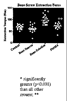
FIG. 1 is a graph of extraction torque results illustrated that the present
Mg0-MKP-
sugar-based product (Bone Solutions) had significantly (p<0.001) greater
extraction
torque (mean 97.5+/- 17.7 Nm) than control, Ca-based product and PMMA. PMMA
had
significantly (p<0.05) greater extraction torque than Ca-based product.
Best Mode for Carrying Out the Invention
The invention provides a bio-material for in-situ (i.e. in vivo) attachment of
biological structures to each other and to manmade structures. The bio-
adhesive also
facilitates the repair of bone, ligaments, tendons and adjacent structures.
Also provided
is a bone substitute for surgical repair. The invented formulation is usable
at a myriad of
temperatures, pH ranges, humidity levels, and pressures. However, the
formulation is
designed to be utilized at all physiological temperatures, pH ranges, and
fluid
concentrations. The mixture typically is injectable, prior to setting and
exhibits neutral
pH after setting. It is absorbed by the host over a period of time.
The mixture is particularly useful in situations (such as plastic surgery)
whereby
the use of metallic fasteners and other non-bloabsorbable materials are to be
assiduously avoided. The material also is useful when a certain amount of
expansion or
swelling is to be expected after surgery for example in skull surgeries. It is
a good
platform for bone-formation. The material can be also used as an anchoring
device or
CA 02580948 2012-10-24
6
grafting material
Generally, the bio-adhesive is derived from the hydrated mixture which
comprises: KH2PO4, a metal oxide, sugar and a calcium containing compound.
Exemplary formulations include the following:
Formulation I *
Potassium phosphate (i.e.KH2PO4) 61%
MgO (calcined) 31%
Ca10(PO4)6(OH)2 4%
Sucrose C12H22011 (powder) 4%
*All values are weight percentages
Water is added up to about 40 weight percent of the formulation, preferably
between 22-25 weight percent.
Formulation II*
KH2PO4 54%
MgO (calcined) 33%
Calcium-containing compound 9% (whereby the compound is
Ca10(PO4)6(0F)2)
Sucrose C12H22011 (powder) 4%
*All values are weight percentages
Water is added up to about 40 weight percent of the formulation, preferably
between about 22-25 weight percent.
Formulation III*
KH2PO4. 44%
MgO (calcined) 44%
Calcium-containing compound 8% (whereby the compound is
Ca10(PO4)6(OH)2 or CaSiO3,
Sucrose C12F122011 (powder) 4%
*All values are weight percentages
Water is added up to about 40 weight percent of the formulation, preferably
between about 36-38 weight percent.
Formulation IV*
KH2PO4 45%
MgO (calcined) 45%
Calcium-containing compound 9% (whereby the compound is
Ca10(PO4)3(OH)2, CaSiO3, or combinations
thereof)
CA 02580948 2012-10-24
7
Sucrose C12F122011 (powder) 1%
*All values are weight percentages
Water is added up to about 40 weight percent of the formulation, preferably
between 22-25 weight percent.
Formulation V
KH2PO4 46%
MgO (calcined) 45%
Ca10(PO4)6(OH)2 8%
Sucralose 2%
*All values are weight percentages
Water is added up to about 40 weight percent of the formulation, preferably
between 22-25 weight percent.
Formulation VI
KH2PO4 61%
MgO (calcined) 32%
Caio(PO4)6(01-)2 4%
Dextrose 1.5%
a- Ca3(PO4)2 1.5%
*All values are weight percentages
Water is added up to about 40 weight percent of the formulation, preferably
between 22-25 weight percent.
Formulation VII
KH2PO4 50%
MgO (calcined) 35%
Ca10(PO4)6(0F1)2 7%
Ca3(PO4)2 3%
Dextrose 5
*All values are weight percentages
Water is added up to about 40 weight percent of the formulation, preferably
between 22-25 weight percent.
Formulation VIII
KH2PO4 61%
CA 02580948 2012-10-24
8
Metal oxide 32% (wherein the metal oxide is MgO, Ca,
FeO or combination thereof),
Ca10(PO4)8(OH)2) 6%
Sugar 1%
*All values are weight percentages
Water is added up to about 40 weight percent of the formulation, preferably
between 22-25 weight percent.
Formulation IX
KH2PO4 54%
Phosphoric Acid 4%
Metal oxide 32% (wherein the metal oxide is MgO,
ZrO, FeO or combination thereof),
Ca10(PO4)8(0F1)2) 7%
Sucrose 3%
*All values are weight percentages
Water is added up to about 40 weight percent of the formulation, preferably
between 22-25 weight percent.
Formulation X
KH2PO4 45%
MgO (calcined) 45%
Ca10(PO4)6(OH)2 10%
Water is added up to about 40 weight percent of the formulation, preferably
between 22-
25 weight percent.
While the above formulations and weight percents are the most preferred
proportions, a range of dry constituents can also be used. For example, a
suitable range
for the phosphate (i.e. MKP) is generally between 20-70 weight percent,
preferably
between about 40-65 weight percent. In some situations it maybe preferable to
use the
phosphate at a range between about 40-50 weight, while in others in may be
preferably
to use a range of about 50 and 65.
A suitable range for the metal oxide (i.e. MgO) is generally between about 10-
60,
preferably between 10-60, and even more preferably between 30-50 weight
percent. In
some situations it maybe preferable to use between about 35 and 50 weight
percent.
Calcium containing compounds can be added in various weight percentages.
CA 02580948 2012-10-24
9
The calcium containing compound(s) is preferably added at about 1-15 weight
percent,
although higher percentages can be employed.
Sugars (and/or other carbohydrate containing substances) are generally present
at weight percent between 0.5 and 20, preferably about 0.5-10 weight percent
of the dry
composition.
Water (or another aqueous solution) can be added in a large range of weight
percents generally ranging from about 15-40 weight percent.
For some embodiments (i.e. formula III) it has been found that adding water at
a
weight percent of about 37 weight percent produces a creamy textured material
that Is
extremely easy to work with has excellent adhesive properties and is easily
injectable
through a syringe.
It is important to note that these are exemplary weight percents and that the
ranges may vary with the addition of various fillers, equivalents and other
components or
for other reasons.
A salient feature of the present invention is the ratio between MKP (MKP
equivalent, combination, and/or replacement) and the metal oxide. A preferred
embodiment has a weight percent ratio between MKP and MgO between about 4:1
and
0.5:1, more preferably between approximately 2:1 and 1:1. In such a preferred
embodiment the inventor surmises that the un-reacted magnesium is at least
partly
responsible for the in vivo expandability characteristics of the bio-adhesive.
Specifically the metal oxide (i.e. magnesium oxide) reacts with water and
serum
and in and around the living tissue to yield Mg(OH)2 and magnesium salts. It
has been
found that one embodiment of the material generally expands to between 0.15
and 0.20
percent of volume during curing in moisture. The expansion of the material is
believed
to increase the adhesive characteristics of the material. For example, the
disclosed
material has been shown to effectively attach soft tissues like ligaments to
bone, the
expansion of the material improving adhesion through mechanical strength.
MgO is the preferred metal oxide (metal hydroxide or other equivalent),
however,
other oxide and hydroxide powders can be utilized in place of or in addition
to MgO,
including but not limited to: FeO, Al(OH)3, Fe203, Fe304, ZrO, and Zr(OH)4,
zinc oxides
and hydroxides, calcium oxide and hydroxides and combinations thereof.
MKP is preferred, but for some applications other compounds may be substituted
for (or added to) MKP, including but not limited to: phosphoric acid and
phosphoric acid
CA 02580948 2012-10-24
salts like sodium, aluminum phosphate, mono-ammonium phosphate and di-ammonium
phosphate.
Calcium-Containino Compound
A calcium containing compound is essential to the invention as it increases
both
5 the bio-compatibility and bio-absorption of the biomaterial. The calcium
compound(s)
can be selected from a variety of biocompatible calcium containing compounds
including
but not limited to tricalcium phosphates. Suitable tricalcium phosphates
include a-
Ca3(P042, 13-Ca3(PO4)2, and Ca10(PO4)6(0F)2.
In general, suitable calcium containing compounds include but are not limited
to:
10 tricalcium phosphates, biphasic calcium phosphate, tetracalcium
phosphate, amorphous
calcium phosphate ("ACP"), CaSiO3, oxyapatite ("OXA"), poorly crystalline
apatite
("PCA"), octocalcium phosphate, dicalcium phosphate, dicalcium phosphate
dihydrate,
calcium metaphosphate, heptacalcium metaphosphate, calcium pyrophosphate and
combinations thereof.
Preferred calcium containing compounds include: tricalcium phosphates, ACP,
dicalcium phosphate, dicalcium phosphate dihydrate and combinations thereof.
a-Ca3(PO4)2,11-Ca3(PO4)2, and Ca10(PO4)6(OH)2, equivalents and combinations
thereof, being the most preferred. A preferred tricalcium phosphate is a
pharmaceutical
or food grade tricalcium phosphate manufactured by Astaris (St. Louis, MO).
Calcium containing compounds increase the bio-compatibility and bioabsorption
of the bio-adhesive. However, calcium containing compounds vary in their
degrees of
bioabsorption and biocompatibility. Some characteristics even vary within the
various
tricalcium phosphate compounds.
It may be advantageous to combine various calcium containing compounds to
manipulate the bio-compatibility and bioabsorption characteristics of the
material. For
example Ca10(PO4)6(OH)2 (HA") is stable in physiologic conditions and tends to
be
relatively poorly absorbed while R-Ca3(PO4)2 is more readily absorbed. The two
can be
combined (i.e. bi-phasic calcium phosphate) to form a mixture having
characteristics
somewhere between HA and II-Ca3(PO4)2. A number of calcium containing compound
combinations can be envisioned.
Sugars, Sugar Substitutes, Sweeteners, Carbohydrates and Equivalents
A salient aspect of a preferred embodiment is the incorporation of at least
one
CA 02580948 2012-10-24
11
sugar or sugar like substance to the bio-material matrix. Inventor discovered
that sugar
containing bio-materials have significant osteoproliferative properties as
well as
enhanced adhesive capabilities. It is believed that a sugar like sucrose may
be replaced
or supplemented with other sugars and sugar related compounds.
Suitable sugars or sugar related compounds include but are not limited to:
sugars, sugar derivatives (i.e. sugar alcohols, natural and artificial
sweeteners (i.e.
acesulfame-k, alitame, aspartame, cyclamate, neohesperidine, saccharin,
sucralose and
thaumatin), sugar acids, amino sugars, sugar polymers glycosaminoglycans,
glycolipds,
sugar polymers, sugar substitutes including sugar substitutes like sucralose
(i.e.
SplendaO, McNeil Nutritionals LLC, Ft. Washington, PA), corn syrup, honey,
starches,
and other carbohydrate containing substances.
Exemplary sugars include but are not limited to: sucrose, lactose, maltose,
cellobiose, glucose, galactose, fructose, dextrose, mannose, arabinose,
pentose,
hexose. Preferably the sugar additive is a polysaccharide, more preferably a
disaccharide like sucrose.
One preferred additive is sugar combined with a flow agent like starch. An
exemplary additive is approximately 97.weight percent sucrose and 3 weight
percent
starch.
The sugar compound, like the other components, can be in a variety of forms
including but not limited to dry forms (i.e. granules, powders etc.), aqueous
forms,
pastes, and gels. It may prove preferable to use a powdered.
The inventor has shown that the invented sugar containing bio-material possess
surprisingly good adhesive qualities. In fact, the invented composition
outperformed
current state of the art materials. (discussed below, See Example I and III).
It is
believed that the sugar improves the physical (and possibly the chemical)
bonding of the
cement to objects. The improved adhesion of sugar containing phosphate cements
is
particularly well suited for attachment of soft tissue like ligaments and
tendons to bone
without the need for intrusive non-absorbable devices like screws and pins.
The
elimination of non-absorbable devices reduces post-operative complications and
preferably promotes bone growth around the repaired site.
Surprisingly and unexpectedly, it was discovered that a sugar containing
composition greatly enhanced formation of new bone. It is believed that the
sugar and
other compounds of the composition provide near ideal conditions for new bone
CA 02580948 2012-10-24
12
formation. This assertion is supported by surprising and unexpected test
results shown
in Example II.
Bone Graft Material
In one embodiment the composition of present invention provides a bone
substitute and a platform for bone formation. An advantage of the substance is
its
gradual absorption by the body without rejection or reaction to contacted
structures. A
further advantage of the invented composition is its significant
osteoproliferative
properties. In fact, in studies the invented composition enhanced bone
formation to
such a surprising degree, so much so that it is believed that the composition
may also be
osteoinductive which is completely unexpected and unprecedented for a multi-
purpose
biomaterial without the use of growth factors. The bio-material is also
believed to have
micro and macro pores.
Additional Embodiments
The formulations disclosed herein may incorporate additional fillers,
additives
and supplementary materials. The supplementary materials may be added to the
bio-
material in varying amounts and in a variety of physical forms, dependent upon
the
anticipated use. The supplementary materials can be used to alter the bio-
material in
various ways.
Supplementary materials, additives, and fillers are preferably biocompatible
and/or bioresorbable. In some cases it may be desirous for the material to be
osteoconductive and/or osteoinductive as well. Suitable biocompatible
supplementary
materials include but are not limited to: bioactive glass compositions,
calcium sulfates,
coralline, polyatic polymers, peptides, fatty acids, collagen, glycogen,
chitin, celluloses,
starch, keratins, nucleic acids, glucosamine, chondroitin, and denatured
and/or
demineralized bone matrices. Other suitable supplementary materials are
disclosed in
U.S. Patent No. 6,331,312 issued to Lee and U.S. Patent No. 6,719,992 issued
to
Constanz .
In another embodiment of the invention the bio-material contains a
radiographic
material which allows for the imaging of the material in vivo. Suitable
radiographic
materials include but are not limited to barium oxide and titanium.
The bio-material described herein may prove ideal for creating bioresorbable
implants and devices which can be resorbed by the body overtime, reducing
CA 02580948 2012-10-24
13
complications while promoting bone reformation. The bio-material can also be
used to
coat various implant parts.
In yet another embodiment the invented bio-material contains a setting
retarder
or accelerant to regulate the setting time of the composition. Setting
regulators are
preferable biocompatible. Suitable retarders include but are not limited to
sodium
chloride, sodium fluosilicate, polyphosphate sodium, borate, boric acid,
.boric acid ester
and combination thereof.
A preferred retarder composition comprises: a sugar (sucrose) and boric acid
in a
weight percent ratio of between 0.5:1 and 1:0.5, preferably at a ratio of
approximately
1:1. This setting regulators is preferably added at less than 5 weight % of
the dry binder
matrix.
The disclosed bio-material may also be prepared with varying degrees of
porosity. Controlling porosity can be accomplished through a variety of means
including:
controlling the particle size of the dry reactants, and chemical and physical
etching and
leaching. A preferred embodiment increases porosity of the bio-material by
addition of
1-20 weight percent of an aerating agent, preferably about 1-5 weight percent.
Suitable
aerating agents include but are not limited: carbonates and bicarbonates such
as:
calcium carbonate, sodium carbonate, sodium bicarbonate, calcium bicarbonate,
baking
soda, baking powder, and combinations thereof.
The biomaterial may be used as delivery system by incorporating biologically
active compounds into the bio-material (i.e. antibiotics, growth factors, cell
etc.). A
porous bio-adhesive increases the effectiveness of such a delivery system.
Cationic antibiotics, especially aminoglycosides and certain peptide
antibiotics
may be most desirable when incorporating drugs into the bio-material. Suitable
aminoglycosides include but are not limited to: amikacin, butirosin,
dideoxykanamycin,
fortimycin, gentamycin, kanamycin, lividomycin, neomycin, netilmicin,
ribostamycin,
sagamycin, seldomycin and epimers thereof, sisomycin, sorbistin, spectinomycin
and
tobramycin. Using inorganic salts like sulfates, phosphates,
hydrogenphosphates
maybe preferable, sulfates being the most preferable. Further information
about using
antibiotics and growth factors in bio-materials can be found in U.S. Patents
No.
6,485,754, issued to Wenz .
Growth factors include but are not limited to growth factors like transforming
growth
factor TGF-11.
CA 02580948 2012-10-24
14
The disclosed bio-material composition may also be seeded with various living
cells or cell lines. Any known method for harvesting, maintaining and
preparing cells
may be employed. See U.S. Patents Nos: 6,719,993 issued to Constanz, 6,585,992
issued to Pugh and, 6,544,290 issued to Lee.
One embodiment of the invention has been shown to be extremely useful as a
scaffold for hard tissue growth and possibly soft tissue growth as well. In
addition,
tissue-producing and tissue-degrading cells may be added to the composition
included
but not limited to: osteocytes, osteoblasts, osteoclasts, chondrocytes,
fibroblasts,
cartilage producing cells, and stem cells. Methods of isolating and culturing
such cells
are well known in the art.
The invented composition can incorporated into an orthopedic kit comprising:
the
material (MKP, metal oxide, calcium containing compounds etc.) in dry form, an
activator
solution (water or other aqueous solution), and any medical devices (i.e.
syringes, knives
etc.), implants, or other agents needed during an operation using the invented
composition. The material and activator solution will preferably be present in
a
predetermined, optimized ratio. Other embodiments of such an orthopedic kit
can also
be envisioned. The biomaterial and other kit components are preferably
sterilized by
techniques well known in the art.
Substance Preparation
A metal oxide powder is a salient ingredient in the invented mixture.
Optionally,
the oxide is subjected to a calcinated process. Calcination durations and
temperatures
are determined empirically, depending on the final characteristics and setting
times
desired. Generally, however, calcination temperatures of up to 1300 C for up
to several
hours are typical.
After calcination, the oxide powder is mixed with MKP, a calcium containing
compound, and sugar. One method for sizing and homogenizing the various
powders is
via vibratory milling. Another homogenization method utilizes a ribbon mixer
wherein the
particles are ground to a fine size. Dry compounds are disclosed herein,
however,
aqueous versions (or other forms i.e. gels etc) of some of the bio-materials
components
can also be utilized. Generally, pharmaceutical grade compounds are utilized.
Sterilization of the various components may be required using sterilization
techniques
known in the art.
CA 02580948 2012-10-24
Upon homogenization wherein all of the constituents are contained in a dry
homogeneous mixture, water (or other aqueous solution) is generally added up
to about
40% of the weight of the resulting slurry although the amount of water can be
adjusted to
form a bio-material of varying viscosity. The slurry is mixed for between 1-10
minutes
5 depending upon conditions. Mixing can be achieved by a variety of
techniques used in
the art including hand and electric mixing. See, U.S. Patent 6,533,821 issued
to present
inventor for further details.
The bio-material can be created in injectable, paste, puddy and other forms.
The
slurry is produced at the user site. The consistency of the material can be
manipulated
10 by varying the amount of water added to the dry mixture. Increasing the
water content
generally increases the flowability while decreasing the water content tends
to thicken
the slurry. The material can be prepared in a myriad of forms.
Working times can be increased or decreased by varying the temperatures of
bio-material components. Higher temperature components tend to react and set
quicker
15 than cooler components. Thus regulating the temperature of the water (or
other
reactants) can be an effective way to regulate working time.
Bonding occurs primarily between the adhesive and bone. However, the
adhesive also bonds to itself, or to soft tissue. The inventor has found that
the use of a
phosphoric acid instead of water increases the bonding strength of the
material. The
molarity of the phosphoric acid can vary, as long as the eventual pH of the
slurry is not
hazardous to the patient, or contraindicative to healing. Generally, a slurry
pH of
between 6 and 8 is appropriate, however other slurry pHs may be employed
depending
on desired results.
Attachment
/5 The attachment of the bio-adhesive to various structures can be
accomplished in
a number of ways including but not limited to: injection, spraying, and other
application
means. The attachment means will vary according to the desired application and
the
form of the adhesive. One exemplary method is described in instant inventors
U.S.
Patent Application No. 6,533,82" .
Example I
An experiment comparing the adhesive qualities of a prior art bone filler
(NORIAN Skeletal Repair System, Paoli, PA) and a preferred embodiment of the
CA 02580948 2012-10-24
16
present bio-adhesive having the weight percent formula: 54% MKP, 33% magnesium
oxide, 9% Ca10(PO4)6(OH)2, and 4% Sucrose mixture (the sugar mixture being 97%
sugar and 3% starch).
The goal of the study was to determine if an injectable MgO-MKP-sugar based
formulation of the present invention had adhesive properties for bone to bone
and
tendon to bone using clinically relevant models. Biomechanical studies were
performed
using a canine cadaver model of anterior cruciate ligament repair and femur
fracture.
Tissue adhesion was quantified with mechanical pull-out and three-point
bending
studies. Sixteen knee joints with femurs and Achilles tendons from 8 mid-sized
dogs
were harvested and three tissue contructs for testing were prepared.
ACL Model: A) Bone to Bone. Bone-patellar ligament grafts were cut and the
patella
bone press-fit into a 7mm diameter bone tunnel in the femur at the ACL
footprint to
mimic human ACL reconstruction. The ligament end served as the anchor for pull
out
mechanical testing. B) Tendon to Bone. Achilles tendon grafts were placed
through a
7mm diameter tibial bone tunnel initiated at the ACL footprint and exiting the
lateral tibial
cortex to mimic human ACL reconstruction. Anchoring screws or sutures were not
used
to augment these repairs. Treatment groups were: 1) Press-fit (Control; n=16);
2)
Calcium based injectable formulation (n=8) (Negative paste control) (NorianC)
Skeletal
Repair System- Synthes, Paoli, PA); 3) MgO-MKP-sugar based bioadhesive. Limbs
were paired for groups 2 and 3. Product was prepared and injected into the
bone defects
surrounding the bone or tendon grafts in the bone tunnels and allowed to cure
overnight.
Grafts were mechanically tested in tension for peak load to failure at
lmm/sec.
Fracture Model: A 1cm long oblique osteotomy was made in the midshaft of the
femur diaphysis and four materials tested to hold the fracture in reduction:
1) Blood clot
(freshly clotted equine blood); 2) cyanoacrylate glue (Ross Super Glue Gel-
Ross
Products, Columbus, OH); 3) Calcium based injectable formulation (NorianO
Skeletal
Repair System- Synthes, Paoli, PA); 4) MgO-MKP-sugar based injectable
formulation.
Additionally, four intact femurs were tested to failure. Groups 3 and 4 were
tested in
paired limbs. Groups 1 and 2 were tested in paired limbs; one half before and
one half
after application of the paste products in groups 3 and 4. First tested
products were
readily removed by scraping. Injectable pastes and cyanoacrylate were applied
liberally
to the fractured bone ends, held together for 15 minutes until hardened, and
allowed to
CA 02580948 2012-10-24
17
cure overnight. Blood clot was applied immediately before testing. Femurs were
tested in
3-point bending under displacement control at 0.1mm/sec for peak load to
failure.
Stiffness and stress to failure were calculated from the slope of the linear
portion of the
load deformation curve and after estimation of bone area at the fracture with
calipers.
Fractures which fell apart before testing were recorded as 0 N to failure.
Data in the ACL model were analyzed with the paired Student's t-test for
calcium vs
magnesium formulations and for press fit vs formulation. Data in the fracture
model were
analyzed with a 1-factor ANOVA for treatment group. Significance was set a
p<0.05.
RESULTS: In the ACL model, both the calcium based formulation and the MgO-
MKP-sugar based formulation had significantly greater pull out force than
press-fit
(friction) within the tunnel for both patellar bone and Achille's tendon
(p<0.004). The
MgO-MKP-sugar based formulation had the greatest adhesive properties,
significantly
greater than the calcium based formulation for both bone (2.5-fold;p<0.0) and
tendon
(3.3-fold;p<0.0). (Table 1)
In the fracture model, blood clot and calcium based formulation had no
adhesive
properties (0 N load to failure) in all specimens. Blood clot was unable to
hold the two
ends of the femur in apposition. The calcium based product held the femur ends
in
apposition, but separation occurred prior to testing. MgO-MKP-sugar based
formulation
and cyanoacrylate failed at significantly greater loads (p<0.0001) and
cyanoacrylate
failed at significantly greater loads (127 N; p<0.01) than the MgO-MKP-sugar
based
formulation (37.7 N). Intact femurs failed at much greater loads with any bone
adhesive
achieving less than 10% of original bone strength.
Table 1. ACL Model ¨ Peak Mean (+/- SEM) Tensile Load (N) to Failure.
Groups Press-fit Ca-based MgO-MKP-sugar
Formulation based Formulation
(Norianq
Bone- 41.6 +/- 427.7 1025.6 +/-118.2a
Bone 16.8a +/103.9a
Tendon- 12.9 +/- 101.6 +/- 338.2 +/-69.9a
Bone 0.03a 23.1 b
Table 2. Mean (+1- SEM) Biomechanical Properties to Failure in Femur
Osteotomies
Repaired with Potential Bone Glues
CA 02580948 2012-10-24
18
Groups Peak Peak Stiffness
Load Stress (N/mm)
(N) (N/mm2)
Blood Clot 0 +1-0 0 +/-0 0 +/-0
Ca-based 0 +/-0 0 +/-0 0 +/-0
Formulation
(Noriane)
MgO-MKP based 37.7 +/- 0.09 +/- 148.7 +/-
Formulation 27.4
Cyanoacrylate 127.0 0.3 +/- 783 +1-
+1-
Intact Femur 1455.8 4.18 +/- 666.8 +1-
+1-
In bone and tendon pullout from a bone tunnel, paste formulations provide some
adhesion due to cement properties (ie hardened filler). However, the MgO-MKP-
sugar
based formulation had additional and substantial adhesive properties of over
1000 N in
bone that should exceed. forces put on the construct in vivo. In femur
fracture
reconstruction, the MgO-MKP-sugar based formulation provided bone adhesion,
but not
as great as our nonbiodegradable positive control glue. Repaired construct
strength was
still < 10% of intact femur strength, but may provide fragment containment and
osteoconduction.
A biodegradable MgO-MKP-sugar based, injectable formulation adhered bone
and tendon within bone tunnels sufficiently to significantly augment, or
potentially be
used independently, in ACL reconstructions. Adhesion of bone ends may be
sufficient to
contain fracture fragments in comminuted fracture repair and may be useful if
osteoconduction and biodegradation profiles complement fracture healing as
anticipated.
Example II OSTEOPROFLIFERATIVE RESULTS: Formula II
ANIMALS:
Species/breed: Equine/Mixed Breed
Initial age: A minimum of 3 years maximum of 20 years at
start of acclimatization
CA 02580948 2012-10-24
19
Initial weight: Approximately 800-1200 kg at acclimation
Sex: geldings, mares
Identification of animals: Individual neck collar, ear tag or halter tag
Pretreatment: Vaccinations: Eastern, Western Encephalitis,
Influenza; West Nile Virus and tetanus.
De-wormed post arrival at Ohio State Finley
Research farm. Animals will have had no
previous compound exposure.
SITE DESCRIPTION:
This study will be conducted at the Ohio State University Alice Finley
Memorial farm
(Finley farm) and the Veterinary Teaching Hospital (VTH). Evaluation will take
place at
the Veterinary teaching hospital. The facility's animal accommodations,
laboratory
support areas, record keeping, and anticipated compliance are to be
satisfactory to meet
the requirements of this protocol.
CA 02580948 2012-10-24
MANAGEMENT:
Floor space per animal: Animals will be housed in box stalls for the
duration of the study.
Feeding and watering method: Hay and grain is fed twice/day. Water will be
provided ad libitum.
Housing: Bedded box stalls at the Finley farm or VTH.
Environmental control: Finley farm box stalls are in a barn that is
not
temperature regulated.
VTH box stalls are sheltered in a building and are
temperature regulated.
Feed: Approximately 3 lbs. grain/animal/day. Hay
will
be offered at approximately 15 lbs twice daily and
more as necessary.
Water: Water will be checked daily and cleaned if
necessary.
DESIGN:
Experimental Study; Nested Paired Design; Each horse serves as its own
control.
5 Horses, limb, and medial or lateral splint bone are assigned in a
controlled block design.
Eight horses, bilateral MtII and MtIV fractures (24 splint bones). One medial
and one
lateral splint (Mtl I and MtIV) will be treated with MgO-MKP-sugar injectable
formulation
(n=16). The contralateral splint will be injected with either Calcium-based
injectable
formulation [Comparative treatment] or receive no injection (Untreated
control) Table 3
10 the result is 4 groups of 8 limbs each: 1) Untreated Natural healing
(control), 2) Calcium-
based nonadhesive injectable product [Treatment comparison], 3) Magnesium-
based
adhesive injectable test product.
Table 3. signment of metatarsi (splints) to treatment groups (n=8 per group)
HORSES METATARSAL II -TREATMENT METATARSAL IV-TREATMENT-
None Bone Solutions Bone Scource Bone Solutions
Product Product Product
MgO-MKP-sugar Ca-based MgO-MKP-sugar
340 X - right X - left X - left X - right
352 X - left X -right X - right X - left
354 X - right X - left X - left X - right
362 X - left X - rigy X - ri9ht X - left
_ _ _ _ _
CA 02580948 2012-10-24
21
365 X - right X - left X - left X - right
366 X - left X - right X - right X - left
369 X - right X - left X - left X.- right
377 X - left X - right X - right __ X - left
PROCEDURE:
Inclusion Criteria: Horses (aged 3-20 yrs) must be healthy on physical
examination and
complete blood count, and be sound with no palpable or radiographic
abnormalities of
the metatarsus.
Blinding: Splint and limb assignments will be recorded. All radiographic, qCT,
biomechanical testing and histomorphology will be performed with samples coded
in a
blinded fashion.
Fracture Model ¨ Fractures (Mt (Splint) II and Mt (Splint) IV) will be
performed under
general anesthesia at day 0. Horses will be administered procaine penicillin
(22,000units/kg) intramuscularly and gentamicin (6.6 mg/kg) intravenously 30
minutes
prior to anesthesia. Horses will be sedated with xylazine HCI (1mg/kg),
induced with
ketamine (2mg/kg) and maintained in dorsal recumbency on isoflurane and oxygen
to
effect. The splint bones are directly under the skin at the locations for
these bone
defects. After aseptic preparation, small 2-cm incisions will be made over the
smooth
palpable surface of the splint bones; 15 cm distal to the palpable
tarsometatarsal joint. A
curved spatula is placed under the splint bone and a nitrogen-driven
oscillating bone
saw used to create a 3-piece fracture containing a triangular fragment [ 90 ,
1.5-cm
arm]. The bone saw removes a 1mm width of bone. The incisions are flushed
liberally
with saline to remove bone dust and dried. Bleeding will be arrested on the
bone surface
by pressure or radiofrequency cautery. The triangular piece of bone will be
placed back
into the parent defect according to assignment. If the bone is assigned to
receive
injectable paste, it will be mixed according to manufacturer's
recommendations, ¨0.5ml
will be placed onto the cut bone surface and the triangular piece glued back
into place.
The fragment will be press fit into place for 30 minutes to assure curing or
permit blood
clot in the control specimens. A layered closure of the incision will be
performed, a
sterile bandage applied and horses recovered. Sterile bandages are maintained
for 2
weeks,
Material Preparation: Bone Solutions product (MgO-MKP-sugar based) and Bone
Source product (Ca based) were mixed with a metal spatula iust prior to
application in
CA 02580948 2012-10-24
22
order of Table 3 and applied into the fracture gap with a metal spatula. Both
products
were applied after 2 minutes of mixing and reapplied as needed to position
sufficient
material into the fracture bed.
OUTCOME ASSESSMENTS:
Clinical Assessments - Horses will be monitored daily for clinical signs of
any reaction to
the procedures or therapy. Rectal temperature (T), heart rate (HR) and
respiratory rate
(RR) will be recorded daily for 1 week following surgery and following
injections and then
weekly until termination of the study at 8 weeks.
Pain ¨ Horses will be monitored for pain by assessing physical parameters (T,
HR, RR),
lameness scores (0-5) while in the stall.
Swelling Surgical site swelling will be assessed by score [0-4; 0= no swelling
and 1=
minimal, 2=mild, 3= moderate, and 4= marked swelling]. Surgical site drainage
will be
assessed by drainage score of drainage character (color, viscosity) [0-4; 0=no
drainage,
1=0-25% of the bandage surface stained with drainage, 2=26-50% of the bandage
surface stained with drainage, 3=51-75% of the bandage surface stained with
drainage;
4=76-100% of the bandage surface stained with drainage].
Gait Assessment - Lameness will be scored 0-5 for each hindlimb at the walk on
week ¨
1,1,3,4,5,6,7, and 8. [0=no lameness, 1=minimal lameness, 2 mild lameness, 3
moderate lameness, 4 marked lameness (only placing part of the foot), and 5
=non-
weightbearing lameness.
Euthanasia - Horses will be euthanized at 7 weeks within the guidelines of the
AAEP by
an overdose of intravenous pentobarbital solution after sedation with 500 mg
xylazine
HCI IV and the distal limbs harvested.
Fracture Healing (Bone Adhesion and Union)
Radiographs ¨ Oblique radiographs will be taken before fracture and injection,
and every
other week for 7 weeks until termination. Radiographs will be scored for
fracture
fragment migration (0=none, 1=minimal, 2=mild, 3=marked), bone proliferation
(0=none,
1=minimal, 2=mild, 3=marked), bone remodeling (0=none, 1=minimal, 2=mild,
3=marked), and fracture closure (0=none, 1=minimal, 2=mild, 3=complete). The
width
and length of the fracture callus will be measured and calibrated using a
radiographic
measuring standard included in all films.
CA 02580948 2012-10-24
23
Quantitative Computer Tomography (gC7) - The metatarsus of the distal limbs
will be
screened at 1 cm intervals for soft tissue abnormalities associated with the
fracture
healing process. At and for at least 1cm proximal and distal to the bone
defect sites, 1
mm slices will be obtained. Subsequently, Mt IV and MtII will be harvested,
cleaned of
soft tissue and scanned in cross section in lmm slices from the top to the
bottom of the
callus to determine area, density and mineral content (area x density) of
mineralized
callus. Each slice will be standardized for x-ray attenuation differences for
density
measurements by using potassium phosphate standards. After standardization, a
calculation will be performed to convert potassium phosphate region of
interest (ROI) to
ash density (mg/mm3). Tracings of the ROI will be performed on cross section
views
from bone at the healed fracture site for bone area (amount of bone), density
of bone in
the healing fracture, and density of bone in the callus. Splints will be
mechanical tested
immediately after qCT.
Mechanical Testing Metatarsal II and IV ends will be secured in grips tested
quasi-
statically to failure in 3-pt bending (1.5 mm/sec) using a servohydraulic
materials testing
system. The bones will be positioned in the jig to ensure appropriate and
bending for
both right and left sides. The load/deformation data will be collected and
maximum load
to failure calculated.
Histology - After mechanical testing splint bones will be embedded
undecalcified in
PMMA, sectioned (10um) in the longitudinal frontal plane [EXACKT system, SU],
stained with Masson's Trichrome, and evaluated for callus composition,
maturity, cortical
continuity, and fracture bridging. Assessment of tissue type, such as
cartilage, fibrous
tissue and bone, within the defect will be noted.
Data Analysis: Descriptive statistics will be generated for all outcome
variables. A paired
t-test will be used to evaluate the effect of MgO-MKP-sugar-based (Mg-based)
injectable
paste treatment compared to Calcium-based or no treatment on healing for
objective
data. Scored data will be expressed as median and range and analyzed by Mann
Whitney U Rank test. Differences will be considered significant at p<0.05.
FINDINGS AND CONCLUSIONS
Experimental Design: All 8 horses completed the 7 week healing study as per
the
assignment in Table 3. All horses met the inclusion criteria. Signalments are
listed in
Table 4. All horses underwent surgery to create the triangular metatarsal
fractures and
CA 02580948 2012-10-24
24
application of the assigned treatment. The fragment was press fit into the
parent defect
for 30 minutes and materials seemed cured at surgical closure.
Table 4. Signalment of horses used in this study.
Horse # Breed Sex Approximate Age (yrs) Scale Weight (kg)
340 Morgan/Stand ard bred Female 9 491
352 Thoroughbred Female 9 513
354 Standard bred Female 17 480
362 Paint Female 8 519
365 Standardbred Female 10 528
366 Paint Female 11 534
369 Quarter Horse X Female 7 486
377 Quarter Horse X Female 6 554
OUTCOME ASSESSMENTS:
Clinical Assessments -
Pain and Gait - Horses were not lame at any time point following surgery as
estimated
by lameness score (median 0, range 0) as per protocol. Physical examination
parameters remained within normal limits through out the study.
Incisional Swelling and Drainage ¨ There was no difference in swelling
postoperatively
among the 4 treatment groups and there was no drainage at the incisions at any
time
point. At termination of the study, only one surgical site had a palpable
firm, nonpainful
¨ 2cm enlargement. The interpretation of these data is that the Mg and Ca
materials are
clinically biocompatible, clinically nonirritating. Clinically evident tissue
or bone
proliferation did not occur and therefore was not excessive.
CA 02580948 2012-10-24
Radiographs¨ Radiographs were taken as per protocol before surgery and every
other
week until the termination of the study. Radiographs were evaluated for
fragment gap,
presence of material, bone formation, bone remodelling and bone healing.
Migration of
5 the fragment was assessed as the distance (mm) from the apex of the
fragment to the
apex of the fragment bed as a straight line. The MgO-MKP-sugar treatment
secured the
fragment significantly closer (P<0.05) to the parent fragment bed than either
no
treatment or Ca-treatment immediately after surgery (week 0). Migration of the
fragment
did not occur in the Mg- or Ca-treatments until week 4 in Mtll or until week 2
in MtIV. The
10 fragment migrated less in the MgO-MKP-sugar-treatment as compared to no
treatment
at all time points and this was statistically significant for up to 4 weeks.
(See appendix for
graph and data) Callus formation (bone proliferation at the healing fragment)
was
estimated from the radiographs by measuring the width and height of the new
bone
formed around the fragment at its greatest point and multiplying these numbers
to
15 estimate area of new bone. New bone callus was significantly greater in
the Mg0-MKP-
sugar-treatment (Mg-treament) than both the Ca-treatment and no treatment in
both Mtll
and Mt1V. Significant formation of bone occurred by 4 weeks and persisted
through 7
weeks.
Radiodense material could be identified in the gap between the fragment and
parent
20 bone on the radiographs of some horses at some time points, particularly
the early time
points. (See graph in appendix) product was noted of equal frequency and
amount to
Ca product until week 4 after which less material was noted in general (lower
scores),
but greater in Mg group, and at week 7 only in the MgO-MKP-sugar group.
Bone remodelling around the fragment and parent bone, was significantly
greater in the
25 MgO-MKP-sugar -treatment than in the no treatment or Ca-treatment
groups.
Bone healing around the fragment and parent bone was greater in the MgO-MKP-
sugar
-treatment and this was significant (p<0.05) in all weeks compared to no
treatment and
at weeks 4, 6 and 7 compared to Ca-treatment.
CA 02580948 2012-10-24
26
Euthanasia and Bone Harvest ¨ Horses were euthanized at 7 weeks
postoperatively as
outlined by the protocol. Metatarsi and distal limbs were cut off, labelled,
stored in plastic
and frozen.
Quantitative Computed Tomography -
Intact limbs and metatarsal bones (4 per horse) were scanned [Picker P Helical
CT,
Philips Medical Systems for North America, Bothell, WA]after 7 weeks of
healing. Intact
limbs were scanned in cross section at 1cm slices and each slice evaluated
subjectively
for dystrophic mineralization of the surrounding soft tissue. No abnormal
mineralization
was noted including in the suspensory ligament, tendons or surrounding skin.
Metatarsal
bones were scanned in lmm slices in sagittal section from medial to lateral
and to
include at least 1 cm above the callus to 1 cm below the callus. The central
slice of the
metatarsal scans that transacted the fragment was selected and a region of
interest
traced for the gap, the fragment, and the callus. For the regions of interest
for the gap,
the fragment and the callus, measurements were recorded for density of tissue
and size
of region. Density measurements were then transposed from potassium phosphate
density to ash density using the phantom calculations simultaneously collected
with each
slice. There was a tendency (p<0.08) for the density within the gap between
the
fragment and the parent bone to be greater in the MgO-MKP-sugar -treatment
when
compared to no treatment. There was no difference (P<0.13) in density of the
gap
comparing Mg and Ca treatment. When taken in concert with the scored data from
the
radiographs, this likely reflects the presence of material at 7 weeks. (See
raw data and
tablulated data in appendix) There was no significant difference in density or
the size of
the fragment between groups. There was significantly greater amount of callus
around
the healing fragment in the Mg-treatment compared to no treatment (p<0.01) and
Mg-
treatment compared to Ca-treatment (p<0.02). These data corroborated the
radiographic
measurements of greater callus. In summary these data show that there was no
destruction of the fragment by the materials, no abnormalities In the density
of bone
formed and that the Mg-treatment significantly increased bone formation at the
fragment
site. This osteoproliferative effect seen in this model and species is an
osteoinductive
response to the Mg-product. Further investigation using the highest purity
product and
standard osteoinduction models will confirm this finding.
CA 02580948 2012-10-24
27
Mechanical Testing - Bones were failed in 3-pt bending and measurements
recorded for
peak load to failure (N) and cross sectional diameter (mm). Calculations were
made for
peak stress to failure (N/mm2). There was no significant differences in the
mechanical
testing results among any groups. The size and strength of the healed MtIV was
significantly greater than Mtn. (See appendix for data)
Histology¨ Bones were sectioned in cross section to mimic the plane of the qCT
assessments and to see the fragment and surrounding bone is cross section.
Material
staining brightly was grossly obvious in 6 of the 8 Mg-treated Mt IV bones and
3 or the 8
Mg- treated Mt 11 bones. Material was grossly apparent in 4 of the 8 Ca-
treated Mt IV
bones. Histologic evaluation of the specimens revealed that the tissue types
adjacent to
the fragments and material was fibrous tissue and/or bone. There was no
inflammatory
cells within this adjacent tissue. There was no granulomatous response (influx
of giant
cells). Bone was noted to be directly adjacent to the material. The histology
data
supports the following conclusions. The Mg material is not absorbed and
remained
adhere to the site for 7 weeks. The Ca material was either absorbed or
migrated from
the site by 7 weeks in many of the specimens. Both the Ca and Mg material is
biocompatible and did not incite an inflammatory reaction. The body did not
wall off the
materials. Bone or fibrous tissue, the anticipated healing tissue types were
abundant and
in close proximity to material without effect.
CA 02580948 2012-10-24
28
APPENDIX I-DOSAGE ADMINISTRATION
All animals will receive Bone Source and Bone Solutions Products. Products
will be
mixed immediately prior to placement, using a spatula, into the bone defect to
cover all
surfaces of bone. Bone fragments will be held into position for a minimum of 5
minutes
and allowed to cure for a minimum of 30 minutes before skin closure. Bleeding
will be
controlled on the surface of the bone before applying paste or replacing the
fragment
(untreated control).
APPENDIX II-PHYSICAL EXAMINATION
Inclusion Criteria:
1. Normal on physical examination form (including lameness). Jog with score of
less
than 1
2. Palpation of both metatarsi will be acceptable.
3. Acceptable CBC and chemistry profile
4. Acceptable radiographs of both metatarsi.
Physical examinations will be performed by an appropriately experienced
veterinarian
and will include rectal temperature, evaluation of tongue and gingivitis
including capillary
refill time, heart rate, respiratory rate, thoracic and GI auscultation, and
the assessment
of the general physical condition of each animal.
CA 02580948 2012-10-24
29
APPENDIX III-CLINICAL PATHOLOGY
Hematology, Serum Chemistry will be performed as standard at OSU clinical
pathology
laboratory
Blood samples will be taken for hematological examination, serum chemistry and
plasma
drug exposure. Two types of sterile evacuated tubes will be used for blood
collection.
Tube size will be appropriate for the volume of sample required. A tube with
EDTA
anticoagulant will be used for hematology, a tube with no anticoagulant will
be used for
serum collection and a tube with EDTA will be used for plasma drug exposure.
All tubes
with anticoagulant will be gently inverted after filling.
EXAMPLE III-ADHESION TO STEEL SCREWS INTO BONE-Formula II
A biodegradable monopotassium phosphate, magnesium [Mg] oxide, tricalcium
phosphate, sugar injectable formulation will increase screw extraction torque
and
surface bonding compared to polymethylmethacrylate [PMMA], calcium [Ca]
phosphate
or no bone cement.
Bone cements serve as bone void fillers and can cement structures, such as
implants into bone. Bone cements are used to secure joint implants into bone
cavities',
lute plates and screws onto bone2, and enhance srcew pullout forces3.
Mechanisms of
action for enhancing security of the implants in these applications include
hardening
within the bone cavity and increasing surface contact area. None of the
currently
available cements (biodegradable or nonbiodegradable) claim to adhere implants
to
bone, but this property could further enhance the security of implants in bone
and reduce
micromotion. A MgO-MKP-sugar formulation has demonstrated adhesive properties
for
bone to bone and tendon to bone,4 and may therefore provide adhesion of
implants to
bone. The specific goal of this study was to determine if a MgO-MKP-sugar (Mg-
based)
bone cement had adhesive properties to stainless steel screws compared to a Ca-
based
commercial product and PMMA. Implant security was quantified as peak
extraction
torque. Material distribution and bonding to the implant was assessed with
high-detailed
radiography and undecalcified histology. Extraction torque was selected to
represent
bone-material-implant bonding because interface failure, rather than failure
of the
material or bone, occurs at the loss of implant security.
CA 02580948 2012-10-24
METHODS: Sixteen paired radii were harvested from 8 mid-sized dogs. Four holes
were
drilled, equidistant, from cranial to caudal in the distal diaphysis.5 The
bones were
secured in a jig and drilled perpendicular to the surface with a 2.5 mm drill
bit and the
length of the hole measured with a depth gauge. The holes were manually tapped
to be
5 filled with a 316L stainless steel cortical bone screw [Synthes, Paoli,
Pa] of appropriate
length to a torque of 0.706 Nm [Qdriver2 Torque Screwdeiver, Snap-on Inc.,
Kenosha,
WI] according to the following assignments: Gp1-Control, No material; Gp2- Ca-
based
biodegradable bone filler/cement [Bone Scource; Stryker Inc, Kalamazoo, MI];
Gp3-
PMMA [ SimplexTmP, Stryker Inc., Kalamazoo, MI]; and Gp4- Mg-based
biodegradable
10 bone filler/cement [Bone Solutions, Dallas, TX1. Material was prepared
and used to fill
the assigned holes which were rotated to control for hole position from
proximal to distal.
In rapid succession, the screws were placed and the material allowed to cure
for 96
hrs.The extraction torque (Nm) for each screw was tested and measured using a
Torque
Sensor/Load Cell Display [Transducer Techniques Inc, Temecula, CA] connected
with a
15 torque wrench during derotation of screws. Peak values were recorded
(Nm). Radii were
digitally radiographed and the cemented area around each hole measured using
an
electronic pen [Osirix Medical Imaging Software] and recorded. Screws were
reinserted
and bones were cut into slabs on either side of the hole, sectioned
undecalcified [Exackt
System, Zimmer, Warsaw, IND] cranial to caudal, and stained with Masson's
trichrome
20 stain. Histologic sections were evaluated qualitatively for interface gap,
bone/screw/material contact, and material microscopic appearance.
RESULTS:The Mg-based product (Bone Solutions) had significantly (p<0.001)
greater
extraction torque (mean 97.5+/- 17.7 Nm) than control, Ca-based product and
PMMA.
PMMA had significantly (p<0.05) greater extraction torque than Ca-based
product. (Fig
25 1) An area of cement around the screw was identifiable in all materials,
but significantly
greater (p<0.001) in Mg-based product and PMMA than control or Ca-based
product
[Table 5] and was obvious grossly.
CA 02580948 2012-10-24
31
Table 5. Mean (+1- SEM) area (pixels2) of cement present surrounding screws
placed in
canine radii.
Control Ca-based Mg-based-Bone Solutions PMMA
Bone Scource
0+/-0 519 +1-36 973 +/-100* 1309+/-179*
*P<0.001
Histologically the Ca-based product was granular, dense, homogeneous with a
gap at
the interface. The PMMA was finely granular, homogeneous and in contact at the
interface. The Mg-based productwas granular, nonhomogeneous, in direct contact
with
screw and bone. The material was densely packed at the interface.
DISCUSSION: The Ca-based cement did not provide greater extraction torque on
the
screw due to separation at the interface. PMMA diffused into the surrounding
bone,
provided a tight bond at the screw interface, and greater extraction torque
than Ca-
based cement or control, but is not biodegradable. Mg-based cement diffused
into the
surrounding bone, provided a tight bond at the screw interface, the greatest
extraction
torque and is biodegradable. The mechanism of superior adhesion to the implant
appeared to include expansion and compression against the surface of screw and
bone.
CONCLUSION: A biodegradable magnesium injectable cement was superior at
securing
stainless steel implants in bone.
REFERENCES: 1)Sporer and Paprosky. (2005)36:105;2)Anderson et al. Vet Surg
(2002) 31:3;3)Griffon et a/. Vet Surg (2005):34:223;4)Bertone et al.
(2005)Trans
ORS: 1007;5)Linn et a/..V.C.O.T. (2001)14:1-6.
Having described the basic concept of the invention, it will be apparent to
those
skilled in the art that the foregoing detailed disclosure is intended to be
presented by
way of example only, and is not limiting. Accordingly, the scope of the claims
should not
be limited by the preferred embodiments set forth in the examples, but should
be given
the broadest interpretation consistent with the description as a whole.
Additionally, the
recited order of the elements or sequences, or the use of numbers, letters or
other
designations therefore, is not intended to limit the claimed processes to any
order
except as may be specified in the claims.