Note: Descriptions are shown in the official language in which they were submitted.
CA 02582203 2007-03-16
COMPOSITE JOINT IMPLANT
BACKGROUND OF THE INVENTION
[0001] Total knee replacement ("TKR") is a commonly-used
procedure for correcting deformities and repairing damage to
the knee joint. The procedure used for TKR is generally known
in the art and includes many variations. Generally, such a
procedure includes exposing the knee joint by forming at least
one incision through the soft tissue in the knee area and
retracting the wound. The joint is then resected, which
includes removing the damaged portions of the joint. This
typically includes removing one or both of the femoral
condyles and/or the tibial plateau, which is typically
accomplished by forming a series of cuts according to any one
of various patterns. The cuts are typically made so that the
bone can accept an artificial replacement for the resected
portions of the joint. As the precise anatomy of the knee on
which TKR is preformed varies significantly among patients, it
is necessary to provide artificial replacements for the knee
components in various shapes and sizes. It is also necessary
to form the cuts in the bones of the knee joint to
appropriately accept the implant that best suites the anatomy
of the individual joint as best suited for the patient.
[0002] In order to facilitate the appropriate joint
resection and artificial joint selection, various trial
implants have been developed are used in "trial reduction" of
the resected joint. To assist in trial reductions, a number
of differently sized "trial" joint implants (which are also
referred to as "provisional" implants) are supplied. After a
preliminary estimate of the appropriately sized implant is
made, trial implants are inserted into the resected joint,
usually on both the femur and on the tibia. The implant is
then examined for proper fit, and the joint is tested for
proper kinematics. If the fit of the trial is improper,
different trials are selected in succession until proper fit
-1-
CA 02582203 2007-03-16
is achieved. Selection of differently sized trials may
require further joint resection. Once a proper size
determination has been made, a permanent joint implant of a
size which corresponds to that of the appxopriately-sized
trial is affixed within the joint. In TKR this typically
includes affixing permanent implants into both the femoral and
the tibial components of the knee. A similar trial reduction
procedure is used to determine proper implant fit in a total
hip replacement (THR) procedure.
[00031 Trial femoral components must accurately match the
geometry of the permanent implant to be used in TKR. Further,
femoral trials must be sufficiently rigid to replicate proper
joint kinematics. Costs associated with manufacturing such
trial components has lead to known trial components being made
so as to be reusable throughout multiple procedures. Reuse of
trials requires that the trials be sterilized prior to each
use, which is typically done using an autoclave procedure.
Such a procedure is somewhat rigorous with respect to the
items subjected thereto, which further requires robust
construction of the trials. In response to these
requirements, known trial components have been manufactured
from cast cobalt-chromium (CoCr) or stainless steel ("SS"),
both of which can withstand multiple autoclave cycles and are
sufficiently rigid to provide accurate trial reduction.
However, the processing required to impart the necessary
geometry onto these materials requires many secondary
operations, such as CNC grinding or polishing. The material
properties of CoCr and of SS are such that these secondary
operations require relatively low feed and tool speed rates to
properly create the complex geometries that are part of the
trial. Each of these secondary operations is, thus, costly
and time consuming, leading to a large overall cost increase
of trial components.
[0004] In addition to the cost associated with processing
the cast materials of typical trials, the density of the
-2-
CA 02582203 2007-03-16
material can be quite high, resulting in a relatively heavy
component. Each trial component may weigh approximately 1-1.5
pounds, a weight which becomes problematic due to the methods
employed during TKR and THR procedures. Currently, validated
sterilization methods require each component that mav
potentially enter the sterile field to be steam-sterilized
prior to surgery (typically via an autoclave process) As a
result, all surgical tools that may potentially be used during
TKR and THR procedures are kitted and held in sterilization
trays. The kitting of instruments is based on the surgical
steps for which they are required as part of a particular
procedure. As a result, all instruments required to complete
a step are preferably stored in one tray or case. Multiple
trays are then placed into a sterilization case and the case
is processed through the sterilization process and brought
into the operating room. In the case of femoral trials,
because final determination of femoral size is made
interoperatively, all such devices for a given TKR system are
housed on a single tray and brought into the operating room
together. A typical TKR system can have eight differently
sized trials for both the left and right femoral components,
resulting in sixteen femoral trials being stored in a single
sterilization tray. Based on the average trial weight, the
fully-loaded tray may twenty pounds or more. When combined
with the other trays contained in the sterilization case,
total case weight is significant. The same problem applies
for THR procedures: as with femoral sizing, proximal stem
sizing must be performed interoperatively. Therefore, a
fully-loaded THR tray may also weigh upwards of twenty pounds.
[0005] It is therefore desired to provide a trial component
that has a reduced weight, and which reduces costly process
steps, while retaining the desired characteristics for such a
component.
[0006] As used herein when referring to bones or other
parts of the body, the term "proximal" means close to the
-3-
CA 02582203 2007-03-16
heart and the term "distal" means more distant from the heart.
The term "inferior" means toward the feet and the term
"superior" means toward the head. The term "anterior" means
toward the front part or the face and the term "posterior"
means toward the back of the body. The term "medial" means
toward the midline of the body and the term "lateral" means
away from the midline of the body.
SUNIMARY OF THE INVENTION
[0007] The present invention relates to a fernoral component
for use in connection with knee anthroplasty. The implant
includes a support having a contoured inner bone engaging
surface, and a shell affixed to the support. The shell has an
outer surface spaced so as to provide an articulation surface
for engaging the tibia that substantially replicates the shape
of a femoral condyle, and an inner surface for receiving an
outer surface of the support. The support bone engaging
surface is structured to mate with a prepared surface of the
distal femur and the support spaces the shell outer surface at
a predetermined distance from the prepared surface.
[0008] The femoral component of the present invention may
have a support that is formed from a plastic. Further, the
femoral component may have a shell that is made from a metal,
such as stainless steel or cobalt chrome, which may be formed
using a hydroform process. Preferably, the shell is further
shaped so as to provide an outer profile having a rib
extending therefrom in a direction substantially away from the
articulation surface. Further preferably, the support is made
from a polymeric material and wherein the shell further
includes a folded portion extending orthogonally away from the
rib into a portion of the support.
[0009] In an alternative embodiment, the shell is made from
carbon fiber, which can include either long or short fibers.
Further, the shell may include a layer of polymer overmolded
on the carbon fiber.
-4-
CA 02582203 2007-03-16
[0010] A further embodiment of the present invention
relates to a femoral component for use in connection with a
joint replacement for a patient. The femoral component
includes a support and a shell affixed to the support. The
shell is shaped so as to provide an articulation surface for
the joint and the support is structured to mate with a
prepared surface of the joint and to space apart the shell at
a predetermined distance therefrom.
[0011] In a preferred embodiment, the prepared joint is the
knee, and the articulation surface is formed so as to
replicate the anatomy of an articulation surface of a femoraZ
condyle. In such an embodiment, the support bone engaging
surface is structured to mate with a prepared surface of the
distal femur.
[0012] In an alternative embodiment, the prepared joint is
the hip and the articulation surface is formed so as to
replicate the anatomy an articulation surface of a femoral
head. In such an embodiment, the support surface forms a stem
being adapted to mate with the inside surface of a prepared
femoral canal.
BRIEF DESCRIPTION OF THE DRAWINGS
[0013] The present invention will be better understood on
reading the following detailed description of nonlimiting
embodiments thereof, and on examining the accompanying
drawings, in which:
[0014] FIG. 1 is an isometric view of the trial implant
according to an embodiment of the present invention;
[0015] FIG. 2 is an assembly view of the trial implant
according to an embodiment of the present invention;
[0016] FIG. 3 is a distal to proximal view of an implant
according to an embodiment of the present invention;
[0017] FIG. 4 is a posterior to anterior view of an implant
according to an embodiment of the present invention;
[0018] FIG. 5 is a proximal to distal view of an implant
according to an embodiment of the present invention;
-5=-
CA 02582203 2007-03-16
[0019] FIG. 6 is a lateral view of an implant according to
an embodiment of the present invention;
[0020] FIG. 7 is an isometric view of the outer surface of
an implant according to a further embodiment of the present
invention;
[0021] FIG. 8 is an isometric view of a bone engaging
surface of an implant according to a further embodiment of the
present invention;
[0022] FIG. 9 is a cross section view taken along line 9-9
in FIG. 5;
[0023] FIG. 10 is a hip implant according to an alternative
embodiment of the present invention; and
[0024] FIG. 11 is a cross section view taken.along line 11-
11 in FIG. 10.
DETAILED DESCRIPTION
[0025] In describing the preferred embodiments of the subject
matter illustrated and to be described with. respect to the
drawings, specific terminology will be resorted to for the
sake of clarity. However, the invention is not intended to be
limited to the specific terms so selected, and it is to be
understood that each specific term includes all technical
equivalents which operate in a similar manner to accomplish a
similar purpose.
[0026] Referring to the drawings, wherein like reference
numerals represent like elements, there is shown in FIGS. 1-6,
in accordance with a preferred embodiment of the present
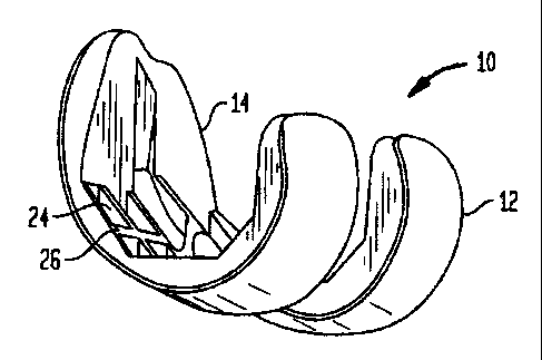
invention, more particularly, a femoral implant 10 used during
a TKR procedure. The particular implant shown is preferably
used as a trial implant; however it may be used as any type of
femoral implant. Generally, the implant of the present
invention has two primary surfaces thereof, including an
articulating surface 12, and a bone engaging surface 14.
Preferably, articulating surface 12 is shaped so as to
approximately replicate the shape of the distal femur and, in
particular, the articulating surfaces of the femoral condyles.
-6-
CA 02582203 2007-03-16
It is not necessary that articulating surface 12 match the
particular anatomy of the knee of the particular patient.
Further, articulating surface is preferably designed to engage
an artificial tibial implant (not shown). The desired general
shape and design for articulating surfaces of femoral implants
is known in the art.
[0027] Bone engagirig surface 14 is formed to match the
surface of the distal femur once the bone has been resected.
Resection of the distal femur may vary by application, but is
generally performed so as to remove one or both of the femoral
condyles. This is generally done by making a series of cuts
in the distal femur, the positioning and formation of which is
known in the art. The femoral implant bone engaging surface
shown in FIGS. 1-6 has a profile that matches one known shape
for the resected distal femur; however, other shapes may be
now known or later contemplated and corresponding shapes for
bone engaging surface 14 would be understood by one having
reasonable skill in the art.
[0028] The geometry of both articulating surface 12 and
bone engaging surface 14 lead to bone eagaging surface 14
being spaced proximally of articulating surface 12 and being
spaced apart at a distance therebetween. Accordingly, implant
has a thickness that is appropriate to provide the'
preferred spacing between articulating surface 12 and bone
engaging surface 14. Preferably, the general shape of implant
10 is similar to that of implants known in the art. In
particular, when implant 10 is to be used as a femoral trial,
it is preferred that implant 10 matches the shape of a
corresponding permanent implant as closely as possible.
[0029] As best shown in FIG. 2, implant 10 is preferably
formed from two separate parts. Support 16 is interposed
within shell 18 and forms bone engaging surface 14 therein.
The outside surface 20 of support 16 is designed to
substantially mate with inside surface 22 of shell 18. Shell
18 forms articulating surface 12, and preferably has a thin,
-7-
CA 02582203 2007-03-16
substantially uniform thickness such that the shape of inside
surface 22 substantially matches that of articulating surface
12. Accordingly, support 16 provides a majority of the
appropriate spacing between articulating surface 12 and bone
engaging surface 14.
[0030] Various materials can be used in formation of shell
18 and support 16. Acceptable materials for shell 18 include
various metals, such as CoCr, SS and aluminum alloys, or
polymeric material, such as polyetheretherketone (PEEK). if a
polymeric material is used to form shell 18, the polymer may
be reinforced with carbon fiber, including long short or micro
fibers, as they are known in the art. Preferably, shell is
formed from a metal, such as CoCr or SS having a thickness
between about 0.015 inches and about 0.065 inches, or aluminum
alloy having a thickness between about 0.030 inches and about
0.080 inches. In a preferred embodiment, shell 18 is formed
from SS and has a thickness of about 0.040 inches.
[0031) Various materials may also be used in the formation
of support 16. Acceptable materials for support 16, include
metal and polymeric material. Metals may include CoCr,
aluminum alloys and SS, and polymeric materials may include
Ultexg, PEEK, polycarbonate, polysulphone, Xylar , and Lexan .
In an embodiment of the present invention, support 16 can be
made from a fiber-reinforced polymeric material. Such
materials may include PEEK reinforced with carbon fibers,
which may comprise long, short or micro fibers. Further,
support 16 is preferably formed with a series of recesses 24
therein. The inclusion of recesses 24 within support 16
reduces the amount of material used to form support 16, which
may reduce the overall cost of implant 10 and/or the weight
thereof. Further, the formation of recesses 24 in support 16
results in the formation of a number of ribs 26 within the
structure of support 16. Ribs may increase the overall
strength of support 16 and, thus, of implant 10, allowing for
less-rigid and, possibly, less expensive materials to be used.
-8_
CA 02582203 2007-03-16
Still further, the inclusion of recesses 24 allows the
material from which support 16 is formed to have a more
uniform thickness. This is advantagecus when forming support
16 using an injection molding process because uriiform material
thickness allows the material throughout the entire part to
cool (and thus, shrink) uniformly. This helps prevent the
part from warping during cooling.
[0032] In a preferred embodiment of implant 10, shell 18 is
formed from a metal, preferably CoCr or SS and support 16 is
formed from a polymeric material, preferably Xylar4D. In such
an arrangement, shell 18 is more preferably formed using a
hydroform process. Hydroform is a process that is generally
known in the art and is useful for imparting complex, three-
dimensional ("3D") shapes into metal. Preferably, shell 18 is
formed using a vertical hydraulic hydroforming press. Such a
process can be carried out by Aero Trades Manufacturing,
located at 65 Jericho Turnpike, Mineola, NY. It is preferred
that a metal subjected to a hydroform process is thin enough
to be accurately formed by the process. It is also preferred
that the material be thick enough to retain the shape imparted
therein. The ideal thickness for shell in this embodiment
will vary by the material and specific geometry used and will
be known by those having reasonable skill in the art. The use
of a hydroform process to form shell 18 reduces the need for
the additional process steps of CNC grinding or polishing, as
are needed with a casting process.
[0033] Generally, the combination of a shell 18 made from
hydroformed metal and a support 16 made from a polymeric
material allows for an implant 10 which is appropriately
shaped and sufficiently rigid to provide acceptable trial
joint reduction, while being lightweight and cost-effective
from a manufacturing standpoint. The lightweight design of
such an implant 10 allows for easy transportation of a number
of such implants 10 when used in a set of trial implants.
Further, the cost-effective manufacture of such implants makes
_q_
CA 02582203 2007-03-16
it reasonable to use each of such implants in only one
surgical procedure. The provision of such disposable trial
implants may eliminate the need to design such an implant to
withstand multiple autoclave cycles, and to withstand multiple
trial reductions, further lowering the manufacturing cost
thereof.
[0034] Shell 18 may be affixed to support 16 by a variety
of methods, including using adhesives. Additionally, fixation
elements such as screws, bolts or rivets may be included
within implant 10 to secure shell 18 to support 16. Further,
corresponding tabs may be formed in appropriate portions of
shell 18 and support 16 to achieve fixation therebetween.
[0035] Referring now to FIGS. 7-8, a further smbodimsnt of
the present invention is shown wherein implant 10 is formed
from support 16 and shell 18 in a manner similar to that of
implant 10 described with reference to FIGS. 1-6. Implant 10
of the present embodiment includes shell 18 having a generally
proximally extending rib or flange 28 extending along at least
a portion of the outer periphery of shell 18 and preferably
the entire outer periphery. The integral formation of rib 28
within the outer periphery of shell 18 increases the rigidity
of shell 16, and accordingly of implant 10 overall. Rib 28
may be formed in a metal shell 16 by hydroforming.
[0036] More preferably, as shown in FIG. 9, shell 18
further includes folded section 30 extending inwardly from the
upper surface of rib 28. Folded section 30 further increases
the rigidity of shell 18 and implant 10, especially with
respect to flexion of implant 10 in the anterior-posterior
direction. Additionally, folded section 30 providesfor a
means of affixation between support 16 and shell 18. In
particular, in a preferred embodiment of the present
invention, shell 18 is formed from hydroformed metal,
preferably CoCr or SS, and support 16 is formed from a
polymeric material. In this embodiment, support 16 is formed
by insert molding the polymeric material onto shell 18. in
-10-
CA 02582203 2007-03-16
such a process, support 16 is formed by injection-molding of a
polymeric material into an appropriately shaped mold into
which a pre-formed shell 18 has been inserted. Because the
molten polymeric material can easily flow into and around any
geometry formed in the shell, including rib 20 and folded
portion 30, direct contact between the polymeric support 16
and the shell 18 may be the primary method of attachment
therebetween. Incorporation of rib 28 and folded portion 30
furthers this attachment because the polymer flows into the
shell, fully encasing the folded portion 30. This direct
contact between the two materials along the periphery of the
shell provides sufficient purchase to fully affix the shell 18
to the support 16.
[0037] Additionally, as shown in FIG. 9, shell 16 may have
post 32 affixed to inside surface 22 thereof. Preferably,
post 32 is either T-shaped, as shown, or includes a stepped"
geometry, as it is known in the art. Inclusion of this form
of post 32 provides additional contact points between shell 18
and support 16. Post 32 may be fabricated to provide geometry
similar to folded portion 32 discussed above, wherein the
contact between post 32 and the hardened polymer comprising
support 16 creates additional purchase, further affixing shell
18 to support 16. Post 32 may be added to inside surface 22
after formation of shell 18 and affixed thereto using welding
or a similar process. In this particular embodiment, implant
may include a plurality of.posts 32.
[0038] In an alternative embodiment of the present
invention, an implant 10 generally similar in structure to
those discussed with respect to FIGS. 1-9 is made from
polymeric reinforced carbon fiber. Carbon fiber is a
reinforcing fiber known for its lightweight, high strength and
high stiffness. Carbon fiber is produced by a high-
temperature stretching process of an organic precursor fiber
based on polyacrylonitrile ("PAN"), rayon, or pitch in an
inert atmosphere at temperatures above 1,800 degrees,
-11-
CA 02582203 2007-03-16
Fahrenheit. Fibers can be transformed by removing more non-
carbon atoms via heat treating above 3,000 degrees Fahrenheit.
After these fibers are produced, they can be utilized in many
different forms. They can be woven into long, dry fabric,
pre-impregnated with resin, wound onto spools for use in
filament winding, or braided and chopped into small fibers.
There are several ways in which to produce components using
carbon fiber; however, all of such processes require the use
of a mold to impart the necessary geometry into the carbon
f iber . The mold used in such a process def ines the shape of
the component. Accordingly, any component that can be molded
can be formed from carbon fiber. For example, femoral trials
can be created using carbon fibers. In a preferred
embodiment, the femoral trial can be molded using a two-part
mold; one mold to define the bone engaging surface 14 and the
other to form the articulating surface 12.
[0039] Molding processes used to form a trial from carbon
fiber include autoclave molding, compression molding, bladder
molding, resin transfer molding ("RTM") roll wrapping,
filament winding, and wet lay-up. Any of these methods. can be
used to produce knee femoral trials for TKR and hip stem
trials for THR. All of these types of molding processes force
the carbon and resin to conform to the desired shape using
heat and/or pressure. Once the part has cured, it maintains
its shape permanently and the composite construction provides
sufficient rigidity to allow the implant 10 to perform
equivalently to a metal trail during trial reduction. The use
of micro carbon fibers reduces manufacturing costs, but also
reduces material strength. Preferably, implant 10 of the
present embodiment is molded from a polymer reinforced with
long fiber, which is then overmolded with a "neat" polymer.
[0040] While robust, the composite construction of the
implant 10 of the present embodiment of the invention
possesses less resistance to the effects of repeated autoclave
cycling than cast CoCr or SS trials. Previously known trials
-12-
CA 02582203 2007-03-16
have been designed to survive multiple autoclave cycles and
retain the rigidity they had before the first use thereof.
Implant 10 of the present embodiment need only possess
sufficient rigidity for a single use and needs not have the
same robustness of reusable trials. Implant 10 of the current
embodiment, however, has a weight that is significantly less
than reusable trials, and thus alleviates many of the problems
associated with the weight thereof.
[0041] Implant 10 of the present embodiment can be formed
using a two-part structure as shown in FIGS. 1-9, wherein
shell 18 includes articulating surface 12, and support 16
includes bone engaging surface 14 and appropriately spaces
apart articulating surface 12 from bone engaging surface 14.
In such an embodiment, shell 18 is preferably affixed to
support 16 using an adhesive or an epoxy compound.
Alternatively, implant 10 can be molded in a unitary form,
having articulating surface 12 and bone engaging surface 14
formed therein.
[0042] Referring now to FIG. 10, an alternative embodiment
of the present invention is shown in which implant 110 is in
the form of a hip stem trial as is used in a THR procedure.
The use of hip stem trials is similar to that of femoral
trials. Generally, implant 10 replicates the shape and joint
kinematics of a permanent implant and is used in trial
reduction of the replacement joint. Implant 110 of the
present invention includes a modular articulating surface 112,
which replicates a resected femoral head and is generally in
the shape of a portion of a sphere. Further, implant 110
includes a bone engaging stem portion having surface 114,
which is appropriately shaped so as to fit within a resected
proximal femoral canal. Support 116 gives shape to bone
engaging surface 114 and appropriately spaces apart
articulating surface 112 therefrom. Implant 110 can be
fabricated using a hydroform process as discussed above by
forming two half-shells with the hydroform process and then
-13
CA 02582203 2007-03-16
assembling the half-shells onto a plastic inner structure.
Alternatively, implant 110 can be formed using a tube
hydroforming process, which can be carried out by Vari-Form,
which is located at, 250 Lothian Ave., Strathory, Ontario, CA.
[0043] Support 116 can be formed from various materials
including metal. In one form of the present embodiment,
support 116 is made from a metal tube, which is subjected to
pressure to impart the appropriate shape therefor.. In an
alternative embodiment, support 116 is made from a molded
polymeric material, which may be fiber reinforced in a manner
similar to other embodiments of the present invention
discussed above. The general shape of the femoral head may be
provided within support 116. In such an arrangement, shell
118 may be affixed thereto to provide implant 110 with
articulating surface 112. Shell 118 can be formed from
various metals including CoCr and SS or molded polymeric
material, which may be fiber reinforced. A metal shell 118
may be formed by hydroforming, as discussed above.
Alternatively, articulating surface 112 may be provided on
support 116 in a unitary fashion.
[0044] Although the various embodiments of the present
invention have been discussed as they apply either to the
human knee and hip joints, one having reasonable skill in the
art upon reading this disclosure would understand that the
present invention can be used to form other joints of human or
animal bodies. Such joints may include the elbow, wrist,
shoulder, etc.
[0045) Although the invention herein has been described
with reference to particular embodiments, it is to be
understood that these embodiments are merely illustrative of
the principles and applications of the present invention. it
is therefore to be understood that numerous modifications may
be made to the illustrative embodiments and that other
arrangements may be devised without departing from the spirit
-14-
CA 02582203 2007-03-16
and scope of the present invention as defined by the appended
claims.
-15-