Note: Descriptions are shown in the official language in which they were submitted.
CA 02583406 2007-03-20
WO 2007/001378 PCT/US2005/033728
MICROFLUIDIC DEVICE FOR DETECTING SOLUBLE MOLECULES
Field of the Invention
This invention relates to microfluidic devices and their use in the detection
of
disease.
Background of the Invention
Sexually transmitted diseases remain a public health issue in many
communities. According to the American Social Health Association, a partner of
the
Center for Disease Control and Prevention, 65 million Americans are currently
infected
with a sexually transmitted disease (STD) and 15 million are infected each
year. Most
STDs are curable, and early detection can prevent complications and the
continued
spread of the disease. Treatment and counseling for these diseases are
available, but
the inability to perform testing at the point-of-care delays results and
hinders the
effectiveness of public health workers to stem these epidemics.
Several problems exist with the testing methods currently available in
clinical
settings. Many tests cannot be performed at point-of-care facilities and
results can take
weeks to obtain. For STD testing, these problems are compounded by social
stigma
and anxiety and convincing patients to make a return visit can be problematic.
Current
methods often require large blood samples, limiting the number of diseases for
which
an individual can be tested per visit. Other tests require uncomfortable
sainple
collection methods. For example, Chlamydia testing in men requires specimens
collected from inside the urethra with a cotton swab. While less unpleasant
testing
methods are available, this method is widely used because it is fast and cost
effective.
Delayed results, availability problems, uncomfortable sample collection and
the
cost of these tests all impede their use at point-of-care facilities, making
it easy to
understand why less than half of American adults between ages 18 and 44 have
been
tested for non-HIV/AIDS STDs. STDs are more prevalent in low income areas
where
patients may not be able to afford testing, but even well funded facilities
cannot afford
to run the most sophisticated tests, like polymerase chain reaction (PCR)
detection, for
every patient in every case. Many point-of-care facilities do not have the
space or
1
CA 02583406 2007-03-20
WO 2007/001378 PCT/US2005/033728
money to dedicate to the equipment required for these tests. In these cases,
doctors will
diagnose their patients by outward symptoms only and sometimes prescribe
antibiotics
unnecessarily because testing is simply not feasible. Many STDs can cause
severe
damage and be spread without ever exhibiting symptoms. Chlamydia, for example,
only expresses symptoms about 25% of the time increasing the risk of
unknowingly
transmitting the disease. Chlamydia and gonorrhea are the main causes of
pelvic
inflammatory disease, the leading cause of sterility among women.
There is a need in the medical community for rapid and inexpensive detection
of STDs and other diseases. Further, it is preferable if these detection
methodologies
can be delivered at the point-of-care in a single patient visit.
U.S. Patents 6,063,589 and 6,527,432 disclose microfluidic devices on
"spinning disks" that are substantially two-dimensional. The "spinning disks"
may
contain multiple detection devices; however, each device may have to be
individually
filled with the biological sample.
U.S. Patent 6,479,239 discloses a device for detecting and identifying
infectious
disease agents using physical separation techniques. Ultracentrifuge tubes
having
successively smaller diameters are used to characterize infectious agents
based on their
size. Secondary detection methods may be used for additional characterization.
U.S. Patent 6,551,841 discloses a microfluidic device capable of detecting
soluble analytes. The sample fluid is forced through the device using a pump
or by
capillary action.
U.S. Patent 6,929,239 discloses a microfluidic device, in a "card"
configuration
capable of conducting multiple simultaneous chemical reactions utilizing an
internal
network of intercomiecting ducts and channels.
Summary Of The Invention
The present disclosure provides a microfluidic-based device for the detection
of
disease (e.g., infectious disease agents) that is inexpensive to manufacture
and operate
2
CA 02583406 2007-03-20
WO 2007/001378 PCT/US2005/033728
and provides rapid results. Ideally, the device disclosed herein may be
configured for
use in conjunction with standard, low cost centrifuges, including basic
clinical
centrifuges nonnally used in the preparation of blood samples at point-of-care
facilities.
In one aspect, the invention provides a non-radial cylindrical microfluidic
device for analyzing the presence or absence of a molecule to be detected in a
fluid
sample. The device comprises a sample reservoir having a sample input port in
fluid
connection with at least one detector array, wherein each detector array
comprises at
least one detector that comprises a reaction chamber comprising an immobilized
capture molecule, and a reagent capable of undergoing a colorimetric reaction
or
displaying an optically detectable signal and capable of reacting with the
molecule to
be detected.
The device of this invention may be used to assess the presence or absence of
a
molecule is biological fluids such as whole blood, blood serum, blood plasma,
seminal
fluid, prostatic fluid, saliva, urine, and spinal fluid.
The fluid sample may be moved from the sample reservoir to the reaction
chamber by any appropriate means including, for example, passively by
capillary
action, or actively by centripetal force arising from centrifugation of the
device or by
gas pressure applied to the sample reservoir. Further, all fluids may be moved
through
the device, ideally using centripetal force arising from centrifugation of the
device, or
by gas pressure.
In one embodiment, the detector may fia.rther comprise a diluent chamber and
diluent, and a mixing cliannel capable of generating turbulent flow, wherein
the mixing
channel is upstream from the reaction chamber. The fluid channel leaving the
diluent
chamber joins the fluid chaimel leaving the sample reservoir upstream from the
mixing
channel. In a related embodiment, the diluent chamber is isolated from the
mixing
channel by a first burst valve.
In another embodiment, the detector(s) contain a sample chamber having a pre-
defined capacity. In a related embodiment, the loading channels connecting the
sample
reservoir to the sample chambers is sloped down in the direction of the sample
reservoir
such that, under centrifugation, excess sample fluid not accommodated by the
sample
3
CA 02583406 2007-03-20
WO 2007/001378 PCT/US2005/033728
chambers flows back into the sample reservoir.
In preferred embodiments, the reagent that is capable of undergoing a
colorimetric reaction or displaying an optically detectable signal is further
capable of
binding to the molecule to be detected.
In one embodiment, the immobilized capture molecule (immobilized within the
reaction chamber) is an antigen, the molecule to be detected is a serum
antibody or
other soluble binding protein, and the reagent is a detectably labeled
antibody capable
of binding to the serum antibodies or other soluble binding protein contained
in the
fluid sample. The detectably labeled antibody may be an enzyme-linked antibody
or an
antibody having a fluorescent or other optically readable tag. The
colorimetric reaction
and/or optically readable tag may be qualitatively assessed by the operator or
may be
qualitatively assessed or measured by an optical sensing device (e.g., a
detector).
Although any antigen may be used in this embodiment, useful antigens include
those that are specific for microorganisms that cause sexually transmitted
diseases
including, for example, Chlamydia spp., Gonorrhea spp., human Papillomavirus,
herpes simplex virus, hepatitis B, syphilis, trichomononiasis, bacterial
vaginosis, and
human immunodeficiency virus.
In another embodiment, the immobilized capture molecule is an antibody
specific for a pre-determined antigen. Although an antibody specific for
almost any
antigenic molecule may be used, particularly useful capture antibodies include
those
that bind to microorganism-specific antigens, such as antigens specifically
associated
with Clzlanaydia spp., Gonorrhea spp., human Papillomavirus, herpes simplex
viras,
hepatitis B, syphilis, trichomononiasis, bacterial vaginosis, and human
immunodeficiency virus.
In this embodiment, the reagent is a detectably labeled antibody capable of
binding to the antigen to be measured contained in the fluid sample. The
detectably
labeled antibody may be an enzyme-linked antibody or an antibody having a
fluorescent or other optically readable tag. The colorimetric reaction and/or
optically
readable tag may be qualitatively assessed by the operator or may be
qualitatively
assessed or measured by an optical sensing device (e.g., a detector) using
well known
4
CA 02583406 2007-03-20
WO 2007/001378 PCT/US2005/033728
detection techniques.
In another embodiment, the immobilized capture molecule is an
oligonucleotide, the molecule to be detected is a polynucleic acid (e.g., DNA
or RNA),
and the reagent is a detectably labeled oligonucleotide.
In another embodiment, the device further contains a wash chamber containing
a wash buffer and a developing chamber containing the reagents, wherein the
wash
chamber is isolated from the reaction chamber by a second burst valve and the
developing chamber is isolated from the reaction chamber by a third burst
valve,
wherein the first burst valve is designed to rupture at a substantially lower
pressure than
the second burst valve and the second burst valve is designed to rupture at
substantially
lower pressure than the third burst valve. The pressure differences that cause
the first,
second, and third burst valves to rupture may be advantageously produced by
different
centrifugal forces. Specifically, the operator may control the centrifugal
force applied
to the device by controlling the revolution speed of the centrifuge.
In a related aspect, the invention provides a method for detecting a molecule
in
a fluid sample comprising introducing the sample into a device of any of the
foregoing
aspects and embodiments, centrifuging the device, detecting the presence or
absence of
the colorimetric response, and relating the presence or absence of the
colorimetric
response to the presence or absence of the molecule to be detected.
It is contemplated that the devices disclosed herein are microfluidic devices
having channel diameters of 10 - 1000 m, preferably 100 - 500 gm, and chamber
capacities of 1 - 1000 l, preferably 10 - 500 l. It is well recognized that
higher
centrifuge speeds (i.e., centrifugal forces) are required as chamiel and
chamber sizes are
reduced. In preferred embodiments, the devices of the invention are designed
to run on
standard clinical blood centrifuges operating at 500 - 1500 rpm.
In another aspect, the invention provides a diagnostic kit for use in
detecting a
molecule associated with a disease state. The kit comprises any of the devices
described above along with instructions for its use.
"Non-radial cylindrical device," as used when referring to devices of the
present
5
CA 02583406 2007-03-20
WO 2007/001378 PCT/US2005/033728
invention, means a device having a length that is greater than its radius. Non-
radial
cylindrical devices are designed to be revolved, such as in a centrifuge,
rather than
rotated around a central axis such as is the case for a "spinning disk"
device. One
distinguishing feature of a non-radial cylindrical device is that the
direction of the force
vector during centrifugation is constant over the entire device. By contrast,
force
vectors on "spinning disk" devices radiate outward in all directions
simultaneously
from the center of the disk.
Brief Description of the Drawings
Further understanding of the principles of the present invention may be had by
reference to the accompanying drawings, wherein:
FIGURE 1 is a perspective view of a microfluidic device in accordance with the
principles of this disclosure;
FIGURES 2A-C are schematic diagrams of microfluidic devices in accordance
with the principles of this disclosure. FIGURE 2A is a schematic diagram of a
detector
array containing three individual detectors. FIGURE 2B is a schematic diagram
of the
reaction region of a single detector. FIGURE 2C is a schematic diagram of the
chamber region of a detector array;
FIGURE 3 is a schematic diagram of a reversible two-step detector;
FIGURE 4 is a schematic diagram of another reversible two-step detector; aiid
FIGURE 5 is a schematic diagram of a microfluidics device lacking a diluent
chamber and mixing channel.
Detailed Desciiption of the Invention and Best Mode
This disclosure provides a microfluidic device that is capable of rapidly
testing
for multiple diseases (or confirmatory testing of the same disease) in
parallel and
requires substantially less blood than do traditional clinical methods. At
these reduced
assay volumes, one vacuum tube, the current standard means of drawing blood,
contains sufficient blood to run dozens of different tests. The device is
particularly
effective for diagnosing STDs, like human papilloma virus (HPV), that can
present an
6
CA 02583406 2007-03-20
WO 2007/001378 PCT/US2005/033728
early detection problem. Advantageously, the device may be used to test for
any
pathogen or foreign material that stimulates the production of antigen
specific
antibodies, allowing several strains to be tested for at once.
The microfluidic diagnostic device described herein solves a number of
logistical problems in point-of-care testing for STD's and other infectious
diseases.
The design ensures standardized dilution which can be calibrated to different
disease
exposure states, removing the need for caregivers to undertake the tedious
task of
micropipetting samples and reagents into dozens of wells. The device may be
able
distinguish diseased, vaccinated and no contact states without performing
several
additional unnecessary dilutions. Elimination of such intermediate steps
reduces the
risk of technician exposure to contagious diseases. In preferred embodiments,
the
device can provide a qualitative visual result, thereby removing the need for
a
photometer or other optical detection device. These improvements make the
microfluidic testing device user friendly and cost effective, reduce the need
for
additional capital investment, and encourage a higher standard of medical
care.
Due to the contained chemistry and high surface to volume ratio inside the
device this test method has the potential to provide faster results than
presently
available alternatives. Testing for several diseases during a regular doctor's
visit
becomes possible, alleviating the problem of patients who do not return for
their test
results. This device allows for rapid detection of a large number of diseases
that
otherwise often require outsourced testing. The low cost of manufacturing will
make
this device ideal for use in low income areas and smaller facilities.
Generally, the operators need only to collect the blood sample, separate the
plasma from cellular fraction (e.g., by centrifugation), and deposit an
aliquot of blood
plasma into the device. Alternatively, the device may contain an in-line
filter to
separate blood fractions thereby enabling the operator to simply deposit an
aliquot of
whole blood into the device before centrifugation. Of course, more extensive
pre-
testing preparation may be performed as required for each particular set of
assay
conditions.
The blood, plasma, or other sample is driven through the device and into the
reaction chamber by capillary action, gas pressure, centrifugation, or any
other
7
CA 02583406 2007-03-20
WO 2007/001378 PCT/US2005/033728
appropriate means. The device may be configured to yield a qualitative visual
response
that is read without the assistance of a photometer or other optical detector
and, in such
case, ideally reports diseased, vaccinated or "no contact" states. No contact
refers to an
individual who has never encountered the antigen, meaning they have never had
the
disease nor have they been vaccinated against it. Quantitative results may be
obtained
through the use of an optical detection system such as a fluorimeter or a
spectrophotometer.
In one preferred embodiment, the device is substantially cylindrical and
physically sized to make it backwards compatible, fitting into standard blood
centrifuges, which are present in nearly all point-of-care locations. The
detection assay
is ideally based on the Enzyme Linked hnmunosorbent Assay (ELISA) type
protocol,
specifically "microELISA" for small fluid volumes; however, other detection
methodologies appropriate to the type of molecule to be detected may be used
(e.g.,
oligonucleotide-based detection methods, etc.).
Device Operation
The microfluidic device employs fabrication techniques to make microELISA
and other micro-reagent testing inexpensive and easy to use in a point-of-care
setting.
The blood, plasma, or other fluid sample is introduced into the sample
reservoir of the
microfluidic device via sample port(s), ideally through a safety cap
containing a needle
that aspirates the sample using capillary force. These ports may
advantageously
incorporate accessory spill chambers to trap excess fluid and to prevent
overflow in the
event that the device is improperly loaded. The fluid reservoir is in fluid
communication with a series of sample chambers that will hold the samples
after
loading until the device is centrifuged.
Ideally, the fluid sample is moved from the sample reservoir to the reaction
chamber by capillary action. However, if necessary, the fluid sample may be
moved by
gas pressure or centrifugal forces. Gas pressure may be applied using any
appropriate
method. For example, the sample port(s) may be adapted to accept a standard
Luer
syringe which, upon depression by the operator, creates a positive gas
pressure forcing
the fluid sample into the reaction chamber. Alternatively, the device may be
fitted with
a cap that contains a pressurized gas (e.g., N2 and CO2) chamber which, when
affixed
8
CA 02583406 2007-03-20
WO 2007/001378 PCT/US2005/033728
to the device, forms a relatively gas-tight seal and causes the pressurized
gas to be
released into the sample reservoir. The cap may serve the additional purpose
of
preventing spillage of the fluid sample during handling, thereby further
reducing the
likelihood of operator exposure. For devices having waste or other chambers
downstream of the reaction chamber, overfilling of the reaction chamber may be
prevented by the inclusion of a burst valve at the exit of the reaction
chamber. This
burst valve is designed to rupture under the forces exerted by centrifugation
but not
under the gas pressure used to load the reaction chamber.
Once the reaction chamber is loaded, the device is typically allowed to
incubate
for a period of time determined based on the detection methodology. Further
processing of the sample and the device also varies based on the detection
methodology
and the features of the device.
Once fluid sample has been loaded into the device and the molecules to be
detected have been allowed to bind to the immobilized capture molecule in the
reaction
chamber, the device is ideally placed into a standard laboratory centrifuge
(e.g., the
same centrifuge used for preparing whole blood samples) for the completion of
the
reaction and detection process.
In one embodiment, each detector contains a chamber holding a washing
solution and a chamber holding a developing solution. The washing solution is
used to
wash the fluid sample and any unbound molecules out of the reaction chamber.
The
developing solution contains the reagents necessary to yield a colorimetric
result that
indicates the presence or absence of the molecule to be detected. The washing
chamber
and the developing chamber may, optionally, be isolated from the reaction
chamber by
burst valves. In one embodiment, the burst valve isolating the washing chamber
is
designed to rupture at a lower pressure than the burst valve isolating the
developing
chamber. Thus, in this configuration, the device must be centrifuged at a
first, slower
speed to effect washing of the reaction chamber, and then centrifuged at a
second,
higher speed to initiate the colorimetric reaction. In another embodiment,
either with or
without burst valves, the washing chamber is connected to the reaction chamber
by a
shorter channel length then is the developing chamber. Thus, under
centrifugation, the
washing solution reaches the reaction chamber first. In yet another
embodiment, the
9
CA 02583406 2007-03-20
WO 2007/001378 PCT/US2005/033728
developing chamber empties directly into the "top" of the washing chamber,
again
causing the washing solution to reach the reaction chamber first. It is
contemplated
that, in this embodiment, there will be an insignificant amount of mixing
between the
washing and developing solutions.
It is further contemplated that, under certain reaction conditions and for
certain
clinical uses, the fluid sample may require having a pre-determined volume, a
diluent
chamber filled with a diluent (e.g., a saline solution), and mixing channels.
The diluent
chambers lie apart from, but may be parallel to, the sample chambers and both
sets of
chambers are in fluid communication with one or more mixing channel.
When the centrifuge is activated the fluid sample and diluent is forced into
the
mixing channels where the mixing process begins, diluting the sample to the
working
concentration. Alternatively, the fluid sample and diluent may be forced
together and
into the reaction chamber using gas pressure as above.
In one embodiment, mixing continues in the reaction chamber where the one-
step microELISA reagents are stored. The diluted sample reacts with the
microELISA
reagents in this chainber under conditions suitably adjusted to permit
immunological
reactions. In other embodiments, each detector further comprises additional
reagent
chambers in fluid contact with the reaction chamber in order that assays other
than one-
step microELISA may be performed. Optionally, these reagent chambers may be
initially isolated from the reaction chamber by burst valves. The burst valves
are
preferably configured to burst or open at pre-set pressures (centrifugal
forces) and may
be configured to burst either substantially simultaneously such that reagent
chambers
are released together, or at different pressures in order to deliver the
various reagents in
a sequential (i.e., pre-defined) order.
Each reaction chamber on the device contains pathogen-specific reagents which
are used to detect any pathogen or pathogen-specific antibodies contained in
the
biological sample. These reaction chambers may contain a single type of
antigen-
specific reagent or multiple antigen-specific reagents for a single pathogen.
Alternatively, the reaction chamber may contain antigen-specific reagents
specific for a
plurality of pathogens. The reaction chambers of a single device may contain
the same
antigen (redundant tests), different antigens for the same pathogen, or
antigens for a
CA 02583406 2007-03-20
WO 2007/001378 PCT/US2005/033728
plurality of pathogens.
In the most preferred embodiments, the pathogen-specific reagents produce a
visible color change in the presence of the pathogen or a pathogenic marker or
other
molecule to be detected. The colorimetric reporter may be read by a photometer
or
other optical detector for quantitative (or qualitative) results or just
visually for a
qualitative test.
Device Desi~n
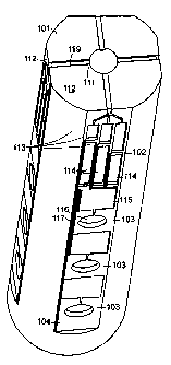
FIGURE 1 is a perspective view of a microfluidic device 10 according to the
principles of the present disclosure. The device 10 comprises of three
regions: the
loading region 101, the chamber region 102, and the reaction region 103. A
single
chamber region 102 and reaction region 103 that are in fluid connection are,
together,
referred to as a perpendicular detector array 104. The device 10 is physically
configured and sized such that it may be inserted into the rotor of a
centrifuge.
Preferably, the device has substantially the same dimensions as a vacuuin tube
blood
container so that the same centrifuge used to isolate blood plasma at the
point-of-care
may be used to run the device 10.
The loading region 101 consists of a sample reservoir 111 and a plurality of
loading channels 112. It is contemplated that loading region 101 contains a
single
loading channel 112 providing a fluid connection between sample reservoir 111
and
each perpendicular detector array 104; however, a plurality of loading
channels 112
may connect to each detector array 104. Further, FIGURE 1 illustrates a device
10
having four perpendicular of detector arrays 104; however, the exact number of
configuration may be modified to each individual application or user's needs
and the
maximum number of arrays will depend upon the overall size of the device 10
and the
size of each individual detector array 104 and practical manufacturing
constraints.
The loading region may optionally contain an overflow chamber (not shown)
either on its surface or disposed below, but in fluid contact with, sample
reservoir 111.
In one embodiment, the overflow chamber is disposed below sample reservoir 111
and
is connected via overflow channe1119.
11
CA 02583406 2007-03-20
WO 2007/001378 PCT/US2005/033728
In another embodiment, loading region 101 is covered by a solid surface (not
shown) that contains an injector port such that the operator may inject a
biological
sample (e.g., a blood, plasma, or other fluid sample) through the injector
port into
sample reservoir 111. The cover may be flat or it may be recessed to provide
additional
volume capacity to sample reservoir 111.
Optionally, a filter may be disposed between fluid reservoir 111 and loading
channels 112 in order to prevent contaminating elements such as blood cells or
other
large particulate matter from entering detector array 104.
Each detector array 104 comprises of one or more (three are illustrated in
FIGURE 1) individual detectors.
FIGURE 1 illustrates a simple detector configuration. Each detector consists
of
a sample chamber 113, an optional diluent chamber 114, optional mixing
channels 115,
and a reaction chamber 116. The sample chamber 113 is in fluid contact with
the
loading channel 112. Both sample chamber 113 and diluent chamber 114 are in
fluid
contact with the mixing channels 115 via perpendicular channels 117. The
reaction
chamber 116 is covered by a transparent or translucent covering such that a
colorimetric reaction may be viewed by the operator or measured using an
optical
detection device.
In one embodiment, loading channels 112 slope down toward sample reservoir
111. In this configuration, the biological sample is loaded into sample
reservoir 111
and sample chambers 113 are allowed to fill by capillary action prior to
centrifugation.
Thus, when placed under centrifugal force, the excess biological sample flows
back
into sample reservoir 111 and further flows through overflow channel 119 into
the
overflow chamber below. This configuration advantageously prevents overfilling
of
sample chambers 113.
The sample chambers 113 of a single detector array may have the same or
different volumes. Sample chambers 113 of different volumes are useful to
perform
serial dilutions of the biological sample against the same detection reagents
or when
using different detection reagents under well characterized conditions or when
serial
dilutions are not necessary. Likewise, the volume of diluent chambers 114 may
be
12
CA 02583406 2007-03-20
WO 2007/001378 PCT/US2005/033728
advantageously modified as desired.
Burst valves 118 may be optionally used to prevent the diluent from escaping
diluent chamber 114 prior to use (i.e., during storage) and/or to control the
flow of
biological sample from sample chamber 113 (i.e., to ensure proper sample
volume
enters the detector. Likewise, burst valve 118 may be placed between mixing
channels
115 and reaction chamber 116 to prevent the escape of reactants from reaction
chamber
116 during handling.
Mixing channels 115 are serpentine in configuration and have tight corners to
promote turbulent flow which facilitates mixing of the biological sample and
the
diluent.
The detectors are configured for each individual application and set of
reagents
to be used.
FIGURES 2A-2C are schematic diagrams showing enlargements of the various
components of detector array 104 and an individual detector. FIGURE 2A
illustrates
detector array 104 having overflow chamber 210. FIGURE 2B illustrates reaction
region 103 of a single detector. FIGURE 2C illustrates chamber region 102 of a
single
detector. Loading channel 112 is divided into a plurality of smaller loading
channels to
conduct the biological sample from loading region 101 (not shown) into
individual
sample chambers 113.
Antigen/Antibody-trap Detectors
In its simplest configuration, the detectors are used to identify antigens of
interest in a biological sample. In this embodiment, an antigen-specific
antibody is
adhered to or otherwise immobilized on the walls of reaction chamber 116.
Reaction
chamber 116 ideally also contains an antigen-specific antibody that is unbound
and
detectably labeled, along with any additional reagents required to detect the
presence of
the antigen of interest. Alternatively, reagents can be stored in other
chambers or
compartments and added to the reaction chamber as needed by use of flow
control
devices like the aforementioned burst valves. For example, the unbound
antibody may
be linked to an enzyme capable of performing a colorimetric reaction as in a
one-step
13
CA 02583406 2007-03-20
WO 2007/001378 PCT/US2005/033728
ELISA format. Thus, under centrifugal force, the fluid sample that is loaded
into
sample reservoir 111 is diluted with the diluent contained in diluent chamber
114 and
mixed in mixing chaimels 115. The plasma sample then flows into reaction
chamber
116 where the antigen of interest is captured by the immobilized antibody and
detected
by the unbound antibody by way of a colorimetric reaction (e.g., such as that
obtained
using horseradish peroxidase and alkaline phosphatase, or any of the other
well known
enzyme-substrate combinations used in immunoassay systems).
In an alternative embodiment, exposure of the test subject to a pathogen may
be
determined by detecting the presence of antigen-specific antibodies in the
plasma or
serum of the test subject. In such cases, the antigen-trap detector described
above may
be slightly modified in that an pathogenic antigen is immobilized on the wall
of
reaction chamber 116 which captures the antigen-specific antibodies in the
biological
sainple. These antibodies may then be detected using, for example, an anti-
human (or
appropriate species depending upon the test subject species) antibody which is
ideally
linked to a detectable label or to an enzyme capable of catalyzing a
colorimetric
reaction, as above.
Two-Step Detectors
FIGURE 3 is a scheinatic diagram of a single detector that may be used in a
"two-step" detection process such as a traditional ELISA assay. In this
detector
configuration, sample reservoir 111 is in direct fluid contact with reaction
chamber 116.
Optionally, sample reservoir 111 contains a filter to separate the cellular
fraction from
the blood plasma/serum or for additional filtration of a plasma/serum sample.
Alternatively, sample reservoir 111 lacking a filter is loaded with a plasma
or serum
sample wherein erythrocytes and other blood cells have been previously
removed. In
addition, it is contemplated that the device can be configured to permit
application of
either an undiluted biological sample or a diluted biological sample.
Reaction chamber 116 ideally contains an iminobilized capture antibody (if a
blood-borne antigen is being assayed) or an immobilized capture antigen (if
antigen-
specific antibodies are being assayed).
The biological sample fills reaction chamber 116 by capillary action or it may
14
CA 02583406 2007-03-20
WO 2007/001378 PCT/US2005/033728
be forced into reaction chamber 116 under pressure (e.g., when sample
reservoir 111 is
adapted to accept a syringe and positive pressure is applied by depressing the
syringe
plunger). The binding reaction is permitted to occur in reaction chamber 116
for a
sufficient time. The fluid contents are retained in the reaction chamber using
a low-
pressure burst valve 352 between reaction chamber 116 and waste chamber 340.
The binding reaction is terminated by centrifugation of the device at a first
speed. A low pressure burst valve 351 is present between washing chamber 320
and
channels 117 that direct the washing buffer into reaction chamber 116.
Preferably, the
low pressure burst valves 351 and 352 are designed to burst at approximately
the same
pressure which is the pressure generated by centrifugation at a first speed.
Optionally,
a fail-safe low pressure burst valve 353 is present to further isolate
reaction chamber
116 from the second step reactants (i.e., the washing solution and the
developing
solution). Burst valves 351, 352, and 353 will rupture at approximately the
same time
allowing the wash buffer to flow into reaction chamber 116 and then all
reaction
chamber 116 contents to flow into waste chamber 340. This leaves only the
bound
elements in the reaction chamber.
Next, the colorimetric reaction is initiated by centrifugation of the device
at a
second speed that is greater than the first speed. The reagents necessary for
the
colorimetric reaction, such as a detectably labeled (e.g., enzyme linked)
secondary
antibody and colorimetric reagents, are contained in the developing solution
within
developing chamber 310. These reagents are released into reaction chamber 116
by the
bursting of high pressure burst valve 354. The reagents are held within
reaction
chamber 116 because waste chamber 340 and accompanying fluid channels 330 that
connect it to reaction chamber 116 are ideally designed to have substantially
the same
volume as the combined volume of reaction chamber 116 and washing chamber 320.
Furthermore, substantially more developing solution may be used than is needed
to
ensure that the reaction chamber is filled during the colorimetric reaction.
As above,
the results of the colorimetric reaction may be viewed and/or measured through
a
transparent or translucent covering on reaction chamber 116.
FIGURE 5 illustrates another two-step detector configuration. The fluid sample
is loaded by capillary action or gas pressure through sample chamber 111 into
reaction
CA 02583406 2007-03-20
WO 2007/001378 PCT/US2005/033728
chamber 116. The fluid sample is allowed to incubate with the immobilized
capture
molecule. Following incubation, the device is centrifuged, causing burst
valves 511
and 512 to rupture allowing the wash solution to flow from wash chamber 440
and the
developing solution to flow from the developing chamber 450 through reaction
chamber 116 into waste chamber 420. By virtue of the shorter channel length,
the wash
buffer reaches reaction chamber 116 before the developing solution. The volume
of
waste chamber 420 and associated channe1560 is substantially the same as the
volume
of wash buffer and reaction chamber 116. Thus, the developing solution is
retained in
reaction chamber 116 because waste chamber 420 and channel 560 is full of
reaction
and wash solutions.
Reversible Two-Step Detectors
FIGURE 4 is a schematic diagram of a reversible two-step detector that is
configured to accept biological samples that require filtration prior to ELISA
detection
(e.g., whole blood). The biological sample is loaded into sample reservoir 111
which is
sealed and the device centrifuged at a first speed sufficient to filter the
sainple through
filter 410, with the filtrate (e.g., serum) passing into reaction chamber 116.
Optionally,
loading channe1112 is in fluid contact with either waste chamber 420 or waste
chamber
430 to accommodate excess filtrate. Likewise, reaction chamber 116 may be in
fluid
contact with the same waste chamber 420 (not shown) or a different waste
chamber
430. The binding reaction within reaction chamber 116 is allowed to proceed
for an
appropriate period of time.
The device is next inverted in the centrifuge (centrifugal force applied in an
upward direction in FIGURE 4) and centrifuged at a second speed. The second
speed
is greater than the first and results in the release of wash buffer contained
within wash
chamber 440 through burst valve 441 that is designed to rupture at the
centripetal force
generated by the second speed. The wash buffer flows through reaction chamber
116
and, along with the unbound reagents, is captured in one or both of waste
chambers 420
and 430. Optionally, a second burst valve 442 is placed between reaction
chamber 116
and one of the waste chambers 420 to ensure that the first waste chamber 430
is filled.
This may improve the fluid handling characteristics of the device. The second
burst
valve 442 is designed to rupture at the second speed or above. The second
speed is
16
CA 02583406 2007-03-20
WO 2007/001378 PCT/US2005/033728
significantly greater than the first speed in order that burst valve 441
remains intact
during the filtration process.
The device is further centrifuged at a third speed that is significantly
greater
than the second speed. The third speed ruptures burst valve 443, releasing the
developing solution from developing chamber 450 into reaction chamber 116 in
order
that the colorimetric reaction occurs. In the event that first waste chamber
430 is not
full and burst valve 442 has not ruptured, the volume of the developing
solution and the
third speed will be sufficient to do so. The principles of the developing
solution and
colorimetric detection are the same as described above.
Other Detector Configurations
It is recognized that the exact configuration of the chambers and channels may
be modified for the particular needs of each individual assay without
departing for the
scope and spirit of this disclosure. For example, the immobilized capture
molecule
may be present in mixing channels 115, obviating the need for reaction chamber
116,
but necessitating one or more waste chambers 420. Likewise, the devices of
this
invention may be configured for detection assays requiring more than two
steps. Such
device configurations utilize additional reactant chambers in addition to
washing
chamber 440 and developing chamber 450 described herein. The timing of release
of
the contents from these additional chambers may be controlled by any
appropriate
means including the additional burst valves as described herein.
Alternative Methods For Detection
Although this disclosure has been exemplified by the use of an ELISA-based
assay to detect the molecule of interest in the fluid sample, any appropriate
detection
assay may be used.
Florescence in situ Hybridization (FISH) may be used to detect and identify
microorganisms by labeling chromosomes or genes with fluorescently labeled DNA
probes that are complimentary to segments of the target organisms' genome. In
this
embodiment the organism to be detected is collected with a swab, or capillary
tube if
present in the blood stream, and moved into the reaction chamber. A reactant
fluid
17
CA 02583406 2007-03-20
WO 2007/001378 PCT/US2005/033728
containing the labeled DNA probes is present in (or introduced into) the
reaction
chamber. The device is incubated under conditions that allow the probes to
penetrate
the organism and bind to the target sites (nucleic acids of interest). The
device is then
centrifuged such that the reaction chamber is emptied of excess fluid and,
preferably,
the reaction chamber is washed with a washing buffer as described above.
Microorganisms are retained in the reaction chamber for later detection either
by the
use of a filter near the exit channel of the reaction chamber (i.e., that
connects the
reaction chamber to the waste chamber), or by making the exit channel
sufficiently
narrow such that the microorganisms cannot pass. If the target organism is
present then
the DNA probes will remain trapped with in them in the reaction chamber, if
they are
not there will be few or no DNA probes present in the reaction chamber when
the test is
complete.
Oligonucleotide ligation reactions may be used to detect nucleic acids in the
fluid sample. The reaction chamber contains immobilized capture
oligonucleotides that
are complimentary to the sequences of interest. The fluid sample is introduced
into the
reaction chamber and complementary strands will bind to the capture
oligonucleotides
causing a change in the optical properties of the reaction chamber. Because of
the
small size of the probes and recent developments in microfabrication and
surface
treatment thousands of different segments could be probed at the same time
using
optical detectors. Future technologies may eliminate the need for an optical
detector.
EIA could also be preformed by using secondary antibodies which are bound to
gold colloids, quantum dots or other visible markers removing the need for
developing
solutions.
Device Manufacture
Compression molding of polymers in photolithographically defined micromolds
may advantageously be used to form the microfluidic channels on the device.
Polymer
embossing and micromolding are techniques that enable the fabrication of
several
hundreds or thousands of inexpensive, disposable parts from one silicon
master. The
technique enables tight tolerances and high quality control. Molding of
engineering
polymer resins are useful techniques for mass production, while soft
lithography is
suitable for rapid prototype development and testing.
18
CA 02583406 2007-03-20
WO 2007/001378 PCT/US2005/033728
Without delving unnecessarily into well known fabrication techniques, it is
noted that using transparency masks, it is possible to obtain resolution as
low as 20 m,
and features of 50 m and larger are easily reproduced (Whitesides et al.,
Annu. Rev.
Biomed. Eng. 3: 335-373, 2001). The desired features are first printed on a
high
resolution transparency. SU-8, a negative photoresist, is spun onto a silcon
wafer. The
transparency is then placed over the photoresist and exposed to UV light. The
UV light
causes the negative photoresist to harden while areas not exposed to UV light
may be
washed away. The image on the transparency is transferred to the SU-8 layer on
the
silicon wafer. Polydimethyl siloxane (PDMS) and a crosslinking agent are mixed
and
poured into the micromold. After curing, the pattern of microchannels and
reservoirs
are transferred to the new media.
All publications and patents cited in this specification are herein
incorporated
by reference as if each individual publication or patent were specifically and
individually indicated to be incorporated by reference. Although the foregoing
invention has been described in some detail by way of illustration and example
for
purposes of clarity of understanding, it will be readily apparent to those of
ordinary
skill in the art in light of the teachings of this invention that certain
changes and
modifications may be made thereto without departing from the spirit or scope
of the
appended claims.
What is claimed is:
19