Note: Descriptions are shown in the official language in which they were submitted.
CA 02586214 2007-05-01
WO 2006/050424 PCT/US2005/039638
APPARATUS AND PROCESSES FOR PREVENTING OR
DELAYING ONE OR MORE SYMPTOMS OF PRESBYOPIA
FIELD OF THE INVENTION
[0001] The present invention generally relates to an apparatus and processes
for preventing or
delaying presbyopia. More particularly, the present invention relates to
processes and apparatus
for ablating epithelial cells in the germinative zone or the pregerminative
zone of the crystalline
lens of the eye so that onset or progression of presbyopia or one or more
symptoms is delayed or
prevented.
BACKGROUND OF THE INVENTION
[0002] Presbyopia is the impairment of vision due to advancing years or old
age. Presbyopia is
what causes a middle-aged or older person to hold a newspaper, magazine, or
book at arm's
length to read it. Many presbyopic individuals wear bifocals to help them cope
with presbyopia.
Presbyopia is typically observed in individuals over 40 years of age.
Presbyopic individuals
suffering from presbyopia may have normal vision, but the ability to focus on
near objects is at
least partially lost over time, and those individuals come to need glasses for
tasks requiring near
vision, such as reading. Presbyopia affects almost all individuals over the
age of 40 to a greater
or lesser degree.
[0003] For an eye to produce a clear image of objects at different distances,
the effective focal
length of the eye is adjusted to keep the image of the object focused on the
retina at the back of
the eye. Accommodation refers to this change in effective focal length.
Accommodation is the
ability of the eye to change its focus and is accomplished primarily by
varying the shape of the
crystalline lens. Accommodation provides the ability to change focus from
distant objects to
near objects. The ability to change focus from distant objects to near objects
is impacted by
presbyopia.
[0004] The shape of the crystalline lens is varied by the use of certain
muscles and structures
within the eye. As shown in Figure 1, the crystalline lens 114 is located in
the forward part of the
eye. The crystalline lens has a generally circular cross-section having two
convex refracting
surfaces. The curvature of the posterior surface of the lens (which is nearer
to the vitreous body)
is greater than that of the anterior surface. The crystalline lens is
suspended by a circular
CA 02586214 2007-05-01
WO 2006/050424 PCT/US2005/039638
assembly of collagenous fibers called zonules 104, which are attached at their
inner ends to the
lens capsule (the outer surface of the crystalline lens) and at their outer
ends to the ciliary body
115, a muscular ring of tissue located just within the outer supporting
structure of the eye, the
sclera 101. The ciliary body 115 is relaxed in the unaccommodated eye and
therefore assumes its
largest diameter.
[0005] During an individual's life, the crystalline lens continues to grow by
epithelial cell
division at the equator of the crystalline lens and formation of
differentiated fiber cells from
some epithelial cells. Presbyopia is believed to occur at least partially
because of continued
growth of the crystalline lens. One result of such growth is a progressive
reduction in the
flexibility of the crystalline lens, thus leading to the continuous decrease
of accommodation. The
growth in size of the lens in the confines of the capsule cause it to lose its
ability to focus.
[0006] Previous approaches to treating presbyopia have been addressed to the
cornea or sclera of
the eye, although there have been suggestions to treat presbyopia by
addressing the crystalline
lens. According to http://www.allaboutvision.com/visionsurgery/presbyopia
surgery 2.htm,
while some surgeons work with the sclera, others think the lens might be the
key to presbyopia
surgery, and they have proposed two techniques but have not yet begun
experiments. One
technique, called photophako reduction (PPR), would employ a laser to create
cavities in the
lens, thereby reducing its size. Another technique, called photophako
modulation (PPM) would
employ a laser to create minute perforations in the lens to soften it. Another
technique involves
using a photodisruptive laser to soften the inside of the crystalline lens to
restore elasticity.
[0007] Other attempts to treat presbyopia have involved addressing the sclera
or zonules. Laser
Presbyopia Reversal (LAPR) involves using infrared lasers for ablation of
parts of the sclera.
Surgeons use the lasers to make spoke-like excisions in the sclera to thin it
and give the lens
more room to function. Another approach called Anterior Ciliary Sclerotomy
(ACS) has
attempted to make the fibers attached to the lens taut by placing several
partial thickness
incisions on the sclera or white part of the eye in a radial pattern. Some
surgeons have placed
silicone implants inside the radial incisions, trying to prevent the
regression. Yet another
technique for tightening the lens fibers involves applying an infrared laser
to strategically thin
the sclera.
2
CA 02586214 2007-05-01
WO 2006/050424 PCT/US2005/039638
[0008] U.S. Patent No. 5,465,737 (Schachar) and other patents issued to the
same inventor
describe treating presbyopia and hyperopia by a method which increases the
amplitude of
accommodation by increasing the effective working distance of the ciliary
muscle in the
presbyopic eye. Schachar states that presbyopia is also arrested by inhibiting
the continued
growth of the crystalline lens by application of heat, radiation or
antimitotic drugs to the
epithelium of the lens.
[0009] Other techniques for treating presbyopia have been suggested, the most
common being
addressed to the cornea or the sclera. U.S. Patent No. 6,258,082 describes a
method and surgical
technique for corneal reshaping and for presbyopia correction. The preferred
embodiments of the
system consists of a scanner, a beam spot controller and coupling fibers and a
basic laser.
Presbyopia is treated by a method which uses ablative laser to ablate the
sclera tissue and
increase the accommodation of the ciliary body.
[0010] U.S. Patent No. 6,263,879 describes treating presbyopia by a method
which uses ablative
lasers to ablate the sclera tissue and increase the accommodation of the
ciliary body. A scanning
system is proposed to perform various patterns on the sclera area of the
cornea to treat
presbyopia and to prevent other eye disorder such as glaucoma.
[0011] U.S. Patent No. 6,491,688 describes a method and apparatus for
presbyopia correction.
The disclosed preferred embodiments of the system consists of a beam spot
controller, a beam
delivery device, a slit lamp, a visible aiming beam and a selected solid state
laser. Presbyopia is
treated by the thermal contraction of the human zonnulas with a temperature
increase of about
(15-50) degree-C generated by the selected lasers. The near infrared laser is
focused and
delivered by a gonio lens to the target zonnulas area and viewed by a surgeon
using a slip lamp.
The selected laser having optimal absorption characteristics is tightly
focused such that only the
target zonnulas is heated, while the cornea, the lens body and the adjacent
areas are not damaged.
[0012] U.S. Patent No. 6,663,619 (VISX Incorporated) discloses an ophthalmic
surgery system
and method for treating presbyopia by performing ablative photodecomposition
of the corneal
surface. A laser system ablates tissue to a predetermined ablation shape, and
the cornea heals
significantly to form a multifocal shape correcting presbyopia. The multifocal
shape corrects for
near-vision centrally and far-vision peripherally. The system and method
enables wide area
treatment with a laser having a narrower beam than the treatment area, and can
be used in the
3
CA 02586214 2007-05-01
WO 2006/050424 PCT/US2005/039638
treatment of many conditions in conjunction with presbyopia such as hyperopia,
hyperopic
astigmatism and irregular refractive aberrations.
[0013] U.S. Patent No. 6,745,775 (Surgilight, Inc.) describes treating
presbyopia by a method
which uses various lasers to remove a portion of the scleral tissue and
increase the
accommodation of the presbyopic patient's eye.
[0014] U.S. Patent Nos. 5,312,320 (VISX, Incorporated) describes controlled
ablation of the
cornea, using ultraviolet laser radiation, wherein irradiated flux density and
exposure time are so
controlled as to achieve desired depth of the ablation. Sculpturing action
results from
precharacterized distribution of flux density across the cross-section of
laser-beam projection, in
the context of beam size, at cornea incidence, to match the area to be
ablated, and the duration of
exposure determines the extent of curvature change. Illustrative techniques
and situations are
disclosed, for myopia correction, for hyperopia correction, and for
astigmatism correction.
[0015] U.S. Patent Nos. 5,711,762 and 5,735,843 (VISX, Incorporated) describes
an argon- =
fluoride excimer laser or other laser source that directs its radiation
through a mask and onto
corneal tissue, or other biological matter, to fonu an ablation therein of
predetermined
configuration and depth by a process of ablative photodecoMposition. The masks
are formed
with a slit, circular, crescent or other openings of widths between 30 and 800
microns, and may
even be formed to provide a graded intensity center to edge. The mask is
reflective or composed
of or faced with an organic polymer to prevent heat build-up.
[0016] U.S. Patent No. 6,325,792 (Swinger and Lai) describes the application
of low energy,
ultra-short (femptosecond) pulsed laser radiation to the patient's eye in one
of a number of
patterns such that the exposed ocular tissue is ablated or excised through the
process of optical
breakdown or photodisruption in a very controlled fashion. Using the laser
inside the eye allows
the surgeon to perform glaucoma operations such as trabeculoplasty and
iridotomy, cataract
techniques such as capsulectomy, capsulorhexis and phacoablation, and
vitreoretinal surgery,
such as membrane resection. The various procedures are accomplished by
controlling energy
flux or irradiance, geometric deposition of beam exposure and exposure time.
[0017] U.S. Patent No. 6,706,036 (Lai) describes a laser-based method and
apparatus for corneal
surgery. The present invention is intended to be applied primarily to ablate
organic materials, and
human cornea in particular. The invention uses a laser source which has the
characteristics of
4
CA 02586214 2007-05-01
WO 2006/050424 PCT/US2005/039638
providing a shallow ablation depth (0.2 microns or less per laser pulse), and
a low ablation
energy density threshold (less than or equal to about 10 mJ/cm2), to achieve
optically smooth
ablated corneal surfaces. Lai states that the surgical system can be used to
perform surgical
procedures including removal of corneal scar, making incisions, cornea
transplants, and to
correct myopia, hyperopia, astigmatism, and other corneal surface profile
defects.
[0018] U.S. Patent No. 5,439,462 (Intelligent Surgical Lasers) describes an
ophthalmic laser
system removing cataractous tissue from the lens capsule of an eye by
phacofragmentation of the
lens tissue for subsequent aspiration of the treated tissue.
[0019] Despite the numerous patents and publications describing treatments for
presbyopia, the
successful treatment of presbyopia has remained elusive. There remains a need
for processes
and apparatus for preventing or delaying presbyopia.
SUMMARY OF THE INVENTION
[0020] Apparatus and processes are provided for preventing or delaying
presbyopia. The
process comprises ablating epithelial cells in the germinative zone or the
pregenninative zone of
the crystalline lens. Preferably, the epithelial cells are ablated
symmetrically and/or along suture
lines of the crystalline lens and/or by an even number of ablation points. The
process can
comprise making ablation points symmetrically around the circumference of the
crystalline lens.
The process can include ablating a desired percentage of the epithelial cells
in the germinative
zone or the pregerminative zone of the crystalline lens. Using the present
apparatus and
processes, epithelial cells can be ablated without forming a cataract or an
astigmatic condition,
and it is not necessary to decrease the equatorial diameter of the crystalline
lens.
[0021] As another aspect, apparatus and processes are provided for preventing
or delaying one or
more symptoms of presbyopia in a patient. The process comprises the steps of
selecting a patient
prior to detecting a symptom of presbyopia in the patient, and ablating
epithelial cells in the
germinative zone or the pregerminative zone of the crystalline lens. The
patient can be selected
based on an increased risk factor for presbyopia, such as hyperopia, or based
on age, for example
wherein the selected patient is less than 40 years of age.
[0022] As yet another aspect, a laser apparatus and processes using the
apparatus are provided
for ablating epithelial cells in a crystalline lens. The apparatus comprises a
laser source
CA 02586214 2007-05-01
WO 2006/050424 PCT/US2005/039638
providing laser light radiation; and a laser delivery system operatively
connected to the laser
source for receiving the radiation from the laser source and generating a
plurality of laser beams.
The laser delivery system can include a fiber optic bundle, diffractive optic,
binary optic, or other
means for providing a plurality of laser beams from a single laser beam. The
laser delivery
system can also include a focus lens for receiving and focusing the plurality
of laser beams. The
laser delivery system can include a plurality of primary lenses having
different focal lengths
and/or a rotator for automatically rotating the plurality of laser beams. The
apparatus can also
include an alignment mechanism which provides visible light at the ablation
points..
BRIEF DESCRIPTION OF THE FIGURES
[0023] Figure 1 shows the interior of a human eye, including the crystalline
lens.
[0024] Figure 2 shows the arrangement of epithelial cells in a crystalline
lens of a human.
[0025] Figure 3 shows the various zones of epithelial cells in a crystalline
lens.
[0026] Figure 4 show a laser system using a laser bundle for ablating a large
number of epithelial
cells in a desired location.
[0027] Figure 5 shows a laser system using a diffractive optic for ablating a
large number of
epithelial cells in a desired location.
[0028] Figure 6 shows a device for easily and precisely changing the laser
spot pattern generated
on a crystalline lens.
[0029] Figure 7 shows a mechanical rotation stage for precisely rotating the
ablation points by
small amounts.
[0030] Figure 8 shows how a focus lens can provide ablation points of a
desired size or at a
desired distance from laser light radiation provided by a fiber optic bundle.
[0031] Figure 9 shows how a focus lens can provide ablation points of a
desired size or at a
desired distance from laser light radiation provided by a diffractive optic.
[0032] Figure 10 shows how laser light radiation is focused at an ablation
point on a crystalline
lens rather than a cornea.
6
CA 02586214 2007-05-01
WO 2006/050424 PCT/US2005/039638
DETAILED DESCRIPTION
[0033] Apparatus and processes are provided for preventing or delaying the
onset or progression
of presbyopia. The onset and progression of presbyopia are typically
manifested through one or
more symptoms of presbyopia, and the present apparatus and processes can be
used to prevent or
delay one or more symptoms of prebyopia. The apparatus and processes can be
used to prevent
or delay the onset of presbyopia before a patient is diagnosed with or begins
to suffer from one
or more symptoms of presbyopia. The apparatus and process can be used to
prevent or delay the
progression of presbyopia so that one or more symptoms of presbyopia does not
become worse
or more noticeable.
[0034] Symptoms of presbyopia include decreased focusing ability for near
objects, eyestrain,
difficulty reading fine print, fatigue while reading or looking at an
illuminated screen, difficulty
seeing clearly up close, less contrast when reading print, need for brighter
and more direct light
for reading, needing to hold reading material further away in order to see it
clearly, and
headaches, especially headaches when using near vision.
[0035] The apparatus and processes address the onset or progression of
presbyopia through the
inhibition of epithelial cell reproduction in the crystalline lens. The
apparatus and processes can
be used to inhibit reproduction of epithelial cells that are about to enter
the germinative zone of
the crystalline lens. These cells are generally found in a pregerminative zone
of the crystalline
lens. Alternatively or additionally, the present apparatus and processes can
be used to inhibit
reproduction of cells already in the germinative zone of the crystalline lens.
[0036] Epithelial cell reproduction can be inhibited in a numerous ways.
Epithelial cell
reproduction can be inhibited by preventing, slowing or stopping epithelial
cell mitosis.
Reproduction of epithelial cells can be inhibited by ablating epithelial cells
in the pregerminative
zone or in the germinative zone of the crystalline lens. As described in more
detail below,
ablation of epithelial cells can promote or establish growth stasis of the
crystalline lens. The
ablation of epithelial cells is performed in a fashion which avoids,
minimizes, or reduces damage
to the lens capsule and to epithelial cells in the central zone or to fiber
cells.
[0037] Epithelial cells in the pregerminative zone and/or in the germinative
zone of the
crystalline lens can be ablated by any suitable technique, but will generally
be ablated using
laser-based surgical techniques. Ablating cells means removing cells,
including by cutting,
7
CA 02586214 2007-05-01
WO 2006/050424 PCT/US2005/039638
extirpating, vaporizing, abrading, or any other suitable technique for
removing cells from a living
tissue. When using a laser-based surgical technique, ablated cells are usually
vaporized.
[0038] Treatment processes will generally include the step of dilating the
pupil in order to
expose more of the crystalline lens. Dilation will facilitate exposure and
treatment of the
peripheral portions of the crystalline lens, including the germinative zone.
Dilation is useful
because the present techniques are to be applied to the lens rather than the
iris, and the iris
normally is disposed above the area of germinative zone and pregerminative
zone.
[0039] Treatment processes can also include the step of visually identifying
the epithelial cells in
the germinative or pregerminative zone of the crystalline lens. The epithelial
cells can be
identified when viewed microscopically by their size or shape. Alternatively
the epithelial cells
in the germinative zone which are in the process of mitosis can be identified
by a biochemical
flag or indicator. Accordingly, an additional step may be the administration
of such a
biochemical flag or indicator.
[0040] Treatment processes can also include one or more of the steps of
determining the growth
rate of a crystalline lens or reproduction rate of epithelial cells in the
crystalline lens, and
estimating the amount of epithelial cells to be ablated for establishing a
growth stasis. If those
rates are determined, an approximation can be made regarding the extent to
which the mitotic
process of epithelial cells should be prevented, slowed or stopped M order to
bring to or near a
stasis the formation of fiber cells from epithelial cells and/or growth of the
crystalline lens.
[0041] Figure 1 shows various structures of the human eye. The outermost layer
of the eye is
called the sclera 101, which is commonly referred to as "the white of the
eye." The sclera 101 is
the tough, opaque tissue that serves as the eye's protective outer coat. Tiny
muscles connect to
the sclera 101 around the eye and control the eye's movements. The sclera 101
maintains the
shape of the eye.
[0042] The cornea 102 is at the front of the eye. Light passes through the
cornea 102 when it
enters the eye. The cornea is arranged in layers, namely epithelium, Bowman's
layer, the
stroma, Descemet's Membrane, and the endothelium. The epithelium is the
cornea's outermost
region. The epithelium blocks the passage of foreign material, provides a
smooth surface that
absorbs oxygen and cell nutrients from tears, then distributes these nutrients
to the rest of the
cornea. The epithelium is filled with thousands of tiny nerve endings that
make the cornea
8
CA 02586214 2007-05-01
WO 2006/050424 PCT/US2005/039638
extremely sensitive to pain when rubbed or scratched. The present apparatus
and processes are
designed to avoid damaging or inflicting pain on the cornea (including the
epithelium layer).
Under the epithelium is a transparent sheet of tissue called Bowman's layer.
Bowman's layer is
composed of strong layered protein fibers called collagen. If injured,
Bowman's layer can form a
scar as it heals. If these scars are large and centrally located, some vision
loss can occur.
Accordingly, the present processes and apparatus are designed to avoid damage
to Bowman's
layer or other layers containing collagen. Under Bowman's layer is the stroma,
which provides
most of the cornea's thickness. It is mostly water and collagen. Collagen
gives the cornea its
strength, elasticity, and form. The collagen's shape, arrangement, and spacing
produce the
cornea's light-conducting transparency. Under the stroma is Descemet's
membrane, a thin but
strong sheet of tissue that serves as a protective barrier against infection
and injuries. Descemet's
membrane includes collagen fibers (different from those of the stroma) and is
made by the
endothelial cells that lie below it. The endothelium pumps excess fluid out of
the stroma. If
endothelium cells are damaged by disease or trauma, they are not repaired or
replicated. If too
many endothelial cells are destroyed, corneal edema and/or blindness may
ensue. Once again,
the present processes and apparatus are designed to avoid damaging the layers
of the cornea
(including the endothelial cells of the cornea) when used to treat a patient
for the prevention of
presbyopia.
[0043] Returning to Figure 1, the choroid 103 (or uveal tract) contains the
blood vessels that
supply blood to structures of the eye. The front part of the choroid 103
contains: ciliary body
115 which is a muscular area and the zonules 104 that are attached to the lens
114. The ciliary
body 115 contracts and relaxes to control the zonules 104, which in turn
control the size of the
crystalline lens for focusing. The iris 105 is the colored part of the eye.
The color of the iris is
determined by the color of the connective tissue and pigment cells. Less
pigment makes the eyes
blue; more pigment makes the eyes brown. The iris is an adjustable diaphragm
around an
opening called the pupil 106. The iris 105 may be moved by dilating the pupils
by
administration of eye drops, for example, mydriatics, such as atropine,
cyclopentolate,
homatropine, phenylephrine, scopolamine, and tropicamide. Ophthalmologists
routinely dilate
patients' eyes as part of eye exams.
[0044] The retina 107 is located at the back of the eye. The retina 107 is the
light-sensing
portion of the eye. The macula 108 is in the center of the retina, and in the
center of the macula
9
CA 02586214 2013-11-12
is an area called the fovea centralis. This area is responsible for seeing
fine detail clearly.
Retinal nerve fibers collect at the back of the eye and form the optic nerve
109, which
conducts the electrical impulses to the brain. The optic nerve 109 is
connected to the sclera
101 at the back of the eye. The spot where the optic nerve and blood vessels
exit the retina is
called the optic disk 110. This area is a blind spot on the retina because
there are no rods or
cones at that location.
100451 The
eye has two fluid-filled sections separated by the crystalline lens 114. The
larger,
back section contains a clear, gel-like material called vitreous humor 111.
The smaller, front
section contains a clear, watery material called aqueous humor 112. The
aqueous humor is
divided into two sections called the anterior chamber (in front of the iris)
and the posterior
chamber (behind the iris). The aqueous humor is produced in the ciliary body
115 and is drained
through the canal of Schlemm 113. If this drainage is blocked, glaucoma can
result.
100461 The
crystalline lens 114 is a clear, biconvex structure about 10 mm (0.4 inches)
in
diameter in an average adult and smaller in children. The crystalline lens
changes shape because
it is attached to muscles in the ciliary body. The crystalline lens 114 is
used for dynamic
focusing. Additional details about the crystalline lens are provided in
Figures 2 and 3 and the
descriptions below, as well as in Kuszak et al., Electron Microscopic
Observations of the
Crystalline Lens, Microscopy Research and Technique 33:441-79 (1996) and
Kuszak et al.,
Biology of the Lens: Lens Transparency as a Function of Embryology, Anatomy,
and
Physiology, In: The Principles and Practice of Ophthalmology (2nd ed.), edited
by Albert DA
and Jacobiec FA. Philadelphia, PA: Saunders, 1999, p. 1355-1408.
[00471 The
present apparatus and processes primarily relate to the anatomy of the
crystalline
lens. The adult human crystalline lens is an asymmetric, oblate spheroid. The
crystalline lens is an
intricate arrangement of highly specialized cells that produce a gradient of
refractive index.
100481
Figure 2 shows the general structure of a crystalline lens 201. The
crystalline lens is a
transparent, biconvex structure with an anterior half that is less spherical,
than the posterior half.
The core of the crystalline lens comprises a nucleus of primary lens fibers
202 which are
elongated along the visual axis. The core is surrounded by a cortex of
elongated secondary lens
fibers 203. At the anterior face of the lens resides a layer of cuboidal cells
204 which make up
CA 02586214 2007-05-01
WO 2006/050424 PCT/US2005/039638
the central zone of the lens 201. An anterior monolayer 205 serves as the germ
cell layer of the
lens, a stratified epithelia-like tissue. However, unlike other stratified
epthelia that have their
stem cells distributed throughout a basal germ cell layer, stem cells of the
lens are sequestered as
a narrow latitudinal band within the lens epithelium, forming the germinative
zone of the
crystalline lens. The germinative zone lies at the periphery of the lens
epithelium just above the
lens equator. Some of the germinative zone cells undergo mitotic division, and
a number of the
daughter cells terminally differentiate to become additional lens fibers.
Differentiating cells in
the process of becoming lens fibers 206 are found outside their germinative
zone in the
transitional zone. Because these are the second lens fibers to develop, they
are referred to as
secondary fibers 203. The epithelial cells of the crystalline lens are covered
by a noncellular
outer covering called the capsule 207.
[0049] Figure 3 generally shows that the lens epithelial cells tend to be
sequestered in distinct
zones within the lens epithelium. A central zone 302 comprises a broad polar
cap of lens
epithelium that covers most of the anterior surface of the lens. Central zone
cells are held in the
GO stage of the cell cycle and, therefore, do not contribute to secondary
fiber formation.
Between the central zone 302 and the germinative zone 304 is a relatively
narrow zone called the
pregerminative zone 303. A small number of pregerminative zone cells undergo
mitosis, and
some of these daughter cells terminally differentiate into secondary lens
fibers. Finally, beyond
the germinative zone is a narrow latitudinal band of cells called the
transitional zone 305.
Transitional zone cells are the cells that have undergone mitosis in the
geiminative zone and
have been selected to terminally differentiate into secondary lens fibers. As
additional
germinative zone cells are recruited throughout life to become secondary lens
fibers, the
transitional zone cells are forced to migrate posteriorly. During the
migration of these nascent
secondary lens fibers, they simultaneously rotate 180 degrees about their
polar axis, and then
elongate bidirectionally until they become mature secondary lens fibers. As
elongation proceeds,
the anterior ends of the initial elongating secondary lens fibers are
insinuated beneath the apical
membranes of the overlying lens epithelium and above the anterior ends of the
primary lens
fibers. Simultaneously, the posterior ends of the same elongating secondary
lens fibers are
insinuated beneath the lens capsule and above the posterior ends of the
primary lens fibers.
Secondary lens fiber elongation is complete, and fibers are considered mature
when they are
11
CA 02586214 2007-05-01
WO 2006/050424 PCT/US2005/039638
arranged end to end as a complete growth shell, rather than as a layer or
stratum, as is typical of
most stratified epthelia.
[0050] As additional secondary lens fibers develop throughout life, their
anterior ends are
insinuated beneath the apical membranes of the lens epithelium and above the
anterior ends of
previously formed lens fibers, while their posterior ends are insinuated above
the capsule and
beneath the basal membranes of the same previously formed lens fibers. The
ends of the lens
fibers meet to form a definitive line called a suture line. In this manner,
lens fibers of every shell
lie atop lens fibers of the previously formed shell and beneath the lens
fibers of the subsequently
formed shell. In addition, the entire lens mass is enclosed in a basement
membrane-like capsule,
that is produced by the basal membrane of the lens epithelial cells and
elongating lens fibers. As
a result of its continuous production throu=hout life, the lens capsule
becomes the thickest
basement membrane in the body.
[0051] Unlike other stratified epithelia, the crystalline lens does not
routinely slough off cells
from its older, uppermost strata. Instead, the older lens cells are
progressively more internalized
throughout life. In this manner, the crystalline lens retains all of its lens
fibers arranged in order
of ascending age from its periphery to its interior.
[0052] At any age, the germinative zone 303 comprises approximately the outer
10% of the
anterior surface of the lens epithelium (additionally, the transitional zone
comprises the most
peripheral segment of this area). The central zone 302 and pregeiminative zone
304 account for
the remaining 90% of the anterior surface area of the lens epithelium.
Although all zones of the
lens epithelium increase in size as a function of age, mitotic activity is
restricted primarily to the
germinative zone.
[0053] As mentioned above, lens epithelial cells are separated into distinct
subpopulations.
Adult lens central zone epithelial cells are cuboidal 204 with an average
height of 3 to 7 pm.
Pregerminative zone cells and germinative zone cells are generally smaller.
The germinative
zone may be identified on the crystalline lens by reference to latitudinal and
longitudinal
coordinates, for example, from 90 degrees (the top of the crystalline lens) to
about 75 to about 80
degrees latitude is the central zone. The germinative zone is from about 0
degrees to about 10
degrees latitude. The longitudinal coordinates can be between 0 and 90
degrees, though
preferably symmetrical longitudinal coordinates are employed.
12
CA 02586214 2007-05-01
WO 2006/050424 PCT/US2005/039638
[0054] The proliferation of fiber cells in the crystalline lens can be reduced
by preventing,
slowing or stopping the mitotic process of epithelial cells before or after
they enter the
germinative zone, such as by ablating such epithelial cells. If the mitotic
process of a significant
percentage of epithelial cells is prevented, slowed or stopped, the epithelial
cells form fewer fiber
cells, and growth of the crystalline lens can be brought to or near a stasis.
Moreover, by
inhibiting the mitotic process, the compaction of epithelial cells within the
crystalline may be
reduced or slowed. These effects can prevent or delay the onset or progression
of one or more
symptoms of presbyopia.
[0055] Ablation of epithelial cells in the germinative zone or the
pregeiminative zone according
to the present techniques does not require a decrease the equatorial diameter
of the crystalline
lens, but rather promotes or establishes growth stasis of the crystalline lens
and may also reduce
the compaction of fiber cells in the cortex and/or nucleus of the crystalline
lens. Preferably
epithelial cells are ablated so as to promote or establish growth stasis in
the crystalline lens, to
avoid or reduce or minimize compaction of the fiber cells of the cortex and/or
nucleus of the
crystalline lens, and/or to maintain the size of the crystalline lens. In some
embodiments, the=
process for preventing or delaying the symptoms of presbyopia can include
ablating epithelial
cells in the crystalline lens without stopping growth of the crystalline lens;
that is, the crystalline
lens may continue to experience some growth and become somewhat larger, but
the symptoms of
presbyopia are prevented or delayed.
[0056] The present processes are primarily designed to symmetrically ablate
epithelial cells in
the pregefininative zone and/or in the germinative zone of the crystalline
lens. When an ablative
point is made on the crystalline lens, preferably there is also one or more
additional ablation
points made to form a symmetric pattern with the first (and any other)
ablation point, such that
the ablation points are symmetrically disposed around the crystalline lens
(more particularly,
around the pregerminative zone or the germinative zone of the crystalline
lens).
[0057] While some types of laser surgery may employ either an even or odd
number of ablation
points (for example, providing holes in the iris for relieving pressure for
glaucoma), it is
contemplated that an even number of ablation points can be preferable for the
present processes.
Where a number of ablation points are made in the crystalline lens, it is
desirable that the
13
CA 02586214 2007-05-01
WO 2006/050424 PCT/US2005/039638
ablation pattern is symmetrical and that the number of degrees between each
ablative point is
approximately the same.
[0058] It is desirable in the present processes to provide symmetrical
ablation of epithelial cells
in the germinative zone and/or the pregerminative zone of the crystalline
lens. This is in contrast
to other types of ophthalmic surgery, where symmetry is not as important or
immaterial. This is
because when lens cells are damaged, cells or fiber growth will move toward
the damaged area,
which may result in disruption of visual clarity. By symmetrically ablating
the epithelial cells,
movement of cells and fiber growth will tend to be relatively uniform in the
crystalline lens,
which should avoid or reduce disruption of visual clarity.
[0059] Accordingly, the present processes will preferably yield an even number
of ablation
points (although an odd number may be suitable in some circumstances). More
preferably the
present processes yield a symmetric pattern of an even number of ablation
points around
essentially the entire circumference of the crystalline lens. For example, if
six ablation points
were to be made in the crystalline lens at various degrees longitude, ablation
points could be
made at 90 degrees, 91 degrees, 92 degrees, and at 270 degrees, 271 degrees,
and 272 degrees,
because that would result in having three ablation points on either side,
though the regions at
zero degrees and 180 degrees would not be ablated. However, it would be more
desirable to
have the ablation points at 0 degrees, 60 degrees, 120 degrees, 180 degrees,
240 degrees, and 300
degrees, so that the ablation points were symmetrical around the entire
circumference of the
crystalline lens.
[0060] Preferably, the epithelial cells are ablated symmetrically around the
crystalline lens
and/or an even number of ablation points are made. For example, at least 4
symmetrical ablation
points are made in the crystalline lens, such as at about 0 degree longitude,
about 90 degrees
longitude, about 180 degrees longitude, and about 270 degrees longitude around
the
circumference of the germinative zone or the pregerminative zone. As another
example, at least
12 symmetrical ablation points are made in the germinative zone of the
crystalline lens, such as
at about 0 degree, about 30 degrees, about 60 degrees, about 90 degrees, about
120 degrees,
about 150 degrees, about 180 degrees, about 210 degrees, about 240 degrees,
about 270 degrees,
about 300 degrees, and about 330 degrees (all in degrees longitude), around
the circumference of
the germinative zone or the pregerminative zone.
14
CA 02586214 2007-05-01
WO 2006/050424 PCT/US2005/039638
[0061] By using symmetric ablation, it is believed the risk of causing a
cataract or an astigmatic
condition is reduced. A cataract is a clouding of the crystalline lens. An
astigmatic condition
would occur where the crystalline lens distorts because the progression of
epithelial cells around
the circumference of the crystalline lens has not been uniformly slowed or
stopped. By using
symmetric ablation, it is believed that the likelihood of maintaining optical
clarity of the
crystalline lens is improved.
[0062] It is desirable to make ablation spots in a manner which maintains
optical clarity of the
crystalline lens. To that end, it is desirable to maintain fiber cell growth
toward the suture lines
which are naturally present in the crystalline lens and to avoid growth of the
fiber cells in a
different or haphazard fashion. Suture lines are the end-to-end junctions of
the fiber cells which
are aligned with each other. One technique for improving the likelihood of
maintaining fiber cell
growth toward suture lines is to ablate epithelial cells along the suture
lines of the crystalline
lens.
[0063] Ablating epithelial cells in a symmetrical pattern and/or along suture
lines reduces the
risk that other epithelial cells will repair perceived damage from ablation in
a manner which
interferes with optical clarity. Although the present processes is designed to
ablate epithelial cells
before differentiation, fibers in the process of differentiating rely on
epithelial cells for critical
information. Moreover, ablating some epithelial cells can decrease the
provision of
differentiation support factors, thereby reducing the reproduction and/or
differentiation of
unablated epithelial cells.
[0064] The present processes may comprise making a number of ablation points
in the epithelial
cells of the germinative zone or the pregerminative zone of the crystalline
lens to prevent or
delay the onset or progress of presbyopia or one or more symptoms. The
epithelial cells can be
ablated by making a suitable number of ablation points in the germinative zone
or in the
pregerminative zone, for example 2, 3, 4, 6, 8, 12, 16, 20, 24, 28, 30, 60,
120, 180, 360, 480, 540,
600, 660, 720, 800, 840, or 960 ablation points are made in the germinative
zone or the
pregerminative zone of the crystalline lens. Furthermore it may desirable to
make even larger
numbers or ablation points of ablation points in the germinative zone or in
the pregerminative
zone, for example about 1000, 1800, 2000, 2400, 3000, 3600, 4000, 4800, 5000,
6000, 7000,
CA 02586214 2007-05-01
WO 2006/050424 PCT/US2005/039638
7200, 8000, 8800, 9000, 9600 or 10000. Any two of the foregoing numbers may be
combined to
form a range of ablation points.
[0065] Preferably, epithelial cells in the germinative or pregenninative zone
are sufficiently
ablated so that growth stasis of the crystalline lens is established.
Alternatively, epithelial cells
in the germinative or pregerminative zone are sufficiently ablated to
establish a growth rate that
is about 95% or less of the pre-ablative growth rate, alternatively about 90%
or less.
Alternatively, epithelial cells in the germinative or pregerminative zone are
sufficiently ablated
to establish a growth rate that is at most about 85%, 80%, 75%, 70%, 65%, 60%,
55%, 50%,
45%, 40%, 35%, 30%, 25%, 20%, 15%, 10%, 5%, 3%, 2%, or 1% of the pre-ablative
growth
rate.
[0066] The present processes comprise making a sufficient number of ablation
points without
ablating so many epithelial cells that the function or structure crystalline
lens is seriously
damaged, such as by the formation of a significant cataract or astigmatic
condition. It is
contemplated that not each and every epithelial cell in the germinative zone
will be ablated, but
rather a percentage of such cells. In preferred embodiments, at least about
10% of the epithelial
cells in the germinative zone of the' crystalline lens are ablated.
Alternatively, at least about
0.001%, at least about 0.01%, at least about 0.1%, at least about 1%, at least
about 2%, at least
about 5%, at least about 7%, at least about 10%, at least about 12%, at least
about 15%, at least
about 18%, at least about 20%, at least about 25%, at least about 30%, at
least about 35%, at
least about 40%, at least about 50%, at least about 60%, or more of the
epithelial cells in the
germinative zone and/or the pregerminative zone of the crystalline lens are
ablated.
Alternatively a percentage of the circumference of the germinative or
pregerminative zone may
be ablated. For example, from about 0.001% up to 100% of the circumference of
the crystalline
lens can be ablated.
[0067] The number of ablation points made in the present processes will depend
in part on the
size and shape of the ablation points. For example, fewer ablation points will
usually be made
when the ablation points are larger. Sizes for the ablation points include,
but are not limited to,
ablation points having diameters in the range from about 1.6 microns to about
3000 microns,
alternatively from about 3 microns to about 1000 microns, alternatively from
about 3.12 microns
to about 106 microns. Preferably, ablations points have diameters sizes in the
range of from
16
CA 02586214 2007-05-01
WO 2006/050424 PCT/US2005/039638
about 3 to about 300 microns. Sizes for ablation points further include, but
are not limited to,
ablation points having volumes in the range of from about 14 cubic microns to
about 1.4 X 10⁷
cubic microns, alternatively from about 140 cubic microns to about 1.4 X 10⁶
cubic microns,
alternatively from about 1400 cubic microns to about 1.4 X 10⁵ cubic microns.
The ablation
points can have a shape that is round, square, polygonal, or another shape.
For example, the
ablation points may have the shape of a circle, curved line or crescent, which
may facilitate
ablation of a larger number of epithelial cells at each ablation point. The
ablation points can be
connected to form a larger and/or different shape or pattern. For example, the
ablation points can
be connected to form a line, a ring, or circle that is essentially the entire
circumference of the
crystalline lens.
[0068] The processes may further comprise the step of selecting a patient(s)
prior to the onset of
one or more symptoms of presbyopia, and ablating a number of epithelial cells
in the germinative
zone or in the pregerminative zone of the crystalline lens of the selected
patient(s). A number of
epithelial cells are ablated to prevent or delay one or more (preferably all)
symptoms of
presbyopia. The patient may be selected based on age or based on one or more
risk factors for
symptoms of presbyopia. For example, the patient may be at least 12 years of
age, alternatively
at least 15 years of age, alternatively at least 18 years of age,
alternatively at least 21 years of
age, alternatively at least 25 years of age, alternatively at least 30 years
of age, alternatively at
least 35 years of age, alternatively at least 40 years of age, alternatively
at least 45 years of age,
alternatively at least 50 years of age, alternatively at least 55 years of
age, alternatively at least
60 years of age, alternatively at least 65 years of age, alternatively at
least 70 years of age,
alternatively at least 75 years of age, alternatively at least 80 years of
age. Alternatively, the
patient may be less than 12, 15, 18, 21, 25, 30, 35, 40, 45, 50, 55, 60, 65,
70, 75, or 80 years of
age. The patient may be selected based on an increased risk factor for
symptoms of presbyopia,
such as hyperopia (which puts additional demand on the flexibility of the
lens), occupation
(which is a risk factor if the occupation requires near vision demands),
gender (presbyopia often
occurs earlier in females, ocular disease or trauma (damage to the lens or
ciliary muscles can
accelerate progression of presbyopia), systemic disease (diabetes and multiple
sclerosis increased
risk for presbyopia), drug use (drugs such as alcohol, anti-depressants, and
antihistamines can
decrease the flexibility of the lens), or atmospheric or geographic factors
(higher annual
17
CA 02586214 2007-05-01
WO 2006/050424 PCT/US2005/039638
temperatures and greater exposure to ultraviolet radiation put individuals at
an increased risk for
presbyopia).
[0069] The present apparatus and processes include several techniques for
inhibiting epithelial
cell mitosis, specifically mitosis of those cells that are in the geinfinative
zone of the crystalline
lens. For example, a laser-based approach 'may be used wherein a laser is
employed to inhibit
epithelial cell mitosis ablating by those lens epithelial cells that are
becoming mitoticly active.
Those epithelial cells are generally located in the germinative zone of the
lens.
[0070] The present apparatus will include a laser source capable of generating
a laser beam.
Preferably, the laser source generates short pulses of laser light having a
wavelength which will
not damage the cornea and which will not generate substantial thermal damage
to the eye. In
general, the energy level per pulse for the laser is preferably in the range
of from about 0.1
microjoules to about 1200 microjoules, preferably from about 1 microjoule to
about 120
microjoules, more preferably about 12 microjoules. Numerous commercially
available lasers
'meet these requirements. Using lasers at very short pulse durations with a
relatively predictable
power level is desired, so if the laser is well calibrated, there should not
be significant differences
in the amount of energy provided for each ablation point.
[0071] Preferred laser sources include sources of visible wavelength laser
light and infrared laser
light. Preferably, a YAG laser is used, such as a Nd:YAG (Neodymium:Yttrium
Aluminum
Garnet) laser. Ophthamic Nd:YAG lasers for laser capsulotomy after cataract
surgery include
the 7970 Coherent YAG laser; Ophthalmic Nd:YAG laser YC-1600 available from
Nidek
Incorporated of Fremont, California; and the Alcon 2500 YAG Laser. Nd:YAG
lasers have been
used for ophthalmological surgeries such as posterior capsulotomy and
peripheral iridotomy.
Nd:YAG lasers generate short pulse, low energy, high power, coherent optical
radiation. When
the laser output is combined with focusing optics, the high irradiance at the
target causes tissue
disruption via optical breakdown. Different materials can be included in the
YAG crystals that
emit very specific wavelengths. In medical applications, homium and thulium
are impurities
frequently used in the YAG crystal, but they have slightly different
wavelengths.
[0072] Other laser sources include helium-cadmium lasers, argon ion lasers,
krypton ion lasers,
xenon lasers, iodine lasers, holmium doped yttrium-aluminum garnet lasers,
yttrium lithium
fluoride lasers, excimer lasers, chemical lasers, harmonically oscillated
lasers, dye lasers,
18
CA 02586214 2007-05-01
WO 2006/050424 PCT/US2005/039638
nitrogen lasers, neodymium lasers, erbium lasers, ruby lasers, titanium-
sapphire lasers and diode
lasers. Suitable YAG lasers further include frequency doubled and frequency
tripled YAG
lasers. The wavelength of many YAG lasers can be converted from infrared to
the green or UV
part of the spectrum, by shining them through special crystals. Because these
are conversions
from the original infrared wavelength to the second or third harmonic of the
fundamental
frequency, suitable additional ranges of laser light wavelengths are provided.
[0073] A laser source provides a beam with a characteristic power, which
depends on the
wavelength of the laser light radiation. The laser beam also has a diameter
and a surface area of
contact. For example, if the diameter of the beam is 1.17 cm, the illuminated
surface area will be
one square centimeter, since the area is determined by the equation 7E12 where
r is the radius of
the beam. If a laser source provides a laser beam having a power of 1 watt and
a diameter of
1.17 cm, the beam has an irradiance or intensity of 1 watt per square cm,
since the intensity is
determined by the equation I = P / A, where P is equal to Power (in watts)
divided by A, the area
illuminated by the beam in square centimeters. Therefore, if a beam having a
power of 1 watt
had a diameter of 0.56 cm, the irradiance would be 4 watts / square cm, since
the surface area
illuminated would be 0.25 cm2.
[0074] If this same laser beam was focused with a focus lens to a smaller
diameter and surface
area, the intensity would be greatly increased. For example, if the same beam
(having a power of
1 watt) were focused to a diameter of 15.7 x 10⁻¹¹ square cm, the intensity
would be 1 watt / 15.7
x 1041 square cm, or 6.37 million watts / square cm. From these calculations,
it can be seen that
laser beams having relatively low laser power are capable of producing high
intensities when
focused. For that reason, many laser delivery systems include focus lens(es)
to adjust the size
and intensity of laser beams.
[0075] For the present apparatus, the laser source and laser delivery system
should be selected
and operated in a manner that avoids, reduces or minimizes damage to the
cornea. The
wavelength of the laser light can be a wavelength that is generally not
absorbed by the cornea.
Wavelengths of about 400 nm and longer, alternatively about 632 nm and longer,
are preferred.
Wavelengths of about 1400 nm and shorter are also preferred. The laser
delivery system can
include a focus lens that focuses the laser beam at a desired point of
ablation rather than at
another part of the eye.
19
CA 02586214 2007-05-01
WO 2006/050424 PCT/US2005/039638
[0076] All materials have a damage threshold, which is a level at which the
intensity of the laser
beam will cause the material to begin to vaporize or burn. The threshold is
the level where
damage begins to occur. For materials at room temperature, the damage
threshold is dependent
upon the intensity of the laser light, how long the light is illuminating the
area, and the amount of
laser light absorbed by the material.
[0077] When a laser beam contacts a material, the material may absorb the
radiation and convert
it to heat. The amount of energy from the laser beam absorbed by the material
and converted to
' heat is partially dependent upon the wavelength of the laser beam. In the
case of eye tissue,
wavelengths in the visible part of the electromagnetic spectrum (from about
400 nm to about 700
nm) readily pass through the cornea and the crystalline lens and are absorbed
at the retina. If the
corneal and crystalline lens tissues are healthy, a very small percentage of
laser energy is
absorbed by them. However, at wavelengths shorter than 400 nm (which is the
ultraviolet part of
the spectrum), the tissue that makes up the cornea and crystalline lens absorb
higher percentages
of energy. This is also true for wavelengths longer than 700 urn. The near
infrared part of the
spectrum (from about 700 to about 1400 nm), although not visible to the naked
eye, can pass
through the cornea and lens, but again, with higher levels of absorption than
visible light.
Wavelengths longer than about 1400 urn are, for the most part, completely
absorbed by the
cornea. The Nd:YAG laser operates at 1064 nm in the near infrared part of the
spectrum.
Therefore, a this wavelength, some absorption occurs in the corneal and lens
tissues.
[0078] As discussed above, the intensity of the laser increases as the area
illuminated decreases.
A focus lens used for focusing light has a specific focal length for a given
wavelength of light.
The focal length of a lens refers to the distance from the lens that the light
will converge to the
smallest diameter before diverging, or spreading out again. This is often
referred to as Best
Focus of the lens because it is the best or smallest spot which the focus lens
can make under the
given conditions.
[0079] After a laser beam (or more generally, any light) passes through a
focus less, the diameter
of the laser beam exiting the focus lens gets smaller and smaller until it
reaches the Best Focus.
For example, a YAG laser beam with a diameter of about 10 mm is pointed
directly at the center
of a lens with a nominal focal length of 50 mm. If the beam is measured at a
distance of 25 mm
after that focus lens, the beam is about 5 mm in diameter. In other words, at
half the distance to
CA 02586214 2007-05-01
WO 2006/050424 PCT/US2005/039638
best focus (25 mm away from a lens having a 50 mm focal length), the beam is
half its original
diameter. At a distance of 37.5 mm from the lens, the beam is about 2.5 mm in
diameter. At a
distance of 43.75 mm from the lens, the beam has a diameter of about 1.25 mm.
At a distance of
46.875 mm from the lens, the beam diameter is about 0.625 mm (625 microns). At
a distance of
48.4375 mm from the lens, the beam is about 312 microns in diameter. This
reduction continues
down to best focus where it produces a focus spot approximately 3.2 microns in
diameter (having
an area of approximately 10.24 square microns) at a distance of 50 mm from the
lens. It is not
presently possible to obtain a focus spot of less than about 3 times the
wavelength of the light
being focused. For an Nd:YAG laser at 1.064 microns, the best focus would be
about 3.2
microns.
[0080] Figure 10 illustrates how the distance from the focus lens can be used
to selectively
ablate epithelial cells of the crystalline lens without damaging the cornea or
lens capsule. In the
illustration, a focus lens 1001 focuses a laser beam 1002 that contacts an eye
1003. The focus
lens is positioned to that its "best focus" will be at the epithelial cells of
the crystalline lens 1004.
At the surface of the cornea 1004, the laser beam has a diameter of about 312
microns. At the
crystalline lens 1005, however, the laser beam has a diameter of about 3.12
microns. With a one
watt laser, the intensity would be 1309 watts / square cm at the surface of
the cornea, and 40
million watts / square cm at the lens tissue. This is because the surface of
the cornea is
physically further away from best focus. While a distance of 1.5 mm may not
seem like a long
distance, it is a significant distance in this context, near the best focus of
a focus lens having a
focal length of 50 mm. Although the distance that separates the two tissues
(1.5 mm) is
relatively small, the intensities of the laser beam at these two planes are
significantly different.
[0081] The value of 1.5 mm for separation from the surface of the cornea to
the lens tissue is
used as an example, and the actual separation for a patient may vary slightly.
Accordingly, it is
contemplated that the present apparatus and processes may also steps or
apparatus for measuring
the separation, particularly the thickness of the cornea, and adjusting the
ablation or laser
delivery system based upon the measured thickness. Suitable techniques for
measuring corneal
thickness include an optical low coherence reflectometry (OLCR) pachymeter or
an ultrasonic
pachymeter.
21
CA 02586214 2007-05-01
WO 2006/050424 PCT/US2005/039638
[0082] Even if cornea and the crystalline lens tend to absorb the laser light,
the cornea is able to
dissipate the absorbed laser energy as heat without damage, since the laser
light has an intensity
(about 1309 watts / square cm) which is below the damage threshold of the
cornea tissue.
However, at about 40 million watts / square cm, the epithelial cells of the
crystalline lens are
vaporized because the damage threshold of that tissue has been exceeded. In
short, by
positioning the focus lens a precise distance from the crystalline lens in the
eye, the present
processes and apparatus can ablate epithelial cells without damaging the
cornea.
[0083] When the damage threshold of a tissue has been exceeded, tissue is
ablated and form a
cloud of vaporized material. A percentage of laser light is absorbed in the
vaporized tissue. This
means that a portion of a laser pulse may tend to heat the vaporized material,
creating a plasma.
This would be seen as a flash of white light at or near best focus. The plasma
continues to heat
and expand, vaporizing more surrounding tissue as long as the laser pulse
continues to heat it.
The heat generated by the plasma interacts with the lens tissue differently
than the pure laser
light does and tends to heat the surrounding lens tissue which creates more
thermal damage than
vaporization. However, by limiting the length of the laser pulse in time, it
is possible to
minimize the undesired thelinal damage by reducing the amount of remaining
laser energy that
tends to heat the plasma that formed by the leading edge of the pulse.. Laser
beams having pulse
lengths below about 1 microsecond tend to be more ablative than thermal.
Shorter pulse lengths
tend to do less thermal damage, but require higher average power from the
laser to produce the
desired ablation. Accordingly, it is desirable to provide a laser beam having
a sufficiently high
intensity to ablate epithelial cells in a relatively short pulse. For example,
pulse durations of
about 100 microseconds or less, alternatively about 10 microseconds or less,
alternatively about
1 microsecond or less, alternatively about 100 nanoseconds or less,
alternatively about 10
nanoseconds or less, alternatively about 1 nanosecond or less, alternatively
about 100
picoseconds or less, alternatively about 1 picosecond or less, alternatively
about 100
femtoseconds or less, alternatively about 10 femtoseconds or less are
contemplated.
[0084] The present apparatus may include a laser source which provides laser
light in pulses.
The laser source may include a laser-generating element that produces pulses
of laser light
having a selected pulse length and/or pulse rate. Pulses can be generated by a
laser internally
using various methods, such as by pulsing the excitation mechanism or Q-
switching, or the laser
can be run in a continuous wave (CW) mode and modulated externally using
deflectors or
22
CA 02586214 2007-05-01
WO 2006/050424 PCT/US2005/039638
modulators such as acousto-optic or electro-optic types. The laser source may
include a pulse-
selection element operatively coupled to the laser-generating element, such as
when the laser-
generating element produces a continuous beam of laser light rather than a
pulse or produces
pulses which are different from what is desired. Suitable pulse-selection
elements are
commercially available from Neos Technologies and Intra-action Corporation.
The pulse length
and pulse rate are selected in conjunction with laser wavelength and energy
level so that the
application of laser light ablates the desired epithelial cells without unduly
damaging
surrounding tissue or the cornea. Any suitable pulse length may be employed in
the present
processes and apparatus. Laser light may be applied to the crystalline lens in
pulse(s) having a
length on the order of nanoseconds, for example, tens or hundreds of
nanoseconds.
Alternatively, the pulse length may be on the order of microseconds,
picoseconds, or
femtoseconds. With a femtosecond laser, each pulse of laser light has a pulse
length on the order
of femto seconds (or quadrillionths of a second).
[0085] Short pulse lengths are desirable to avoid transferring heat or shock
to material being
lasered, which means that ablation can be performed with virtually no damage
to surrounding
tissue. Further, a femtosecond laser can be used with extreme precision.
Femtosecond pulse
generating lasers are known to the art. Lasers of this type are capable of
generating pulse lengths
presently as short as 5 femto seconds with pulse frequencies presently as high
as 10 KHz.
[0086] While the present processes may be applied using conventional laser
equipment as
described above, novel laser-based surgical apparatus can facilitate the
inventors have also
developed. A laser source such as a Nd:YAG laser source can be operatively
coupled to laser
delivery system that generates a plurality of laser beams, so that a plurality
of ablation points on
the crystalline lens may be generated simultaneously. The laser source can
include the lasing
medium, electronic controls, power supply and internal optics for pulsed or
continuous wave
operation; the laser source provides a beam to external components which may
include lenses, or
more mirrors and beam splitting optics to couple the laser energy to the laser
delivery system.
For example, the laser beams may be generated by a fiber optic bundle, a
diffractive optic, or a
binary optic. Fiber optic bundles are groups of optical fibers bound together,
typically at the
ends only, and encased in a flexible protective jacket. The ends of a fiber
optic bundle can
include almost any number of optical fibers and can be arranged into different
shapes and
configurations.
23
CA 02586214 2007-05-01
WO 2006/050424 PCT/US2005/039638
[0087] In addition to a suitable laser source, the laser apparatus also
comprises a laser delivery
system which provides a plurality of laser beams. The laser delivery system
may include a fiber
bundle, diffractive optic or other discrete components for generating multiple
beams and any
mechanical apparatus for holding, rotating or positioning elements of the
delivery system. The
laser delivery system can also include a beam intensity controller which can
regulate the energy
of each laser pulse so that the ablation as another way controlling ablation.
The laser source
and/or laser delivery system can be operatively connected to a computer
controller which is
programmed to control the generation of laser beams. The computer controller
can be connected
to the focus lens and programmed to move the focus lens to provide a precise
change in the
separation of ablation points.
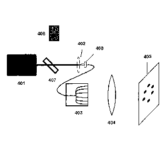
[0088] Figure 4 illustrates a laser apparatus in which laser light radiation
(a laser beam) from a
laser source 401 is transferred into the proximal end of a bundle of single
mode fiber optics using
a positive lens, referred to herein as a launching lens 402. The distal end of
optical fiber bundle
403 of this laser delivery system provides a plurality of laser beams. Single
mode optical fibers
typically have core diameters of 9 microns or less, allowing only one type of
laser energy
distribution to propagate through them. The number of optical fibers in the
bundle can vary.
Each optical fiber will receive a portion of the total energy from the laser
source. Consideration
for the amount of energy required for the treatment process from each optical
fiber, the amount
of available energy from the laser and the potential damage threshold of the
proximal end of the
optical fibers are factors in how many actual fibers are used. For this
example, six individual
optical fibers are represented. A modified version of this would be to launch
the laser energy
into a single optical fiber. This optical fiber would then be split into two
optical fibers. The two
optical fibers are then split again to give four optical fibers. This process
can be continued to
achieve the desired number of optical fibers at the distal end. This process
is preferred for more
uniform laser intensity at each distal end of the optical fibers.
[0089] At the distal end 403 of the fiber optic bundle, each optical fiber is
separated and
positioned in a holder made of a material that will set the position and
separation of each optical
fiber end with respect to each other and allow for the polishing of all the
optical fibers as a unit.
Materials such as, but not limited to, aluminum, nylon or plastic for example,
may be used to
join the optical fibers into a fiber optic bundle. The ends of the optical
fibers are typically
affixed using a suitable epoxy. Energy launched into the proximal end of the
fiber optic bundle
24
CA 02586214 2007-05-01
WO 2006/050424 PCT/US2005/039638
will exit the distal ends and propagate towards the primary focus lens. The
distance from the
distal end of the fiber optic bundle to the primary focus lens should be such
that all laser energy
from the sum of the optical fibers falls within the clear aperture of the lens
to minimize loss.
[0090] The laser delivery system of Figure 4 also includes a focus lens 404.
The primary focus
lens will image the laser light from the fibers at the focal length of the
primary focus lens. The
focused laser beams will form spots at Best Focus in an image plane 405. For
example, if the
ends of 9 micron core optical fibers are separated radially on a 3.75 mm
diameter, the primary
focus lens will produce an image of approximately 9 micron spots with the same
relative
separations at the focus of the lens. This is an example of 1:1 image relay.
Altering the focal
length of the primary focus lens can effect the separation of the spots
produced. Figure 8
illustrates how a pair of optical fibers 801 offset from each other at an
angle 0 (theta) can interact
with a focus lens 802 downfield from the optical fibers 801. The optical
fibers 801 emit laser
beams 804 and 807 which pass through and are focused by the focus lens 802.
Each of laser
beams 804 and 807 contacts the focus lens 802 at an angle off-center. The
focus lens 802
provides beams 805 and 806 whose diameters are narrowing and whose irradiance
is increasing
as they approach a focal point. The focus lens 802 can be computer-controlled
and/or motorized
So that the distance from the optical fiber can be adjusted, thereby adjusting
the separation
between the ablation points. The motor actuation of the focus lens can be done
by any means,
such as electrical gear devices or piezoelectric activators.
[0091] The fiber optic bundle can be held in a mechanical stage that can
rotate the device, for
example, along the 3.75 mm diameter in which the fiber end lie. This allows
for the spots being
imaged on the eye tissue to rotate as well. It also allows for the
illumination of new tissue for
therapy along a fixed diameter. This rotational device can be manual or
motorized. These
devices can be equipped with digital readouts that can give rotational
information of the fiber
optic bundle or the diffractive optic with a high degree of accuracy which
directly con-elates to
the rotation of the focus spots in the circular pattern.
[0092] A visible aiming system can be utilized to target the invisible Nd:YAG
laser radiation on
or in close proximity to the germinative zone or another target zone. Figure 4
also shows how
visible laser energy that is below the damage threshold of eye tissue can be
introduced into the
fiber optic bundle device using an alignment mechanism which provides visible
light at the
CA 02586214 2007-05-01
WO 2006/050424 PCT/US2005/039638
ablation points, allowing the physician the ability to view the relative
position of where the laser
energy from the ablative laser source will hit. The laser apparatus in Figure
4 also includes an
alignment laser 406, which is a low powered visible laser device, such as a
laser diode in the
visible spectrum at 630 inn, that provides a light beam into the fiber optic
bundle by use of a
dicroic splitter 407, a coated mirror, or similar device or means for
providing more than one light
beam to a single location. These devices can be manufactured with coatings
that will reflect
visible light and transmit near infrared light at 1064 nm, typically generated
from a YAG laser.
For example, a mirror can be coated on one side so that it reflects light from
one side and is
transparent to light from the other side. The dicroic splitter 407 and
alignment laser 406 can be
positioned in such a way that the visible light beam will be co-linear with
the ablative laser
beam. Because the materials used in the fiber bundle device can pass both
visible and near
infrared laser energy, both lasers can utilize the same components without one
affecting the
other. Figure 4 depicts the fiber optic bundle device (which includes a SMA
connector 408 at the
proximal end and a flexible protective jacket 403 at a distal end). The
dicroic splitter 407 makes
it possible for the alignment laser 406 to be on or off while therapy is being
performed.
[0093] Figure 5 shows the use of a single optical element 502 such as a
diffractive optic or
binary optic to generate a plurality of laser beams. This optic alters phase
relationship of the
laser beam resulting in a distribution of the laser energy in a desired
pattern. For example, a set
of six focus spots are founed in a circular pattern at 3.75 mm for a primary
focus lens 503 with a
focus of 2 inches. Each diffractive optic 502 is designed with fixed
characteristics, and the
pattern of spots imaged by the primary focus lens 503 at the image plane 504
is fixed. Figure 9
illustrates how a laser source 901 can interact with a diffractive optic 902
and a focus lens 903
downfield from the laser source 901. The laser source 901 provides laser beam
904 which
passes through the diffractive optic 902 which can diffract the light to
provide more than one
laser beam. In Figure 9, two laser beams 905 and 906 are provided by the
diffractive optic 902,
though a greater number of laser beams may. be provided by other suitable
diffractive optics.
The focus lens 903 provides beams 907 and 908 whose diameters are narrowing
and whose
irradiance is increasing as they approach a focal point. As mentioned above,
altering the location
of the focus lens in relation to the focal length of the lens of the laser
delivery system will
increase or decrease the diameter, or separation, of the focus spots. The
location of the focus
lens 903 can be computer-controlled and/or motorized so that the distance from
the optical fiber
26
CA 02586214 2007-05-01
WO 2006/050424 PCT/US2005/039638
can be adjusted, thereby adjusting .the separation between the ablation
points. The motor
actuation of the focus lens can be done by any means, such as electrical gear
devices or
piezoelectric activators.
[0094] The diffractive optic or binary optic can be held in a rotation stage
as illustrated for focus
lenses in Figure 6. Again, by rotating the optic, the six focus spots can be
positioned at different
positions along a fixed diameter.
[0095] Figure 5 also shows the use of an alignment mechanism which provides
visible light at
the ablation points. An alignment beam 506 is provided as described in Figure
4, although the
diameter of the focus spot pattern may fall on a slightly different
circumference because of the
shorter wave length of the alignment laser. This can be compensated for by the
addition of a
negative correction lens in the alignment beam path before the dicroic
splitter if it is determined
that the differences in each circumference is significant to this application.
Figure 5 shows the
optical system with a dicroic splitter 505 as described in connection with
Figure 4, though other
devices or means may be used which provide more than one light beam to a
single location. As
discussed earlier, the dicroic splitter permits the alignment beam to remain
in the on state during
therapy with no adverse effects.
[0096] The fiber optic bundles and diffractive and binary optics described
herein are exemplary
of a broader class of optical components contemplated for generating a
plurality of ablation
points. For example, other elements such as beamsplitters, diffraction
gratings, binary optics,
diffractive optics, micro optics, filters, gratings, and others are
contemplated. A preferred
apparatus has an even number of laser beam-generating elements, for example an
even number
of optical fibers in a fiber optic bundle or an optic that produces an even
number of focus spots.
A laser-guiding element can be made to generate these spots that are equal
distance around the
circumference from each other to keep the desired symmetry.
[0097] It is also possible to produce spots that are offset from the center
line. In previous
examples, the laser beam was directed at the center and normal to the lens. In
other words, the
laser beam hits the lens at a 90 degree angle. This will produce a focus spot
that is collinear, that
is, on the same center line as the laser. A line could be drawn from the spot
at best focus,
through the center of the lens and back down the center of the laser beam all
the way back to the
laser. However, the laser can be adjusted off this centerline and then pointed
back towards the
27
CA 02586214 2007-05-01
WO 2006/050424 PCT/US2005/039638
focusing lens such that the beam still hits the center of the lens. A line
drawn down the center of
the beam to the lens and compared to the original centerline, will be an
offset of a few degrees.
[0098] If the angle between the original centerline and the new beam path is
offset by 10
degrees, the focusing lens will still produce a focus spot at the same
distance from the lens.
However, because the beam is entering the center of the lens at an angle, the
focus spot now
produced is also offset a few degrees. The lens tries to bend the laser beam
back to the original
centerline, but pointing the laser to the lens at an angle causes the focused
spot to appear off to
one side of the centerline. If a second laser is set up on the other side of
the original centerline
and pointed to the center of the lens, a second focus spot will be produced on
the opposite side of
the original centerline. The separation of the two focus spots are a
combination of the total angle
between the two beams, now 20 degrees, and the focal length of the lens. The
longer the focal
length of the lens, the greater the separation of the two spots at focus.
10099] For the optical fiber bundles discussed above, if the fiber ends are
tipped at an angle with
respect to the focus lens (as shown in Figure 8), the lens will produce
separate spots as described
above. However, if the fibers are not angled the lens can also be used to
image the fiber tips
which will also produce individual spots. The smallest focus spots that can be
made are the
diameters of the tips of the fibers for the non-angled fibers. This is
referred to an image relay
system. The diffractive optic works more like the example of the two lasers. A
single beam is
pointed to the center of the diffractive optic at 90 degrees of incidence. The
diffractive optic
causes 50% of the beam to steer off of the center line of the lens in one
direction and 50% of the
beam to steer off at the same angle but in the opposite direction. Just like
in the example, the
lens produces two focus spots because the two beams produced by the
diffractive are coming at
the lens at an angle. The separation of the two spots is a combination of the
grating period of the
diffractive optic and the focal length of the lens. For the diffractive
system, if the diffractive
remains constant, changing the focal length of the lens changes the spot
separation.
[00100] A diffractive optic can be made to produce two spots when used
with a focusing
lens. The lens has a focal length of 50 mm. The separation of the two spots
produced is lOmm
apart. Increasing the focal length of the lens will increase the separation of
the two spots as
described in the example of the two lasers pointing at a common lens. The
longer the focal
length of the lens, the greater the separation of the two focus spots. By
increasing or decreasing
28
CA 02586214 2007-05-01
WO 2006/050424 PCT/US2005/039638
the focal length of the lens in the diffractive system described, the
separation can be adjusted for
each individual patient. The diffractive is rotationally sensitive, that is,
the two beams it
produces rotate about a central axis as the diffractive is rotated. This
allows for positioning the
two spots at any angle on a fixed circumference. Again, changing the focus
lens to a lens having
a different focal length would allow for positioning the two spots on
different circumferences to
meet the needs of different patients.
[00101] Other techniques may be used to create a pattern of ablation
points on the
crystalline lens. For example, a mask can be used with a scanning mechanism or
a laser beam
having a wider diameter to created a desired pattern, as described in U.S.
Patent Nos. 6,263,879.
A mask can be used to limit the laser beam to a defined pattern, thereby
creating a desired
ablation pattern, as described in U.S. Patent Nos. 5,711,762 and 5,735,843.
For example, a mask
having slits with a circular or crescent shape may be used.
[00102] Figure 6 shows a lens array for the laser delivery system which
can be used to
easily and precisely change the primary focus lens in the path of the laser
light. The lens array
comprises a housing 601 having openings for a plurality of focus lens. The
housing 601 shown
in Figure 6 has six openings for focus lenses 602a through 602f. In the lens
array, each circle
602a, 602b, 602c, 602d, 602e, and 602f, represents a single focus lens (such
as those represented
as focus lens 404 in Figure 4 and focus lens 503 in Figure 5). As shown in the
side view of
Figure 6, the lenses 602a, 602b, 602c, and 602d may be held out from the
housing 601 (in the
side view, lens 602e and 602f are hidden by lenses 602b and 602c). Figure 6
shows the lens
602a through 602d held out at different distances, however, it is preferable
that the lenses be held
at the same distance from the housing or within the housing itself. The lens
array is located after
the fiber optic bundle or the diffractive optic. The lens array is rotatable
such that all the laser
light beam illuminates only one lens of the lens array. The lens array can be
used to alter the
circumference of the focus spot pattern. The side view of this device shows
how different focal
length lenses can be offset in simple holders so that the image plane remains
constant by fixing
each lens at different distances to the image plane to compensate for the
different focal lengths of
these primary focus lenses. The focal lengths may vary, for example, by 0.01
to 2 millimeters.
[00103] The laser delivery system can also include a mechanism for sliding
the focus lens
closer to or farther from the patient. The focus lens could be adjusted by a
sliding means, such
29
CA 02586214 2007-05-01
WO 2006/050424 PCT/US2005/039638
as where a lens would be disposed on a sliding adjustment stage that would
move in the
longitudinal direction, either closer to or farther from the eye to be
treated. The lens would be
disposed on a slide having a micrometer-scale adjustment capacity. For
example, the distance of
the lens from the eye to be treated could be adjusted from an initial focal
length of 50 millimeters
to an adjusted focal length of 51 millimeters.
[00104] The laser delivery system can also include a mechanism to rotate
the laser beams
so that an additional set of ablations can be made on a single patient. Means
for rotating the laser
beams include automated and manual devices and include wheels or turntables in
which the laser
element is in the middle or the circumference. Such mechanisms for rotation
facilitate making
ablations to the crystalline lens in more than one step. For example, if 1,000
ablation points on
the crystalline lens are to be made, the practitioner may wish to make 500 at
one time, followed
by 500 at a second time. It may be desirable to have a smaller number of laser
beam-generating
elements connected to a single laser source in order to avoid excessive power
requirements for
the laser source. For this option, it may be desirable to dispose a plurality
of laser spot-
generating elements on a wheel that can be rotated. Preferably, the wheel can
be accurately and
.precisely rotated, for example, with precision on a micrometer scale. The
doctor or other
medical practitioner who is performing the procedure could then locate that
wheel at a second
position for a crystalline lens, and then by just using a micrometer on this
slide, make a one
millimeter adjustment for this stage. The patient would remain in the same
location.
[00105] Figure 7 shows a mechanical rotation stage for precisely rotating
the ablation
points. The center of the device is hollow so that fixtures for the fiber
optic bundle, diffractive
optic or binary optic can be affixed in such a way that the device can be
rotated with a minimal
amount of off-axis steering. Off-axis steering can cause the spots to rotate
out of position at the
target area. It is worth noting that because the laser beam passes through the
diffractive optic or
binary optic, it is generally unaffected by off-axis steering, whereas this
problem greater
consideration for the fiber optic bundle array.
[00106] The apparatus may further comprise a digital display that is
operatively connected
to a rotation wheel equipped with an encoder or other feedback mechanism so
that the position of
the laser spot-generating element (the optic) can be set to a precise degree.
The digital display
can indicate where the laser spots will be generated to a significant degree
of accuracy, for
CA 02586214 2007-05-01
WO 2006/050424 PCT/US2005/039638
example to one-hundredth of a degree. Using such a digital display, the laser
spot-generating
element can be rotated, for example from zero to 359.99. This provides precise
feedback as to
where the laser spot-generating elements are in rotation around that
circumference, so one could
always keep track. Alternatively, the rotation of the laser spot-generating
elements can be
motorized. A small motor could be placed on a rotation wheel with the feedback
and the doctor
can either drive it to that next position or enter the desired degrees of
rotation and the laser spot-
generating element will rotate to the entered position.
[00107] For example, it may be desirable to make 64 ablation points. One
option is to
have 64 laser spot-generating elements associated with a single laser source.
There would just be
one fire of a laser and the treatment is done. However, the consequence is
that the power of the
laser source is divided into 64 parts. If the laser does not have sufficient
power to provide
enough energy for each of those 64 spots, then the procedure will not be as
effective as desirable.
One solution is to provide a larger laser, since the power of the laser is
limiting how many spots
can be generated. Another solution is to divide the laser into a lesser number
of laser spot-
generating elements, and apply the laser spots to the crystalline lens in more
than one step, as
described herein.
[00108] An option for adjusting the spacing of ablation points comprises
substituting a
focus lens having a different focal length. The operator can make a slight
adjustment. Those
slides can also be motorized and it can also have feedback with a digital
display providing high
precision. A high degree of accuracy can be provided with linear or rotational
stages having
encoders built into them.
[00109] A translation stage in an operational system can be used to
customize the spot
diameter for different size eyes of different patients. With an array of
lenses in place, the right
lens position can be selected and then an adjustment can be made to offset the
focus so that the
subject remains at the same place. Offsets for a focal length change of the
lens are relatively
small, typically on the order of only a few millimeters for the methods
described here.
[00110] There is substantial imagery laying, magnifications, and optics in
between the
optical fibers, the tips of the optical fibers, and the location of the
subject. Relaying the image of
the ends of the fibers and magnification has to be taken into account. If the
magnification is
changed, for example from of 1:1 to 1:8, the ablation points will be further
apart. The result is to
31
CA 02586214 2013-11-12
effectively de-magnified, or make a larger spot array in the image plane. One
of ordinary skill will
recognize that the size of the circular spot made by the array of fibers on
the far end or the output end
will be changed by changing the magnification of the optical system between
the subject and the ends of
the fiber.
[00111]
Another way is to take two or more optical fibers and manipulate them
independently of each
other such that the light being emitted to a lens hits the lens on a slight
angle and if each of those are on
slightly different angles, the lens will image two spots, effectively the same
diameters, and that
separation is a function of how much the angle of the fiber is tipped as it
shines onto the lens. It is
contemplated that a single optical fiber split into two may be used, which is
commercially available
equipment. This would require very precisely positioning those angles such
that if a line were drawn
directly through the center of the eye, if one optical fiber is rotated 3
degrees so that it hits the lens on a 3
degree angle, instead of straight down the middle, and rotate the other fiber
is rotated 3 degrees, it is
necessary to ensure that both of those optical fibers are very precisely 3
degrees of angle towards the
lens. If one of those angles is 3 degrees and one of is different, for example
2.8 degrees, one spot is
going to be drawing a different arc than the other, the result will be
essentially two different
circumferences being ablated. The undesirable result would be asymmetric
pattern of ablation points but
rather a pattern that is egg-shaped or oval. There are other processes for
generating multiple spots using
tips of optical fibers, for example, by manipulating their magnification in an
array or bundle or using
individual optical fibers and manipulating them individually and changing
their angles with an
impingement angle to the lens.
[00112] Other techniques for making a plurality of ablation points are also
contemplated.
For example, a laser apparatus having a scanning feature may be used. The
scanning feature moves the
laser beam so that the ablation point is moved. Scanning laser apparatus are
known in the art and are
used to generate lines by moving the laser beam back and forth. Ablation spots
can be made by moving
the laser beam in a scanning motion, preferably under computer control to
create a desired pattern of
ablation. For example, a computer controlled scanning mirror can move or scan
a laser beam in the X-Y
direction at the focal plane. The scanning can be carried out in a variety of
patterns. For example, dots,
circles, or curved lines may be created. Additional description and
illustration of scanning laser
techniques can be found in U.S. Patent Nos. 6,325,792 and 6,706,036, though
ablation of
32
CA 02586214 2013-11-12
the cornea is illustrated in those patents rather than ablation of epithelial
cells of the crystalline lens.
1001131 Optionally, a biochemical approach may be used alternatively to or
additionally
with the laser-based approach, where one or more biochemically active agents
are used to inhibit
epithelial cell mitosis. Suitable biochemically active agents may include
Taxol, Nocodazole or other
spindle inhibitors, or Hydroxyurea or other nucleotide synthesis inhibitors. A
suitable biochemically
active agent may be in admixture with an organic or inorganic carrier or
excipient suitable for
administration to the eye. Alternatively, the biochemically active agent may
be ingested or injected.
Preferably, the active chemical agent is administered through eye drops or an
eye salve.
[00114] For example, the laser-based approach may be used to prepare cells,
and
thereafter eye drops containing a suitable biochemically active agent may be
employed. Alternatively, a
light-activated drug may be used.
100115]
[001161 While the present invention has been described and illustrated by
reference to
particular embodiments, it will be appreciated by those of ordinary skill in
the art that the invention
lends itself to many different variations not illustrated herein. For these
reasons, then, reference should
be made solely to the appended claims for purposes of determining the true
scope of the present
invention.
[00117] Although the appendant claims have single appendencies in accordance
with U.S.
patent practice, each of the features in any of the appendant claims can be
combined with each of the
features of other appendant claims or the main claim.
33