Note: Descriptions are shown in the official language in which they were submitted.
CA 02586409 2007-04-26
TRI-BENDING SPHINCTERTOME
FIELD OF THE INVENTION
[0001] The present invention relates to surgical cutting devices, and in
particular to
multidirectional cutting devices.
BACKGROUND OF THE INVENTION
[0002] Laparoscopic surgery is one type of minimally invasive surgery in which
a surgeon uses
numerous trocar ports to access a tissue site of interest within the abdominal
cavity of a patient.
The benefits of laparoscopic surgery, as compared to open incisional,
abdominal surgery, include
less pain, shorter recovery time, less scarring, and lower cost. Endoscopic
surgery affords
another way to access the abdominal cavity via natural openings (mouth, anus,
vagina, urethra)
of the body and through the peritoneal lining of the abdominal cavity.
Obviously, the size and
shape of instruments that may be passed through a body lumen in order to
perform a medical
procedure in the abdominal cavity are greatly restricted due to the anatomical
properties of the
lumen.
[0003] General surgeons, gastroenterologists, and other medical specialists
routinely use flexible
endoscopes for intralumenal examination and treatment of the upper
gastrointestinal (GI) tract,
via the mouth, and the lower GI tract, via the anus. In these procedures, the
physician advances
the flexible endoscope through the lumen, periodically pausing to articulate
the distal end of the
endoscope using external control knobs, to redirect the distal tip of the
endoscope. In this way,
the physician may navigate the tortuous passageway of the upper GI past the
pharynx, through
the esophagus and gastro esophageal junction, and into the stomach. The
physician must take
great care not to injure the delicate mucosal lining of the lumen, which
generally may stretch
open to a diameter in the range of about 15-25 mm, but normally has a non-
circular cross
sectional configuration when relaxed.
[0004] During such translumenal procedures, a puncture must be formed in the
stomach wall or
in the gastrointestinal tract to access the peritoneal cavity. One device
often used to form such a
puncture is a needle knife which is inserted through the working channel of
the endoscope, and
which utilizes energy to penetrate through the tissue. A sphinctertome can
then be inserted and
1
'! I '
CA 02586409 2007-04-26
reinserted into the tissue to expand the puncture made by the needle knife to
form a
multidirectional incision in the tissue. While effective, such a procedure for
forming
multidirectional incisions in tissue is time consuming and can also cause
unnecessary distress to
the tissue
[0005] Accordingly, there remains a need for improved surgical cutting
devices.
SUMMARY OF THE INVENTION
[0006] The present invention provides various devices and methods for forming
a cut in tissue.
In one aspect, a device for cutting tissue is provided that includes a
flexible elongate member and
a first tissue cutting wire that extends along the member. The first tissue
cutting wire can have a
first portion that is constrained with respect to the member, a distal portion
that is anchored to the
member, and an unconstrained portion that is adjacent to a first bend region
on the member. The
device can also include second and third tissue cutting wires that extend
along the member and
which have first portions that are constrained with respect to the member,
distal portions that are
anchored to the member, and unconstrained portions that are adjacent to second
and third bend
regions on the member, respectively. The unconstrained portions of the first,
second, and third
wires can be adapted to move from a delivery configuration to a cutting
configuration to effect
cutting of tissue upon the selective application of tension the wires.
[0007] The wires can have a variety of orientations with respect to the member
to effect
formation of a multidirectional cut. In one embodiment, the unconstrained
portions of the first,
second, and third wires can be circumferentially spaced from one another about
the member, and
can be positioned at the same longitudinal position on the member.
[0008] The first portions of the wires can be constrained, and the distal
portions of the wires
anchored, using a variety of techniques. In one embodiment, the wires can be
constrained within
and anchored to lumens or guide channels formed in the elongate member. In
another
embodiment, the wires can be constrained and anchored by a plurality of spaced
retaining
members positioned on the member. Additionally, the distal portions of the
wires can be
anchored to the member at a position that is offset from a longitudinal axis
of the member to
facilitate bending of the member.
2
i
CA 02586409 2007-04-26
[0009] The device can also include a variety of other features, such as a
central working channel
that extends between the proximal and distal ends of the member. The central
working channel
can have an incising element disposed therein and/or can be adapted to receive
a guide device or
an incising element.
[0010] In another aspect, a device for cutting tissue includes a flexible
elongate member having
proximal and distal ends, a first guide channel extending along a portion of
the member, a second
guide channel extending along a portion of the member and that is angularly
spaced along a
circumference of the member relative to the first guide channel, and a third
guide channel that
extends along at least a portion of the member and that is angularly spaced
along a circumference
of the member relative to the second guide channel. The first guide channel
can include a first
tissue-cutting wire that has a proximal end slidably disposed therein and an
exposed portion that
extends distally beyond the first guide channel. Similarly, the second and
third guide channels
can include second and third tissue-cutting wires having proximal ends
slidably disposed therein
and exposed portions that extend distally beyond the second and third guide
channels,
respectively. The device can also include an actuator coupled to the proximal
ends of the first,
second, and third wires. The actuator can be adapted to selectively apply
tension to the first,
second, and third wires that is effective to bend the member and expose a
portion of the wire
which extends distally beyond the guide channels to cause the device to assume
a cutting
configuration in which the wire is exposed relative to the member.
[0011] The exposed portions of the first, second, and third tissue-cutting
wires can be positioned
at a variety of locations along the member. In one embodiment, the exposed
portions of the first,
second, and third tissue-cutting wires can be circumferentially spaced about
the member and
optionally positioned at the same longitudinal position on the member.
[0012] The guide channels can have a variety of configurations, and in one
embodiment the
guide channels can be lumens that are formed in or along the flexible elongate
member. In
another embodiment, the guide channels can be formed from a plurality of
spaced retaining
members located on the elongate member. The device can also include a variety
of other
features to facilitate cutting tissue, such as at least one central working
channel that extends
between the proximal and distal ends of the member. The central working
channel can be
3
" "I
CA 02586409 2007-04-26
adapted to have an incising element movably disposed therein and/or to receive
a guide device or
incising element.
[0013] Methods for cutting tissue are also provided. In one aspect, a method
for cutting tissue
includes inserting a selectively bendable, elongate flexible cutting member
into a lumen of a
body. The member can have at least three tissue-cutting wires extending
therealong, and the
wires can be at least partially exposed with respect to the member. The method
can further
include selectively applying tension to at least one of the wires to cause the
member to bend
adjacent to the exposed portion of the wire thereby separating the wire from
the member so that
it assumes a tissue-cutting orientation. Energy can be delivered to the
wire(s) when it is in the
tissue-cutting orientation to effect cutting of a tissue.
[0014] For example, and in one embodiment, tension can be selectively applied
to a first of the
wires. This causes the member to bend in a first direction to separate a first
tissue-cutting wire
segment from the member such that the wire segment is in a position to form a
cut in tissue.
Thereafter tension can be selectively applied to a second of the wires to
cause the member to
bend in a second direction to separate a second tissue-cutting wire segment.
In this position, the
second wire segment can then form a second cut in the tissue, and the second
cut can be formed
in a different direction than the first cut. Tension can then be applied to a
third of the wires to
cause the member to bend in a third direction to expose a third tissue-cutting
wire segment. A
third cut can be formed in the tissue, and the third cut can be formed in a
direction that is
different than the direction of the first and second cuts. When the first
tissue-cutting wire
segment is positioned at an angle relative to the second tissue-cutting wire
segment, and the
second tissue-cutting wire segment is positioned at an angle relative to the
third tissue-cutting
wire segment, the method can further include rotating the member within the
tissue to position
the second and third tissue-cutting wire segments at the tissue to effect
cutting.
[0015] The method can also include a variety of other steps to facilitate the
formation of a cut in
tissue, such as forming an incision in the tissue prior to the insertion of
the member using an
incising element disposed within a central working channel of the member or
controlling an
amount of tension applied to the wires such that the device can be positioned
at a desired
4
' '1 '
CA 02586409 2007-04-26
=
location in tissue. Additionally or alternatively, the method can include
using a guidewire to
position the member within the tissue.
BRIEF DESCRIPTION OF THE DRAWINGS
[0016] The invention will be more fully understood from the following detailed
description
taken in conjunction with the accompanying drawings, in which:
[0017] FIG. lA is a perspective view of one embodiment of a surgical cutting
device in a
delivery configuration;
[0018] FIG. 1B is a perspective view of the device of FIG. 1A in a cutting
configuration;
[0019] FIG. 1C is a perspective view of a distal end of a member of the device
of FIG. 1B;
[0020] FIG. 1D is cross-sectional view of a mid-portion of the member of the
device of FIG. 1B
taken across line B-B;
[0021] FIG. 2A is a schematic illustrating the device of FIG. lA forming a
first cut in tissue;
[0022] FIG. 2B is a schematic illustrating the device of FIG. 1A forming a
second cut in tissue;
and
[0023] FIG. 2C is a schematic illustrating the device of FIG. lA forming a
third cut in tissue.
DETAILED DESCRIPTION OF THE INVENTION
[0024] Certain exemplary embodiments will now be described to provide an
overall
understanding of the principles of the structure, function, manufacture, and
use of the devices
and methods disclosed herein. One or more examples of these embodiments are
illustrated in the
accompanying drawings. Those of ordinary skill in the art will understand that
the devices and
methods specifically described herein and illustrated in the accompanying
drawings are non-
limiting exemplary embodiments and that the scope of the present invention is
defined solely by
the claims. The features illustrated or described in connection with one
exemplary embodiment
may be combined with the features of other embodiments. Such modifications and
variations are
intended to be included within the scope of the present invention.
CA 02586409 2007-04-26
[0025] The present invention provides devices and methods that can be used to
form
multidirectional cuts in tissue, such as tridirectional cuts. The tissue
cutting devices disclosed
herein generally include a flexible elongate member with at least first,
second, and third wires
that are at least partially constrained within or along a portion of the
member. A distal portion of
the wires is anchored on or within a portion of the elongate member. When
tension is applied to
at least one of the wires, such as by an actuator at a proximal end of the
device, the elongate
member bows while an unconstrained portion of the wire(s) becomes spaced from
and exposed
relative to the adjacent portion of the elongate member, assuming a tissue-
cutting configuration.
[0026] Such a device is useful, for example, in transgastric surgical
procedures where it is
necessary to form incisions in tough stomach tissue. After puncturing the
tissue, for example
using a needle knife that can be integrated with or separate from the elongate
member, the
elongate member is advanced through the puncture to a desired position. The
elongate member
can then be oriented in the cutting configuration, and energy can be applied
to the exposed wire
to form a cut in tissue. The device can then be manipulated within the tissue
(e.g., rotated) and
tension can be applied to another wire. This causes the elongate member to
bend in a different
direction and expose a different wire in a different cutting configuration
that is effective to cut
tissue in a different direction. A second cut can then be formed in the tissue
in a direction that is
different from the first cut. The device can then again be manipulated in the
tissue and tension
can be applied to the third wire to form a third cut in the tissue, which
extends in yet another
direction. One skilled in the art will appreciate that while the tissue
cutting devices disclosed
herein are described primarily in the context of transgastric surgeries, they
are applicable to a
variety of surgical procedures, including intestinal surgeries to remove
polyps and/or cancer
treatment procedures.
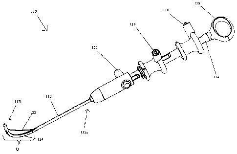
[0027] FIGS. 1A-1D illustrate one embodiment of a device 110 for forming a
tridirectional cut
in tissue. As shown, the device 110 includes a flexible elongate member 112
having proximal
and distal ends 112a, 112b and first, second, and third guide channels 130,
132, 134 that extend
along at least a portion of the member 112 and house first, second, and third
tissue-cutting wires
122, 123, 124. The wires 122, 123, 124 each have first portions that can be
constrained within
the guide channels 130, 132, 134 and distal portions that can be anchored
within the guide
channels 130, 132, 134. Each wire 122, 123, 124 also has an unconstrained
portion that extends
6
CA 02586409 2007-04-26
outside of the guide channels 130, 132, 134. The unconstrained portion of the
wires 122, 124,
134 can be recessed within grooves 136, 137, 138 formed on the member 112 when
the device
110 is in the delivery configuration. In use, tension can selectively and
individually be applied to
the wires 122, 123, 124 to cause the wires 122, 123, 124 to sequentially or
simultaneously move
from the delivery configuration to the tissue-cutting configuration to effect
the cutting of tissue.
Once the wires 122, 123, 124 are in the cutting configuration, energy can be
selectively and
individually applied thereto to effect the sequential or simultaneous cutting
of tissue.
[0028] FIG. lA illustrates the device 110 in the delivery configuration, in
which the member 112
has a relatively straight or linear configuration and the unconstrained
portions of the wires 122,
123, 124 are located adjacent to and in close proximity to the member 112. The
unconstrained
portions of the wires 122, 123, 124 can also rest within the grooves 136, 137,
138 that form
recesses on the member 112 to facilitate ease of delivery to the target site
in the tissue.
Following the placement of the device 110 within tissue, the wires 122, 123,
124 can be
selectively and individually tensioned to move from the delivery configuration
to the tissue
cutting configuration. As tension is applied, the member 112 in the bend
region Q, adjacent to
the wires, is caused to bend or bow such that the wire 122 separates from the
member 112.
Although FIG. 2C illustrates only one wire (122) under tension to cause the
adjacent section of
the member 112 to bend toward it, it is understood that each of the wires 123,
124 can be
selectively and independently tensioned to effect bending of the member 112 in
other directions,
as shown in FIGS. 2A-2C and as will be discussed in more detail below.
[0029] The flexible elongate member 112 can have virtually any configuration
that allows it to
be laparoscopically or endoscopically inserted to a surgical site. As shown,
the member 112 is
substantially cylindrical and sufficiently strong to be inserted into tissue,
yet flexible enough to
bend upon the application of tension to the wires 122, 123, 124. The member
112 can include
openings (openings 126a, 126b, 128a, 128b are shown in FIG. 1C) that
correspond to openings
in the guide channels 130, 132 to allow a portion of the wires 122, 123 to
exit and reenter the
guide channels 130, 132 and the member 112, such that they are unconstrained
from the guide
channels 130, 132. The member 112 can also include a groove or other receiver
(groove 136,
137, 138 is shown in FIG. 1D) that is formed in the outer surface of the
member 112 between the
openings 126a, 126b, 128a, 128b. The grooves can be adapted to hold the
unconstrained
7
i
CA 02586409 2007-04-26
portions of the wires 122, 123, 124 adjacent to the member 112 in a recessed
condition when the
device 110, or at least a portion thereof, is in a delivery configuration.
This is particularly
advantageous in that it protects the wires 122, 123, 124 and tissue from
damage during insertion
of the device 110. The distal end 112b of the elongate member 112 can be
adapted to facilitate
insertion into tissue, and as shown in FIGS. 1A-1C, the distal end 112b can be
tapered and/or
rounded. Alternatively or additionally, the distal end 112b of the member 112
can include
markings (not shown) formed thereon to facilitate positioning of the device
110 within tissue.
While the member 112 can have a variety of sizes, and the size of the member
12 can depend
upon the application of the device 110 and the type of tissue to be cut, in an
exemplary
embodiment, the member 112 can have a diameter in the range of about 2.0 mm to
3.5 mm, and
more preferably about 2.4 mm.
[0030] One skilled in the art will appreciate that the device 110 can also
include a variety of
features to facilitate the formation of a cut in the tissue. As shown in FIG.
1D, the member 112
can include at least one working channel 134 that can be centrally positioned
and that extends
therethrough. The working channel 134 can be adapted to hold a guide device,
such as a
guidewire, to facilitate positioning of the device 110 within tissue.
Optionally or additionally,
the working channel 134 can house an incising element (not shown) for forming
an initial
incision within the tissue. One skilled in the art will appreciate that the
incising element can
have a variety of configurations and can be integrated within the working
channel or it can be a
separate device that is inserted within the working channel. One exemplary
incising element that
can be used with the devices disclosed herein is the incising element employed
in the integrated
guidewire needle knife that is disclosed in commonly owned application
entitled "Integrated
Guidewire Needle Knife," filed concurrently herewith, the disclosure of which
is incorporated by
reference herein.
[0031] Referring back to FIGS. 1A-1B, a handle 114 can be located on the
proximal end 112a of
the member 112 to facilitate manipulation and handling of the device 110. The
handle 114 can
have any configuration that allows a user to conveniently hold and operate the
device 110. In
one embodiment however, the handle 114 has a substantially elongate shape and
includes a
thumb ring 116 to facilitate grasping. The handle 114 can also include at
least one actuator that
enables tension to be selectively applied to the tissue-cutting wires 122,
123, 124. In one
8
CA 02586409 2007-04-26
=
embodiment, the handle 114 also includes first, second, and third sliding
actuator levers 118,
119, 120 that are coupled to the first, second, and third wires 122, 123, 124,
respectively. In
alternate embodiments, rotatable knobs or dials can be used to selectively
apply tension to the
wires. A locking mechanism (not shown) can also be associated with the tension
applying
mechanism to hold the wires 122, 123, 124 in position once tension is applied.
In use, the levers
118, 119, 120 can be moved in the proximal direction to apply tension to the
wires 122, 123,
124, causing the wires 122, 123, 124 and the member 112 move from a delivery
configuration to
a tissue-cutting configuration. Once in the tissue-cutting configuration, the
wires 122, 123, 124
can be locked in position using the locking mechanism.
[0032] While a variety of locking mechanisms can be used, one exemplary
locking mechanism
can include a clamp that is effective to clamp down onto the wires and thereby
prevent
movement of the wires such that the device is held in the desired orientation.
The clamp can
have a variety of shapes and sizes, and it can be positioned at various
locations on the device. In
one embodiment, the clamp can be disposed within the handle. In an initial
position, the clamp
is spaced apart from the wires to allow free movement thereof. Once the wires
are tensioned and
the bend regions bent, the clamp can be moved until it engage the wires within
the handle. The
clamp will thus prevent movement of the wires when the clamp is in the locked
position. In
order to lock the clamp, the clamp can include a mating element formed thereon
and configured
to engage a corresponding mating element formed in the handle. For example,
the clamp can
include threads formed therein that are configured to mate with corresponding
threads in the
handle. As a result, rotation of the clamp about the handle will cause the
clamp to move between
the initial and locked positions. While the exemplary mating technique
includes threads, one
skilled in the art will appreciate that various other mating techniques can be
used.
[0033] The handle 114 can also optionally be adapted to facilitate the
delivery of energy to the
wires 122, 123, 124 to cut tissue. An energy source (not shown), such as a
battery that is in
electrical communication with the wires, can be disposed within the handle.
Alternatively, the
handle can be adapted to be coupled to an external energy source, such as a
generator or an
outlet. The handle can also include a mechanism that facilitates the selective
delivery of energy
to the wires, such as a button or knob that can cause activation of the energy
source. While the
exemplary embodiment illustrates an elongate handle 114, one skilled in the
art will appreciate
9
CA 02586409 2007-04-26
that the handle can have any configuration that allows a user to selectively
apply tension as well
as energy to the wires, such as a joystick control.
[0034] As noted above, first, second, and third guide channels 130, 132, 134
extend along at
least a portion of the member 112 for constraining portions of the wires 122,
123, 124 relative to
the member 112. The guide channels 130, 132, 134 can extend along the member
112 in a
variety of orientations to help effect multidirectional bending of the device,
however as shown,
the guide channels 130, 132, 134 are circumferentially spaced about the member
112 (e.g.,
radially disposed about the circumference of the member 112). The
circumferential spacing of
the guide channels 130, 132, 134 and hence the wires 122, 123, 124 enables the
tridirectional
bending of the member 112. The exposed portions of each of the wires 122, 123,
124 can be at
the same longitudinal level of the member 112, as shown in FIGS. 2A-2C, or
they can be at
different longitudinal levels. As a result, the member 112 has a bend zone Q.
This
configuration, as will be described below, enables the device 110 to be
inserted into tissue, and
then configured into a cutting configuration to form a cut in one direction
using wire 122. The
member 112 can then be configured in a cutting configuration to cut tissue in
another direction
with wire 123, and a third direction with wire 124, such that a tridirectional
cut is formed in the
tissue.
[0035] The circumferential spacing of the guide channels 130, 132, 134 and
hence the wires 122,
123, 124, also influences the shape of the resulting cut. While the guide
channels 130, 132, 134
can be separated by virtually any range of angles, in one embodiment the guide
channels 130,
132, 134 are equally spaced (i.e., by about 120 ) about the member 112. This
configuration can
result in the formation of a substantially triangular cut in tissue.
[0036] In particular, FIG. 2C illustrates one embodiment in which at least a
proximal portion of
the guide channel 130 is disposed within a proximal portion of the member 112.
The guide
channel 130 can extend through the proximal portion of the member 112 to an
opening 126a,
which marks the termination of a proximal portion of the guide channel 130. A
wire, such as
wire 122 shown in FIG. 2C, can extend through the guide channel 130, which
maintains it in a
constrained position, emerge through the opening 126a, and extend along the
member 112 in an
external, unconstrained position until it is anchored back within another
opening 126b. The
CA 02586409 2007-04-26
opening 126b can form the entry to a distal portion of the guide channel 130,
or it can simply
form a termination point for the wire 122.
[0037] Guide channels 132 and 134 are circumferentially spaced and angularly
offset from guide
channel 130 and are similarly constructed. That is, guide channels 132 and 134
each have a
proximal portion that constrains a wire and an opening (opening 128a is shown
in FIG. 2C)
through which the wire emerges to an external, unconstrained position. Each of
guide channels
132, 134 can also have an opening (opening 128b is shown in FIG. 2C) analogous
to opening
126b within which a distal portion of the wire can be anchored. While the
exemplary
embodiment illustrates guide channels 130, 132, 134 that are positioned at the
same longitudinal
position on the member 112, in other embodiments the guide channels can be
longitudinally
separated and radially offset from one another by a range of degrees, such as,
for example, about
90 or about 180 , depending upon the type of cut desired.
[0038] The guide channels 130, 132, 134 can have a variety of configurations,
however as
shown in FIG. 1D, the first and second guide channels 130, 132, 134 are
lumens. In other
embodiments, however, the guide channels can be retaining members, such as
guides, clips, or
fasteners, that are located on the outside of the member. The retaining
members can be relatively
closely spaced to hold the wires in a constrained position and more widely
spaced to form the
unconstrained portion of the wires.
[0039] As noted above, a distal portion of each of the wires 122, 123, 124 is
anchored to the
member 112, for example, within a distal portion of its respective guide
channel 130, 132, 134.
A variety of techniques can be used to anchor the distal portions of the wires
122, 123, 124, such
as welding, knots, adhesives, or other fasteners. In one embodiment, the
inside of the guide
channels can include hooks or some other fastener, and the distal end of the
wires can be
attached to such hooks or fasteners. The distal ends of the wires 122, 123,
124 can also be
anchored within the guide channels 130, 132, 134 at a position that is offset
from the longitudinal
axis of the member 112. As a result, and upon the application of tension to
the wires 122, 123,
124 the wires 122, 123, 124 can act as a lever arm to facilitate bending of
the member 112. The
proximal ends of each of the wires 122, 123, 124 can be disposed within the
guide channels 130,
132, 134 and coupled to a tension applying mechanism, such as the actuation
levers 118, 119,
11
Ir.,
CA 02586409 2013-11-26
120 located on the handle 114 as described above. A proximal portion of the
wires 122, 123,
124 can also be in electrical communication with, for example, a source of RF
energy.
[0040] The wires 122, 123, 124 can be formed from a variety of materials that
are suitable to cut
tissue. In one embodiment, the wires 122, 123, 124 are made from a conductive
material such
that the tissue can be cut via electrosurgical energy. Exemplary materials can
include stainless
steel, nitinol, carbon steel, aluminum, and combinations thereof. One skilled
in the art will
appreciate that the wires can also have a variety of sizes depending upon the
type of tissue to be
cut. In one embodiment, however, the wires can have a diameter in the range of
about 0.005
inch to 0.025 inch, and more preferably about 0.015 inch.
[0041] While the exemplary embodiment illustrates a device 110 having three
wires 122, 123,
124, one skilled in the art will appreciate that the device can have any
number of wires in any
number of configurations to form a variety of multidirectional cuts in tissue.
[0042] The device disclosed herein can be used in surgical procedures in the
manner described
below. Following preparation of the patient as known in the art, the member
can be inserted into
a natural or created orifice to a target site. As noted above, the device 110
is typically inserted in
the delivery configuration shown in FIG. 1A, where the member 112 has a
substantially linear
configuration and the wires 122, 123, 124 are located adjacent to the member
112 and optionally
recessed within the grooves 136, 137, 138. A variety of techniques can be used
to insert the
device into the orifice, and in one embodiment a guide device, such as a
guidewire, can be
positioned within a central working channel of the device and used to guide
the device to the
target site. Once at the target site, the guide device can be removed from the
working channel
and optionally replaced by an incising element. Alternatively, in embodiments
where the device
includes an additional working channel for a treatment device, the incising
element can be
inserted in such a working channel while the guide device remains in place. In
either
embodiment, the cutting device can then be used to form an initial perforation
in the tissue. A
variety of incising elements can be used, however in one exemplary embodiment
an integrated
guidewire needle knife device can be used, such as that disclosed in commonly
owned United
States patent application publication no. 2007/0255303 entitled "Integrated
Guidewire Needle
Knife".
12
I dl
CA 02586409 2007-04-26
[0043] Following the formation of the initial perforation, the device can be
further advanced
through the perforation so that an exposed portion of a wire to be used to cut
tissue is properly
positioned adjacent to the tissue to be cut. Thereafter, the member is moved
to the cutting
configuration. This can be effected by applying tension to one or more of the
wires, which
causes the portion of the member that is adjacent to the unconstrained portion
of the wire to bend
or bow, such that the unconstrained portion of the wire moves from a position
adjacent to the
member to a position spaced a distance away from the member and in a cutting
configuration.
For example, FIG. 2A illustrates the device 110 where tension is applied to
wire 122 such that
the bend region Q is bowed and the wire 122 is in the cutting configuration.
In alternate
embodiments, tension is simultaneously applied to the wires 122, 123, and/or
124 such that bend
region Q is bowed and the wires 122, 123 and/or 124 are in the cutting
configuration. Once the
wire is positioned in the cutting configuration, the locking mechanism can
optionally be
activated to maintain the position of the wire(s).
[0044] At this point, energy, such as RF energy, can be delivered to the
tensioned wire(s) by, for
example, using an energy delivery mechanism on the handle. This causes the
wire(s) to
penetrate the tissue, forming a cut in the tissue. For example, FIG. 2A
illustrates the device 110
being used to form a first cut CI in tissue 170 using wire 122. Following the
formation of the cut
C1, the energy delivery can cease, and the locking member can optionally be
released, causing
the member to return to its delivery configuration. The device can then be
slightly rotated within
the tissue such that the second wire 123 is aligned with the direction of
intended cutting, which is
different than the direction of the first cut C1. Tension can be applied to
the second wire 123, if
it is not tensioned already, to cause a portion of the device Q that is
adjacent to the second wire
123 to bend while the second wire 123 extends a distance away from the member
112 to assume
a tissue cutting configuration. Energy can then be delivered to the second
wire 123 to form a
second cut C2, as shown in FIG. 2B. Following formation of the second cut C2,
the application
of energy to the wire 123 can cease, and the tension can optionally be
released, causing the
member 112 to return to its delivery configuration. The device can then be
rotated again such
that the third wire 124 is aligned with the intended direction of the third
cut, which will be
different than the direction at least of the first and second cuts C1, C2.
Tension can then be
applied to the third wire 124, if the third wire is not already tensioned,
such that it assumes a
tissue-cutting configuration, and energy can be delivered to the third wire
124 to cause a third cut
13
CA 02586409 2007-04-26
C3 to be formed in tissue, as shown in FIG. 2C. The resulting shape from the
first, second, and
third cuts CI, C2, C3 is substantially triangular.
[0045] Although the above methods use the sequential tensioning of the wires
122, 123, 124 to
form a multidirectional cut in tissue, one skilled in the art will appreciate
that the wires can also
be simultaneously tensioned and energy applied to one, two or all of the
tensioned wires to form
a cut in tissue. The simultaneous tensioning of the wires is particularly
advantageous in that it
can facilitate positioning the device in tissue. For example, and referring
back to FIG. 2B, as
energy is applied to the wire 122, tension can be selectively applied to the
wire 123. This results
in the device bending at a location that is approximately midway between the
planes of wire 122
and wire 123. Additionally, and upon the application of energy to two or more
of the tensioned-
wires, the device can be used to form a large incision in tissue, such as to
cut a large otomy in an
organ in order to gain access to the adjacent space.
[0046] Cutting devices, including components thereof, can be designed to be
disposed after a
single use, or they can be designed to be used multiple times. In either case,
however, the device
can be reconditioned for reuse after at least one use. Reconditioning can
include any
combination of the steps of disassembly of the device, followed by cleaning or
replacement of
particular pieces, and subsequent reassembly. By way of example, the devices
disclosed herein
can be reconditioned after the device has been used in a medical procedure.
The device can be
disassembled, and any number of the particular pieces (e.g., the wires, the
member, or the
handle) can be selectively replaced or removed in any combination. Upon
cleaning andJor
replacement of particular parts, the device can be reassembled for subsequent
use either at a
reconditioning facility, or by a surgical team immediately prior to a surgical
procedure. Those
skilled in the art will appreciate that reconditioning of a cutting device can
utilize a variety of
techniques for disassembly, cleaning/replacement, and reassembly. Use of such
techniques, and
the resulting reconditioned cutting device, are all within the scope of the
present application.
[0047] One skilled in the art will appreciate further features and advantages
of the invention
based on the above-described embodiments. Accordingly, the invention is not to
be limited by
what has been particularly shown and described, except as indicated by the
appended claims.
14