Note: Descriptions are shown in the official language in which they were submitted.
CA 02587432 2012-10-18
WO 2006/053216 PCT/US2005/040930
Devices and Methods for Selecting Intraocular Lenses
Background of the Invention
Field of the Invention
[0001] The present invention relates generally to devices and methods for
selecting an
intraocular lens and more specifically to devices and methods of finding
suitable powers and/or
locations of intraocular lenses to be implanted into an eye in order to obtain
a predetermined
refractive outcome, taking into account such parameters as the asphericity of
the cornea and/or
the intraocular lens.
Description of the Related Art
[0002] US 5,968,095 refers to a method of preoperatively
selecting the power of an intraocular lens (IOL) to be implanted into an eye
having a lens haptic
plane. The method involves selecting eye parameters to construct an eye model
for finding a
correct representation of the intraocular lens as axially positioned in the
eye following surgical
implantation. However, this method is not designed to be applicable when any
of the optical
surfaces is aspheric. In particular this method is not applicable when using
aspheric lenses
designed to reduce or eliminate the spherical aberration of the cornea. Other
commonly applied
methods to determine IOL power, such as the widely used SRK/T formula, and
other widely
applied methods such as the Hoffer Q and Holladay 1 and Holladay 2 formulas,
suffer the same
shortcoming in being based on thin lens vergence calculations and/or spherical
lens surfaces.
Paul-Rolf Preussner et al. disclose an alternative method of predicting
outcome of choice of IOL
model and power in J Cataract Refract Surg, 2004, Vol.30, pp. 2077-2083.
[0003] As aspheric IOLs capable of correcting spherical aberrations now are
becoming
available on the market (e.g., Tecnis brand of IOL, available from AMO Inc.,
Santa Ana, CA),
there is a demand to obtain reliable methods to select aspheric IOL powers in
order to achieve the
desired patient outcome in terms of spectacle correction and/or image quality.
Brief Description of the Drawings
[0004] Embodiments of the present invention may be better understood from
the following
detailed description when read in conjunction with the accompanying drawings.
Such
embodiments, which are for illustrative purposes only, depict the novel and
non-obvious aspects
CA 02587432 2012-10-18
= .
WO 2006/053216 PCT/US2005/040930
of the invention. The drawings include the following figures, with like
numerals indicating like
parts:
[0005] FIG. 1 is a graphical representation of elements of an eye
model used in various
embodiments of the present invention.
[0006] FIG. 2a and 2b are magnified views of the corneal region of
the graphical
representation shown in FIG. l
[0007] FIG. 3 is a flow chart showing a method of selecting an IOL
according to one
embodiment of the invention.
[0008] FIG. 4 is a flow chart showing a method of selecting an IOL according
to another
embodiment of the invention.
[0009] FIG. 5 is a graphical representation of the elements of
computing system for selecting
an IOL according to embodiments of the present invention.
[0010] FIG. 6 is printout of the formulas programmed into each cell
of an Excel spreadsheet
used to provide the ray tracing program in accordance with embodiments of the
present
invention.
[0011] FIG. 7 is illustrates the numerical result of the
calculation in each cell of an Excel
spreadsheet used to provide the ray tracing program in accordance with
embodiments of the
present invention.
[0013] FIG. 8A-8D are through-focus MTF plots used to determine maximum MTF of
an
10L.
Detailed Description of the Drawings
[0014] The present invention is directed to reliable methods and devices for
selecting spherical
and aspheric intraocular lenses (IOLs) that provide a predetermined refractive
outcome for
patients in need of cataract or refractive surgery. Embodiments of the
invention may be
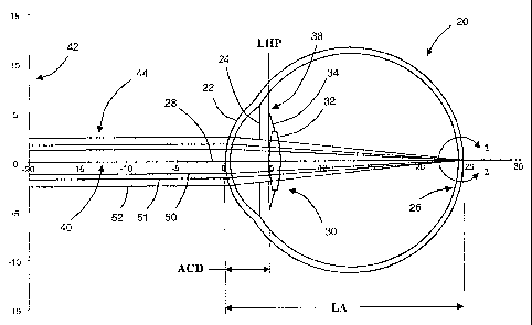
understood by reference FIG. 1, which is a graphical representation of a model
of an eye 20
comprising a cornea 22, an iris 24, a retina 26, and an optical axis 28. An
IOL 30 is disposed
within the eye 20, the IOL 30 comprising an optic 32 and one or more haptics
34 having distal
ends 38. In general, the eye model may consist of the dimensional parameters
illustrated by the
geometry shown in FIG. 1, for example, the axial length of the eye (AL) and
the anterior chamber
depth (ACD). Other dimensional parameters that may be included in the eye
model that are not
shown in FIG. 1 include, but are not limited to, the corneal radius (CR), the
corneal power (K),
and crystalline lens thickness (LT). The eye model may also include various
other parameters
-2-
CA 02587432 2012-10-18
WO 2006/053216 PCT/US2005/040930
such as, for example, the refractive indices of the various portions of the
eye 20 and/or the IOL
30. In certain embodiments, the distal ends 38 of the haptics 34 are disposed
within a plane
defined a lens haptic plane (LHP). In other embodiments, other information of
the IOL 30 may
be included in the eye model such as, for example, an effective principal
plane of the optic 32 or
other information regarding the optic 32 useful in determining the performance
optic 32 and/or
the location of the optic 32 within the eye 20.
[0015] The graphical representation of the eye model illustrated in FIG 1 also
has a coordinate
system containing a horizontal axis 40 and a vertical axis 42, which are
labelled in units of
millimetres. The graphical representation illustrated in FIG. 1 also shows a
plurality of rays 44
entering cornea 22 and the IOL 30 of the eye model. The plurality of rays 44
comprises a
paraxial ray 50 that is disposed near the optical axis 28 and a marginal ray
52 that is disposed
near edge of the opening formed by the iris 24. The plurality of rays 44
additionally comprises
an averaged ray 51 disposed between the paraxial ray 50 and the marginal ray
52, for example, at
a height at the pupil that is IN-2- or 1/2 times the height of the entrance
pupil height. In some
embodiments, the eye model additionally contains information regarding an
object or source
represented by the plurality of rays 44 entering the eye 20, for example, the
distance of the source
or object from the eye 20 and/or the extent of the source or object in units
of length or arc length.
[0016] Referring to FIG. 2a, which is a magnified view of the region around
retina 16, the rays
50-52 may come to focus at different points along the optical axis 28, which
are labelled in the
figure as marginal focus, best focus, and paraxial focus. As illustrated in
the figure, the distance
between the marginal focus and the marginal focus may be used to define a
longitudinal spherical
aberration (LSA). Such a result may be produced, for example, when the
surfaces of the IOL 30
are spherical. Alternatively, one or more of the surfaces of the IOL 30 may
comprise an aspheric
profile that is configured to reduce or eliminate spherical aberrations
produced either by an IOL
made of spherical surfaces or by at least portions of the eye 20 (e.g., the
cornea 22). In such
embodiments, as illustrated in FIG. 2b, the rays 50-52 focus to a common or
substantially
common point along the optical axis 28.
[0017] Embodiments of the invention may be used in conjunction with an eye
model such as
that illustrated in FIG. 1 to select or determine a characteristic of an IOL
to be implanted into the
eye of a subject or a class of subjects, for examples subjects of a particular
age group or
condition (e.g., a class of subjects who have had a LASIK or similar
procedure). In certain
embodiments, measurements from a subjects eye (e.g., AL, ACD, CR, LT) may be
used in
conjunction with statistical data and/or an analytical tool (e.g., a ray trace
program or algorithm)
-3-
CA 02587432 2007-05-11
WO 2006/053216 PCT/US2005/040930
to determine the characteristic of the IOL. The characteristic of the IOL
resulting from
embodiments of the invention may include the thickness of the IOL, the power
of the IOL, the
asphericity of the IOL, and/or the location of the IOL within the eye of the
subject or subjects.
[0018] Referring to FIG. 3, in certain embodiments, a method 100 of selecting
an IOL
comprises one or more of the following operational blocks 110-180. The method
100 comprises
an operational block 110, which comprises determining physical characteristic
of the eye such as
an axial eye length or a pupil size at a desired light level. The method 100
further comprises an
operational block 120, which comprises determining a desired postoperative
condition such as a
postoperative refraction and/or spherical aberration. The method 100 further
comprises an
operational block 130, which comprises determining an aspheric representation
of the corneal
curvature or curvatures. The method 100 also comprises an operational block
140, which
comprises selecting an IOL with one or more predetermined characteristics
(e.g., with a
predetermined power or asphericity) and determining the location of a plane of
fixation of the
IOL following implantation (e.g., the lens haptic plane or LBP). The method
100 additionally
comprises an operational block 150, which comprises employing the results of
operational blocks
110-140 to establish an eye model. The method 100 also comprises an
operational block 160,
which comprises computing, by means of an analytical tool (e.g., a ray tracing
routine) with said
eye model, an expected postoperative condition such as an expected
postoperative refraction
and/or spherical aberration. The method 100 further comprises an operational
block 170, which
comprises, in the case the expected postoperative condition is not within the
desired
postoperative condition, selecting another IOL with different power and/or
asphericity and
repeating operational blocks 150 and 160 until the expected postoperative
condition is within the
desired postoperative condition. The method 100 may also comprise an
operational block 180,
which comprises selecting, for implantation, an implantable IOL of the nearest
power and
asphericity available or designing an implantable IOL that results in the
desired postoperative
refraction and spherical aberration.
[0019] The method 100 may also include transforming the measured axial length
to a human
population average scale by adding to the value a transformation constant. The
axial length
measured by ultrasound is not the same as the optical axial length, and as the
axial length
measured by one piece of equipment may differ from that measured by another
one, there is a
benefit to obtaining instrument independent measurements. Measurement of axial
eye length for
an individual patient may be obtained by ultrasound A-scan or the newer
partial coherence inter-
ferometry (PCI) principle, available with the Zeiss IOLMaster. Regardless of
the instrument
-4-
CA 02587432 2012-10-18
WO 2006/053216 PCT/US2005/040930
and/or method used, the axial eye length may first be transformed to a human
population average
(HPA) scale. In certain embodiments, an underlying assumption for the HPA
scale is that the
mean axial length is about constant in any large group of adults.
Transformation is discussed in
more detail by Norrby et al. (J Cataract Refract Surg 2003; 29:100-105) and
the HPA scale is
introduced by Norrby et al. (J Cataract Refract Surg 2005; 31:1338-1344).
Transformation amounts to the addition of a correction constant
to the measured axial eye length. The correction constant is generally
regarded as instrument spe-
cific, for example, as described in Norrby et al. (J Cataract Refract Surg
2005; 31:1338-1344). A
general outline of a routine to obtain a common scale for axial lengths may
include the following
steps. First the postoperative anterior chamber depth consistent with axial
length, corneal radius,
postoperative refraction and IOL power implanted are calculated by thick lens
ray tracing for a
number of individual cases. The mean of the calculated anterior chamber depths
may be
calculated and compared with a previous study with the same lens (e.g.,
Pharmacia CeeOn
809C brand of IOL published in Koranyi et al.: J Cataract Refract Surg, 2002;
28:243-247).
The measured axial lengths may then be transformed by addition of a
constant value, and the mean anterior chamber depth was calculated anew. This
process may be
iterated until the calculated mean anterior chamber depth coincided with that
of the other.
[0020] The pupil size may be measured preoperatively at the desired light
level, e.g. mesopic
light (dusk). The pupil size at dusk is about 4 mm, but variations between at
least about 2 mm to
6 mm or more can occur.
[0021] The aspheric representation of the anterior corneal is typically
derived from corneal
topography, most commonly based on the so-called Placido disk principle.
Resulting height maps
can be used to fit an aspherical description of the surface by a least squares
optimization. Slit
based methods such as implemented on the Orbscan brand of topography systems
(Bausch &
Lomb) may be used for the same purpose (Holladay et al. J Refract Surg 2002;
18:683-691). The
Orbscan can also be used to obtain an aspherical description of the posterior
corneal surface in
the same mariner. Instruments based on the Scheimpflug principle, such as
Nidek EAS-100, may
also be used to obtain anterior and posterior curvatures of the corneal
surfaces. By rotating the
slit and taking multiple pictures the topography of both surfaces can be
obtained. The newly
presented Oculus Pentacam eye scanner, which is also based on the Scheimpflug
principle,
achieves this within a couple of arc seconds, and is suitable for use with the
method 100.
[0022] Independent of the measurement system used, the topography for the
purpose of the
method 100 is conveniently described as a conicoid surface of revolution,
characterized by the
-5-
CA 02587432 2007-05-11
WO 2006/053216 PCT/US2005/040930
aspheric constant k value (conic constant), optionally extended with
additional polynomial terms.
Preferably, k values are obtained for both anterior and posterior corneal
surfaces, optionally in
combination with additional polynomial terms.
[0023] The method 100 may be used to calculate an amount of at least one of a
postoperative
refraction and a postoperative spherical aberration for a lens that is
implantable into the eye of a
subject. Preferably the calculations are carried out using a ray tracing
program or procedure,
although other calculating means may also be used, such as an optics design
program. One
benefit of the method 100 is that it is capable of reducing the amount of
computation necessary
when using a ray tracing procedure and yet produces reliable information for
lens power
selection. Accordingly, only limited numbers of rays needs to be employed with
the routine
rather than the great number of rays normally traced for the purpose of
optical design (several
software packages are commercially available, e.g., those sold under the brand
names of
Zemax , OSLO , Code V ), which are cumbersome to employ, although they could
be used
for the purpose of the IOL power calculation and assessment of the resulting
image quality.
[0024] In one aspect of the invention a ray entering the pupil at ikri of the
entrance pupil
height is employed. This ray is here termed the focusing ray. Alternatively a
ray at the full pupil
diameter (marginal ray) and a ray with close to zero ray height (paraxial ray)
are traced. Focus is
in this case assumed to be at the midpoint of the foci of the marginal and the
paraxial rays. The
distance between the foci of the marginal and the paraxial rays, the
longitudinal spherical
aberration (LSA), can also be used as a simple metric for image quality. The
smaller LSA is the
better the image quality is.
[0025] According to one embodiment of the method 100, one or more of the
surfaces of an IOL
such as the IOL 30 are described by the formula:
1 2
Y
x= _______________________________ +a4y4 +a6y6 + ...................... ( 1 )
1 +1)2 y2
wherein R is the radius of curvature at the apex, k the conic constant, y the
radial distance
from the optical axis and x the sag in the direction of light propagation.
Depending on the value
of k the first term is a conic section and describes a:
hyperbola k< 0
parabola k=0
prolate ellips 0 < k< 1
-6-
CA 02587432 2012-10-18
WO 2006/053216 PCT/US2005/040930
circle k = 1
oblate ellips k > 1
[0026] The coefficients for the additional polynomial terms a4, a6, etc. can
either be set to
zero, in which case the surface is a conicoid of revolution, or be given
positive or negative non-
zero values to modify the simple conic section rotational surface.
Alternatively, the method 100
may be used with other forms of the above equation or other definitions of
terrns such as conic
constant.
[0027] A method to design intraocular lenses for the purpose of correcting
average corneal
spherical aberration obtained from pooled corneal data of a an elected patient
group is further
explained in the U.S. Patent Number 6,609,793. Corneas of the
normal population are in the prolate range (0 < k < 1) however, the method 100
is applicable to
all types of aspheric IOLs, such as IOLs with a hyperbolic (including
parabolic) or oblate
(including spherical) surfaces.
[0028] According to one aspect, patients having had corneal refractive surgery
to correct
myopia can have a hyperbolic anterior surface ( k 5_ 0), while those having
had corneal refractive
surgery to correct hyperopia can have an oblate anterior surface ( k 1)
(Buehren et al., Scientific
poster 144, AA0 2004, New Orleans). The method 100 demonstrates satisfying
capacity in
obtaining careful prediction of IOL powers also for such patients, including
estimating the
resulting retinal image quality in terms of LSA, although surfaces deviating
considerably from
prolate may be required.
[0029] The method 100 may further comprise obtaining the corneal apex radius,
typically both
anterior and posterior corneal apex radii, from the topography, or from
corneal radius measured
by conventional keratometry (at about 3mm diameter) and corrected to the value
at the apex by
the method described by Dubbelman et al. (Vision Res 2005; 45:117-132).
[0030] There are both indirect and direct methods available to preoperatively
determine the
location of the lens haptic plane (LHP), i.e. the distance from the anterior
cornea to the LHP.
Direct methods include ultrasound biomicroscopy, optical coherence tomography
and
Scheimpflug photograptly as taught in US 5,968,095. Newer,
commercially available equipment having the capacity to conduct such direct
measurements
includes the following systems, which are available from the listed companies:
Artemis
(Ultralink LLC), Visante OCT (Zeiss), and Pentacam (Oculus).
-7-
CA 02587432 2012-10-18
WO 2006/053216 PCT/US2005/040930
[0031] Alternatively, the location of the lens haptic plane may be obtained
with a prediction al-
gorithm that includes preoperatively measured parameters such as axial eye
length (AL), corneal
radius (CR) or, alternatively, corneal power (K), anterior chamber depth
(ACD), and crystalline
lens thickness (LT). Norrby et al. (J Cataract Refract Surg 2005; 31:1338-
1344) have studied
prediction algorithms of the general type:
LHP =
a+bxAL+cxACD+dxLT+exCR
+ f x AL2 + gxACD2 +hxLT2+ixCR2 (2)
+ jxALxACD+kxALxLT +1xALxCR
+ ACDxLT + nxACDxCR+ axLTxCR
[0032] One finding of the study mentioned is that AL and ACD measured with one
piece of
equipment can deviate systematically from that measured with another piece of
equipment
(Norrby et al. J Cataract Refract Surg 2003; 29:95-99; see also Koranyi et al.
J Cataract Refract
Surg 2002; 28:243-247, and Norrby, J Cataract Refract Surg 2001; 27:1656-
1661).
To correct measured AL and ACD the concept of a
Human Population Average (HPA) scale was devised (Norrby et al. J Cataract
Refract Surg
2005; 31:1338-1344). Algorithms containing LT and ACD in general were found to
be
unreliable when employing measurements obtained with different pieces of
equipment, despite
correction of ACD to the HPA scale (Norrby et al. J Cataract Refract Surg
2005; 31:1338-1344).
Also the early attempts to model LHP in terms of and LT and ACD (Non-by and
Koranyi, J
Cataract Refract Surg 1997; 23:254-259, US 5,968,095)
were found unreliable. Regression forniulas containing CR and AL in linear,
quadratic and cross-terms, with or without the constant a, in accordance with
the general
formula above, gave consistent results independent of the measurement
equipment used, when
AL was transfornied to the HPA scale. A preferred algorithm is
LHP = 2.486 + 0.2174x (AL + AAL) ¨ 0.4213x CR (3)
wherein AL is the measured axial eye length, AAL is the transformation
constant (ranging from
0.2 mm to 1.0 mm depending on equipment used) and CR is the keratometric
corneal radius (at
about 3mm diameter); (see also S Norrby et al. J Cataract Refract Surg 2005;
31:1338-1344). The
position of the IOL 30 in the eye is determined by its vault height, i.e. the
distance between the
LHP and the anterior apex of the IOL 30, where the LHP coincides with the
plane of contact
between the IOL haptics and ocular tissue (e.g. the capsular bag). The vault
height is considered
-8-
CA 02587432 2007-05-11
WO 2006/053216 PCT/US2005/040930
to be positive if the anterior IOL apex is posterior to LHP and negative if
the anterior IOL apex is
anterior to LHP.
[0033] The present invention also relates to an improved eye model, which
admits simple ray
tracing procedures to evaluate suitable intraocular lenses for implantation
and to select a lens
available in terms of refractive power and/or asphericity. The eye model
includes values of the
axial eye length based on a measured axial eye length transfon-ned to the
human population
average scale by addition of a transformation constant; the pupil size at a
desired light level, an
aspheric representation of the corneal curvature and a value of the lens
haptic plane location (the
plane of fixation of an implantable IOL following implantation). Routines of
how to obtain the
mentioned necessary values for the eye model from an individual are described
above. Besides
admitting a significant calculation simplicity, the invented eye model
provides estimations that
are substantially independent from what type of biometric instrumentation that
are used for the
eye axial length.
[0034] In certain embodiments, a method comprises determining the optical
quality of an eye
following the implantation of an implantable IOL. The method may be based upon
using the
above described eye model with an aspheric IOL and a ray tracing routine, for
example, in which
a marginal ray and a paraxial ray are used to calculate the longitudinal
spherical aberration
(LSA). If an undesired high value of LSA is obtained from the method, another
lens with another
power and/or asphericity is selected and the method is repeated until a lens
is found that provides
a predetermined optical quality, as represented by a low LSA.
[0035] Referring to FIG. 4, in certain embodiments, a method 200 of selecting
an IOL
comprises one or more of the following operational blocks 210-270. Where
appropriate, aspects
of the method 100 discussed above herein may also be applied to embodiments of
the method
200. The method 200 comprises an operational block 210, which comprises
determining one or
more ocular dimensions based on one or more measurements of at least one eye.
The method
200 also comprises an operational block 220, which comprises selecting a
desired refractive
outcome. The method 200 comprises an operational block 230, which comprises
selecting an
IOL (e.g., the IOL 30) having at least one of a power, an aspheric profile,
and a lens plane. The
method 200 comprises an operational block 240, which comprises establishing an
eye model
based on one or more characteristics of the at least one eye. The method 200
comprises an
operational block 250, which comprises determining a location of the lens
plane. The method
200 comprises an operational block 260, which comprises performing a
calculation to determine
a predicted refractive outcome based on the eye model and a ray tracing
algorithm. The method
-9..
CA 02587432 2012-10-18
WO 2006/053216 PCT/US2005/040930
200 comprises an operational block 270, which comprises comparing the
predicted refractive
outcome to the desired refractive outcome. The method 200 comprises an
operational block 280,
which comprises, based on the comparison, repeating the calculation with an
IOL having at least
one of a different power, a different aspheric profile, and a different lens
plane. The method 200
comprises an operational block 290, which comprises selecting an implantable
IOL configured
for implantation into the eye of a subject.
[0036] Referring to operational block 210, the method 200 incorporate one or
more ocular
dimensions based, for example, the eye model illustrated in FIG. I. In certain
embodiments, the
method 200 may incorporate data from a database of eyes or from a plurality of
eyes belonging to
subject belonging to a particular population such as a population of cataract
patients or subjects
that have received a corneal treatment for vision correction. Such data is
illustrated, for instance,
in U.S. Patents 6,609,793 and 6,830,332 and U.S. Patent Application
Publication 2004/088050.
[0037) Referring to operational block 220, the desired refractive outcome may
be, for example,
providing a subject with distant vision and/or near vision. This may include
providing the
subject sufficient visual acuity that there is no need for external corrective
spectacles or contact
lenses for near and/or distant vision. Alternatively, the refractive outcome
may be less stringent
in terms of the degree of correction. For example the refractive outcome might
to provide
sufficient visual acuity such that normal vision is provided by the use of
external corrective
spectacles or contact lenses having a correction of less than about 3
Diopters, preferably less than
2 Diopters, and more preferably less than 1 Diopter. In some embodiments the
desired refractive
outcome is reduction of spherical aberrations or other higher order
aberrations that would have
been created by the use of, for example, and IOL having only spherical
surfaces. Alternatively or
additionally, the desired refractive outcome is reduction of spherical
aberrations or other higher
order aberrations induced by the cornea or some other part of the eye. Such
criteria are discussed
in U.S. Patents 6,609,793.
[0038] Referring to operational block 250, the lens plane may be lens haptic
plane (LHP)
illustrated, for example, in FIG. 1. Alternatively, the lens plane may be some
other that is
appropriate for determining, for example, the power, asphericity, and/or
location of an IOL in
within the eye of a subject. For example, the lens plane used in the method
200 may be an
effective principal plane of the optic 32. In such embodiments, a distinction
may be made
between lenses of various manufactures so that effect of different geometries
on IOL
performance may be taken into account.
-10-
CA 02587432 2007-05-11
WO 2006/053216 PCT/US2005/040930
[0039] Referring to operational block 260, calculation of a predicted
refractive outcome is
based not simply on measurements and correlation databases, such as those used
in currently
existing formulas such as Holladay 1 and 2, Hoffer Q, and SRK/T and a ray
tracing algorithm.
Rather, the current method 200 calculates a predicted refractive outcome based
on a ray tracing
or wavefront analysis in addition to using measurement and correlation
databases. This approach
has been found by the inventor to provide a more reliable way of providing a
patient a lens with
the correction power to provide normal vision as well as provide the
possibility of correcting for
higher order ocular aberrations such as spherical aberrations. The one or more
ocular dimensions
may include, for example, any of the dimension of any of the elements of the
eye 20 illustrated in
FIG. 1
[0040] In certain embodiments, a computer system 300 for selecting an IOL for
placement into
the eye of a subject comprises a processor 302 and a computer readable memory
304 coupled to
the processor 302. The computer readable memory 304 has stored therein an
array of ordered
values 308 and sequences of instructions 310 which, when executed by the
processor 302, cause
the processor 302 to select an implantable IOL configured for implantation
into the eye of a
subject. The array of ordered values 308 may comprise data used or obtained
from the methods
100, 200 or other methods consistent with embodiments of the invention. For
example, the array
of ordered values 308 may comprise one or more ocular dimensions of an eye or
plurality of eyes
from a database, a desired refractive outcome, parameters of an eye model
based on one or more
characteristics of at least one eye, and data related to an IOL or set of IOLs
such as a power, an
aspheric profile, and/or a lens plane. The sequence of instructions 310 may
include one or more
steps of the methods 100, 200 or other methods consistent with embodiments of
the invention.
In some embodiments, the sequence of instructions 310 includes determining a
location of the
lens plane of an IOL, performing one or more calculations to determine a
predicted refractive
outcome based on an eye model and a ray tracing algorithm, comparing a
predicted refractive
outcome to a desired refractive outcome, and based on the comparison,
repeating the calculation
with an IOL having at least one of a different power, a different aspheric
profile, and a different
lens plane.
[0041] The computer system 300 may be a general purpose desktop or laptop
computer or may
comprise hardware specifically configured performing the task of selecting an
IOL for placement
into the eye of a subject. In some embodiments, the computer system 300 is
configured to be
electronically coupled to another device such as a phacoemulsification console
or one or more
instruments for obtaining measurements of an eye or a plurality of eyes. In
other embodiments,
-11-
CA 02587432 2007-05-11
WO 2006/053216 PCT/US2005/040930
the computer system 300 is a handheld device that may be adapted to be
electronically coupled to
one of the devices just listed.
[00421 A number of examples will now be presented demonstrating how methods
and devices
according to embodiments of the invention may be used to determine a suitable
lens for a patient
in terms refractive power and/or reduced aberrations. These examples also
demonstrate that these
methods can be used to estimate the visual quality of the patient in terms of
the longitudinal
spherical aberration (LSA) of the retinal image. The examples given
demonstrate that methods
according to the invention are applicable for different k values of the cornea
and the importance
of considering pupil size and how consideration of k values for both anterior
and posterior cor-
neal surfaces effect the predictability of the methods.
Example 1. Demonstration of the ray tracing technique for the focusing ray
[0043] In this and the following examples, a ray tracing procedure is used in
determining
various lens parameters such as, for example, IOL optic power and LHP. The ray
tracing
procedure utilized is in the form of a Microsoft Excel spreadsheet; however,
any ray tracing
program or routine may, in general, be utilized in various embodiments of the
invention. In the
ray tracing discussed here, a meridional ray impinges on a surface and follows
a straight line, as
expressed by
y=yo+ut (4)
where yo is the radial height at the origin, u the angle (in radians) between
the ray and the opti-
cal axis and t the distance, along and parallel with the optical axis, between
the origin and the
intersection with the surface.
[0044] The condition for intersection is that the radial height y at the
surface and of the ray is
the same. The calculation can be set up in an Excel spreadsheet and the Goal
Seek (or Solver)
utility can be used to find the value for t for which there is zero difference
between the heights
of the surface and of the ray. In this examples, the additional polynomial
terms ( a4y4 ,a6y6) are
set equal to zero for simplicity, but the method is valid for non-zero values
also.
[0045] The slope of the surface at the point of intersection is found by
numeric differentiation.
From the slope, the angle of the normal is found, and Snell's law of
refraction is applied to find
the angle of the refracted beam. The intersection of the refracted beam with
next surface is
sought as before, the slope at the point of intersection is found as before,
Snell's law of refraction
is again applied to find the angle of the beam leaving this surface, etc.
until after refraction at the
-12-
CA 02587432 2007-05-11
WO 2006/053216 PCT/US2005/040930
last surface the intersection between that beam and the optical axis (focus)
is sought. This
calculation can be set up as a macro program to perform these calculations.
[0046] A ray traced at 1/-sh.- .=-,0.7 of the height of the entrance pupil
height is an average ray in
the sense that it divides the pupil into two surfaces of equal area, one outer
annular ring and a
central circle. It is here termed the focusing ray and its intersection with
the optical axis is
adopted as one definition of best focus.
[0047] An alternative definition of best focus is the midpoint between a
marginal ray (i.e. a ray
entering at the margin of the pupil) and a paraxial ray (i.e. entering
infinitesimally close to the
optical axis at the pupil). The distance between the foci of the marginal and
paraxial rays, the
longitudinal spherical aberration (LSA) is a simple metric for optical quality
of the image foinied
at the photoreceptor layer of the retina. The sign convention is here taken
that if the paraxial ray
focuses posterior to the marginal ray, the spherical aberration is termed
positive. Conversely if
the paraxial ray focuses anterior to the marginal ray, the spherical
aberration is negative. The best
image quality is when LSA is zero. The smaller the absolute value of LSA, the
better the image
quality.
[0048] The entrance pupil (on the first spectacle lens surface) is 5mm in this
example.
A. CORNEA
Surface Apex radius (mm)
anterior 7.7 0.82
posterior 6.8 0.66
B. LENS
Surface Apex radius (mm)
anterior 12.154 -5
posterior -12.154 -5
[0049] Coefficients a4, a6, etc. are all set equal to zero in this example.
C. AXIAL DISTANCES
Object Spectacle Vertex Corneal Anterior Intraocular Axial Trans f.
di stance lens distance thickness chamber lens thick- length const.
thickness depth ness
6m 2mm 12mm 0.5mm 4.9mm 1.13mm 23.77mm 0.23mm
-13-
CA 02587432 2007-05-11
WO 2006/053216 PCT/US2005/040930
[0050] Anterior chamber depth is defined here and in subsequent examples as
the distance from
the anterior apex of the cornea to the anterior apex of the lens (whether the
natural lens or an
IOL). The transformation constant, here assumed to be 0.23, transforms the
measured axial
length to the human population average (HPA) scale.
D. REFRACTIVE INDICES
Air Spectacle Cornea Aqueous Intraocular Vitreous
lens lens
1 1.5 1.376 1.336 1.458 1.336
[0051] The macro program "Sub trace()" is run to determine the ray path with
the given input,
followed by "Sub spectacle()" to find the spectacle power giving zero ray
height at the retina, i.e.
the power to focus the image on the photoreceptor layer of the retina. Because
changing the
spectacle power changes the ray incidence on the cornea, "Sub trace()" is run
again followed by
"Sub spectacle()". Repeating this sequence a few times results in sufficient
accuracy in the final
result.
[0052] FIG. 6 illustrates the formulas programmed into each cell of the Excel
spreadsheet used
to provide the ray tracing program, while FIG. 7 illustrates the numerical
result of the calculation
in each cell. The Sub trace() and Sub spectacle() routines used in the
spreadsheet model are as
follows:
Sub trace()
Range("D12").GoalSeek Goal:=0, ChangingCell:=Range("D10")
Range("E12").GoalSeek Goal:=0, ChangingCell:=Range("El 0")
Range("F12").GoalSeek Goal:=0, ChangingCell:=Ran ge ("F10")
Range("G12").GoalSeek Goal:=0, ChangingCell:=Range("G10")
Range("H12").GoalSeek Goal:=0, ChangingCell :=Range("H10")
Range(112").GoalSeek Goal:=0, ChangingCell:=Range(110")
Range("J12").GoalSeek Goal:=0, ChangingCell:=Range("J10")
End Sub
Sub spectacle()
Range("J8").GoalSeek Goal:=0, ChangingCell:=Range("D4")
End Sub
-14-
CA 02587432 2007-05-11
WO 2006/053216 PCT/US2005/040930
[0053] In the example, the spectacle power becomes +0.01D. FIG. 8.
Example 2. Selecting power of a spherical IOL
[0054] The entrance pupil (on the first spectacle lens surface) is 5mm in this
example.
E. CORNEA
Surface Apex radius (mm)
anterior 7.87 0.82
posterior 6.40 0.66
[0055] The radii apply at the center of the cornea. Corneal radius determined
with a
keratometer applies at a circle of about 3mm diameter. With the k-values
given, 7.90mm and
6.42mm, respectively, would have been measured.
F. INTRAOCULAR LENSES
G. Power Front radius
Back radius Thickness Vault height
(D) (mm) (mm) (mm) (mm)
20.0 12.154 -12.154 1.10 0.03
20.5 11.856 -11.856 1.11 0.03
21.0 11.572 -11.572 1.12 0.02
[0050 Vault height is the distance from LHP to the anterior surface of the
lens (positive if the
lens surface is posterior to LHP).
H. AXIAL DISTANCES
Object Spectacle lens Vertex Corneal LHP
Axial length
distance thickness distance thickness
6m 2mm 12mm 0.574mm 4.36mm 23.92mm
[0057] LHP was calculated by the formula
LHP = 2.486+ 0.2174x(AL+AAL)-0.4213xCR
[0058] where CR is the measured corneal radius (7.90mm), AL is the measured
axial length
(23.69mm) and AAL is the transformation constant, here assumed to be 0.23mm.
(AL + AAL) is
the axial length transformed to the human population average (HPA) scale,
which is the value
given in the table. The anterior chamber depth is LHP plus the vault height
for the specific IOL
chosen.
-15-
CA 02587432 2007-05-11
WO 2006/053216 PCT/US2005/040930
I. REFRACTIVE INDICES
Air Spectacle Cornea Aqueous Intraocular Vitreous
lens lens
1 1.5 1.376 1.336 1.458 1.336
RESULTS
IOL (D) Spectacle (D)
20.0 +0.40
20.5 +0.03
21.0 -0.37
[0059] A surgeon would probably choose to implant the 21.0D lens. Slight
myopia is often
preferred.
[0060] Using the midpoint between marginal and paraxial ray foci as focusing
criterion the
following results are obtained.
RESULTS
IOL (D) Spectacle (D)
20.0 +0.39
20.5 +0.01
21.0 -0.38
[0061] These results are for all practical purpose equal to those obtained
with the focusing ray
as focusing criterion. The axial defocus of the marginal and paraxial rays in
relation to the
focusing rays are given in the following table.
DEFOCUS IN RELATION TO FOCUSING RAY
IOL (D) Marginal ray Paraxial ray
20.0 -0.267 +0.259
20.5 -0.278 +0.268
21.0 -0.289 +0.279
[0062] The marginal ray thus focuses anterior and the paraxial ray posterior
to the focusing ray,
indicating that the optical system has overall positive spherical aberration.
The near symmetry in
-16-
CA 02587432 2007-05-11
WO 2006/053216 PCT/US2005/040930
relation to the focusing ray is another indication of the agreement between
the two focusing cri-
teria in this example.
Example 3. Selecting power of an aspherical IOL
[0063] A generalized aspheric surface may be characterized using Equation (1),
discussed in
greater detail above herein. The entrance pupil (on the first spectacle lens
surface) is 5mm in this
example.
J. CORNEA
Surface Apex radius (mm)
anterior 7.87 0.82
posterior 6.40 0.66
[0064] The radii apply at the center of the cornea. Corneal radius determined
with a
keratometer applies at a circle of about 3mm diameter. With the k-values
given, 7.90mm and
6.42mm, respectively, would have been measured.
K. INTRAOCULAR LENSES
Power (D) Anterior surface Posterior surface Thicknes Vault
Radius k Radius k s (mm) height
(mm) (mm) (mm)
20.0 12.154 -12.154 1.13 0.01
20.5 11.856 -11.856 1.13 0.00
21.0 11.572 -7 -11572 -7 1.14 0.00
21.5 11.301 -11.301 1.15 -0.01
22.0 11.043 -11.043 1.16 -0.01
[0065] Vault height is the distance from LHP to the anterior surface of the
lens.
L. AXIAL DISTANCES
Object Spectacle lens Vertex Corneal LHP Axial
distance thickness distance thickness length
6m 2mm 12mm
0.574mm 4.36mm 23.92mm
[0066] LHP was calculated by the formula,
LHP = 2.486 + 0.2174 x(AL + AAL)-0.4213xCR ,
-17-
CA 02587432 2007-05-11
WO 2006/053216 PCT/US2005/040930
where CR is the measured corneal radius (7.90mm), AL is the measured axial
length
(23.69mm) and AAL is the transformation constant, here assumed to be 0.23mm.
(AL+ AAL) is
the axial length transformed to the human population average (HPA) scale,
which is the value
given in the table. The anterior chamber depth is LHP plus the vault height
for the specific IOL
chosen.
M. REFRACTIVE INDICES
Air Spectacle Cornea Aqueous Intraocular Vitreous
lens lens
1 1.5 1.376 1.336 1.458 1.336
RESULTS
IOL (D) Spectacle (D)
20.0 +1.19
20.5 +0.87
21.0 +0.57
21.5 +0.26
22.0 -0.04
[0067] Using the midpoint between marginal and paraxial ray foci as focusing
criterion, the
expected spectacle refraction is -0.11D with the 22.0D IOL. The focus of the
marginal ray is
+0.067mm in relation to the focusing ray, i.e. focuses posterior to the
focusing ray. The focus of
the paraxial ray is -0.118mm in relation to the focusing ray, i.e. focuses
anterior to the focusing
ray. This system thus exhibits negative spherical aberration, reversing the
focusing order of the
rays.
Example 4. Demonstrating the influence of k-value
[0068] The entrance pupil (on the first spectacle lens surface) is 5mm in this
example.
[0069] The average k-value in the human population is 0.82, with a standard
deviation of 0.18
(Dubbelman, M., Weeber, H. A., van der Heijde, G. L. and Volker-Dieben, H. J.
Radius and
asphericity of the posterior corneal surface determined by corrected
Scheimpflug photography.
Acta Ophthalmol Scand 2002; 80: 379-383).
[0070] For illustration a 20.5D spherical lens and a 21.5D aspherical lens
with the following
designs are chosen.
-18-
CA 02587432 2007-05-11
WO 2006/053216 PCT/US2005/040930
N. INTRAOCULAR LENSES
Power Anterior surface Posterior surface Thickness
Vault height
(D) Radius k a4 a6 Radius k a4 a6 (mm) (mm)
(mm) (mm-4) (mm-6) (mm) (mm-4) (mm.6)
20.5
11.856 1 0 0 -11.856 1 0 0 1.11 0.03
spherical
21.5
11.301 -4 -1404 1.10 -11.301 -4 0 0 1.15 -0.01
aspherical
[0071] Assume that the surgeon has come to these lens powers with a
calculation method that
does not take corneal asphericity into account. Which influence will variation
of up to 3 standard
deviations of corneal asphericity have on postoperative refraction?
[0072] The keratometrically (at 3mm diameter) measured anterior corneal radius
is assumed to
be 7.90mm. The posterior radius is unknown, but is as in previous examples
assumed to be
6.42mm (at 3mm diameter) and have a k-value of 0.66. The shape of the
posterior surface is
further assumed to be independent of that of the anterior surface and remain
unchanged when the
k-value of the anterior surface is varied.
O. ANTERIOR CORNEAL SURFACE
Surface characierZytics Q. Apex
SD k type radias
(mm)
-3 0.28 prolate 7.79
_9 0.46 prolate 7.82
-1 0.64 prolate 7.84
-0 0.82 prolate 7.87
+1 1.00 sphere 7.90
+2 1.18 oblate , 7.92
+3 1.36 oblate 7.95
R. AXIAL DISTANCES
Object Spectacle Vertex Corneal LHP Axial
distance lens distance thickness length
thickness (trans-
formed)
6m 2mm 12mm 0.574mm 4.36mm 23.92mm
-19-
CA 02587432 2007-05-11
WO 2006/053216
PCT/US2005/040930
S. REFRACTIVE INDICES
Air Spectacle Cornea Aqueous Intraocular Vitreous
lens lens
1 1.5 1.376 1.336 1.458 1.336
RESULTS
Spectacle correction (D) with
SD k Spherical Aspherical
IOL IOL
20.5D 21.5D
-3 -0.72 0.21 0.23
-2 -0.54 0.15 0.15
-1 -0.36 0.09 0.07
0 -0.18 0.03 -0.01
+1 0.00 -0.04 -0.10
+? 0.18 -0.10 -0.18
+3 0.36 -0.17 -0.27
[0073] This example shows that the effect of neglecting corneal asphericity in
IOL power
calculation has effect on the postoperative refraction for spherical as well
as for aspherical IOLs.
Example 5. Finding the influence of pupil size
[0074] The entrance pupil is defined on the first spectacle lens surface and
is varied in this
example.
[0075] For this example a 20.5D spherical lens and a 21.5D aspherical lens
with the following
designs are chosen.
T. INTRAOCULAR LENSES
Power Anterior surface Posterior surface Thick-ness Vault
height
(D) - Radius k a4 a6 Radius k a4 a6 (mm)
(mm)
(nun) (mm-4) (rnm.6) (mm) (mm-4) (mm-6)
20.5
11.856 1 0 0 -11.856 1 0 0 1.11
0.03
spherical
21.5
11.301 0 -1.10 1.10-6 -11.301 1 0 0 1.15 -0.01
aspherical
-20-
CA 02587432 2007-05-11
WO 2006/053216 PCT/US2005/040930
[0076] Normally pupil size is not considered in IOL power calculation. About
4mm is common
at mesopic light conditions (dusk), but individual variations from 2mm up to
6mm or even wider
are known. What could the consequences be for patients depending on pupil
size?
[00771 The following additional parameters are assumed.
U. CORNEA
Surface Apex radius (nom)
anterior 7.87 0.82
posterior 6.40 0.66
V. AXIAL DISTANCES
Object Spectacle lens Vertex Corneal LHP Axial
length
distance thickness distance thickness
(transformed) _
6m 2mm 12mm 0.574mm 4.36mm 23.92mm
W. REFRACTIVE INDICES
Air Spectacle Cornea Aqueous Intraocular Vitreous
lens lens
1 1.5 1.376 1.336 1.458 1.336
X. RESULTS
Postoperative refraction (D) with
Pupil (mm) Spherical IOL Aspherical IOL
20.5D 21.5D
2 +0.62 +0.02
3 +0.49 +0.06
4 +0.29 +0.10
+0.03 +0.15
6 -0.32 +0.20
f00781 This example shows that the pupil size can have large effects on
postoperative
refraction, in particular in an eye with much spherical aberration, i.e. in
the normal case an eye
with a spherical IOL. The aspherical IOL in this example corrects for most of
the spherical
aberration of the cornea, but not all, hence there is some effect of pupil
size on postoperative
CA 02587432 2007-05-11
WO 2006/053216 PCT/US2005/040930
refraction. However, if the corneal aberrations were perfectly corrected by
the IOL, there would
be no effect of pupil size on postoperative refraction.
Example 6. Consequence of not knowing the posterior corneal curvature
[0079] The entrance pupil (on the first spectacle lens surface) is 5mm in this
example.
[0080] For this example a 20.5D spherical lens and a 22.0D aspherical lens
with the following
designs are chosen.
Y. INTRAOCULAR LENSES
Anterior surface Posterior surfaceVault
= Power Thickness
radius k radius k
height
(D) (mm)
(mm) (mm) (mm)
20.5
11.856 1 -11.856 1 1.11 0.03
spherical
??.0
11.043 -7 -11.043 -7 1.16 -0.01
aspherical
[0081] Coefficients a4, a6, etc. are all set equal to zero in this example.
[0082] In the normal case only the anterior radius of the cornea is measured,
known and used in
IOL power calculation. Corneal thickness, posterior radius and posterior
asphericity is generally
not known. What are the consequences of making assumptions about these unknown
quantities?
[0083] Assume as before that the corneal curvature (at 3mm) measured by
keratometry was
found to be 7.90mm.
Z. CORNEAL CASES
Anterior surface Posterior surface
Thickness Ratio of radii
Apex radius Apex radius
k k (mm) (posterior/anterior
(mm) (mm)
Case 1 7.87 0.82 6.40 0.66 0.574 0.81
Case 2 7.87 0.82 6.40 1.00 0.574 0.81
Case 3 7.87 0.82 7.30 0.82 0.574 0.93
Case 4 7.87 0.82 6.40 0.66 0.000 0.81
Case 5 7.87 0.82 6.95 1.00 0.574 0.88
Case 6 7.87 1.00 6.40 1.00 0.574 0.81
-22-
CA 02587432 2007-05-11
WO 2006/053216 PCT/US2005/040930
[0084] Case 1 is considered to have the proper values for all variables. The
ratio of radii is
taken from Dubbelman et al. Acta Ophthalmol Scand 2002; 80:379-383. In Case 2
the posterior
surface is assumed to be spherical. In Case 3 the posterior surface is assumed
to be concentric
with the anterior surface and having the same asphericity, which leads to the
ratio of radii given.
In Case 4 the corneal thickness is neglected. In Case 5 the ratio of radii is
assumed to follow the
classic Gullstrand model, i.e. 6.8/7.7. In Case 6 both surfaces are assumed
spherical.
[0085] The following additional parameters are assumed.
AA. AXIAL DISTANCES
Object Spectacle lens Vertex distance Corneal
Axial length
distance thickness thickness
(transformed)
6m 2mm 12mm 0.574mm 23.92mm
BB. REFRACTIVE INDICES
Air = Spectacle Cornea Aqueous Intraocular Vitreous
lens lens
1 = 1.5 1.376 1.336 1.458 1.336
CC. RESULTS
Postoperative refraction (D) with
Spherical IOL Aspherical IOL
20.5 D 22.0 D
Case 1 +0.03 -0.04
Case 2 +0.11 +0.04
Case 3 -0.74 -0.78
Case 4 +0.25 +0.18
Case 5 -0.42 -0.47
Case 6 +0.04 +0.03
[0086] Whether the IOL is spherical or aspherical this example shows that the
posterior corneal
radius, i.e. the assumed ratio of radii, has the largest influence (Cases 3
and 5). Putting corneal
thickness equal to zero (Case 4) causes less than a quarter of dioptre
increase in refraction.
Neglecting posterior corneal asphericity (Case 2) has little influence, and
simultaneously
disregarding asphericity of both surfaces (Case 6) has close to negligible
influence. This result is
-23-
CA 02587432 2007-05-11
WO 2006/053216 PCT/US2005/040930
coincidental though. Other initial asphericities would give different results
as can be inferred
from Example 4.
Example 7. Alternative calculations using optical design programs
[0087] The entrance pupil (on the first spectacle lens surface) is 5mm in this
example.
[0088] For illustration a 20.5D spherical lens and a 22.0D aspherical lens
with the following
designs are chosen.
DD. INTRAOCULAR LENSES
Anterior surface Posterior surface
Power Thickness
radius k radius
(D) (mm)
(mm) (mm)
20.5
11.856, 1 -11.856 1 1.11
spherical
22.0
11.043 -7 -11.043 -7 1.16
aspherical
[0089] Using the optical design software OSLO alternative focusing criteria
were evaluated
= Minimum on-axis spot size
= Minimum RMS OPD on axis
= Maximum MTF at 20 cycles/mm
= Maximum MTF at 50 cycles/mm
[0090] Calculations are monochromatic assuming the following refractive
indices.
EE. REFRACTIVE INDICES
Air Spectacle Cornea Aqueous Intraocular Vitreous
lens lens
1 1.5 1.376 1.336 1.458 1.336
[0091] The keratometrically (at 3mm diameter) measured anterior corneal radius
is assumed to
be 7.90mm. The posterior radius is unknown, but is as in previous examples
assumed to be
6.42mm (at 3mm diameter) and have a k-value of 0.66. The apex radii are
slightly steeper due to
the asphericity.
FF. CORNEA
Surface Apex radius (mm)
-24-
CA 02587432 2007-05-11
WO 2006/053216 PCT/US2005/040930
anterior 7.87 0.82
posterior 6.40 0.66
[0092] Other parameters are as follows.
GG. AXIAL DISTANCES
IOL type Object Spectacle Vertex Corneal Aqueous Vitreous
distance lens distance thickness thickness thickness
thickness
(m) (mm) (mm) (mm) (mm) (mm)
Spherical 3.811 18.425
6 2 12 0.574
Aspherical 3.771 18.415
[0093] Vitreous thickness includes an assumed 0.25mm retinal thickness.
RESULTS
FOCUSING Spectacle power (D) with
CRITERION Spherical IOL of Aspherical IOL of
20.5D power 22.0D power
Minimum spot size -0.27 +0.03
Minimum RMS OPD -0.01 -0.05
Max MTF @ 20c/mm -0.16 +0.01
Max MTF @ 50c/mm +0.26 -0.01
Focusing ray +0.03 -0.04
[0094] It can be seen that the Minimum RMS OPD criterion, which is a commonly
accepted
definition of best focus, agrees well with the focusing ray for both the
spherical and the
aspherical IOLs. The considerable amount of spherical aberration in case of
the spherical IOL
causes the various focusing criteria to disagree.
[0095] The through-focus MTF plots (output by the OSLO program) at 20 and 50
cyc/mm used
to determine maximum MTF are shown in FIGS. 9A-9D. The horizontal line at the
top is the
diffraction limited MTF of the system at the spatial frequency given.
Example 8. Correcting extreme corneal aberrations by adjusting the shape of
the IOL
[0096] The entrance pupil (on the first spectacle lens surface) is 4mm in this
example.
[0097] The k-value can vary considerably outside the normal range (see Example
4) in persons
who have undergone corneal refractive surgery. Correction of myopia tends to
make the corneal
CA 02587432 2007-05-11
WO 2006/053216 PCT/US2005/040930
spherical aberration more positive (towards oblate), while correction of
hyperopia tends to make
the corneal spherical aberration more negative (towards hyperopic) (Buehren et
al., Scientific
poster 144, AAO 2004, New Orleans).
[0098] Consider two eyes, one originally ¨5D axially myopic and the other +5D
axially
hyperopic. They thus differ in axial length and proportionally in anterior
chamber depth.
However, their corneas and lenses are originally assumed to be identical.
Their refractive state is
characterized by the spectacle spherical equivalent (SE) and longitudinal
spherical aberration
(LSA). The anterior chamber depth was estimated from clinical data for eyes of
corresponding
lengths.
[0099] For this example the following refractive indices are assumed.
HH. REFRACTIVE INDICES
Air Spectacle Cornea Aqueous, Crystalline Intraocular
lens vitreous lens lens
1 1.5 1.376 1.336 1.4274 1.458
[0100] The eyes can now be summarized as follows.
1. ORIGINAL STATUS OF THE EYES
Refractive state Cornea Crystalline lens Ocular
distances
Original SE LSA Anterior Posterior
Anterior Posterior Thick- AL ACD LHP
ametropia (D) (mm) Apex Apex Thick- Apex Apex
ness (mm) (mm) (mm)
radius k radius k ness radius k radius k (11111)
(mm) (mm) (mm) (mm) (mm)
Myopic -5.0 0.084 7.870 0.82 6.400 0.66 0.574 10.670
-3 -5.848 -2 3.76 25.43 3.47 - 4.74
Hyperopic +5.0 0.023 7.870 0.82 6.400 0.66 0.574
10.670 -3 -5.848 -2 3.76 21.62 2.96 - 3.91
[0101] LHP was calculated from the formula
LHP = 2.486+ 0.2174x (AL+ AAL)-0.4213xCR
[0102] in which the transformation constant AAL was set to 0.25mm and the
corneal radius at
3mm CR is 7.896mm with the apex radius and k-value as given in the table.
[0103] Assume that these eyes undergo corneal refractive surgery to make them
emmetropic.
Besides correcting the spherical equivalent the myopic eye is assumed to
become one unit of k-
value towards oblate, and the hyperopic eye is assumed to become one unit of k-
value towards
hyperopic. The myopic correction further results in decrease of the central
thickness of the cornea
amounting to 0.060mm, while the hyperopic correction does not cause any change
of the central
-26-
CA 0258 7 4 32 2 012 -10 -18
=
WO 2006/053216 PCT/US2005/040930
thickness of the cornea. The decrease in corneal thickness causes a
corresponding decrease in
AL. ACD and LHP in the myopic case. The following situation ensues.
2. STATUS OF THE EYES AFTER CORNEAL REFRACTIVE SURGERY
Refractive state Cornea Crystalline lens Ocular
distances
__________________________________________________________ - '
Original SE LSA Apex Apex Thick- Apex
Apex Nick- AL ACD LHP
ametropia (m) ,min,r )
radius k
radius k ness radius k radius k nen Or (min) (inn)
(RIM) (IRM) (mm) (mm) (mm)
Hyperopic oto -9522 6.967 " -0.18 6.400 - 0.66 0.574 - 10.670 -3 -
5.848 -2 3.76 21.62 2.96 3.91
[0104] Note that the myopic eye now has considerable positive spherical
aberration (LSA) and
that the surgery of the hyperopic has even reversed the sign and resulted in
considerable negative
spherical aberration (LSA) of the entire eye.
[0105] Assume that these eyes several years later are eligible for cataract
surgery. The aim of
the surgery is emmetropia (with the target at 6m) and elimination of spherical
aberration. The
following lenses are designed for this purpose.
Original fOL Anterior surface Posterior surface
ametropia Power 'thickness Nitwit radius k a4 A6 radius k a4 a6
(13) (mm) height onro (mm') (inm4) (mm) (mm) (mmt
(inm)
Myopic 23.20 1.19 -0.02 10.468 -5.45 -1 4g5) 3 .10.468
1 0 0
Hyperopic 20.23 1.13 0.00 12.012 2.45 8.8010'
-1.40.1F- -12.012 j- 1 0 0
[0106] The situation is now characterized as follows.
3. STATUS OF THE EYES AFTER CATARACT SURGERY
Refractive state Cornea Ocular distances
Anterior Posterior
Original SE LSA.AL ACD LHP
ametropia (D) (nun) (mm) (mm) (mm)
(mm) (mm) (trun)
Myopic 0.00 0.00 8.794 1.82 6.400 0-66 0.514 25.37 4.66
4.68
Hyperopic 0.00 0.00 6.967 -0.18 6.400 0.66 0.574 21.62 3.91 3.91
[0107] This example shows that intraocular lenses can be designed to correct
rotationally
symmetrical aberrations, i.e. sphere and spherical aberration, for eyes having
extreme corneal
spherical aberration.
[0108] The above presents a description of the best mode contemplated of
carrying out the
present invention, and of the manner and process of making and using it, in
such full, clear,
concise, and exact terms as to enable any person skilled in the art to which
it pertains to make
and use this invention. This invention is, however, susceptible to
modifications and alternate
constructions from that discussed above which are fully equivalent. The scope
of the claims
-27-
CA 02587432 2012-10-18
WO 20061053216
PCDUS2005/040930
should not be limited by the preferred embodiments or the examples, but should
be given
the broadest interpretation consistent with the description as a whole.
-28-