Note: Descriptions are shown in the official language in which they were submitted.
CA 02587584 2007-05-15
=
=
"1"./ Ci5 2 2 11. P. 0 I.3 6 IPEWS
EXPRESS MAIL NO. EV484961828US =
" Docket No. 68799-17 "'
===
FOOT ORTHOSIS SUPPORT DEVICE METHOD AND APPARATUS
BACKGROUND OF THE INVENTION
Orthotics and lower limb orthosis devices, in one form, are made for patients
by
practitioners for a custom fit to accommodate a patients lower limb support
needs. It
has been found, in the practice of forming foot orthotics or orthoses, that
the logistics of
transporting product from the factory to the practitioner is time-consuming,
as the
practitioner must first send a mold back to the manufacturing facility.
Further, there is
an issue of maintaining product at the practitioners location whereby
constantly
sending molds to the manufacturer can deplete the practitioners supply of
stock mold
materials. Therefore, in one form it is desirable to have an embodiment where
a semi-
rigid device used to make a negative mold of a patient's foot is not sent back
to the
orthosis manufacturer, but rather, can be stored at the practitioners location
for reuse.
Further, storage space is generally not plentiful at a practitioners business
location,
and maintaining inventory of foot molding products can be very challenging. In
one
form, the embodiments below disclose a convenient method of storing and
stacking the
flexible members.
The description relates to a flexible support device that is adapted to be
used in
assisting in the molding casting process. In general, a negative shape of the
patient's
foot is cast for purposes of creating a dynamic ankle foot orthosis. It should
be noted
that the foot cast is for the lower leg including the ankle portion, as well
as the lower
foot region of a patient, essentially the biomechanical structures below the
knee of a
patient.
Another area of the disclosure relates to pediatric orthotics utilizing a
flexible
support device. In areas where custom orthotics are not appropriate for
various
budgetary reasons, a mild support system is advantageous for various young
people
with foot misalignments. Therefore, pre-made orthotics have provided a service
where
foot support is appropriate.
During pronation of a foot there are three significant segments of the foot
that
must be controlled. The heel area during pronation tends to shift into
eversion.
Eversion is an anatomical condition where the heel, with respect to the ankle,
is
= repositioned and rotates about a longitudinal axis laterally outwardly.
The longitudinal
arch must maintain a proper biomechanical position and alignment. During
pronation
SEA 1722349v5 61799.17
MENDED SHEET
CA 02587584 2007-05-15
=
=
PCTSUSOS/2-11-1114-7 peal1EI-106 wail%
the arch moves medially and distally to a flat position, more soin the medial
direction.
Finally, the forefoot will shift laterally outwardly to abduction. Therefore,
all three of
these occurrences happen in conjunction and the heel and the arch in the
forefoot will
shift commensurate with the misalignment of each general foot region.
It should be noted that during collapse of the mid foot longitudinal arch, the
skin
surface of the heel will remain substantially intact with the weight-bearing
surface, but
the upper portion of the heel will move laterally inwardly, rotating about a
substantially
longitudinally extending axis.
Therefore, an effective orthotic or orthosis device must address all three of
these
simultaneously while providing for movement and general athletic motions of
the
patient. The device should address these misalignment issues and be
comfortable and
wearable by the patient
A further embodiment of the invention is to have an off-the-shelf non-
customized
device for the patient as well. Further, because patients that are young are
growing
and outsize these devices in a relatively short amount of time, there is an
economic
incentive to make a less expensive device which will have a limited lifetime
irrespective
of the use and wear of the device.
Deep foot orthotics are problematic in that they have not often been
comfortable
to patients. Therefore, the challenge has been to provide a comfortable off-
the-shelf
foot orthotic that provides support and adapts to various patients' feet
without specific
molding.
Pre-made inserts have been problematic because of the instability associated
with them.
BRIEF DESCRIPTION OF THE DRAWINGS
Fig. 1 is an upper front isometric view of a flexible support device;
Fig. 2 is an isometric view of a flexible support device showing the various
regions of the device;
Fig. 3 shows an assortment of sizes of flexible support devices stacked in a
convenient volumetrically efficient fashion;
Fig. 4 shows sizing of a flexible support device with respect to a patienVs
foot;
Fig. 5 shows a second stockinette positioned over the flexible support device
=
and generally around the foot of the patient;
SEA 17223490 68799-17 2
AMENDED SHEET
=
CA 02587584 2007-05-14
WO 2006/055474 PCT/US2005/041147
Fig. 6 shows positioning of a member that aids in the prevention of injury
when
removing a cast;
Fig. 7 shows flexible strips in a flexible state wrapped around the foot of
the
patient;
Fig. 8 shows the practitioner positioning the ankle region of the patient for
proper
alignment;
Fig. 9 shows a method of removing the cast from the leg of the patient;
Fig. 10 shows removal of the flexible support device from the cast;
Fig. 11 shows a method of altering the flexible support device to accommodate
a
patient with a narrow foot;
Fig. 12 shows a method of overlapping the flexible support device to
accommodate a narrower foot of a patient;
Fig. 13 shows a method of repositioning the flexible support device by
applying
heat to a localized area;
Fig. 14 shows alteration of the flexible support device to accommodate a
particular patient;
Fig. 15 is an isometric view of a rigid shell device:
Fig. 16 schematically shows another device adapted to measure the lower limb
portion of a patient;
Fig. 17 shows a casting device operating in conjunction with an insert adapted
to
cooperatively function in a manner to get a proper measurement of the foot of
a patient;
Fig. 18 shows another modular arrangement of devices adapted to properly
measure the lower limb of a patient for purposes of creating an orthotic or
orthosis;
Fig. 19 shows an exploded view of an orthotic support device having a soft
inner
shell and a harder outer shell;
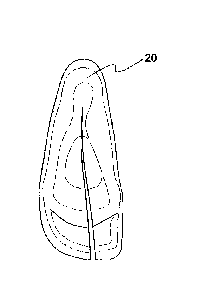
Fig. 20 shows an isometric view of an orthotic support with a soft inner shell
having edge portions protruding beyond the edge portions of the outer shell;
Fig. 21 shows a bottom view of an orthotic support device.
DETAILED DESCRIPTION OF THE PREFERRED EMBODIMENTS
In general, the specification below will first describe one form of casting a
lower
limb orthotic/orthosis device whereby a flexible support device is employed.
Thereafter,
with reference to Figs. 15-18, a second embodiment is shown whereby the second
embodiment can be used in various forms to provide the practitioner numerous
options
3
CA 02587584 2007-05-14
WO 2006/055474 PCT/US2005/041147
for producing an orthotic/orthosis device. In general, the shell as shown in
Fig. 15 can
be used as a casting device, or alternatively as a measuring device to measure
the
general contours of a patient's foot where only the measurements need to be
sent to a
manufacturing facility for production of an orthosis (or simply used to
provide a specific
size and shape of orthosis premade). Further, the embodiments as shown in
Figs. 15-
18 can be used in a modular-type arrangement were modular components are
arranged to provide a wide variety and proper fit to the patient. Finally, the
embodiments as shown in Figs. 19-21 show a system where a rigid shell is
employed
with an interior soft shell, where in this version, the rigid outer shell is
essentially the
end product that provides support for the patient and the interior soft shell
can absorb
localized protrusions and indentations for a more comfortable fit for the
patient.
As shown in Fig.1 the flexible support member 20 is shown. As shown in this
figure, an axis system is defined where the arrow indicated at 22 indicates a
longitudinal axis. Likewise, the orthogonal arrow 24 indicates a lateral axis.
Finally, the
arrow that is orthogonal to the two mentioned arrows is indicating a vertical
axis 26.
As further shown in Fig. 1, the flexible foot support 20 has a medial region
generally indicated at 28 and a lateral region generally indicated at 30.
Further, the
longitudinally forward region is generally referenced as a distal region 32
and the
opposed longitudinal region is commonly referred to as a proximal region 34.
In
addition to the aforementioned regions, a plantar region indicated at 36
defines the
general upper surface that comes in contact with the lower portion of a
patient's foot.
The medial lateral wrap region generally indicated at 38 is a substantially
vertical region
that is adapted to encompass the calcaneus (a portion of the heel bone), the
medial
arch which is sometimes referred to as the longitudinal arch, and the
navicular.
Further, the medial lateral wrap region is adapted to cover the first
metatarsal head and
the fifth metatarsal head, the base of the fifth metatarsal head and the
peroneal arch.
As shown in Fig. 2, the plantar region 36 is approximately defined as the
central
region within the encompassed section 40. The hatched region around the upper
perimeter is substantially defined as the medial lateral wrap region as
described above.
The plantar region 36 is defined to have various regions as shown in Fig. 2.
Beginning
in the longitudinally rearward section, the heel depression indicated at 42 is
defined as
a region adapted to be depressed to a patient's heel during a molding process.
The
heel depression region 42 provides a foundation for the medial lateral wrap
region 38
as described further below and this region of the material is adapted to work
in
4
CA 02587584 2007-05-14
WO 2006/055474 PCT/US2005/041147
conjunction with this longitudinally rearward portion to correct various
skeletal
biomechanical misalignments such as pronation, supination, and varus-valgus.
The
peroneal arch is a region 44 where the surface raises somewhat vertically. The
peroneal arch is distal to the heel depression to help control the heel
(calcaneus
position), and is right behind the base of the fifth metatarsal. This region
helps support
the arches of the foot and overall foot alignment. It should be noted that the
peroneal
arch region 44 is a vertical indentation which is represented in the outer
surface of the
flexible support devices 20. This can be advantageous for providing feedback
to a
practitioner when casting to denote a certain position. Further, the region 44
is a
potential reference point to aid instruction when instructing a practitioner
to properly
exercise a molding process and aligning the bone structure of a patient
described
further herein below.
Also shown in Fig. 2, the medial arch region 46 is defined generally as a
raised
region in the central portion of the plantar region 36. As with the peroneal
arch region
44, the medial arch region has a raised region which a practitioner can use to
grab
when conducting and creating a mold upon a patient as described further herein
below.
The flexible support member 20 by default has a raised medial arch region. It
should
be noted that the member is flexible and described further herein below. The
medial
arch is useful in aligning the avicular navicular and assisting in properly
aligning the foot
to a solid functional biomechanical neutral position as opposed to a pronated
foot or
supinated foot. The metatarsal arch indicated at 48 is a raised region adapted
to
support the metatarsals, particularly the central metatarsals 2, 3 and 4.
Further shown in Fig. 2 is the metatarsal depression generally indicated at
50.
This region is defined as a region that supports and aligns the metatarsal
heads.
The most forward distal region indicated at 52 is the toe rise region. This
region
is divided into a drop first toe subregion 54 and a two-five subregion 56. The
drop toe
subregion 54 is positioned slightly vertically lower with respect to the two-
five subregion
56. From the sulcus, the two-five subregion 56 slopes downwardly in the
longitudinally
forward direction toward the distal area and downwardly to the laterally
outward region
indicated as 58. This region helps align the foot and allows propreaceptive
input for the
client so that the foot may be aligned properly. Specifically, the surface
allows the
client to become aware of his feet and his foot placement. Therefore the
raised region
brings this awareness to the client during the casting process, allowing for a
better
mold.
5
CA 02587584 2007-05-15
=
%PEWS
P CT/ ILO ,.S 0 / 4.1. 1 4.7
=
There will nowbe discussion of the molding process, during Which a
practitioner
will take a mold of the lower foot region of a patient. As shown in Fig. %the
first step in
the molding process is to choose the proper size of a flexible support device.
Fig. 3
shows an assortment of sizes of flexible support devices to accommodate a wide
variety of patients. As shown in this figure, the variety of flexible support
devices 20 are
adapted to be stacked as shown. In other words, the cavity region of a larger
flexible
support device will support the next smaller size. In a storage location, the
flexible
support devices are arranged in a stacked position whereby an outer surface of
an
immediately smaller flexible support device is engaged in a cavity region of
the
immediately larger flexible support device so the plurality of flexible
support devices are
arranged in a stacked manner. This allovirs for storage of quite a few
flexible support
devices in a practitioners office. Further, this stacking method facilitates
in sizing up
the proper flexible support member 20 so the practitioner can easily identify
which size
would be appropriate. For example, if the practitioner chooses one of the
central sizes
and it is does not properly fit the patient, the practitioner can gauge the
difference of
size required and skip a set number of increments smaller or larger to gauge
the
approximate appropriate dm for the particular patient.
Thereafter (or prior to sizing), a stockinette is placed on the patient's foot
as
shown in Fig. 4. A stockinette is defined broadly as a flexible cover to
provide some
protection and at least partial separation between respective inner and outer
portions of
the stockinette. In one form the stockinette is made from a fabric-like
material, similar
to an expandable sock. The foot is then placed into a properly sized amble
support
device 20. The various plantar surfaces described above with reference to Fig.
2 must
be aligned with the corresponding anatomy of the patient's foot. In
particular, the heel
region of the patient should be pressed firmly against the substantially
vertical surface
of the proximal region 34 of the medial lateral wrap region 38. Referring back
to Fig. 2,
the proximal location of the medial lateral wrap region 38, generally
indicated at 39, is
referred to as the heel cup region. One advantageous aspect of the heel cup
region 39
is that it provides an initial foundational support when molding. When not
providing a
vertical support region in the heel cup region, the prior art support members
will move
with respect to the foot to improper locations. This leads to improper casting
and an
eventual poor support device. Therefore, having the heel cup region 39 aids in
preventing an improper casting.
SEA 1722349v5 61799.17 6
AMENDED SHEET
=
CA 02587584 2013-06-11
= Now referring to Fig. 5, it can be seen that the heel region 70 of the
patient is
pressed firmly against the heel cup region 39 of the flexible support devices
20. After
the heel is properly aligned in the rearward, proximal location of the
flexible support
device 20, the practitioner must check the distal regions to ensure that the
metatarsal
heads are not crossing the total sulcus. Referring back to Fig. 2, the total
sulcus
indicated at 45 is the distal methead proximal toe rise area indicated at the
laterally
extending line designated by 45. Although other portions of the anatomy could
be
aligned to the flexible support device, this region is accessible to view by
the
practitioner and generally, the intermediate plantar surface regions will be
properly
positioned corresponding to the anatomy of the patient. Referring back to Fig.
4, it is
advantageous to have the overall length of the flexible support device
slightly longer
than the toes 72 of the patient. In other words, this region is not critical
for a proper
mold and therefore the extra space indicated at 73 between the toes 72 and the
forward
vertical region 33 will not generally be a problem in molding. In one form,
the gap
region between the forward surface of the patient's toes 72 and the forward
vertical
region 33 is approximately a quarter of an inch or greater.
Fig. 5 further shows the application of a second stockinette 74. In one
application, a second stockinette is applied over the flexible support device
20 and the
initial layer of stockinette. The second layer of stockinette is advantageous
for removal
of the layer of the cast that is to be applied which is described below.
Further, it has a
second advantage of aiding the removal of the flexible support device 20 after
the mold
has at least partially cured and taken a substantially rigid form (also
described further
below). Further, the second stockinette 74 increases the net volume of the
positive
mold which represents the foot and ankle region, the first and second
stockinettes and
the flexible support device 20.
As shown in Fig. 6, a cutting strip 76 that is shown beneath the second
stockinette 74 is "wormed in" down a portion of the patient's leg and foot
region. In one
form, this is located in the forward central region to facilitate a buffer
region when
removing the mold. This cutting strip is applied underneath one of the two
stockinettes.
In another form the cutting strip is applied in the outer surface of the
stockinette 74.
Appling the cutting strip 76 is an optional process for aiding in the removal
of the mold if
a non-flesh-cutting element is used to remove the mold. It in another version,
the
cutting strip is taped or otherwise attached to the inner stockinette.
7
CA 02587584 2013-06-11
= As shown in Fig. 7, pliable molding strips 95 are wrapped around the
second
stockinette 74 (see Fig. 6) in one form the molding strips are fiberglass, but
any
substance that is initially pliable and can harden to a mold after a few
minutes would
suffice. The preset state of the fiberglass rolling is generally a condition
where the
fiberglass tape is wet and wrapped around the entire ankle foot region with
the
stockinette applied thereon as shown in Fig. 6. The molding strips 95 are
defined
broadly to cover all materials that have the ability to be applied in a very
flexible form
fitting manner and harden to at least a semi-rigid state to preserve a
negative mold of
the outer surface of the inner structure members (e.g. the contour of the
lower limb and
the flexible support device). When the molding strips are all applied to the
foot region
they collectively form a wrap 97 as shown in Fig. 7.
Before the wrap 97 hardens, the practitioner engages in an alignment and
feature definition process. This process essentially positions the foot into a
proper
neutral biomechanical position to form a proper mold. As described above, the
features
of the flexible support device 20, given its flexibility, allow the
practitioner to have a
greater amount of control over the manipulation of the position of the various
features of
the foot and lower limb regions of the patient. In other words, without some
flexibility of
the flexible support device 20, the anatomical features of the foot would not
be
manipulated. However, the flexible support device is sufficiently rigid to
allow a
distribution of pressure upon adjacent regions of the foot and lower ankle
region that
the practitioner is not in direct contact with. The goal is to have the mold
formfitting to
the contours of the patient's foot and maintaining the correct overall
biomechanical
alignment.
The aforementioned arch regions as shown in Fig. 2 assist in aligning the
arches
to form a proper mold. As shown in Fig. 7, the hindfoot is stabilized with the
hand
indicated at 90. The thumb is on the navicular bone of the patient and supply
a vertical
force indicated by arrow 92 helps to define a longitudinal arch. The fingers
span the
instep region 94 and the forward tip portions of the practitioner's fingers
grasp the
calcaneus region and in some cases help remove the pronation of the patient.
If the
patient does not have a pronation problem, the left-hand 90 will support the
ankle
region so it is properly neutrally aligned. The palm of the hand 90 is on the
medial side
of the foot and the fingers extend around the back of the heel.
The practitioner's other hand 99 of the practitioner brings the forefoot to
the
neutral position. A neutral position must be executed about a longitudinal
axis so the
8
CA 02587584 2007-05-14
WO 2006/055474 PCT/US2005/041147
portion of the foot is properly positioned. Further, the medial and lateral
alignment must
be properly aligned as well. It is important to keep the heel vertical,
therefore there may
be some sacrifice in keeping the forefoot horizontal in order to properly
align the heel
region of the patient's foot. The heel alignment is the base, and given the
individual's
range of motion, the best biomechanical alignment is obtained. The flexible
support
device provides a more gradual transition from the forefoot to the rear foot
because the
rigidity and flexibility of the flexible support device 20 will allow any
manipulation to
extend longitudinally rearwardly and supply a force along the surface of the
foot. In
other words, even though the practitioner will exert a force on the distal
region of the
foot, this force is distributed longitudinally rearwardly to the heel region
because of the
flexibly controlled deformation of the flexible support device. Without the
flexible
support device 20, any manipulation by the practitioner's fingers will create
a localized
depression upon the wrap 97. However, with the flexible support device
contained
thereunder, any manipulation is not directly applied but it is more uniformly
distributed
around the adjacent regions were pressure is applied. Given that the flexible
support
device already has a preset form of key features and depressions as described
in Fig.
2, these features are better maintained. The flexible support device provides
a more
natural transition of manipulation from the rearward portion of the foot to
the forward
portion of the foot.
As shown in Fig. 8, the process of the alignment and feature definition
process is
substantially complete and the wrap 97 is beginning to cure to some degree and
taking
a solidified form. At this point the practitioner has the ability to
manipulate localized
regions for a better detailed fit. As shown in Fig. 8, the practitioner is
contouring the
heel to a proper alignment. Of course the practitioner may choose to contour
other
regions to take a proper neutral biomechanical set form. It should be noted
that when
pressure is applied the medial lateral wrap region 38 as shown in Fig. 2, the
precured
wrap 97 disperses pressure and aids in not allowing "flesh displacement". In
other
words, particularly in younger patients with more "fleshy" feet that contain
greater fat
deposits, the medial lateral wrap region 38 allows a more proper distribution
of pressure
when the wet flexible wrap 97 is applied therearound. The flexible support
device 20
having a central chamber region aids in positioning the patient's foot from
the beginning
of the molding procedure. In other words, instead of having a substantially
planar
device without sidewalls, the medial lateral wrap region aids in initially
positioning the
foot so the margin of error is reduced for the alignment of the various arch
and
9
CA 02587584 2007-05-14
WO 2006/055474 PCT/US2005/041147
depression regions 42-50 discussed in Fig. 2 and the corresponding anatomical
portions of the foot. The patient's foot is channeled into this chamber region
and there
are less manipulation and alignment issues for the practitioner to be
concerned with
when performing the mold.
As shown in Fig. 9, the cast is removed by incising the front portion with any
conventional type of tool. Any particular chosen method of cutting the cast
after it has
cured can be employed. It should be noted that the flexible support device 20
aids in
the removal of the cured cast from the patient's foot because it allows for a
distribution
of pressure around the lateral regions of the foot during removal. The
flexible support
device 20 further minimizes distortion during cast removal when the forward
central
region of the cast must be expanded and pried laterally outwardly to allow the
foot and
ankle to be interposed and removed therein between the cut portion. Minimizing
the
deformation of the cast is useful when the cast is not fully cured, which can
be a
problem when colder water is used when beginning the curing process of the
cast, or
other reasons that may lead to a slow curing process.
Fig. 10 illustrates one particular use of the flexible support device 20. As
shown
in this figure, the flexible support device 20 is removed from the cast 80.
This allows for
reuse of the flexible support device. This removal can be executed by the
practitioner
and the practitioner can thereafter properly store the flexible support device
in the
manner as shown in Fig. 4. This is particularly advantageous because in one
form of
prior art practice, the entire cast is sent to a third-party company which
makes the final
orthosis support device. This allows the practitioner to refrain from sending
support
devices adapted to be positioned on the lower portion of the patient's foot to
be mailed
along with the cast to a third party fabricator. This depletes the supply of
support
devices for the practitioner, who makes the cast at a location which is
generally not the
location where the final orthosis support device is created. Therefore with
the present
invention, the practitioner maintains his supply of the flexible support
devices as shown
in Fig. 4.
There will now be a discussion of various adjustments that can be made during
the molding process with initial reference to Fig. 11. As shown in this
figure, the flexible
support device 20 is incised in a substantial longitudinal direction. Now
referring to Fig.
12, the flexible support device can be overlapped at the region indicated at
91 to
provide for a patient's foot that may be narrower in the lateral direction.
This provides
flexibility for various shapes feet of patients. It should be noted that when
a shorter and
=
CA 02587584 2013-06-11
.wider foot is required to be molded, the medial lateral wrap regions 38 have
a certain
amount of flexibility to allow this wider foot to be contained in the chamber
region of the
flexible support device 20. The support infrastructure of the various interior
surfaces
described on Fig. 2 is maintained even when a wider foot is entered in the
chamber
region of the flexible support device 20.
In the situation where there are bony prominences or extreme shapes of a
patient's foot which require special accommodation, reference is made to Figs.
13 to
16. As shown in Fig. 13, a heating element 100 supplies heat to a specific
location 12 of
the flexible support.device 20. It should be noted that any area of the
flexible support
device can be heated and manipulated to accommodate any specific situation
with a
patient. It is often at the discretion of the practitioner to accommodate
various extreme
anatomical features of the patient, or any disfigurements. Therefore, by way
of
example, a formation process is shown in Figs. 13-16. Fig. 13 shows the
heating of the
medial region where the first prominent method would be slightly extruded on
the
patient. As shown in Fig. 14, the region 102 is biased laterally outwardly to
accommodate this feature of the patient. Of course various methods of
alteration are
available, such as an alteration to the flexible support member 20, where the
base of
the fifth metatarsal bone region is heated so the material becomes plastic
and
formable where the practitioner can manipulate the region outwardly to
accommodate an oUtward extension of the patient's foot in this region.
It should be noted that the flexible support device 20 is particular adapted
for
external posting. During this process, shim-like devices are positioned either
externally
of the wrap or in some cases wrapped internally thereunder. The shim-like
devices
provide a consistent support surface for maintaining the foot position in a
certain natural
alignment positioned for molding. As described above, the application of
pressure of
the shim allows for a more consistent natural transition of forefoot to rear
foot, given the
rigidity of the flexible support device and the flexibility of the device as
well. It should
further be noted that the various features as shown in Figs. 1 and 2 provide
assurance
that the internal arches of the patient's foot are maintained in the
manipulation of the
patient's foot during the molding process.
The embodiments as shown in Fig. 15 relate to a rigid outer shell device
(control
module 120) that can be used for casting or only as a measuring device for
determining
proper orthoses for the lower leg. In general, the full lower shells comprise
a support
module shown in Fig. 15 having an approximate section that extends up above
the
11
CA 02587584 2007-05-14
WO 2006/055474 PCT/US2005/041147
ankle. This is made of a flexible material such as plastic in a similar manner
as the
flexible support device 20 described above and has a central slit region that
allows for it
to be adjustable. The key features are molded into this module, such as an
arch or
other anatomical regions as described in shown in Figs. 1 and 2. Further,
prominent
features of the foot are compensated for as well.
In one form, the control module 120, which is one form of a flexible support
device, can be used to assist in casting whereby modules are placed around the
patient's foot and squeeze tight for proper fitting. Thereafter, the
practitioner, using
standard mold casting techniques that are described above, is able to create a
correct
negative cast of the patient's foot. Thereafter, this cast is sent to a
facility (or executed
on site) whereby the control module is a known fixture of a cast and making an
improper positive model of the patient's foot can be avoided. Thereafter,
there is a
positive model (mold) that is used to create an orthosis support device.
It should be noted that it is advantageous to have the lateral lower portion
extend
over the foot as well as have the proximal section extend up the lower calf of
the patient
to control foot position during the casting.
A second application is to use the control module as a sizing shell, whereby
no
casting is conducting by the practitioner, but the particular size of the
control module is
relayed back to the manufacturer of the final braces to eliminate casting and
the
physical mailing of the cast which is expensive and causes a time delay.
To facilitate the communication of the proper sizing of the foot without
taking a
cast, marking indications, such as shown in Fig. 15, can be employed whereby
the
ridge 130 will engage certain coinciding locations with certain markings 132
which
would be indicated by certain measurements. As shown in Fig. 16, this could be
accomplished with a strap like system having a base support 140 having a
plurality of
flexible measuring devices 142.
Another element of the apparatus is to have modular components as shown in
Fig. 17. As shown in this figure, there is a rigid foot structure component
150 having a
central lower cavity region 152 that is adapted to receive an orthotic-like
insert 154.
The theory is that the practitioner can fit the shell to the patient and
further have the
flexibility of fitting one of the stock orthotic molds to the patient as well.
The shell can
have various lines 156 or other adjustment features to quantify the position
and
orientation of the orthotic-like insert 154.
12
CA 02587584 2007-05-14
WO 2006/055474
PCT/US2005/041147
As shown in Fig. 18, the modularity can further extend to having a lower semi-
rigid fitting module 160 and then an upper semi-rigid module 162. These
modules can
work in combination and be taken from a plurality of modules that could be
stacked in a
manner similar to Fig. 3 to properly fit the patient.
This concept can be taken further to having an off-the-shelf type orthotic
with
mix-and-match components to properly fit the patient.
Now referring to the embodiment shown in Figs. 19- 21, the apparatus 220
comprises an outer support shell 222 and an inner liner 224. In general, this
embodiment employs a rigid shell to provide a proper orthosis devise for the
patient and
further uses an inner soft shell liner to accommodate various bio-structural
variations
between patient to patient. In general, this embodiment does not employ
casting, but
rather the plastic shell itself is provided as the end product for the user to
wear for lower
limb support.
The outer shell has an interior chamber region and the inner liner has an
exterior
surface that is adapted to engage the inner surface of the outer shell 222.
The outer
support shell 222 comprises a perimeter support region 226 having medial and
lateral
sections 228 and 230. The outer support shell 222 further has a heel cup 232
in the
rearward portion of the apparatus 220. The perimeter support region 226 is
positioned
in a location that is an approximate support location for the patient. In
other words, the
outer shell provides a rigid support to control the biomechanical positioning
and
alignment of the patient.
The outer support shell is made of ,a rigid material such as plastic, but does
provided a certain amount of flexibility or comfort to account for various
foot positions
which the patient may be in without allowing the foot to completely collapse.
The embodiment shown in Figs. 19 - 21 is adapted to be an off-the-shelf type
orthotic utilizing proprioceptive feedback (sensory feedback); this is
important in
providing the patient with a heightened sense of foot position to aid in
proper alignment
of his other feet.
The perimeter region 226 provides a certain amount of flexibility; when it is
depressed by hand with a modest grip, the size will deflect inward or
outwardly a few
millimeters. It is important to note that this flexibility provides
functionality for
accommodating a wide range of patients' feet. Further, the flexibility allows
for a
footwear device such as a shoe or a boot to press upon the outer surface of
the outer
support shell to provide a better and more accommodating fit. The outer
support shell
13
CA 02587584 2007-05-14
WO 2006/055474 PCT/US2005/041147
222 has an overall thickness between .5 and 3 millimeters in the broader
range. A
more preferable range is between 1/16 of an inch to 90 thousandths of an inch.
In one
form, a polyethylene base plastic is used to mold the outer support shell 222.
Of
course other materials providing flexibility and strength can be employed.
As shown in Fig. 21 the under portion of the outer support shell 222 comprises
a
variety of support sub region surfaces. It should be noted that this underside
of the
surface correlates to a raised region on the chamber region of the outer
support shell
222. As shown in this figure, the peroneal surface 244 is located on the
laterally
outward region; the metatarsal raised region 246 is located in the forward
medial region
and adapted to engage the metatarsals of the patient. The medial longitudinal
arch
indicated at 248 is adapted to provide the common support in the laterally
inward
medial region of the patient. These arches are accentuated to some degree to
give
proprioceptive feedback to the patient so he or she will be induced to
mentally align his
or her foot to enhance his or her development of voluntary control of foot
alignment.
As shown in Fig. 20, the outer support shell 222 has a rearward upper
perimeter
ridge region 250. This region is positioned vertically below the perimeter
ridge 266 of
the inner support liner to provide a blending of pressures from the perimeter
region 250.
In other words, by positioning the perimeter ridge 266 above the lower rigid
ridge 250,
less direct edge pressure is applied to the foot region of the patient. The
inner liner 224
provides the smooth transition to prevent that focused edge pressure that
would
otherwise be present and create discomfort with the patient. The upper portion
of the
inner liner allows for an automatic adjustment of the pressure, so the upper
perimeter
ridge region 250 need not be custom to the patient; rather, the apparatus 220
is self-
adjusting to each patient.
The inner liner 224 protects the side of the foot as it shifts positions from
the
proximal edge of the support shell. In other words, the patient is less likely
to engage
the perimeter rigid region 250 and have their flesh have a localized pressure
developing
an irritation.
There will now be a description of the inner liner 224 with initial reference
to Fig.
19. As shown in this figure, the inner liner 224 comprises a base region 260
and a
forward region 262. The inner liner 224 further has an upper region 264 that
comprises
a perimeter ridge 266. The construction of this inner liner 224 is generally
made from
foam. In one form, the inner liner 224 is made from closed-cell 5-pound
density foam
from ethylene vinyl acetate.
14
CA 02587584 2007-05-15
=
P C T S 43 5 41 .1 4- 7 ..11:1217113 6 1PENUS
=
=== The forward region of the outer support shell 222 has a lateral
region 270. As
shown in Fig. 20, the inner liner 224 has an extension region 272 that extends
longitudinally forward from the lateral region 270. The inner liner 224 is
adapted to
= extend out
and cover the metatarsal head. The support shell is adapted to be . =.
terminated just prior to the fifth metatarsal head on the lateral side. It
should be noted =
in Fig. 20 that the medial side is adapted to be cut back before the first
metatarsal
head.
By having the outer support shell 222 provide the rigid structure so the
extension
region 272 is positioned at substantially right angles from the lateral region
to the
plantar region, the inner shell provides some rigidity to prevent abduction of
the foot
when the foot pronates. This is a condition when the medial longitudinal arch
of the
patient collapses.
Now referring to Fig. 19, the general area indicated at 80 is defined as a
pivot
region where the metatarsal heads approximately end and, in an operating
=
environment, the patient will pivot when walking or running. It should be
noted that the
lateral region 270 terminates prior to this pivot region 80 to not interfere
with the
pivoting action. However, the region 272 of the inner shell 224 being more
flexible and
made from foam-like material will accommodate the pivoting action during
walking or
running (or other bipedal motion).
Therefore, it can be appreciated that the apparatus 220 is well suited to
prevent
pronation of a patient's foot which is a common joint misalignment
biomechanical issue
in many young patients. The medial section 228 of the perimeter support region
226
=¨=== will have a tendency to apply a pressure on the medial region to
prevent the pronation
described above. Further, with the cup region orientated where the rearward
surface
extends in a plane that is substantially orthoganal to the longitudinal axis
and the
medial region in a plane orthogonal to the lateral axis, additional support is
provided
and added rigidity is a benefit to prevent eversion of the heel.
The depth allows the flexible support shell to function property because
having
the vertical region indicated at 227 in Fig. 19 allows for a greater moment of
inertia
when a moment is applied about a lateral axis such as a pressure from the
patients foot
in the lateral region 270. This is particularly advantageous because less
material and
structure is required to provide that rigidity, creating a fighter more
compact orthotic.
= =
= The final component of providing I proper biomechanIcal alignment for the
patient is preventing the forefoot from abducting laterally outwardly with
respect to the
SEA 1722349v9 68799-17 15
AMENDED SHEET
CA 02587584 2013-07-10
WO 2006/055474 PCT/US2005/041147
heel region of the patient. As described above in greater detail the lateral
region 270
provides a base region for supporting the portion of the inner liner extension
region 272
to aid in supporting in controlling the abduction. As described above, the
flexible foam
insert provides flexibility during running where it will actually collapse to
a certain
degree to provide the range of motion for the patient.
The apparatus 220 is particularly useful in an environment of footwear such as
a
shoe where the upper perimeter ridge 266 of the inner liner 224 is adapted to
position
laterally outwardly with respect to the center chamber region of the shoe. In
other
words, the extension region 272 is easily repositioned and grasped laterally
outwardly
by the patient and the perimeter region of the patient's foot will easily
glide past the
outer support shell 222 and be positioned in proper foot position in the shoe.
It should
further be noted that given the overall length of the apparatus, it will fit
properly in a
shoe and not be positioned vertically forward with respect to the shoe to
prevent
movement of the soft liner inner liner 224.
In one form, a layer on the upper surface of the inner liner 224 can be
applied to
aid in breathability of the apparatus 220. Further, the coefficient of
friction between the
foot and stocking of the patient can be adjusted to prevent discomfort such as
excessive footwear which may cause blisters or the like.
A further modification can be employed where the lower surface of the outer
support shell 222 can be filled with some form of material to provide extra
support and
rigidity. In one form, the aforementioned arch regions can be enhanced and
amplified
to facilitate the proprioceptive feedback to the patient.
One form of manufacturing the outer support shell 222 is employing common
thermal sheet forming techniques such as draping. However, many forms of
manufacture can be employed such as injection molding, milling etc.
The scope of the claims should not be limited by the preferred embodiments
set forth herein, but should be given the broadest interpretation consistent
with the
description as a whole.
16