Note: Descriptions are shown in the official language in which they were submitted.
CA 02588267 2007-05-09
VESSEL SEALING INSTRUMENT
WITH OPTIMIZED POWER DENSITY
TECHNICAL FIELD
The present disclosure relates to a forceps used for both endoscopic
and open surgical procedures that includes an electrode assembly that allows a
user to selectively seal and/or cut tissue. More particularly, the present
disclosure
relates to a forceps that applies a unique combination of mechanical clamping
pressure and electrosurgical energy to effectively seal and sever tissue
between
sealed tissue areas.
BACKGROUND
Open or endoscopic electrosurgical forceps utilize both mechanical
clamping action and electrical energy to effect hemostasis. The electrode of
each
opposing jaw member is charged to a different electric potential such that
when the
jaw members grasp tissue, electrical energy can be selectively transferred
through
the tissue. A surgeon can either cauterize, coagulate/desiccate and/or simply
reduce or slow bleeding, by controlling the intensity, frequency and duration
of the
electrosurgical energy applied between the electrodes and through the tissue.
Certain surgical procedures require more than simply cauterizing
tissue and rely on the combination of clamping pressure, electrosurgical
energy
1
CA 02588267 2007-05-09
and gap distance (i.e., distance between opposing jaw members when closed
about tissue) to "seal" tissue, vessels and certain vascular bundles. More
particularly, vessel sealing or tissue sealing is a recently-developed
technology that
utilizes a unique combination of radiofrequency energy, clamping pressure and
precise control of gap distance to effectively seal or fuse tissue between two
opposing jaw members or sealing plates. Vessel or tissue sealing is more than
"cauterization", which involves the use of heat to destroy tissue (also called
"diathermy" or "electrodiathermy"). Vessel sealing is also more than
"coagulation",
which is the process of desiccating tissue wherein the tissue cells are
ruptured and
dried. "Vessel sealing" is defined as the process of liquefying the collagen,
elastin
and ground substances in the tissue so that the tissue reforms into a fused
mass
with significantly-reduced demarcation between the opposing tissue structures.
To effectively seal tissue or vessels, especially thick tissue and large
vessels, two predominant mechanical parameters must be accurately controlled:
1)
the pressure applied to the vessel; and 2) the gap distance between the
conductive
tissue contacting surfaces (electrodes). As can be appreciated, both of these
parameters are affected by the thickness of the vessel or tissue being sealed.
Accurate application of pressure is important for several reasons: to oppose
the
walls of the vessel; to reduce the tissue impedance to a low enough value that
allows enough electrosurgical energy through the tissue; to overcome the
forces of
expansion during tissue heating; and to contribute to the end tissue
thickness,
which is an indication of a good seal. It has been determined that a typical
fused
vessel wall is optimum between about 0.001 and about 0.006 inches. Below this
2
CA 02588267 2007-05-09
range, the seal may shred or tear and above this range the tissue may not be
properly or effectively sealed.
With respect to smaller vessels, the pressure applied becomes less
relevant and the gap distance between the electrically conductive surfaces
becomes more significant for effective sealing. In other words, the chances of
the
two electrically conductive surfaces touching during activation increases as
the
tissue thickness and the vessels become smaller.
Typically, and particularly with respect to endoscopic electrosurgical
procedures, once a vessel is sealed, the surgeon has to remove the sealing
instrument from the operative site, substitute a new instrument through the
cannula
and accurately sever the vessel along the newly formed tissue seal. As can be
appreciated, this additional step may be both time- consuming (particularly
when
sealing a significant number of vessels) and may contribute to imprecise
separation
of the tissue along the sealing line due to the misalignment or misplacement
of the
severing instrument along the center of the tissue seal.
Several attempts have been made to design an instrument that
incorporates a knife or blade member, which effectively severs the tissue
after
forming a tissue seal. For example, commonly-owned U.S. App. Ser. Nos. 10/472,
295, 10/460,942 and 10/991157 all disclose instruments that include a
mechanical
cutting mechanism for selectively cutting tissue along a tissue seal. These
instruments have enjoyed great success in the operating field.
3
CA 02588267 2007-05-09
Sealing and electrical cutting on the same instrument is a recently
developed technology that provides different advantages over mechanically
cutting
tissue. However, electrical cutting of tissue has proven difficult for
manufacturing
due to the dimensions between electrodes being relatively small. The
electrodes
may produce heat formation and electrical charging during the seal cycle that
detrimentally affects the cut performance. This may manifest itself by
damaging
tissue within the cut zone and minimizing hydration by forcing conductive
fluids
from the cut area.
SUMMARY
Accordingly, the present disclosure is directed to an electrode
assembly for use with an instrument for sealing and cutting vessels and/or
tissue.
In one embodiment the assembly includes a pair of opposing first and second
jaw
members at least one of which being movable relative to the other from a first
position wherein the jaw members are disposed in spaced relation relative to
one
another to a second position wherein the jaw members cooperate to grasp tissue
therebetween.
Each jaw member includes an insulator and at least one electrically
conductive tissue sealing surface extending along a length thereof, each
tissue
sealing surface being adapted to connect to a source of electrosurgical energy
such that the tissue sealing surfaces are capable of conducting
electrosurgical
energy through tissue held therebetween to effect a seal.
4
CA 02588267 2007-05-09
The first jaw member includes at least one electrically conductive
cutting element disposed within the insulator of the first jaw member, the
electrically conductive cutting element disposed in general vertical
registration to
the insulator on the second jaw member defining at least one cutting zone
between
the at least one electrically conductive tissue sealing surfaces and the
cutting
element.
An insulative material is included that is disposed between at least
one of the electrically conductive tissue sealing surfaces of the first jaw
member
between at least one eiectrically conductive tissue sealing surface of the
first jaw
member and the cutting element, the insulative material configured to reduce
the
exposed surface area of at least one electrically conductive tissue sealing
surface
of the first jaw member to optimize the power density for at least one of
sealing and
cutting tissue.
In another embodiment of the present disclosure a method of
focusing energy to a specific area within tissue for use with an instrument
for
sealing and cutting vessels and/or tissue is provided. The method includes
providing a pair of opposing jaw members each having an insulation and a pair
of
electrically conductive tissue sealing surfaces at least one of which being
movable
relative to the other from a first position wherein the jaw members are
disposed in
spaced relation relative to one another to a second position wherein the jaw
members cooperate to grasp tissue therebetween. Each jaw member includes at
least one electrically conductive tissue seaiing surface extending along a
length
thereof, the jaw members adapted to connect to a source of electrosurgical
energy
5
CA 02588267 2007-05-09
such that the electrically conductive tissue sealing surfaces are capable of
conducting electrosurgical energy through tissue held therebetween to effect a
seal. The at least one electrically conductive tissue sealing surface having
an
exposed surface area and the first jaw member includes a cutting element
disposed between electrically conductive tissue sealing surfaces. The method
further includes positioning the opposing first and second jaw members about
tissue and disposing an insulative material upon at least one electrically
conductive
tissue sealing surface of the first jaw member, the insulative material
configured to
reduce the exposed surface area of the at least one electrically conductive
tissue
sealing surface of the first jaw member. The method also includes adjusting
the
position of the insulative material on the electrically conductive tissue
sealing
surfaces to focus electrosurgical energy to a specific area between the
cutting
element and the electrically conductive tissue sealing surfaces of the jaw
member.
BRIEF DESCRIPTION OF THE DRAWINGS
Various embodiments of the subject instrument are described herein
with reference to the drawings wherein:
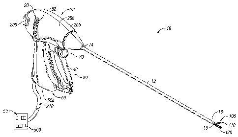
FIG. 1A shows a right, perspective view of an endoscopic bipolar
forceps having a housing, a shaft and a pair of jaw members affixed to a
distal end
thereof, the jaw members including an electrode assembly disposed
therebetween;
FIG. 1 B shows a left, perspective view of an open bipolar forceps
showing a pair of first and second shafts each having a jaw member affixed to
a
distal end thereof with an electrode assembly disposed therebetween;
6
CA 02588267 2007-05-09
FIG. 2 shows a cross-sectional view of a vessel sealing instrument
showing one embodiment of a cut-zone configuration having an optimized power
density;
FIG. 3 shows an enlarged cross-sectional view of the vessel sealing
instrument showing a cut-zone configuration having an optimized power density;
FIG. 4 shows a cross-sectional view of the vessel sealing instrument
showing a vessel held between the jaw members; and
FIG. 5 shows an alternate embodiment of the vessel sealing
instrument having additional insulative material.
DETAILED DESCRIPTION
For the purposes herein, vessel/tissue cutting or vessel/tissue
division is believed to occur when heating of the vessel/tissue leads to
expansion
of intracellular and/or extra-cellular fluid, which may be accompanied by
cellular
vaporization, desiccation, fragmentation, collapse and/or shrinkage along a so-
called "cut zone" in the vessel/tissue. By focusing the electrosurgical energy
and
heating in the cut zone, the cellular reactions are localized creating a
fissure.
Localization is achieved by regulating the vessel/tissue condition and energy
delivery, which may be controlled by utilizing one or more of the various
geometrical electrode and insulator configurations described herein. The cut
process may also be controlled by utilizing a generator and feedback algorithm
(and one or more of the hereindescribed geometrical configurations of the
electrode and insulator assemblies), which increases the localization and
maximizes the so-called "cutting effect".
7
CA 02588267 2007-05-09
For example, the below-described factors may contribute and/or
enhance vessel/tissue division using electrosurgical energy. Each of the
factors
described below may be employed individually or in any combination to achieve
a
desired cutting effect. For the purposes herein the term "cut effect" or
"cutting
effect" refers to the actual division of tissue by one or more of the
electrical or
electro-mechanical methods or mechanisms described below. The term "cutting
zone" or "cut zone" refers to the region of vessel/tissue where cutting will
take
place. The term "cutting process" refers to steps that are implemented before,
during and/or after vessel/tissue division that tend to influence the
vessel/tissue as
part of achieving the cut effect.
For the purposes herein the terms "tissue" and "vessel" may be used
interchangeably since it is believed that the present disclosure may be
employed to
seal and cut tissue or seal and cut vessels utilizing the same inventive
principles
described herein.
It is believed that the following factors either alone or in combination,
play an important role in dividing tissue:
= Localizing or focusing electrosurgical energy in the cut zone during the
cutting process while minimizing energy effects to surrounding tissues;
= Focusing the power density in the cut zone during the cutting process;
= Creating an area of increased temperature in the cut zone during the cutting
process (e.g., heating that occurs within the tissue or heating the tissue
directly with a heat source);
8
CA 02588267 2007-05-09
= Pulsing the energy delivery to influence the tissue in or around the cut
zone.
"Pulsing" involves as a combination of an "on" time and "off" time during
which the energy is applied and then removed repeatedly at any number of
intervals for any amount of time. The pulse "on" and "off' time may vary
between pulses. The pulse "on" typically refers to a state of higher power
delivery and pulse "off' typically refers to a state of lower power delivery;
= Spiking the energy delivery creates a momentary condition of high energy
application with an intent to influence the tissue in or around the cut zone
during the cut process. The momentary condition may be varied to create
periods of high energy application;
= Conditioning the tissue before or during the cutting process to create more
favorable tissue conditions for cutting. This includes tissue pre-heating
before the cutting processes and tissue rehydration during the cutting
process;
= Controlling the tissue volume in or around the cut zone to create more
favorable conditions for tissue cutting;
= Controlling energy and power delivery to allow vaporization to enhance and
or contribute to the cutting process. For example, controlling the energy
delivery to vaporize both intracellular and/or extracellular fluids and/or
other
cellular materials and foreign fluids within the cut zone;
= Fragmenting the tissue or cellular material during the cutting process to
enhance tissue division in the cut zone;
= Melting or collapsing the tissue or cellular material during the cutting
process to enhance tissue division in the cut zone. For example, melting
the tissue to create internal stress within the tissue to induce tissue
tearing;
9
CA 02588267 2007-05-09
= Controlling tissue temperature, arcing, power density and/or current density
during the cutting process to enhance tissue division in the cut zone;
= Applying various mechanical elements to the tissue such as pressure,
tension and/or stress (either internally or externally) to enhance the cutting
process; and
= Utilizing various other tissue treatments before or during the cutting
process to enhance tissue cutting, e.g., tissue sealing, cauterization and/or
coagulation.
Many of the electrode assemblies described herein employ one or
more of the above-identified factors for enhancing tissue division. For
example,
many of the electrode assemblies described herein utilize various geometrical
configurations of electrodes, cutting elements, insulators, partially
conductive
materials and semiconductors to produce or enhance the cutting effect. In
addition, by controlling or regulating the electrosurgical energy from the
generator
in any of the ways described above, tissue cutting may be initiated, enhanced
or
facilitated within the tissue cutting zone. For example, it is believed that
the
geometrical configuration of the electrodes and insulators may be configured
to
produce a so-called "cut effect", which may be directly related to the amount
of
vaporization or fragmentation at a point in the tissue or the power density,
temperature density and/or mechanical stress applied to a point in the tissue.
The
geometry of the electrodes may be configured such that the surface area ratios
between the electrical poles focus eiectrical energy at the tissue. Moreover,
the
geometrical configurations of the electrodes and insulators may be designed
such
CA 02588267 2007-05-09
that they act like electrical sinks or insulators to influence the heat effect
within and
around the tissue during the sealing or cutting processes.
Referring now to the various figures, FIG. 1A depicts a bipolar
forceps 10 for use in connection with endoscopic surgical procedures and Fig.
1 B
depicts an open forceps 100 contemplated for use in connection with
traditional
open surgical procedures. For the purposes herein, either an endoscopic
instrument or an open instrument may be utilized with the electrode assembly
described herein. Obviously, different electrical and mechanical connections
and
considerations apply to each particular type of instrument; however, the novel
aspects with respect to the electrode assembly and its operating
characteristics
remain generally consistent with respect to both the open or endoscopic
designs.
Fig. 1 A shows a bipolar forceps 10 for use with various endoscopic
surgical procedures and generally includes a housing 20, a handle assembly 30,
a
rotating assembly 80, a switch assembly 70 and an electrode assembly 105
having
opposing jaw members 110 and 120 which mutually cooperate to grasp, seal and
divide tubular vessels and vascular tissue. More particularly, forceps 10
includes a
shaft 12 which has a distal end 16 dimensioned to mechanically engage the
electrode assembly 105 and a proximal end 14 which mechanically engages the
housing 20. The shaft 12 may include one or more known mechanically engaging
components which are designed to securely receive and engage the electrode
assembly 105 such that the jaw members 110 and 120 are pivotable relative to
one
another to engage and grasp tissue therebetween.
11
CA 02588267 2007-05-09
The proximal end 14 of shaft 12 mechanically engages the rotating
assembly 80 (not shown) to facilitate rotation of the electrode assembly 105.
In the
drawings and in the descriptions which follow, the term "proximal", as is
traditional,
will refer to the end of the forceps 10 which is closer to the user, while the
term
"distal" will refer to the end which is further from the user. Details
relating to the
mechanically cooperating components of the shaft 12 and the rotating assembly
80
are described in commonly-owned U.S. Patent Application Serial No. 10/460,926
entitled "VESSEL SEALER AND DIVIDER FOR USE WITH SMALL TROCARS
AND CANNULAS" filed on June 13, 2003 the entire contents of which are
incorporated by reference herein.
Handle assembly 30 includes a fixed handle 50 and a movable
handle 40. Fixed handle 50 is integrally associated with housing 20 and handle
40
is movable relative to fixed handle 50 to actuate the opposing jaw members 110
and 120 of the electrode assembly 105 as explained in more detail below.
Movable handle 40 and switch assembly 70 are of unitary construction and are
operatively connected to the housing 20 and the fixed handle 50 during the
assembly process. Housing 20 is constructed from two components halves 20a
and 20b which are assembled about the proximal end of shaft 12 during
assembly.
Switch assembly is configured to selectively provide electrical energy to the
electrode assembly 105.
As mentioned above, electrode assembly 105 is attached to the distal
end 16 of shaft 12 and includes the opposing jaw members 110 and 120. Movable
handle 40 of handle assembly 30 imparts movement of the jaw members 110 and
12
CA 02588267 2007-05-09
120 from an open position wherein the jaw members 110 and 120 are disposed in
spaced relation relative to one another, to a clamping or closed position
wherein
the jaw members 110 and 120 cooperate to grasp tissue therebetween.
Referring now to Fig. 1B, an open forceps 100 includes a pair of
elongated shaft portions 112a and 112b each having a proximal end 114a and
114b, respectively, and a distal end 116a and 116b, respectively. The forceps
100
includes jaw members 120 and 110 which attach to distal ends 11 6a and 11 6b
of
shafts 11 2a and 11 2b, respectively. The jaw members 110 and 120 are
connected
about pivot pin 119 which allows the jaw members 110 and 120 to pivot relative
to
one another from the first to second positions for treating tissue. The
electrode
assembly 105 is connected to opposing jaw members 110 and 120 and may
include electrical connections through or around the pivot pin 119. Examples
of
various electrical connections to the jaw members are shown in commonly-owned
U.S. Patent Application Serial Nos. 10/474,170, 10/116,824, 10/284,562
10/472,295, 10/116,944, 10/179,863 and 10/369,894, the contents of all of
which
are hereby incorporated by reference herein.
Each shaft 112a and 112b includes a handle 117a and 117b
disposed at the proximal end 114a and 114b thereof which each define a finger
hole 118a and 118b, respectively, therethrough for receiving a finger of the
user.
As can be appreciated, finger holes 118a and 118b facilitate movement of the
shafts 11 2a and 11 2b relative to one another which, in turn, pivot the jaw
members
110 and 120 from the open position wherein the jaw members 110 and 120 are
disposed in spaced relation relative to one another to the clamping or closed
13
CA 02588267 2007-05-09
position wherein the jaw members 110 and 120 cooperate to grasp tissue
therebetween. A ratchet 130 is included for seiectively locking the jaw
members
110 and 120 relative to one another at various positions during pivoting.
More particularly, the ratchet 130 includes a first mechanical interface
130a associated with shaft 112a and a second mating mechanical interface
associated with shaft 112b. Each position associated with the cooperating
ratchet
interfaces 130a and 130b holds a specific, i.e., constant, strain energy in
the shaft
members 11 2a and 11 2b which, in turn, transmits a specific closing force to
the jaw
members 110 and 120. It is envisioned that the ratchet 130 may include
graduations or other visual markings which enable the user to easily and
quickly
ascertain and control the amount of closure force desired between the jaw
members 110 and 120.
As best seen in Fig. 113, forceps 100 also includes an electrical
interface or plug 200 which connects the forceps 100 to a source of
electrosurgical
energy, e.g., an electrosurgical generator (not shown). Plug 200 includes at
least
two prong members 202a and 202b which are dimensioned to mechanically and
electrically connect the forceps 100 to the electrosurgical generator 500 (See
Fig.
1 A). An electrical cable 210 extends from the plug 200 and securely connects
the
cable 210 to the forceps 100. Cable 210 is internally divided within the shaft
112b
to transmit electrosurgical energy through various electrical feed paths to
the
electrode assembly 105.
14
CA 02588267 2007-05-09
One of the shafts, e.g., 112b, includes a proximal shaft
connector/flange 119 which is designed to connect the forceps 100 to a source
of
electrosurgical energy such as an electrosurgical generator 500. More
particularly,
flange 119 mechanically secures electrosurgical cable 210 to the forceps 100
such
that the user may selectively apply electrosurgical energy as needed.
It is envisioned that the cutting element may be substantially dull and
only capable of cutting tissue through electrosurgical activation. Moreover,
the
cutting element may be disposed within the insulator of the first or second
jaw
member. As mentioned hereinbefore the potential of the cutting element and the
electrically conductive tissue sealing surfaces may be altered depending upon
a
particular desired surgical effect.
FIGS. 2-4 show an electrode assembly 105 for use with an
instrument for sealing and cutting vessels and/or tissue. Electrode assembly
105
includes a pair of opposing first 140 and second 150 jaw members at least one
of
which is movable relative to the other from a first position wherein the jaw
members
140, 150 are disposed in spaced relation relative to one another to a second
position wherein the jaw members cooperate to grasp tissue "t" therebetween
(FIG.
4). Each jaw member 140, 150 includes at least one electrically conductive
tissue
sealing surface or electrode 142, 142', 152, 152' extending along a length
thereof,
each tissue sealing surface 142, 142', 152, 152' is adapted to connect to a
source
of electrosurgical energy such that the electrically conductive tissue sealing
surfaces 142, 142', 152, 152' are capable of conducting electrosurgical energy
CA 02588267 2007-05-09
through tissue "t" held therebetween to effect a seal. The electrically
conductive
tissue sealing surfaces 142, 142', 152, 152' have an exposed surface area.
Electrode assembly 105 also includes at least one electrically
conductive cutting element 160 disposed within the insulator 144(not shown),
154
of the first or second jaw member 140, 150. Cutting element 160 is disposed in
general vertical registration to jaw member 140 and extends outwardly from the
insulator 154 of jaw member 150. Cutting element 160 defines at least one
cutting
zone between the electrically conductive tissue sealing surfaces 142, 142',
152,
152' and cutting element 160. Cutting element 160 may include an insulative
material 162 disposed thereupon. Insulative material 162 is configured to work
in
conjunction with other factors described below to focus the energy intensity
(or
direction of the power density) to facilitate tissue sealing and/or cutting.
Electrode assembly 105 further includes an insulator or insulative
material 170 disposed upon electrically conductive tissue sealing surface 152.
The insulative material 170 is configured to reduce the exposed surface area
of
electrically conductive tissue sealing surface 152, which localizes current
density or
energy intensity. The other electrically conductive surfaces 142, 142', and
152'
may also include an insulated material disposed thereon. Manipulating the
amount
of insulative material on the electrically conductive tissue sealing surfaces
142,
142', 152, 152' or on the electrically conductive cutting element 160 is
believed to
provide optimal power density for sealing and/or cutting. The insulative
material
may include, but is not limited to, glass, ceramic, polymeric and other
suitable
materials.
16
CA 02588267 2007-05-09
In order to effect tissue sealing, using the configuration shown in
FIGS. 2-4, electrically conductive tissue sealing surfaces 142, 142' should
maintain
a positive polarity while sealing surfaces 152, 152' should maintain a
negative
polarity. This configuration, in conjunction with a neutral electrically
conductive
cutting element 160, is desirable to effect tissue sealing. However, numerous
alternative configurations are also within the scope of the present
disclosure.
In order to effect tissue cutting, using the configuration shown in
FIGS. 2-4, electrically conductive tissue sealing surfaces 142, 142', 152 and
152'
may have a negative polarity while electrically conductive cutting element 160
maintains a positive polarity. However, as mentioned above, this configuration
is
merely indicative of one possible tissue cutting configuration. For example,
the
polarities of the sealing surfaces 142, 142', 152, 152' and cutting element
160
could be reversed to yield a similar result.
Electrode assembly 105 may be configured in a variety of different
arrangements. In one embodiment, the exposed surface area of electrically
conductive tissue sealing surface 152 and electrically conductive cutting
element
160 is substantially the same. Alternatively, the exposed area of electrically
conductive tissue sealing surface 152 may be greater than the exposed area of
electrically conductive cutting element 160. The electrode (in FIG. 2 either
sealing
surface 152 or cutting element 160) with the smallest ratio of exposed surface
area
will have the highest concentration of current density or power. By
manipulating
this ratio additional control over the sealing and cutting processes is
achieved.
17
CA 02588267 2007-05-09
In operation, electrode assembly 105 is used to focus energy/power
density to a specific area within tissue by controlling the exposed surface
area on
one or all of the electrodes 142, 142', 152, 152', the electrically conductive
cutting
element 160 or both the electrodes 142, 142', 152, 152' and cutting electrode
160.
By varying the ratio of the exposed surface area of electrodes 142, 142', 152,
152'
and the cutting element 160, energy may be focused within the cut zone. For
instance, if the surface area of each electrode 142, 142', 152, 152' or
cutting
element 160 is substantially equivalent (i.e., a 1:1 ratio), the power density
created
will be centered in the area between any 2 electrodes. Any variance in this
ratio
will shift the power density towards the electrode with the smallest surface
area.
Moreover, the energy may be focused where tissue has a smaller cross-sectional
area thus facilitating tissue division. The distance between the first 140 and
second 150 jaw members and the position of the insulative material 162, 170
may
be manipulated to focus energy to a specific area depending upon a particular
tissue type or surgical purpose.
Referring now to FIG. 5, an example of an alternate embodiment of
electrode assembly 205 is shown. In this configuration additional insulative
material 262' and 270' is provided that minimizes the exposed surface areas of
electrodes 252 and 252' and cutting element 260 thus increasing the power
density
within the cutting zone.
As can be appreciated, the various geometrical configurations and
electrical arrangements of the aforementioned electrode assemblies allow the
18
CA 02588267 2007-05-09
surgeon to initially activate the two opposing electrically conductive tissue
contacting surfaces and seal the tissue and, subsequently, selectively and
independently activate the cutting element and one or more tissue contacting
surfaces to cut the tissue utilizing the various above-described and shown
electrode assembly configurations. Hence, the tissue is initially sealed and
thereafter cut without re-grasping the tissue.
However, the cutting element and one or more tissue contacting
surfaces may also be activated to simply cut tissue/vessels without initially
sealing.
For example, the jaw members may be positioned about tissue and the cutting
element may be selectively activated to separate or simply coagulate tissue.
This
type of alternative embodiment may be particularly useful during certain
endoscopic procedures wherein an electrosurgical pencil is typically
introduced to
coagulate and/or dissect tissue during the operating procedure.
A switch 70 may be employed to allow the surgeon to selectively
activate one or more tissue contacting surfaces or the cutting element
independently of one another. As can be appreciated, this allows the surgeon
to
initially seal tissue and then activate the cutting element by simply
activating the
switch. Rocker switches, toggle switches, flip switches, dials, etc. are types
of
switches that can be commonly employed to accomplish this purpose.
These switches can be placed anywhere on the instrument or may be
configured as a remote switch, e.g., handswitch or footswitch. The switch may
also
cooperate with a smart sensor 501 (or smart circuit, computer, feedback loop,
etc.)
19
CA 02588267 2007-05-09
that automatically triggers the switch to change between the "sealing" mode
and
the "cutting" mode upon the satisfaction of a particular parameter (Fig. 1A).
For
example, the smart sensor may include a feedback loop that indicates when a
tissue seal is complete based upon one or more of the following parameters:
tissue
temperature, tissue impedance at the seal, change in impedance of the tissue
over
time and/or changes in the power or current applied to the tissue over time.
An
audible or visual feedback monitor may be employed to convey information to
the
surgeon regarding the overall seal quaiity or the completion of an effective
tissue
seal. A separate lead may be connected between the smart sensor and the
generator for visual and/or audible feedback purposes.
The generator 500 delivers energy to the tissue in a pulse-like
waveform. It has been determined that delivering the energy in pulses
increases
the amount of sealing energy which can be effectively delivered to the tissue
and
reduces unwanted tissue effects such as charring. Moreover, the feedback loop
of
the smart sensor can be configured to automatically measure various tissue
parameters during sealing (i.e., tissue temperature, tissue impedance, current
through the tissue) and automatically adjust the energy intensity and number
of
pulses as needed to reduce various tissue effects such as charring and thermal
spread.
It has also been determined that RF pulsing may be used to more
effectively cut tissue. For example, an initial pulse from the cutting element
through the tissue (or the tissue contacting surfaces through the tissue) may
be
delivered to provide feedback to the smart sensor for selection of the ideal
number
CA 02588267 2007-05-09
of subsequent pulses and subsequent pulse intensity to effectively and
consistently
cut the amount or type of tissue with minimal effect on the tissue seal. If
the
energy is not pulsed, the tissue may not initially cut but desiccate since
tissue
impedance remains high during the initial stages of cutting. By providing the
energy in short, high energy pulses, it has been found that the tissue is more
likely
to cut.
Alternatively, a switch may be configured to activate based upon a
desired cutting parameter and/or after an effective seal is created or has
been
verified. For example, after effectively sealing the tissue, the cutting
element may
be automatically activated based upon a desired end tissue thickness at the
seal.
As mentioned in many of the above embodiments, upon compression
of the tissue, the cutting element may act as a stop member and create a gap
"G"
between the opposing conductive tissue contacting surfaces. Particularly with
respect to vessel sealing, the gap distance'is in the range of about 0.001 to
about
0.006 inches. As mentioned above, controlling both the gap distance "G" and
clamping pressure between conductive surfaces are two important mechanical
parameters that need to be properly controlled to assure a consistent and
effective
tissue seal. The surgeon activates the generator to transmit electrosurgical
energy to the tissue contacting surfaces and through the tissue to effect a
seal. As
a result of the unique combination of the clamping pressure, gap distance "G"
and
electrosurgical energy, the tissue collagen melts into a fused mass with
limited
demarcation between opposing vessel walls.
21
CA 02588267 2007-05-09
Once sealed, the surgeon activates the cutting element to cut the
tissue. As mentioned above, the surgeon does not necessarily need to re-grasp
the tissue to cut, i.e., the cutting element is already positioned proximate
the ideal,
center cutting line of the seal. During the cutting phase, highly concentrated
electrosurgical energy travels from the cutting element through the tissue to
cut the
tissue into two distinct halves. As mentioned above, the number of pulses
required
to effectively cut the tissue and the intensity of the cutting energy may be
determined by measuring the seal thickness and/or tissue impedance and/or
based
upon an initial calibrating energy pulse which measures similar parameters. A
smart sensor (not shown) or feedback loop may be employed for this purpose.
As can be appreciated, the forceps may be configured to
automatically cut the tissue once sealed or the instrument may be configured
to
permit the surgeon to selectively divide the tissue once sealed. Moreover, it
is
envisioned that an audible or visual indicator (not shown) may be triggered by
a
sensor (not shown) to alert the surgeon when an effective seal has been
created.
The sensor may, for example, determine if a seal is complete by measuring one
of
tissue impedance, tissue opaqueness and/or tissue temperature. Commonly-
owned U.S. Application Serial No. 10/427,832 which is hereby incorporated by
reference herein describes several electrical systems which may be employed to
provide positive feedback to the surgeon to determine tissue parameters during
and after sealing and to determine the overall effectiveness of the tissue
seal.
From the foregoing and with reference to the various figure drawings,
those skilled in the art will appreciate that certain modifications can also
be made
22
CA 02588267 2007-05-09
to the present disclosure without departing from the scope of the present
disclosure. For example, cutting element may be dimensioned as a cutting wire
that is selectively activatable by the surgeon to divide the tissue after
sealing.
More particularly, a wire is mounted within the insulator between the jaw
members
and is selectively energizable upon activation of the switch.
The forceps may be designed such that it is fully or partially
disposable depending upon a particular purpose or to achieve a particular
result.
For example, the electrode assembly may be selectively and releasably
engageable with the distal end of the shaft and/or the proximal end of shaft
may be
selectively and releasably engageable with the housing and the handle
assembly.
In either of these two instances, the forceps would be considered "partially
disposable" or "reposable", i.e., a new or different electrode assembly (or
electrode
assembly and shaft) selectively replaces the old electrode assembly as needed.
The electrode assembly could be selectively detachable (i.e.,
reposable) from the shaft depending upon a particular purpose, e.g., specific
forceps could be configured for different tissue types or thicknesses.
Moreover, a
reusable forceps could be sold as a kit having different electrodes assemblies
for
different tissue types. The surgeon simply selects the appropriate electrode
assembly for a particular tissue type.
The forceps could also include a mechanical or electrical lockout
mechanism that prevents the sealing surfaces and/or the cutting element from
23
CA 02588267 2007-05-09
being unintentionally activated when the jaw members are disposed in the open
configuration.
Although the subject forceps and electrode assemblies have been
described with respect to preferred embodiments, it will be readily apparent
to
those having ordinary skill in the art to which it appertains that changes and
modifications may be made thereto without departing from the spirit or scope
of the
subject devices. For example, although the specification and drawing disclose
that
the electrically conductive surfaces may be employed to initially seal tissue
prior to
electrically cutting tissue in one of the many ways described herein, the
electrically
conductive surfaces may be configured and electrically designed to perform any
known bipolar or monopolar function such as electrocautery, hemostasis, and/or
desiccation utilizing one or both jaw members to treat the tissue. Moreover,
the
jaw members in their presently described and illustrated formation may be
energized to simply cut tissue without initially sealing tissue which may
prove
beneficial during particular surgical procedures. Moreover, the various
geometries
of the jaw members, cutting elements, insulators and semi-conductive materials
and the various electrical configurations associated therewith may be utilized
for
other surgical instrumentation depending upon a particular purpose, e.g.,
cutting
instruments, coagulation instruments, electrosurgical scissors, etc.
While several embodiments of the disclosure have been shown in the
drawings, it is not intended that the disclosure be limited thereto, as it is
intended
that the disclosure be as broad in scope as the art will allow and that the
specification be read likewise. Therefore, the above description should not be
24
CA 02588267 2007-05-09
construed as limiting, but merely as exemplifications of preferred
embodiments.
Those skilled in the art will envision other modifications within the scope
and spirit
of the claims appended hereto.