Note: Descriptions are shown in the official language in which they were submitted.
CA 02589301 2007-05-29
INTRAVASCULAR OBSTRUCTION REMOVING TOOL
BACKGROUND OF THE INVENTION
Technical Field
[0001] The present invention relates to a removing tool used for removing
intravascular
obstruction, which is formed disturbing a blood flow in a blood vessel of a
living body
vessel, from an imier vascular wall.
Related Art
[0002] As a rule, an intravascular obstruction is occasionally formed and
disturbs a blood
flow in a human body vessel. When a tlirombus, for example, is formed in a
vessel as
such an intravascular obstruction, a quantity of blood flowing to a peripheral
portion
becomes deficient to cause an ischemic disorder. As disclosed in patent
document 1, a
known treatment method for treating this ischemic disorder is to noimalize the
blood flow
by softening and reinoving the thrombus through injecting thrombolytics into
the vessel.
[0003] In addition, for example, when a venous valve, such as of a lower limb,
for
preventing the backflow of venous blood becomes failure, this venous blood
backflow-
preventing valve becomes an intravascular obstruction to make the blood flow
worse,
resulting in an occur-rence of a varix. A known method, which is disclosed in
patent
document 2, for treating this varix is what is called a stripping method for
removing the
vein in which the varix occuiTed. According to this stripping method, a
removing tool is
inserted into the vein as a removing target by partial incision in the skin to
cramp a part of
the vein and, then, draw the partial vein quickly, thus removing the vein from
surrounding
tissues.
CA 02589301 2007-05-29
2
Patent document 1: Japanese Patent Application Publication No. 1993-220152
(page 4, Fig.
1)
Patent document 2: Japanese Patent Application Publication No. 2002-291755
(pages 5
and 6, Figs. 4 and 5)
DISCLOSURE OF THE INVENTION
Problems to be Solved by The Invention
[0004] The treatment method, as disclosed in patent document 1, that utilizes
tluombolytics causes a high cost of the treatinent due to a high price of
thrombolytics. In
addition, removal of the tluombus off the inner vessel wall using
tluombolytics is
occasionally imperfect depending on the status of tluombus, thereby leaving
part of the
thrombus on the iiuler vessel wall. When this status occurs, it is possible
that the
thrombus is again forined by the part of the tlirombus left on the inner
vessel wall. On the
other hand, such tlu-ombolytics caiulot dissolve any intravascular obsti-
uctions other than
the thrombus and, therefore, camiot be used for the treatment of, for example,
the above-
described varix, which is caused by a failed backflow-preventing valve of the
vein.
[0005] According to the method for the treatment disclosed in patent document
2,
removing the vein with the varix formed therein from the surrounding tissues
causes
damage to the tissues during removal, resulting in stresses such as
hemorrliage and pains.
Moreover, the method for the treatment disclosed in patent document 2 can only
be used
for the case where the varix occurs in a removable vessel such as a lower limb
vein.
[0006] The present invention has been made in view of the above-described
problems, and
it is an object of the present invention to remove a variety of intravascular
obstiuctions
forined in the vessel from the iiuier vessel wall without using an expensive
drug while
securing the vessel, and to thereby reliably treat disorders caused by
intravascular
obstructions without increase in cost for treatment and without stresses.
CA 02589301 2007-05-29
3
Means for Solving the Object
[0007] In order to achieve the above object, in the present invention, the
removing tool for
intravascular obstructions comprises a tubular member which has a removing
portion for
removing an intravascular obstruction from the iiuler vessel wall by
contacting to the
intravascular obstructions, and a guide member which guides the removing
portion to a
position where the intravascular obsti-uction is forined in the vessel.
[0008] Specifically, a first invention is drawn to the removing tool for
removing an
intravascular obstruction foi7ned in the vessel in the living body from the
inner vessel wall.
[0009] The removing tool comprises the tubular member, which has the removing
portion,
which is inserted into the vessel and then contacts to an intravascular
obstruction to remove
the intravascular obstruction from the inner vessel wall, and an operating
portion held by
an operator for operating the removing portion outside a living human body in
the
condition where the unit is comlected to the removing portion and the removing
portion
has been inserted into the vessel, and the guide member, which is inserted
into the tubular
member to guide the removing portion to the position where the intravascular
obstruction
is fonned in the vessel.
[0010] According to this configuration, when a variety of intravascular
obstructions such
as the thrombus and the venous blood backflow-preventing valve are forined in
the vessel,
it becomes possible to guide the removing portion of the tubular member to the
position in
the vessel where the intravascular obstructions have been forined, by using
the guide
member. Such a condition where the removing portion has been inserted into the
position
in the vessel where an intravascular obstiuction has been forined enables easy
operation of
the removing portion by holding the operating portion of the tubular member by
the
operator in the exterior of the living body. The operation of the removing poi-
tion enables
complete removal of intravascular obstiuctions from the intei7lal vessel wall
by contacting
the removing poi-tion to intravascular obstructions.
[0011] According to a second invention, in the first invention, the operating
portion is
CA 02589301 2007-05-29
4
comlected to suction means for applying a negative pressure to an interior of
the operating
portion and a place near the removing portion of the operating portion has a
thorough hole
communicating with the interior of the operating portion.
[0012] According to this configuration, when the negative pressure is applied
to the
interior of the operating portion by actuating the suction means, the
intravascular
obstiuction removed off the intei71a1 vessel wall by the removing portion is
sucked into the
inside tluough the thorough hole of the operating portion.
[0013] According to a third invention, in the first or the second invention,
the tubular
member has an extending portion extending to the exterior of the living body
in the
condition where the removing portion is located in the position where the
intravascular
obstruction is formed in the vessel.
[0014] According to this configuration, when the removing portion of the
tubular member
is inserted to the position where the intravascular obsti-uction is forined in
the vessel, the
extending portion of the tubular member extends to the exterior of the living
body. This
enables the operator to hold both the extending portion and the operating
portion by hand
and to use the extending portion and the operating portion to operate the
removing portion
from both sides of the vessel in the longitudinal direction, making it
possible to stabilize
the removing portion in the vein during the operation.
[0015] According to a fourth invention, in the third invention, an end of the
extending
portion in the direction of insertion has a tapering plane tapering toward the
end side.
[0016] According to this configuration, when the tubular member is inserted
into the
vessel, the tapered plane of the tubular member contacts to the intenlal
vessel wall, thereby
making the insertion smooth.
[0017] According to a fifth invention, in any one of the first to the fourth
inventions, an
outer circumferential plane of the removing portion has a projecting portion
projecting
from the outer circumferential plane to extend spirally.
[0018] According to this configuration, since the projecting portion extends
spirally, one
CA 02589301 2007-05-29
movement of the removing portion, which is inserted into the position where
the
intravascular obstruction is formed in the vessel, to the longitudinal
direction of the vein
brings the tips of the projecting portion into frequent contact to the
intravascular
obstruction. Hence, the intravascular obstruction can be removed efficiently
from the
5 iiuler vessel wall. In addition, since the projecting portion is spirally
shaped, a spiral
groove is formed on the outer circumference of the removing portion, resulting
in that the
intravascular obstruction, when removed from the inner vessel wall, is
contained in the
groove instead of being attached to the tips of the projecting portion. As a
result, the tips
of the projecting portion can be always exposed, enabling make more efficient
removal of
obstructions.
Effects of the Invention
[0019] According to the first invention, the removing portion, which removes
an
intravascular obstruction from the inner vessel wall, can be guided by the
guide member to
the position where the intravascular obstiuction is formed in the vessel.
Therefore, a
variety of intravascular obstructions can be removed from the inner vessel
wall by using
the removing portion. In this step, when intravascular obsti-uctions are
removed from the
inner vessel wall, no expensive di-ug is required while securing the vessel.
Therefore,
disorders caused by intravascular obstructions can be reliably treated at a
low cost and with
a low stress.
[0020] According to the second invention, the operating portion is internally
applied with
a negative pressure and the place near the removing portion of the operating
portion has a
thorough hole communicating with the interior of the operating portion.
Therefore,
intravascular obstructions removed from the imier vessel wall are sucked into
the interior
of the removing portion from the thorough hole to be reliably removed from the
vessel.
[0021] According to the third invention, the tubular member has an extending
portion
extending to the exterior of the living body. Therefore, the removing poi-tion
can be
stably operated from both the longitudinal sides of the vessel to enable it to
remove more
CA 02589301 2007-05-29
6
reliably intravascular obstructions from the iiuler vessel wall.
[0022] According to the fourth invention, the end of the extending portion in
the direction
of insertion has a tapering plane tapering toward the end side. Therefore, the
tubular
member can be smoothly inserted into the vessel to enable to remove
intravascular
obstructions with a low stress.
[0023] According to the fifth invention, the removing portion has a projecting
portion
extending spirally to the outer circumferential plane of the removing portion.
This
enables reliable removal of intravascular obstiuctions from the inner vessel
wall, without
the need for increasing the frequency of operation and movement of the
removing portion
in the vessel. Moreover, it is possible to expose always the tips of the
projecting portion,
enabling efficient removal of intravascular obstructions.
BRIEF DESCRIPTION OF THE DRAWINGS
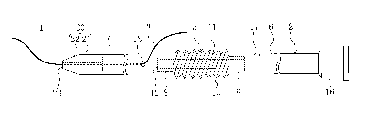
[0024] Fig. 1 is a side view of a removing tool for intravascular obsti-
uctions, according to
an embodiment of the present invention.
Fig. 2 is a side view of the removing tool for intravascular obsti-uctions,
sliowing
the status where a suction apparatus is connected to the removing tool.
Fig. 3 is a partial sectional view of the removing tool for intravascular
obsti-uctions,
showing an enlarged removing portion of a tubular member.
Fig. 4 is a diagram illustrating the case where a venous blood backflow-
preventing
valve is removed from an imier vessel wall: Fig. 4(a) shows the condition of a
guide wire
inserted into a vein; and Fig. 4(b) shows the condition of the tubular member
inserted into
the vein.
Fig. 5 is a diagram coiTesponding to Fig. 1, according to a modified example 1
of
the embodiment.
Fig. 6 is a diagram corresponding to Fig. 4(b), according to a modified
example 2
of the embodiment.
CA 02589301 2007-05-29
7
Fig. 7 is a diagram corresponding to Fig. 1, according to a modified example 3
of
the embodiment.
Fig. 8 is a diagram corresponding to Fig. 1, according to a modified example 4
of
the embodiment.
Fig. 9 is a diagram corresponding to Fig. 1, according to a modified example 5
of
the embodiment.
Description of the reference numerals.
[0025]
1 Removing tool
2 Tubular member
3 Guide wire (guide member)
5 Removing portion
6 Operating portion
7 Extending portion
10 Projecting portion
15 Suction apparatus (suction means)
17 Thorough hole
22 Tapered plane
A Vein
B Venous blood backflow-preventing valve (intravascular obstructions)
DESCRIPTION OF THE PREFERRED EMBODIMENTS
[0026] Preferred embodiments of the present invention will be described in
detail as
follows in conjunction with the drawings.
[0027] Embodiment 1
Fig. 1 shows a removing poi-tion 1 for intravascular obstructions, according
to
CA 02589301 2007-05-29
8
embodiment 1 of the present invention. The removing portion 1 has a tubular
member 2
to be inserted into the vessel and a guide wire 3 as a guide member for
guiding the tubular
member 2 in the vessel.
[0028] The tubular member 2, as a whole, is formed to extend in the direction
of insertion
in the vessel, the total length being set at from approximately 700 mm to 800
mm. hi
addition, the tubular member 2 has a cylindrical removing portion 5 for
removing
intravascular obstructions foimed in the vessel from the inner vessel wall, a
long operating
portion 6 extending from one end in an axial direction of the removing portion
5, and a
long extending portion 7 extending from the other end in the axial direction
of the
removing portion 5.
[0029] The removing portion 5 is made by molding a resin material such as an
elastomer
resin. The length of the removing portion 5 is set at from approximately 20 mm
to 30
min in the axial direction. As also shown in Fig. 3, the removing poi-tion 5
is set so that
the imier diameter is the saine throughout the axial direction, from one end
to the other end,
while the outer diameter has smaller ends in the axial direction. The removing
portion 5
has at each end in the axial direction a small diameter portion 8 that is
thimler than a
middle portion. The middle portion of the removing portion 5 in the axial
direction has a
projecting portion 10 integrally formed, projecting from an outer
circumference of the
removing portion 5, and extending spirally along the outer circumference. The
projecting
portion 10 has a triangle section having an apex on the projecting tip. Thus,
the outer
circumference of the removing portion 5 has a spirally extending groove 1. Fui-
ther, the
extending portion 7 side of the removing portion 5 is blocked by a blocking
portion 12.
[0030] For the operating portion 6, a resin material softer than the resin
material
constituting the removing portion 5 is foi7ned into a tube-like shape to allow
for easy
bending during insertion into the vessel. The inner diameter of the end of the
removing
poi-tion 5 side of the operating poi-tion 6 is set slightly smaller than the
outer diameter of
the small diameter portion 8 of the removing portion 5. The small diameter
portion 8 of
CA 02589301 2007-05-29
9
the operating portion 6 side of the removing portion 5 is held in the
condition of insertion
into and fitting to the interior of the end of the operating portion 6. This
makes the
removing portion 5 integral with the operating portion 6. The other end of the
operating
portion 6 opposite to the removing portion 5 has a connecting portion 16 to be
connected to
a suction apparatus 15 (shown in Fig. 2) mentioned later. The coiulecting
portion 16 is
formed in a tubular shape having a diameter larger than that of the middle
portion of the
operating portion 6 in the longitudinal direction. In addition, a surrounding
wall of the
operating portion 6 has a thorough hole 17 in the vicinity of the end of the
operating
portion 6 at the removing portion 5 side in order to provide a communication
between the
interior and the exterior of the operating portion 6.
[0031] For the extending portion 7, the same resin material as that of the
operating portion
6 is forined into a tubular shape. The iiuler diameter of the end of the
extending portion 7
at the removing portion 5 side is slightly smaller than the outer diameter of
the small
diameter portion 8 of the removing poi-tion 5, while the small diameter
portion 8 of
removing portion 5 at the extending portion 7 side is held in the condition of
insei-tion into
and fitting to the interior of the end of the extending portion 7. This makes
the removing
portion 5 integral with the extending portion 7. In addition, the surrounding
wall of the
extending portion 7 has an inlet hole 18 in the vicinity of the end of the
extending poi-tion 7
at the removing portion 5 side in order to insert the guide wire 3
therethrough.
[0032] On the other hand, the end of the extending portion 7 opposite to the
removing
portion 5 has an end cap portion 20. The end cap poi-tion 20 also constitutes
the tubular
member 2. For the end cap portion 20, a resin material harder than the
extending portion
7 is fonned into a roughly cylindrical shape. The imier diameter of the end
cap portion 20
is slightly smaller than the imier diameter of the removing portion 5 and the
iiuier diameter
of the extending portion 7. The end cap portion 20 has an insertion portion 21
to be
insei-ted into the side of the extending portion 7 opposite to the removing
portion 5. The
outer diameter of the insertion portion 21 is slightly larger size than the
imier diameter of
CA 02589301 2007-05-29
the extending portion 7. The insertion portion 21 is held in the condition of
insertion into
the extending portion 7. This makes the extending portion 7 integral with the
end cap
portion 20. The side of the end cap portion 20 opposite to the insertion
portion 21 has a
tapered plane 22 tapering toward a distal position of the insertion portion
21. The tapered
5 plane 22 is formed to be continuous with respect to the end of the extending
portion 7 in
the condition in which the insertion portion 21 is inserted into the extending
portion 7.
The end of the tapered plane 22 is coiulected to a flat plane 23 extending in
the direction
approximately orthogonal to the axial direction of the end cap portion 20. The
flat plane
23 has an opening through which the guide wire 3 is inserted.
10 [0033] The suction apparatus 15 coiuiected to the connecting portion 16
constitutes the
suction means of the present invention and is coiulected to the coiulecting
portion 16
through a connecting pipe 25. The suction apparatus 15 has a k.nown structure
commonly
used in the medical field.
[0034] On the other hand, the guide wire 3 is made by twining a multiplicity
of fine steel
wires and has a flexibility enabling easy bending during insertion into the
vessel. The
diameter of the guide wire 3 is smaller than the imler diameter of each
portion of the
tubular member 2, and the length is longer than the length of the tubular
member 2.
[0035] Next, the treatment of a varix formed in a vein A of a lower limb by
using the
above-constituted removing tool 1 for intravascular obstiuctions will be
described with
reference to Fig. 4. The varix results from a deteriorated flow of blood
caused by a failed
venous blood backflow-preventing valve B in vein A, turning the venous blood
backflow-
preventing valve B into an intravascular obstruction. On the other hand, in
removing tool
1 used for this case, the outer diameters of the operating portion 6 and the
extending
portion 7 are slightly smaller than the iiuier diameter of the vein A and the
distance
between the tips of the projecting portion 10 in the radial direction of the
removing portion
5 is slightly larger than the imier diameter of the vein A.
[0036] First, partial skin incision is carried out to expose the vein A having
the varix at a
CA 02589301 2007-05-29
11
further central side (an upstream side in the blood flow direction) than the
varix. Next, as
shown in Fig. 4(a), the exposed part of the vein A is incised to make a
central side-incised
portion Cl. Then, the guide wire 3 is inserted into the vein A at the
peripheral side (a
downstream side in the blood flow direction) through the central side-incised
portion Cl.
At this time, the operator operates the guide wire 3a at the proximal side
thereby
facilitating selective insertion of the end of the guide wire 3 into a desired
vein branch
among a multiplicity of vein branches.
[0037] When the end of the guide wire 3 is sufficiently past the point of the
vein A where
the varix is forined, insertion of the guide wire 3 is discontinued. Then, a
point of the
skin coiTesponding to the end of the guide wire 3 is partially incised, and a
point of the
vein corresponding to the end of the guide wire 3 is also incised, thus fon-
i7ing a peripheral
side-incised portion C2 (shown in Fig. 4(b)). Then, the end of the guide wire
3 is pulled
out of the skin tluough the peripheral side-incised portion C2 of the vein A
and the
dissected position of the skin.
[0038] Next, the tubular member 2 is inserted into the vein A through the
central side-
incised portion Cl of the vein A. At this time, first, the end of guide wire
3, which is
projected from the central side-incised portion Cl, is inserted into the
extending portion 7
through the end cap portion 20 of the tubular member 2. Then, the end cap
portion 20 is
inserted into the vein A through the central side-incised portion Cl, and the
extending
portion 7, removing portion 5, and the operating portion 6 are inserted into
the vein A by
hand in this order. By this operation, the end cap portion 20 and the
extending portion 7
are guided by the guide wire 3 to pass tlu=ough the point of the vein A where
the varix is
foimed, and finally to project to the exterior of the skin through the
peripheral side-incised
portion C2. The end cap portion 20 projecting tluough the peripheral side-
dissected part
C2 is pulled out of the skin and the operating portion 6 is pushed to the
interior of the vein
A in the direction of insertion so that the extending portion 7 is guided by
the guide wire 3
further toward the direction of insertion. The removing portion 5, which is
continuous to
CA 02589301 2007-05-29
12
the extending portion 7, is also indirectly guided by the guide wire 3, which
enables it to
position the removing portion 5 in the vicinity of the failed venous blood
backflow-
preventing valve B.
[0039] In this state, the operating portion 6 and the extending portion 7
respectively
project from the central side-incised portion Cl and the peripheral side-
incised portion C2
of vein A to the exterior of the skin. The operator can handle both the
operating portion 6
and the extending portion 7 by hand to allow the removing portion 5 to move
back and
forth in the vein A in the extension direction for the vein A. Thus, the
removing portion 5
can be operated from both sides of the axial direction, and hence, the
removing portion 5
can be stabilized in vein A during operation.
[0040] Operating the removing portion 5 in the above-described maiuler brings
the tips of
the projecting portion 10 into contact with the blood backflow-preventing
valve B, thereby
removing the blood backflow-preventing valve B off the inner wall of the vein
A. In
removing the blood backflow-preventing valve B off the iiuier wall of the vein
A, since the
distance between the tips of the projecting portion 10 is larger than the
inner diameter of
the vein A in the radial direction of the removing portion 5, the tips of the
projecting
portion 10 reliably come into contact with the blood backflow-preventing valve
B.
Further, since the projecting portion 10 is spirally shaped, only a single
movement of the
removing portion 5 provides frequent contact between the tips of the
projecting portion 10
and the blood backflow-preventing valve B.
[0041] Moreover, when a negative pressure is applied to the interior of the
operating
portion 6 by actuating the suction apparatus 15 while moving the removing
portion 5 in the
vein A, the blood backflow-preventing valve B removed from the inner wall of
vein A is
sucked into the tubular member 2 tluough the thorough hole 17 to the suction
apparatus 15
tlirough the operating portion 6, thus removing the blood backflow-preventing
valve B
from the interior of the vein A. The suction apparatus 15 may be actuated
following
removal of the blood backflow-preventing valve B from the imier wall of vein
A.
CA 02589301 2007-05-29
13
[0042] Next, although not shown, the case where the treatment of an ischemic
disease
caused by a tluombus foi7ned in the vein of a lower limb by using the removing
tool 1 for
intravascular obstructions will be described below. The removing tool 1 used
in this case
is such that the size of each portion is set in the same inanner as in the
case of the treatment
of the varix as described above.
[0043] First, pai-tial skin incision is cai7ied out to expose a further
central side of the vein
where the varix is fonned than the thrombus followed by incision of the
exposed part of
the vein. Then, the guide wire 3 is inserted from the central side-incised poi-
tion of the
vein toward the peripheral side, and when the end of the guide wire 3 is
sufficiently past
the thrombus, the insertion of the guide wire 3 is discontinued.
[0044] Subsequently, in the same manner as that of the treatment of the varix
as described
above, when the tubular member 2 is inserted into the vein, the end of the
tubular member
2 is guided by the guide wire to penetrate through the tluombus and the
removing portion 5
of the tubular member 2 is guided by the guide wire 3 to the position in the
vein where the
tluombus is formed. Then the operator handles the operating portion 6 of the
tubular
member 2 by hand to allow the removing portion 5 to move back and forth in the
vein in
the extending direction for the vein. By this step, the tips of the projecting
portion 10 of
the removing portion 5 contacts the tlirombus to remove the thrombus from the
inner wall
of the vein. In removing the thrombus from the iiuler wall of the vein A,
reliable removal
becomes possible by designing the size and shape of the projecting portion 10,
similarly to
the case of removing the blood backflow-preventing valve B as described above.
In
addition, sucking the thrombus removed from the iiuier wall of the vein by
using the
suction apparatus 15 results in removal of the thrombus from the interior of
the vein.
[0045] As described above, the removing tool 1 is comprised of the tubular
member 2,
which has the removing portion 5 for removing the blood flow-preventing valve
B and the
tlirombus, which prevent blood flow, from the iiuier wall of the vein A, and
the guide wire
3 for guiding the removing portion 5 in the vein A. Thus, the removing portion
5 can be
CA 02589301 2007-05-29
14
reliably guided by the guide wire 3 to the position in the vein A where the
blood backflow-
preventing valve B or the thrombus is forined, enabling the removing portion 5
to remove
the blood backflow-preventing valve B or the tlu=ombus from the imler wall of
the vein A.
By this step, when diseases caused by a variety of intravascular obstructions
foi-med in the
vein A are treated, it is not required to use an expensive tlirombolytics and
to extract and
remove the vein A. This suppresses an increase in treatment cost and the
treatment can
be reliably conducted in low stresses.
[0046] Also, the thorough hole 17 is provided on the circumferential wall of
the operating
portion 6 in the vicinity of the removing portion 5, and a negative pressure
is applied to the
interior of the operating portion 6 from the suction apparatus 15. Thus, the
blood
backflow-preventing valve B or the thrombus removed from the imier wall of
vein A can
be reliably removed from the interior of the vein A.
[0047] In treating the varix, the operating portion 6 and the extending
portion 7 are
projected from the skin to allow the operator to hold the portions so that the
removing
portion 5 can be handled at both ends in the axial direction. This makes the
removing
portion 5 stable in vein A during the operation to achieve reliable removal of
the blood
flow-preventing valve B from the iiuler wall of the vein A.
[0048] Since the end cap portion 20 is fonned in a tapered shape, when the
tubular
member 2 is inserted into the vein A, the tubular member 2 is not caught in
the inner wall
of the vein A. Thus, the tubular member 2 can be smoothly inserted into the
vein A to
enable it to remove the blood backflow-preventing valve B and the tluombus in
low
stresses.
[0049] In addition, since the removing portion 5 has the spirally for-ined
projecting portion
10, only a single movement of the removing portion 5 in vein A, for example,
provides
frequent contact between the tips of the projecting portion 10 and the blood
backflow-
preventing valve B or the tlu-ombus, thereby providing efficient removal of
the blood
backflow-preventing valve B or the tlu=ombus. In addition, since the spiral
groove 11 is
CA 02589301 2007-05-29
provided on the outer circumference of the removing portion 5, the blood
backflow-
preventing valve B or the thrombus removed from the iiuler wall of vein A is
not left on
the tips of the projecting portion 10 but contained in the groove 11. As a
result, the tips
of the projecting portion 10 can be always exposed. This alone realizes
efficient removal
5 of the blood backflow-preventing valve B or the thrombus.
[0050] Further, since the spiral groove 11 is in a spiral shape, the blood
backflow-
preventing valve B or the thrombus contained in the groove 11 are, through the
back-and-
foi-th movement of the removing portion 5 in the vein A, moved through the
groove 11 to
the operating portion 6 side and the extending portion 7 side of the groove 11
to be finally
10 discharged out of the groove 11. This reliably prevents clogging of the
projecting portion
10.
[0051] According to this embodiment, the guide wire 3 is inserted into only
the cap
portion 20 and the extending portion 7. However, as in a modification example
1 shown
in Fig. 5, the guide wire 3 may be inserted throughout the longitudinal
direction of the
15 tubular member 2.
[0052] Further, according to this embodiment, the tubular member 2 has the
extending
portion 7. This extending portion 7 may be omitted as in a modification
example 2
shown as shown in Fig. 6. hl this case, the end of removing portion 5 opposite
to the
operating portion 6 is open so that the guide wire 3 is inserted across the
removing portion
5 and the operating portion 6.
[0053] According to this embodiment, the projecting portion 10 is fonned
integrally with
the main body of the removing portion 5. However, as in a modification example
3
shown as shown in Fig. 7, the projecting portion 10 may be separately provided
using, for
example, elastomer resin, followed by adhesion to a cylindrical unit 30 with,
for example,
an adhesive. The cylindrical unit 30 is composed of the same material as that
of the
operating portion 6 and the extending poi-tion 7. Both ends of the cylindrical
unit 30 in
the longitudinal direction has small diameter portions 31 similar to the small
diameter
CA 02589301 2007-05-29
16
portion 8. Each of the small diaineter portions 31 is inserted into the
operating portion 6
and the extending portion 7 so that the cylindrical unit 30, the operating
portion 6, and the
extending portion 7 are integral with each other.
[0054] In addition, as in a modification example 4 shown in Fig. 8, the
operating portion 6
of the tubular member 2, the main body of the removing portion 5, and the
extending
portion 7 may be integrally formed, and the projecting portion 10 may be such
that as in
the modification example 3, a separately provided member is adhered to the
main body of
the removing portion 5. hltegral forining of the operating portion 6, the main
body of the
removing portion 5, and the extending portion 7 provides approximately
uniforin softness
tluoughout the tubular member 2. As a result, during insertion into the vein
A, when, for
example, the tubular member 2 passes a bent portion of the vein A, there is
substantially no
change in an inserting force. This improves the operability of the removing
tool 1.
[0055] As in a modification exainple 5 shown in Fig. 9, the removing portion 5
may be
placed at the end of the tubular member 2 in the direction of insertion. The
small
diameter portion 8 of the removing portion 5 is inserted into and fitted to
the interior of the
end of the tubular member 2. On the end side of the removing portion 5 in the
direction
of insertion, a tapered plane 50 is formed to taper distally. The end of the
tapered plane
50 is coiinected to a flat plane 51 extending in the direction approximately
orthogonal to
the axial direction of tubular member 2. On the flat plane 51, an opening
tluough which
the guide wire 3 passes is forined. Since in the removing tool 1 according to
the
modification example 5 the removing portion 5 is located at the end of tubular
member 2 in
the direction of insertion, only a single incised portion is necessary on the
skin so that the
guide wire 3 is inserted into the vein A tlirough the incised portion followed
by inser-tion of
the removing portion 5 and the tubular member 2 and then by holding of the
tubular
member 2 to operate the removing portion 5. Thus, since only a single incised
portion is
necessary, the treatment involves low stresses.
[0056] According to this embodiment, the projecting portion 10 is continuous
to the
CA 02589301 2007-05-29
17
removing portion 5 in a circumferential direction. The removing portion 5 may,
for
example, have projections not continuous in the circumferential direction.
Further, the
sectional shape of the projecting portion 10 is not restricted to a triangle,
but may be, for
example, a rectangle.
[0057] In treating the varix, following removal of the blood backflow-
preventing valve B
from the imier wall of vein A, the removing portion 5 may be further allowed
to move back
and forth to cause the projecting portion 10 to pass the iiuler wall of vein A
with friction.
By this way, following removal of the tubular member 2 and the guide wire 3
out of the
vein A, when the vein A is held with pressure from the exterior of the skin,
because of the
losing of the blood backflow-preventing valve B and friction of the inner wall
of the vein
A, the vein A can be completely crashed to adhere iiuler walls to each other.
This blocks
the blood flow in vein A, thereby degenerating the vein A.
[0058] When the blood backflow-preventing valve B or the thrombus is removed
from the
imier wall of the vein A, physiological saline, for example, may be introduced
into the vein
A. Specifically, for example, a syringe, not shown, filled with physiological
saline is
connected to the connecting portion 16 of the operating portion 6, and after
the blood
backflow-preventing valve B or the thrombus is removed from the iiuier wall of
vein A by
the removing portion 5, the syringe is operated to supply the physiological
saline to the
vein A through the thorough hole 17. This facilitates the movement, in the
vein A, of the
blood backflow-preventing valve B or the thrombus removed from the inner wall
of vein A.
Thus, the blood backflow-preventing valve B and the thrombus can be more
reliably
sucked by suction apparatus 15.
[0059] According to this embodiment, the suction means is constituted by the
suction
apparatus 15. This suction means may be constituted by a syringe, for example.
INDUSTRIAL APPLICABILITY
[0060] As described above hereinbefore, the removing tool for intravascular
obstructions
CA 02589301 2007-05-29
18
according to the present invention can be used for, for example, treating a
varix caused by
a failed blood backflow-preventing valve in the vein.