Note: Descriptions are shown in the official language in which they were submitted.
CA 02589374 2007-05-29
WO 2006/071441 PCT/US2005/043482
ANTIBODIES DIRECTED TO GPNMB AND USES THEREOF
FIELD OF THE INVENTION
The present invention relates to antibodies with specificity to GPNMB, and
uses of
such antibodies. In particular, the present invention provides fully human
monoclonal
antibodies that specifically bind to GPNMB, and uses thereof. Nucleotide
sequences
encoding, and amino acid sequences comprising, heavy and light chain
immunoglobulin
molecules, particularly sequences corresponding to contiguous heavy and light
chain
sequences spanning the framework regions and/or complementarity determining
regions
(CDRs) are provided. The present invention also provides immunoconjugates
comprising
anti-GPNMB antibodies and methods of using such immunoconjugates. The present
invention further provides bi-specific antibodies comprising an anti-GPNMB
antibody
component and an anti-CD3 component, and methods of using such bispecific
antibodies.
BACKGROUND OF THE INVENTION
GPNMB
A putative transmembrane glycoprotein called "tutib" (Acc. No. X76534 EMBL) ,
= referred to herein as GPNMB, was identified and described by Weterman et
al., (Int J
Cancer 60:73-81, 1995) as differentially expressed in low-metastatic human
melanoma
cancer cell lines and xenografts, compared to a more aggressive melanoma cell
line.
GPNMB shares 33% identity with the precursor of pMe117 melanocyte-specific
protein
(Kwon et al., 1991, PNAS 88:9228-9232). GPNMB is 71% homologous to a dendritic
cell-
associated transmembrane protein, DC-HIL (Shikano et al., 2001 Biol. Chem.
276:8125-
8134). GPNMB is also known as the hematopoietic growth factor inducible
neurokinin-1
protein HGFIN (Bandari et al, Reg. Peptides 111:169-178) and the bone-related
gene
osteoactivin (Owen et al. Crit Rev Eukaryot Gene Expr 2003, 13(2-4):205-220)
It was also reported that nmb could reduce the metastatic potential of a
highly
metastatic mnb-negative melanoma cell line (Wetennan, 1995). GPNMB was
considered a
candidate glioblastoma tumor marker after public database mining and
expression profiling
(Loging et al., 2000, Genome Research 10:1393-1402). This gene was found
overexpressed in lung tumors (US Patent Publication No. US20030064947), as
well as
breast, rectal and colon cancers (US Patent Publication No. US2003100720).
NCBI SAGE
data also shows overexpression of this gene in stomach and pancreatic
carcinoma. The
mouse ortholog has been shown to be highly upregulated in a neural stem cell
line NSC,
1
CA 02589374 2007-05-29
WO 2006/071441
PCT/US2005/043482
derived from the TSC2 knockout model for Tuberous Sclerosis Complex Syndrome
(International Publication No. WO 2003/080856).
Antibodies
Antibodies, also known as immunoglobulins, are typically tetrameric
glycosylated
proteins composed of two light (L) chains (about 25 kDa) and two heavy (H)
chains (about
50-70 kDa). The amino-terminal portion of each chain includes a variable
domain of about
100 to 110 or more amino acids primarily responsible for antigen recognition.
The carboxy-
terminal portion of the L and H chain has one and three or four constant
domains,
respectively that are primarily responsible for effector function. There are
two types of
human L chains, classified as kappa and lambda. H chains are classified as mu,
delta,
gamma, alpha, or epsilon based upon the constant domain amino acid sequence,
defining
the antibody's isotype as IgM, IgD, IgG, IgA, and IgE, respectively. Isotypes
may be
further divided into subclasses e.g. IgGi, IgG2, IgG3, IgG4.
Immunoglobulins can be produced naturally in vivo by B lymphocytes. Each clone
of B cells produces antibody with an antigen receptor having a unique
prospective antigen
binding structure. The repertoire of antigen receptors, approximately 107
possibilities, exists
in vivo prior to antigen stimulation. This diversity is produced by somatic
recombination,
i.e., the joining of different antibody gene segments. Immunoglobulin H chain,
kappa L
chain and lambda L chain are encoded by three separate genetic loci and each
locus has
multiple copies of at least 3 types of gene segments encoding variable (V),
constant (C) and
joining (J) regions, the heavy chain gene also includes a diversity (D)
region. The selection
of specific V, C and J regions (and D for the heavy chain) from amongst the
various gene
segments available (45 heavy chain V; 35 kappa V; 23 heavy chain D; 6 heavy
chain J; 5
kappa J) generates approximately 10 11 possible specificities of gennline
sequences
exhibited in B cells. The joining of V, C and J regions can result in the loss
or addition of
residues at the junctions. The L and H chain V region of human antibodies
consists of
relatively conserved framework regions (FR) that form a scaffold for three
hypervariable
regions also known as complementary determining regions (CDR). From the amino
terminus of either the heavy or light chain, the V domain is made up of FR and
CDR
regions in the following order: FR1-CDR1-FR2-CDR2-FR3. Joining of the V domain
with
a D (heavy chain only) and J domain adds CDR3-FR4. The CDRs are generally
responsible
for antigen binding.
2
CA 02589374 2007-05-29
WO 2006/071441
PCT/US2005/043482
The specificity of monoclonal antibodies have made them attractive agents for
targeting cancer in vivo with the hopes of eradicating disease while sparing
normal tissue.
The approach, which initially utilized mouse monoclonal antibodies has
encountered
limitations to potential effectiveness such as immunogenicity; inefficient
effector functions
and short half-life in vivo. Technologies were developed for: chimeric
antibodies which
sought to utilize the antigen binding variable domains of mouse monoclonal
antibodies
combined with the constant regions of human antibodies (Boulianne, et at. 1984
Nature
312:643-646; Morrison et at, 1984 PNAS USA 81:6851-6855); humanized antibodies
which
grafted antigen binding complementary determining regions (CDRs) from mouse
antibodies
to human immunoglobulin (Jones, et at, 1986 Nature 321: 522-525; Riechmann, et
at, 1988
Nature 332:323-327; Verhoeyen, et at, 1988 Science 239:1534-1536; Vaughan,
eta!, 1998
Nature BiotechnoL 16:535-539); and phage display libraries of single chain
scFvs or Fab
fragments of antibodies (de Haard, et at, 1999 J BioL Chem. 274: 18218-18230;
Knappik-,
et at, 2000 J. Mol. Biol. 296:57-86; Sheets, et at, 1998 PNAS USA 95:6157-
6162; Vaughan,
et at, 1994 Nature Biotechnol 14:309-314, 1996; Griffiths et at EMBO J.
13:3245-3260).
Additionally, transgenic animals having human iinmunoglobulin genes and
nonfunctional
endogenous genes have been developed for immunization and production of fully
human
monoclonal antibodies (Fishwild, et at, 1996 Nature Biotechnol 14:845-851;
Mendez, et at,
1997 Nature Genet. 15:146-156; Nicholson, et at, 1999 J. Immunol 163, 6898-
6906).
Single Chain Antibodies: Single chain Fv antibodies (scFvs) were first
described in
the late 1980's (Bird etal., Science 242:423-426 (1988); Huston et at., Proc.
Natl. Acad.
Sci. USA 85:5879-5883 (1988)). A polypeptide linker, typically ranging in
length from 5 to
27 amino acid residues, is used to join the C-terminus of the variable light
chain domain
(VL) to the N-terminus of the variable heavy chain domain (VH). Alternatively,
the linker
joins the C-terminus of the VH to the N-terminus of the VL. Both formats (VL-
VH and VH-
VO have been used successfully in the literature. The most common linker used
in the
literature is the (Gly4Ser)3 15 amino acid linker, however there are several
other linkers that
have been utilized, including a 25 amino acid linker called 205C (Pantoliano
et at.,
Biochemistry 30:10117-10125 (1991)). Single chain antibodies are currently in
the clinic;
one of the most advanced is h5G1.1 or Pexelizumab. This scFv is specific for
human C5
complement and is being used in clinical trials for cardiac patients
undergoing
cardiopulmonary bypass surgery (Shernan et at., Ann. Thorac Surg. 77:942-949
(2004)).
3
CA 02589374 2007-05-29
WO 2006/071441
PCT/US2005/043482
Bispec0c Antibodies (bi-Abs): An area of mAb research where considerable
progress has been made is in the development of bispeeific antibodies (biAbs).
There are
distinct advantages to developing therapeutic antibody molecules with dual
specificity. For
example, biAbs can serve as mediators to target immune effector cells such as
CTLs to
unwanted cells (Baeuerle et al., Cun-. Opin. Mol. Ther. 5:413-419 (2003)). In
another
example, chemically linked bispecific antibodies directed against Fe gamma
receptors
CD16, CD64, and CD89, were significantly more effective in vitro than
conventional IgG
antibodies (Peipp and Valerius, Biochem. Soc. Trans. 30:507-511(2002)). One of
the
challenges in developing biAbs as viable therapeutics has been producing large
enough
quantities of a stable moiety for clinical applications. Another challenge has
been in
determining the right combination of validated targets and the underlying
biology that
would lead to a therapeutic product. For recent reviews on the difficulties
experienced with
biAbs, see (Kontennann, Acta Phannacol Sin 26:1-9 (2005); Peipp and Valerius,
Soc.
Trans. 30:507-511 (2002)).
Bispecific Single Chain Antibodies (bi-scFi): A notable type of biAb that can
be
made is a bi-specific single chain antibody or bi-seFv. For a review on the
generation of bi-
sav's see (Kipriyanov and Le Gall, Curr Opin Drug Discov Devel 7:233-242
(2004)). Bi-
scFvs are typically comprised of 4 variable domains, 2 heavy (VH) and 2 light
(VI), which
are derived from 2 different antibodies. The 4 domains are linked together
with 3 short
linkers, ranging in length from 5-27 amino acids. The biological activity of
this type of
antibody depends on several features concerning the construction of the
molecule. For
example, both the linker sequences between the antibody V domains and the
order of the 4
antibody V domains themselves (for the 2 antibodies) can vary, as well as the
expression
system that is used; all of which can greatly affect the solubility and
biological activity of
the various resulting products (Kipriyanov et al., J. Mol. Biol. 330:99-
111(2003); Le Gall
et al., Protein Eng. Des. Sel. 17:357-366 (2004); Pavlinkova et al., Clin
Cancer Res.
5:2613-1619 (1999)).
Cylotoxie T lymphocytes: -Under normal circumstances, T cells are activated
when
the CD3/T cell receptor (CD3/TCR) complex binds to a relevant MHC molecule
associated
with a specific Ag peptide. Engagement of CD3/TCR with MHC results in
intracellular
signals necessary to trigger an immune response against a pathogen or tumor.
Similar
signals that cause T cell activation can also be achieved by antibodies that
can bind certain
structures of the CD3/TCR complex. In the literature, it has been shown that
biAbs
4
CA 02589374 2007-05-29
WO 2006/071441
PCT/US2005/043482
recognizing both the TCR/CD3 complex and tumor associated antigen (TAA) can
trigger
the activation program in CTLs in the presence of target cells (Chapoval et
al., J. Immunol
155:1296-1303 (1995)).
Recombinant technologies are being utilized to enable further improvements
upon
antibody molecules with the goal of enhancing in vivo efficacy. Such
technologies provide,
for example, for optimizing molecular size, affinity, phamiacokinetics,
toxicity, specificity,
valency, effector functions, direct and indirect aiming, combination therapy,
and various
prodrug approaches.
It would be desirable to have an antibody suitable for in vivo targeting of
GPNMB
expressing pathologies and to enable therapeutic efficacy.
SUMMARY OF THE INVENTION
The current invention provides human monoclonal antibodies that specifically
bind
GPNMB as well as variants, derivatives and antigen binding fragments of such
antibodies.
The invention provides preferred somatic recombinations of human antibody gene
segments to provide specificity for GPNMB and genetically engineered anti-
GPNMB
antibody variants and derivatives that originate from these gene segments. In
addition, the
current invention provides multiple affinity matured human antibodies with
binding
specificity for GPNMB.
In one embodiment, the present invention provides an antibody, or binding
fragment
thereof, that binds to GPNMB, wherein said antibody, or binding fragment
thereof,
neutralizes a GPNMB-induced activity, and wherein said antibody, or binding
fragment
thereof, cross-reacts with a fully human anti-GPNMB antibody selected from the
group
consisting of Mab1.2.1, Mab1.10.1, and Mab2.22.1 or an antibody in the same
antigen-
binding bin as fully human anti-GPNMB antibody Mab1.2.1, Mab1.10.1, or
Mab2.22.1.
In another embodiment, the present invention provides an antibody, or binding
fragment thereof, that binds to GPNMB, wherein said antibody, or binding
fragment
thereof, neutralizes a GPNMB-induced activity, and wherein said antibody, or
binding
fragment thereof, cross-reacts with a fully human anti-GPNMB antibody selected
from the
group consisting of Mab2.3.1 and Mab1.15.1 or an antibody in the same antigen-
binding
bin as fully human anti-GPNMB antibody Mab2.3.1 or Mab1.15.1.
In yet another embodiment, the present invention provides an antibody, or
binding
fragment thereof, that binds to GPNMB, wherein said antibody, or binding
fragment
thereof, neutralizes a GPNMB-induced activity, and wherein said antibody, or
binding
5
CA 02589374 2007-05-29
WO 2006/071441
PCT/US2005/043482
fragment thereof, cross-reacts with fully human anti-GPNMB antibody Mab2.10.1
or an
antibody in the same antigen-binding bin as fully human anti-GPNMB antibody
Mab2.10.1.
In one embodiment, the present invention provides naked IgG1 anti-GPNMB
antibodies that have cytotoxic effect to cells overexpressing GPNMB. In a
specific
embodiment, the present invention provides methods of treating or preventing
diseases
associated with overexpression of GPNMB comprising administering to a subject
in need
thereof a composition comprising a naked IgG1 anti-GPNMB antibody and an
immunomodulator (such as, but not limited to, interferons and cytokines).
In another embodiment, the present invention provides immunoconjugates that
comprise an anti-GPNMB antibody or a fragment thereof, and a cytotoxic agent.
In a
specific embodiment, the cytotoxic agent is auristatin E (dolastatin-10) or a
derivative
thereof. Methods of using such immunoconjugated are also provided.
In one embodiment, the present invention provides bispecific antibodies
comprising
an anti-GPNMB component and an anti-CD3 antibody component, which enable the
cytotoxic killing of target tumor cells by T cells. In another embodiment, the
present
invention provides single chain Fv antibody conjugated to a cytotoxic agent.
In a specific
embodiment, the cytotoxic agent is auristatin E (dolastatin-10) or a
derivative thereof.
Methods of using such bispecific antibodies and conjugated single chain Fv
antibodies are
also provided.
Amino acid sequences for anti-GPNMB human monoclonal antibodies of the
invention and nucleic acid sequences encoding them are provided.
Compositions comprising human anti-GPNMB antibodies, including therapeutic
compositions comprising same, and methods of use are provided. Particularly,
therapeutic
immunoconjugates comprising anti-GPNMB antibodies and a cytotoxic or
cytostatic agent
- 25 for treating GPNMB expressing cancers and other GPNMB related disorders
are provided.
Dosage regimens are also provided.
Additional aspects of the disclosure will be set forth in part in the
description which
follows, and in part will be obvious from the description, or may be learned
by practicing
the invention.
BRIEF DESCRIPTION OF THE FIGURES
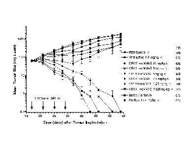
Figure 1: Tumor growth inhibition and complete regression of SK-MEL-2
xenografts in athymic mice after treatment with 2.50 to 20 mg/kg i.v. every 4
days for 4
6
CA 02589374 2007-05-29
WO 2006/071441
PCT/US2005/043482
treatments. The responses of tumor-bearing animals to reference drugs such as
vinblastine
(1.7 mg/kg i.v. q4d X4) and paclitaxel (24 mg/kg i.v. q2d X4) are also shown.
Control
groups are treated with either phosphate-buffered saline (PBS) or
physiological saline.
Figure 2: Indirect immunotoxin killing of UACC-62 melanoma cells by anti-
GPNMB antibodies
Figure 3: Inhibition of colony formation of UACC-62 cells incubated with
Auristatin E (AE) conjugated anti-GPNMB antibodies.
Figure 4: Tumor growth inhibition and complete regression of SK-MEL-2
xenografts in athymic mice after treatment with CR011-vcMMAE 5.0 mg/kg i.v.
every 4
days for 4 treatments. The lack of responses of tumor-bearing animals to
unconjugated
CRO 1 1 or to free monomethylauristatin E demonstrate that the intact
immunoconjugate is
essential for anti-tumor effects.
Figure 5: Tumor size reduction and complete regression of SK-MEL-2 xenografts
in athymic mice after treatment with 1.25 to 20 mg/kg i.v. every 4 days for 4
treatments.
The responses of tumor-bearing animals to reference drugs such as Vinblastine
(1.7 mg/kg
i.v. q4d X4) and paclitaxel (24 mg/kg i.v. q2d X4) are also shown. Control
groups are
treated with either phosphate-buffered saline (PBS) or physiological saline.
Figure 6: The serum concentration-time profile of the antibody of CR011-vcMMAE
after intravenous administration of 1 and 10 mg/kg in athymic mice. Detection
was
achieved with a sandwich ELISA assay, which employed the CR01 1 antigen
(CG56972,
GPNMB) and a horseradish peroxidase-conjugated anti-human globulin. Results
shown are
the serum concentrations expressed as [tg/mL (left x-axis) and micromolarmolar
concentration (right X-axis).
Figure 7: Aggregate responses, expressed as percent cures, were recorded for
test
animals treated with 5 different, graduated dosing intervals (i.e., 0, 1, 4,
8, and 16 days
between treatments). The slope of the line is not significantly different from
0 (p< 0.2904).
Figure 8: The proportions of complete regressors as a function of dosing
interval
and stratified by cumulative dose. For each group, n 6 mice/group. Athymic
mice bearing
established SK-MEL-2 tumor implants (day 14, 80 mg) were treated i.v. with
CR011-
veMMAE and the incidence of complete regressions is recorded.
Figure 9: Effects of ectopic expression of GPNMB or sensitivity to CR011-
vcMMAE. HEK293 cells are transfected with empty vector (vector) or GPNMB-
containing
plasmid (GPNMB) as described in Materials and Methods. A. Cell lysates are
prepared
7
CA 02589374 2007-05-29
WO 2006/071441
PCT/US2005/043482
from the transfected HEK293 cells and the expression of GPNMB (upper panel) or
actin
(lower panel) is determined by immunoblotting. Lane 1: Empty vector
transfectants. Lane
2: GPNMB transfectants. B. Flow cytometry analysis of GPNMB expression on
empty
vector or GPNMB transfected cells. C. CR011-veMMAE in vitro growth inhibition
of
transfected cells. Cells are treated with various concentrations of CR011-
vcMMAE
(diamonds: vector or circles: GPNMB) or IgG2-veMMAE (triangles: vector or
squares:
GPNMB) for 96 hours. After a clonogenic assay, the surviving fraction is
normalized to the
untreated control and expressed as a percentage of the control using GraphPad
Prism
graphing software. Each treatment is performed in triplicate. A representative
graph from
two independent experiments is shown.
Figure 10: Effect of GPNMB siRNA on endogenous GPNMB expression and
sensitivity to CR011-veMMAE. SK-Mel-2 cells are transfected with 50 nM of
control
siRNA or siRNA targeting GPNMB. A. Cell lysates are prepared from the
transfected SK-
Mel-2 cells 2 and 4 days post-transfection and the expression of GPNMB (upper
panel) or
actin (lower panel) is determined by immunoblotting. Lane 1: Mock
(oligofectamine)
transfection. Lane 2: Control siRNA transfection. Lane 3: GPNMB siRNA
transfection. B.
Flow cytometry analysis of GPNMB expression 2 and 4 days after transfection.
SK-Mel-2
cells are transfected with mock, control siRNA or GPNMB siRNA as indicted in
the
Materials and Methods. C. CR011-veMMAE in vitro growth inhibition of mock
(diamonds), control siRNA (circles) or GPNMB siRNA (triangles) transfected SK-
Mel-2
cells is determined by a clonogenic assay as described in Materials and
Methods. The
surviving fraction is normalized to the untreated control and expressed as a
percentage of
control using GraphPad Prism graphing software. Each treatment is performed in
triplicate.
A representative experiment from two independent studies is shown.
Figure 11: FACS analysis of SK-MEL-2 with isotype control, hybridoma IgG2
(B2), recombinant IgG2 (B19) and recombinant IgG1 (B16) to CG56972/GPNMB
relative
to IgG2 (B2, B19) or IgG1 (Control, B16) controls.
Figure 12: (A) PBMC and mAb (IgG1) mediated ADCC of SK-MEL-2 cells.
ADCC effector functions are measured as described above at 2, 5 and 10 jig/200
[t1 using
target:effector ratios of 10, 30, 60 and 100 as indicated. (B) PBMC and mAb
(IgG2) do not
cause ADCC to SK-MEL-2 cells. ADCC effector functions are measured as
described
above at 0, 2, 5 and 10 [tg/200 1 using target: effector ratios of 10, 30, 60
and 100 as
indicated.
8
CA 02589374 2007-05-29
WO 2006/071441 PCT/US2005/043482
Figure 13: Expression of CG56972 in human cancer cell lines and tissues. RTQ
PCR analysis of (A) human brain cancer cell lines or (B) human brain cancer
glioma and
medulloblastoma biopsies. (C) Microarray analysis of CG56972 expression in
human brain
cancer and oligodendroglioma tissues. Tissues or cell lines are harvested,
mRNA prepared
and RTQ PCR or CuraChip analysis performed as described in Materials and
Methods.
Figure 14: FACS analysis of cell surface binding of CRO 1 1 mAb to CG56972. SK-
MEL-2, XF-498, U-118-MG, SNB-78, SF-539 and SF-268 cells are labeled with a
saturating concentration (10 ug/mL) of CR011 mAb or control IgG2. Bound mAb is
detected by flow cytometry with PE-conjugated goat-anti-human secondary
antibody as
described in Materials and Methods. GM: Geometric mean. The SF-268 cell line
is
CG56972 transcript negative and used as a negative control.
Figure 15: Immunoblot analysis of CG5672 expression in human brain cancer cell
lines. Cell lysates are resolved on Tris-glycine gels and transferred to
membranes.
Immunoblot analysis is carried out with a polyclonal antibody to CG56972
followed by
enhanced chemiluminescence detection as described in Materials and Methods.
Arrowheads
indicate the relative mobility of the p100 and 120 CG56972 species. The SF-268
cell line is
CG56972 transcript negative and used as a negative control.
Figure 16: CR011-vcMMAE in vitro growth inhibition of astocytoma/glioblastoma
cell growth. XF-498, SNB-78, U-118-MG, SF-539, LOXIMVI and SF-268 cells are
incubated with the indicated concentration of CR011-vcMMAE. Cells are also
incubated
with control PK16.3 mAb (data shown in Table I) as described in the Materials
and
Methods. Cell growth was determined by clonogenic assay. The surviving
colonies are
counted and plotted using GraphPad Prism graphing software. The experiment is
perfoimed
in triplicate wells and repeated twice. vA representative experiment is shown.
IC5Os for
cell killing is presented in ng/mL concentrations. The LOXIMVI and SF-268 cell
lines are
CG56972 transcript negative and used as negative controls.
Figure 17: Development of CR01 1 Engineered Antibodies. Four antibody variable
(V) domains (shown in C for the bi-scFv) are derived from the light and heavy
chain variable
domains (VL and Vu) making up the antigen binding sites of CR01 1 and anti-CD3
whole
IgGs. The middle linker joining the 2 individual scFv components together
(shown in dashed
line) may play a key role in determining the resulting activity of each of the
scFv
components, including the effective cytolytic activity provided by the
cytotoxic T cells
engaged by the anti-CD3 scFv component of the bi-scFv.
9
CA 02589374 2007-05-29
WO 2006/071441
PCT/US2005/043482
Figure 18: A. ELISA results for CR01 1 say (squares) and CRO 1 1 x anti-CD3
(L4-
L2-L4 linker set) bi-scFv (diamonds). Both engineered CR011 antibodies bound
to the
GPNMB target. B. Western blotting of 2 of the CR01 1 engineered antibody
products
(arrows). Clone 16 corresponded to the CHOK1 line expressing CRO 1 1 scFv
(monomer),
while clone 17 corresponded to the CHOK1 line expressing CRO 1 1 x anti-CD3
(L4-L2-L4
linker set) bi-scFv (dimer). Clones 16 and 17 are used to produce the
engineered antibody
products.
Figure 19: Flow cytometry analysis of binding of CRO 1 1 scFv and CRO 1 1 x
anti-
CD3 (L4-L2-L4 linker set) bi-scFv products to native GPNMB protein expressed
on the cell
surface of target cells. Human T cells are used as a source of CD3, while SK-
Mel-5 cells
are used as a source of GPNMB.
Figure 20: Cytotoxicity analysis showed that purified CR01 1 x anti-CD3 (L4-L2-
L4
linker set) bi-scFv, but not CR01 1 scFv, causes killing of GPNMB positive SK-
Mel-5
tumor cells by T lymphocytes.
Figure 21: The chemical structure of Maleimidocoaproyl-Valine-Citmllin-
Monomethyl-Auristatin E (veMMAE).
Figure 22: Disulfides on CR01 1 antibody are gently reduced in the presence of
TCEP to generate ¨4 thiols per Ab. veMMAE is then added to antibody solution.
Nucleophilic attack of thiolates on maleimide-groups results in a stable
thioester linkage.
The resulting conjugate is purified from the mixture.
Figure 23: Reaction of veMMAE with NAcCys at pH 7.0 and pH 9.0 in the
presence or absence of TCEP. 1A: VCMMAE converts fully into NAcCys-adduct
following
a incubation in phosphate pH 7 buffer. B-E: Appearance of a side product in a
course of
incubation of veMMAE in borate buffer. F-I: Appearance of side products in
borate pH 9
and in the presence of TCEP.
Figure 24: LCMS identification of the side product with retention time of 9.2
min
not capable of reaction with cystein and therefore, not capable of conjugation
to CR011.
Figure 25: Kinetics of the formation of NAcCys-veMMAE and of the side product
(succinimidyl-vcMMAE) following incubation in borate pH 9.0 buffer in the
presence or
absence of TCEP.
CA 02589374 2007-05-29
WO 2006/071441
PCT/US2005/043482
DETAILED DESCRIPTION OF THE INVENTION
As used herein, the term "antibody" refers to an immunoglobulin or a fragment
or a
derivative thereof, and encompasses any polypeptide comprising an antigen-
binding site,
regardless whether it is produced in vitro or in vivo. The term includes, but
is not limited to,
polyclonal, monoclonal, monospecific, polyspecific, non-specific, humanized,
single-chain,
chimeric, synthetic, recombinant, hybrid, mutated, engineered, and grafted
antibodies.
Unless otherwise modified by the term "intact," as in "intact antibodies," for
the purposes of
this disclosure, the term "antibody" also includes antibody fragments such as
Fab, F(a131)2,
Fv, scFv, bi-scFv, bi-Ab, Fd, dAb, and other antibody fragments that retain
antigen-binding
function, i.e., the ability to bind GPNMB specifically. Typically, such
fragments would
comprise an antigen-binding domain.
As used herein, the terms "antigen-binding domain," "antigen-binding
fragment,"
and "binding fragment" refer to a part of an antibody molecule that comprises
amino acids
responsible for the specific binding between the antibody and the antigen. In
instances,
where an antigen is large, the antigen-binding domain may only bind to a part
of the
antigen. A portion of the antigen molecule that is responsible for specific
interactions with
the antigen-binding domain is referred to as "epitope" or "antigenic
determinant."
An antigen-binding domain typically comprises an antibody light chain variable
region (VL) and an antibody heavy chain variable region (Vu), however, it does
not
necessarily have to comprise both. For example, a so-called Fd antibody
fragment consists
only of a VH domain, but still retains some antigen-binding function of the
intact antibody.
As used herein, the term "repertoire" refers to a genetically diverse
collection of
nucleotides derived wholly or partially from sequences that encode expressed
inununoglobulins.- The sequences are generated by in vivo rearrangement of,
e.g., V, D, and
J segments for H chains and, e.g., V and J segment for L chains.
Alternatively, the
sequences may be generated from a cell line by in vitro stimulation, in
response to which
the rearrangement occurs. Alternatively, part or all of the sequences may be
obtained by
combining, e.g., unreanunged V segments with D and J segments, by nucleotide
synthesis,
randomised mutagenesis, and other methods, e.g., as disclosed in U.S. Pat.
No.5,565,332.
As used herein, the terms "specific interaction" and "specific binding" refer
to two
molecules forming a complex that is relatively stable under physiologic
conditions. Specific
binding is characterized by a high affinity and a low to moderate capacity as
distinguished
11
CA 02589374 2007-05-29
WO 2006/071441
PCT/US2005/043482
from nonspecific binding which usually has a low affinity with a moderate to
high capacity.
Typically, binding is considered specific when the affinity constant KA is
higher than 106 M-
I, or more preferably higher than 108 M-1. If necessary, non-specific binding
can be reduced
without substantially affecting specific binding by varying the binding
conditions. The
appropriate binding conditions such as concentration of antibodies, ionic
strength of the
solution, temperature, time allowed for binding, concentration of a blocking
agent (e.g.,
serum albumin, milk casein), etc., may be optimized by a skilled artisan using
routine
techniques.
As used herein, the term "substantially as set out" refers that the relevant
CDR, VH,
or VL domain of the invention will be either identical to or have only
insubstantial
differences in the specified regions (e.g., a CDR), the sequence of which is
set out.
Insubstantial differences include minor amino acid changes, such as
substitutions of 1 or 2
out of any 5 amino acids in the sequence of a specified region.
As used herein, the term "CR011" refers to a fully human monoclonal antibody
that
specifically binds to GPNMB. In some embodiments, CRO 1 1 refers to those
antibodies that
are identified in Tables 2A-2D of the present application. In some
embodiments, CR01 1
refers to Mab 1.15.1 as described in the instant invention.
The terms "GPNMB" and "CG56972" are used interchangeably herein. As used
herein, the terms "GPNMB" or "CG56972" refer to a transmembrane glycoprotein
that has
an amino acid sequence as set forth in SEQ ID NO: 289, an analog, derivative
or a fragment
thereof, or a fusion protein comprising GPNMB, an analog, derivative or a
fragment
thereof. In certain embodiments, the term "GPNMB" refers to the mature,
processed form
of GPNMB. In other embodiments, the term "GPNMB" refers to the extracellular
domain
of GPNMB.
As used herein, the term "GPNMB activity" refers to one or more activities
associated with GPNMB. To "modulate" GPNMB activity is to alter the baseline
results
observed with, and that can be attributed to GPNMB. To "neutralize" GPNMB is
to cancel
one or more effects, e.g. activity observed with, and that can be attributed
to GPNMB.
As used herein, the term "isolated" refers to a molecule that is substantially
free of
its natural environment. For instance, an isolated protein is substantially
free of cellular
material or other proteins from the cell or tissue source from which it is
derived. The term
"isolated" also refers to preparations where the isolated protein is
sufficiently pure to be
administered as a pharmaceutical composition, or at least 70-80% (w/w) pure,
more
12
CA 02589374 2007-05-29
WO 2006/071441
PCT/US2005/043482
preferably, at least 80-90% (w/w) pure, even more preferably, 90-95% pure;
and, most
preferably, at least 95%, 96%, 97%, 98%, 99%, or 100% (w/w) pure.
As used herein, the term "inhibit" or "inhibition of" refers to reducing by a
measurable amount, or to prevent entirely.
As used herein, the term "Cytotoxic effect" in reference to the effect of an
agent on a
cell, means killing of the cell. "Cytostatic effect" refers to an inhibition
of cell proliferation.
A "cytotoxic agent" refers an agent that has a cytotoxic or cytostatic effect
on a cell, thereby
depleting or inhibiting the growth of, respectively, cells within a cell
population.
As used herein, the terms "prevent," "preventing," and "prevention" refer to
the
inhibition of the development or onset of a disorder associated with aberrant
expression
and/or activity of GPNMB (e.g., cancer) or the prevention of the recurrence,
onset, or
development of one or more symptoms of a disorder associated with aberrant
expression
and/or activity of GPNMB (e.g., cancer) in a subject resulting from the
administration of a
therapy or the administration of a combination of therapies.
As used herein, the tett," "effective amount" refers to a dosage or amount
that is
sufficient to reduce the activity of GPNMB to result in amelioration of
symptoms in a
patient or to achieve a desired biological outcome.
As used herein, the term "prophylactically effective amount" refers to the
amount of
a therapy which is sufficient to result in the prevention of the development,
recurrence, or
onset of a disorder associated with aberrant expression and/or activity of
GPNMB (e.g.,
cancer) or one or more symptoms thereof, or to enhance or improve the
prophylactic
effect(s) of another therapy.
As used herein, a "protocol" includes dosing schedules and dosing regimens.
The
protocols herein are methods of use and include prophylactic and therapeutic
protocols.
As used herein, the terms "subject" and "patient" are used interchangeably. As
used
herein, the terms "subject" and "subjects" refer to an animal, preferably a
mammal
including a non-primate (e.g., a cow, pig, horse, cat, dog, rat, and mouse)
and a primate
(e.g., a monkey, such as a cynomolgous monkey, chimpanzee, and a human), and
more
preferably a human.
As used herein, the terms "therapeutic agent" and "therapeutic agents" refer
to an
agent that can be used in the prevention, treatment, management, or
amelioration of a
disorder associated with aberrant expression and/or activity of GPNMB (e.g.,
cancer) or one
or more symptoms thereof. In certain embodiments, the term "therapeutic agent"
refers to
13
CA 02589374 2007-05-29
WO 2006/071441
PCT/US2005/043482
an antibody that immunospecifically binds to GPNMB. In certain other
embodiments, the
term "therapeutic agent" refers an agent other than an antibody that
immunospecifically
binds to GPNMB.
As used herein, the terms "therapies" and "therapy" can refer to any
protocol(s),
method(s), and/or agent(s) that can be used in the prevention, treatment,
management, or
amelioration of a disorder associated with aberrant expression and/or activity
of GPNMB
(e.g., cancer) or one or more symptoms thereof. In certain embodiments, the
terms
"therapies" and "therapy" refer to anti-cancer therapy, biological therapy,
supportive
therapy, and/or other therapies useful in treatment, management, prevention,
or amelioration
of cancer or one or more symptoms thereof known to one of skill in the art
such as medical
personnel.
As used herein, the terms "treat," -treatment," and "treating" refer to the
eradication,
removal, modification, or control of primary, regional, or metastatic cancer
tissue, or the
reduction or amelioration of the progression, severity, and/or duration of a
disorder
associated with aberrant expression and/or activity of GPNMB or amelioration
of one or
more symptoms thereof resulting from the administration of one or more
therapies. In
certain embodiments, such terms in the context of cancer refer to a reduction
in the growth
of cancerous cells, a decrease in number of cancerous cells and/or a reduction
in the growth,
formation and/or volume of a tumor. In other embodiments, such terms refer to
the
minimizing or delay of the spread of cancer resulting from the administration
of one or
more therapies to a subject with such a disease. Treatment can include, for
example, a
decrease in the severity of a sypmtopm, the number of symptoms, or frequency
of relapse.
Unless otherwise defined, scientific and technical terms used in connection
with the
invention described herein shall have the meanings that are commonly
understood by those
of ordinary skill in the art. Further, unless otherwise required by context,
singular terms
shall include pluralities and plural terms shall include the singular.
Generally,
nomenclatures utilized in connection with, and techniques of, cell and tissue
culture,
molecular biology, and protein and oligo- or polynucleotide chemistry and
hybridization
described herein are those well known and commonly used in the art. Standard
techniques
are used for recombinant DNA, oligonucleotide synthesis, and tissue culture
and
transformation (e.g., electrop oration, lipofection). Enzymatic reactions and
purification
techniques are performed according to manufacturer's specifications or as
commonly
accomplished in the art or as described herein. The foregoing techniques and
procedures
14
CA 02589374 2007-05-29
WO 2006/071441
PCT/US2005/043482
are generally perfouned according to conventional methods well known in the
art and as
described in various general and more specific references that are cited and
discussed
throughout the present specification. (See e.g., Sambrook et al. Molecular
Cloning: A
Laboratory Manual, 2d ed., Cold Spring Harbor Laboratory Press, Cold Spring
Harbor,
N.Y. 1989). The nomenclatures utilized in connection with, and the laboratory
procedures
and techniques of, analytical chemistry, synthetic organic chemistry, and
medicinal and
pharmaceutical chemistry described herein are those well known and commonly
used in the
art. Standard techniques are used for chemical syntheses, chemical analyses,
pharmaceutical preparation, formulation, and delivery, and treatment of
patients.
The current invention provides germline human antibody heavy chain V, D, J
combinations and light chain V, J combinations including nucleotide and amino
acid
sequence of the VH and VL domain FR and CDR regions with specificity for
GPNMB.
Upon exposure to antigen, those B cells with antigen binding specificity based
on
gerrnline sequences are activated, proliferate, and differentiate to produce
immuno globulins
of different isotypes as well as undergo somatic mutation and/or affinity
maturation to
produce immunoglobulins of higher affinity for the antigen. The current
invention provides
the nucleotide and amino acid sequence of such affinity matured V domain FR
and CDR
regions having specificity to GPNMB.
Fab type antibody fragments containing the antigen binding portion of the
antibody
molecule may consist of the L chain covalently linked by a disulfide bond to a
portion of the
H chain which has the V domain and first constant domain. Single chain Fv
antibody
fragment (scFv) has the H variable domain linked to the L variable domain by a
polypeptide
linker. The invention provides antibody fragments such as Fab and scFv
molecules having
sequences derived from germline or affinity matured V domains of antibodies
binding
specifically to GPNMB.
A bispecific or bifunctional antibody is an artificial hybrid antibody having
two
different heavy/light chain pairs and two different binding sites. Bispecific
antibodies can
be produced by a variety of methods including fusion of hybridomas or linking
of Fab'
fragments (see, e.g., Songsivilai & Lachmann, 1990 Clin. Exp. Immunol. 79: 315-
321;
Kostelny et al., 1992 J. Immunol. 148:1547-1553). Bispecific antibodies do not
exist in the
form of fragments having a single binding site (e.g., Fab, Fab', and Fv).
It will be appreciated that such bifunctional or bispecific antibodies are
contemplated and encompassed by the invention. A bispecific single chain
antibody with
CA 02589374 2007-05-29
WO 2006/071441
PCT/US2005/043482
specificity to GPNMB and to the CD3 antigen on cytotoxic T lymphocytes can be
used to
direct these T cells to tumor cells expressing GPNMB and cause apoptosis and
eradication
of the tumor. Bispecific scFv constructs for this purpose are described
herein. The scFv
components specific for GPNMB can be derived from anti-GPNMB antibodies
described
herein. In some embodiments, the anti-GPNMB antibody components disclosed
herein can
be used to generate a biologically active scFv directed against GPNMB. The
anti-CD3 scFv
component of the therapeutic bispecific scFv was derived from a sequence
deposited in
Genbank (accession number CAE85148). Alternative antibodies known to target
CD3 or
other T cell antigens may similarly be effective in treating malignancies when
coupled with
anti-GPNMB, whether on a single-chain backbone or a full IgG.
GPNMB binding human antibodies may include H or L constant domains including
L kappa or lambda constant regions, or any isotype H constant domain. In one
embodiment
of the invention, a human antibody with binding specificity to GPNMB contains
germline
sequences such as the heavy chain V regions: VH1-2 (SEQ ID NO: 308), VH2-5
(SEQ ID
NO: 360), VH3-11 (SEQ ID NO: 361), VH3-21 (SEQ ID NO: 362), VH3-30 (SEQ ID
NO:363), VH3-33 (SEQ ID NO: 364), VH4-31 (SEQ ID NO: 365), VH4-59 (SEQ ID
NO:366) or VH5-51 (SEQ ID NO:367); the heavy chain D region: D1-20 (amino acid
sequences translated by SEQ ID NO: 375), D1-26 (amino acid sequences
translated by SEQ
ID NO:376), D3-10 (amino acid sequences translated by SEQ ID NO:377), D3-16
(amino
acid sequences translated by SEQ ID NO:378), D3-22 (amino acid sequences
translated by
SEQ ID NO: 379), D3-9 (amino acid sequences translated by SEQ ID NO:380), D4-
17
(amino acid sequences translated by SEQ ID NO: 381), D5-24 (amino acid
sequences
translated by SEQ ID NO: 382), D6-13 (amino acid sequences translated by SEQ
ID
NO:383), or D6-19 (amino acid sequences translated by SEQ ID NO: 384); the
heavy chain
J region: JH3b (SEQ ID NO: 385), JH4b (SEQ ID NO:386), JH5b (SEQ ID NO: 387)
or
JH6b (SEQ ID NO: 388); the light chain V kappa regions A2 (SEQ ID NO:373), A3
(SEQ
ID NO: 371), A20 (SEQ ID NO: 370), A27 (SEQ ID NO: 369), A30 (SEQ ID NO:374),
L2
(SEQ ID NO:372) or 01 (SEQ ID NO: 368); and the J region JK1 (SEQ ID NO:389),
JK2
(SEQ ID NO: 390), JK3 (SEQ ID NO: 391), JK4 (SEQ ID NO: 392) or JK5 (SEQ ID
NO:
393). (generally, see Kabat Sequences of Proteins ofImmunological Interest,
National
Institutes of Health, Bethesda, Md. 1987 and 1991; also see Chothia & Lesk
1987 J. Mol.
Biol. 196:901-917; Chothia et al. 1989 Nature 342:878-883). In a particular
embodiment
16
CA 02589374 2007-05-29
WO 2006/071441
PCT/US2005/043482
of the invention human antibodies with binding specificity to GPNMB are
combined
gennline regions as shown in Table 1.
TABLE 1: Human anti-GPNMB antibody gennline region combinations.
Ab VII D JH VL JL
_
1.10.2 VH4-59 D6-19 JH4b A3 JK5
1.15.1 VH4-31 D1-20 JH4b L2 JK1
1.2.2 VH2-5 D3-16 JH4b 01 JK5
1.7.1 VH4-31 D1-20 JH4b L') JK1
2.10.2 VH3-30 D3-10 JH6b A3 JK5
2.15.1 VH3-33 D4-17 JH4b A20 JK4
2.16.1 VH3-11 D6-13 JH3b L2 JK3
2.17.1 VH1-2 D6-19 JH5b A2 JK4
2.21.2 VH3-21 D1-26 JH4b A20 JK5
2.22.1 VH4-31 D3-22 JH6b A30 JK1
2.24.1 VH5-51 D5-24 JH4b A27 JK1
2.3.1 VH1-2 D3-10 JH4b A2 JK4
2.7.1 VH3-33 D3-10 JH4b A20 JK4
2.8.1 VH2-5 D3-9 JH4b 01 JK4
In an embodiment of the invention, the isolated antibody has a heavy chain
variable
region polypeptide comprising an amino acid sequence selected from the group
consisting
of SEQ ID NOs:2, 20, 38, 56, 74, 92, 110, 128, 146, 164, 182, 200, 218, 236,
253, 256,
260, 265, 270, 274, 277, 281 and 285. Such amino acid sequences can be encoded
by
nucleotide sequences selected from the group consisting of SEQ ID NOs: 1, 19,
37, 55, 73,
91, 109, 127, 145, 163, 181, 199, 217 and 235. In another embodiment, the
invention
provides an isolated antibody that specifically binds to GPNMB and has a light
chain
variable region polypeptide comprising an amino acid sequence selected from
the group
consisting of SEQ ID NOs: 11, 29, 47, 65, 83, 101, 119, 137, 155, 173, 191,
209, 227 and
245. Such amino acid sequences can be encoded by nucleotide sequences selected
from the
group consisting of SEQ ID NOs: 10, 28, 46, 64, 82, 100, 118, 136, 154, 172,
190, 208, 226
and 244. In yet another embodiment, the invention provides an isolated
antibody that
specifically binds to GPNMB and has a heavy chain polypeptide comprising an
amino acid
sequence selected from the group consisting of SEQ ID NOs: 2, 20, 38, 56, 74,
92, 110,
17
CA 02589374 2007-05-29
WO 2006/071441
PCT/US2005/043482
128, 146, 164, 182, 200, 218, 236, 253, 256, 260, 265, 270, 274, 277, 281 and
285 and has
a light chain polypeptide comprising an amino acid sequence selected from the
group
consisting of SEQ ID NOs: 11, 29, 47, 65, 83, 101, 119, 137, 155, 173, 191,
209, 227 and
245. In yet another embodiment of the invention, anti-GPNMB antibodies
comprise at least
one CDR of any of the H or L CDR polypeptide sequences SEQ ID NOs: 4, 6, 8,
13, 15, 17,
22, 24, 26, 31, 33, 35, 40, 42,44, 49, 51, 53, 58, 60, 62, 67, 69, 71, 76, 78,
80, 85, 87, 89,
94, 96, 98, 103, 105, 107, 112, 114, 116, 121, 123, 125, 130, 132, 134, 139,
141, 143, 148,
150, 152, 157, 159, 161, 166, 168, 170, 175, 177, 179, 184, 186, 188, 193,
195, 197, 202,
204, 206, 211, 213, 215, 220, 222, 224, 229, 231, 233, 238, 240, 242, 247,
249, 251, 254,
257, 261, 266, 271, 278, 282, 286, 255, 258, 262, 267, 272, 275, 279, 283,
287, 259, 263,
264, 268, 269, 273, 276, 280, 284 and 288.
In particular embodiments, human anti-GPNMB antibodies are Mab1.10.2,
Mab1.15.1, Mab1.2.2, Mab1.7.1, Mab2.10.2, Mab2.15.1, Mab2.16.1, Mab2.17.1,
Mab2.21.2, Mab2.22.1, Mab2.24.1, Mab2.3.1, Mab2.7.1, and Mab2.8.1. These
antibodies
have amino acid sequences and nucleic acid sequences encoding them identified
in this
application as shown in Tables 2A-2D.
TABLE 2A Antibody Nucleotide (DNA) and Amino Acid (AA) Sequences
Gene Segment 1.10.2 1.15.1 1.2.2 1.7.1
H variable DNA SEQ ID NO:1 SEQ ID NO: 19 SEQ ID NO:37 SEQ ID NO:55
H variable AA SEQ ID NO:2 SEQ ID NO:20 SEQ ID NO:38 SEQ ID NO:56
H FR1 SEQ ID NO:3 SEQ ID NO:21 SEQ ID NO:39 SEQ ID NO:57
H CDR1 SEQ ID NO:4 SEQ ID NO:22 SEQ ID NO:40 SEQ ID NO:58
H FR2 SEQ ID NO:5 SEQ ID NO:23 SEQ ID NO:41 SEQ ID NO:59
H CDR2 SEQ ID NO:6 SEQ ID NO:24 SEQ ID NO:42 SEQ ID NO:60
H FR3 SEQ ID NO:7 SEQ ID NO:25 SEQ ID NO:43 SEQ ID NO:61
H CDR3 SEQ ID N0:8 SEQ ID NO:26 SEQ ID NO:44 SEQ ID NO:62
H FR4 SEQ ID N0:9 SEQ ID N0:27 SEQ ID NO:45 SEQ ID N0:63
L variable DNA SEQ ID NO:10 SEQ ID NO:28 SEQ ID NO:46 SEQ ID NO:64
L variable AA SEQ ID NO:11 SEQ ID NO:29 SEQ ID NO:47
SEQ ID NO:65
L FR1 SEQ ID NO:12 SEQ ID NO:30 SEQ ID NO:48
SEQ ID NO:66
L CDR1 SEQ ID NO:13 SEQ ID NO:31 SEQ ID NO:49
SEQ ID NO:67
L FR2 SEQ ID NO:14 SEQ ID NO:32 SEQ ID NO:50
SEQ ID NO:68
L CDR2 SEQ ID NO:15 SEQ ID NO:33 SEQ ID NO:51
SEQ ID NO:69
L FR3 SEQ ID NO:16 SEQ ID NO:34 SEQ ID NO:52
SEQ ID NO:70
L CDR3 SEQ ID NO: 17 SEQ ID NO: 35 SEQ ID NO: 53 SEQ ID NO:
71
L FR4 SEQ ID NO:18 SEQ ID N0:36 SEQ ID NO:54
SEQ ID N0:72
18
CA 02589374 2007-05-29
WO 2006/071441
PCT/US2005/043482
TABLE 2B: Antibody Nucleotide (DNA) and Amino Acid (AA) Sequences
Gene Segment 2.10.2 2.15.1 2.16.1 2.17.1
H variable DNA SEQ ID NO:73 SEQ ID NO:91 SEQ ID NO:109 SEQ ID NO: 127
H variable AA SEQ ID NO:74 SEQ ID NO:92 SEQ ID NO:110 SEQ
ID NO:128
H FR1 SEQ ID NO:75 SEQ ID NO:93 SEQ ID NO:111 SEQ
ID NO:129
H CDR1 SEQ ID NO:76 SEQ ID NO:94 SEQ ID NO:112 SEQ
ID NO:130
H FR2 SEQ ID NO:77 SEQ ID NO:95 SEQ ID NO:113 SEQ
ID NO:131
H CDR2 SEQ ID NO:78 SEQ ID NO:96 SEQ ID NO:] 14 SEQ
ID NO:132
H FR3 SEQ ID NO:79 SEQ ID NO:97 SEQ ID NO:115 SEQ
ID NO: 133
H CDR3 SEQ ID NO:80 SEQ ID NO:98 SEQ ID NO:] 16 SEQ
ID NO:134
H FR4 SEQ ID NO:81 SEQ ID NO:99 SEQ ID NO:] 17 SEQ
ID NO:135
L variable DNA SEQ ID NO:82 SEQ ID NO:100 SEQ ID NO: 118 SEQ
ID NO: 136
L variable AA SEQ ID NO:83 SEQ ID NO:101 SEQ ID NO:] 19 SEQ
ID NO:137 -
L FR1 SEQ ID NO:84 SEQ ID NO:102 SEQ ID NO:120 SEQ
ID NO:138 -
L CDR1 SEQ ID NO:85 SEQ ID NO:103 SEQ ID NO:121 SEQ
ID NO:139 -
L FR2 SEQ ID NO:86 SEQ ID NO:104 SEQ ID NO:122 SEQ
ID NO:140 -
L CDR2 SEQ ID NO:87 SEQ ID NO:105 SEQ ID NO:123 SEQ
ID NO:141
L FR3 SEQ ID NO:88 SEQ ID NO:106 SEQ ID NO:124 SEQ
ID NO: 142
L CDR3 SEQ ID NO:89 SEQ ID NO:107 SEQ ID NO:125 SEQ
ID NO:143
L FR4 SEQ ID NO:90 SEQ ID NO:] 08 SEQ ID NO:126 SEQ
ID NO:144
TABLE 2C: Antibody Nucleotide (DNA) and Amino Acid (AA) Sequences
Gene Segment 2.21.2 2.22.1 2.24.1 2.3.1
H variable DNA SEQ ID NO:145 SEQ ID NO:163 SEQ ID NO:181 SEQ ID NO:199
H variable AA SEQ ID NO:146 SEQ ID NO:164 SEQ ID NO:182 SEQ
ID NO:200
H FR1 SEQ ID NO:147 SEQ ID NO:165 SEQ ID NO:183 SEQ ID NO:201
H CDR1 SEQ ID NO:148 SEQ ID NO:166 SEQ ID NO:184 SEQ ID NO:202
H FR2 SEQ ID NO:149 SEQ ID NO:167 SEQ ID NO:] 85 SEQ ID NO:203
H CDR2 SEQ ID NO:150 SEQ ID NO:168 SEQ ID NO:186 SEQ ID NO:204
H FR3 SEQ ID NO:151 SEQ ID NO:169 SEQ ID NO:187 SEQ ID NO:205
H CDR3 SEQ ID NO:152 SEQ ID NO:170 SEQ ID NO:188 SEQ ID NO:206
H FR4 SEQ ID NO:153 SEQ ID NO:171 SEQ ID NO:189 SEQ ID NO:207
L variable DNA SEQ ID NO:154 SEQ ID NO:172 SEQ ID NO:190 SEQ ID NO:208
L variable AA SEQ ID NO:155 SEQ ID NO:173 SEQ ID NO:191 SEQ
ID NO:209
L FR1 SEQ ID NO:156 SEQ ID NO:174 SEQ ID NO:192 SEQ ID NO:210
L CDR1 SEQ ID NO:157 SEQ ID NO:175 SEQ ID NO:193 SEQ ID NO:211
L FR2 SEQ ID NO:158 SEQ ID NO:176 SEQ ID NO:194 SEQ ID NO:212
L CDR2 SEQ ID NO:159 SEQ ID NO:177 SEQ ID NO:195 SEQ ID NO:213
L FR3 SEQ ID NO:160 SEQ ID NO:178 SEQ ID NO:196 SEQ ID NO:214
L CDR3 SEQ ID NO:161 SEQ ID NO:179 SEQ ID NO:197 SEQ ID NO:215
L FR4 SEQ ID NO:162 SEQ ID NO:180 SEQ ID NO:198 SEQ ID NO:216
19
CA 02589374 2007-05-29
WO 2006/071441
PCT/US2005/043482
TABLE 2D: Antibody Nucleotide (DNA) and Amino Acid (AA) Sequences
Gene Segment 2.7.1 2.8.1
H variable DNA SEQ ID NO:217 SEQ ID N0:235
H variable AA SEQ ID NO:218 SEQ ID N0:236
H FR1 SEQ ID NO:219 SEQ ID NO:237
H CDR1 SEQ ID N0:220 SEQ ID N0:238
H FR2 SEQ ID NO:221 SEQ ID NO:239
H CDR2 SEQ ID N0:222 SEQ ID N0:240
H FR3 SEQ ID NO:223 SEQ ID N0:241
H CDR3 SEQ ID N0:224 SEQ ID N0:242
H FR4 SEQ ID N0:225 SEQ ID N0:243
L variable DNA SEQ ID N0:226 SEQ ID N0:244
L variable AA SEQ ID N0:227 SEQ ID N0:245
L FR1 SEQ ID N0:228 SEQ ID NO:246
L CDR1 SEQ ID N0:229 SEQ ID N0:247
L FR2 SEQ ID N0:230 SEQ ID NO:248
L CDR2 SEQ ID NO:231 SEQ ID N0:249
L FR3 SEQ ID N0:232 SEQ ID N0:250
L CDR3 SEQ ID N0:233 SEQ ID NO:251
L FR4 SEQ ID N0:234 SEQ ID N0:252
VH4-31 derived anti-GPNMB Antibodies:
In a particular embodiment, GPNMB-binding human antibodies of the invention
comprise germline V heavy chain region VH4-31 or are derived therefrom and
have an
amino acid sequence of the formula:
XISGPGLVKPSQX,LSLTCTVS GGSIS SX3X4YX5WX6 WIRX7HPGKGLEWIG
YIYYSGX8TYX9NPSLKS RVX10ISVDTSKNQFSLXIILSSVTAADTAVYYCAR
Where: X1 is E or Q;
X2 is T or N;
X3 is A, F or G;
X4 is N or G;
X5 is Y or F;
X6 is T or S;
X7 is Q or H;
X8 is S or N;
X9 is C, S or Y;
X10 is I or T:
XII isKorT;
(SEQ ID N0:253 ).
In specific embodiments SEQ ID NO:253 is combined with D3-22 or D1-20.
Furthermore the combination of SEQ ID NO:253 with D3-22 or D1-20 is combined
with
JH6b or JH4b and in specific embodiments, after affinity maturation these
GPNMB-binding
human antibodies, for example Mab1.15.1, Mab1.7.1 and Mab2.22.1, have amino
acid
sequences SEQ ID NOs:20, 56 and 164 and can be encoded by nucleotide sequences
SEQ
ID NO:19, 55 and 163.
CA 02589374 2007-05-29
WO 2006/071441
PCT/US2005/043482
Furthermore, in particular embodiments H chain CDR1 sequences are the germline
VH4-31 CDR or affinity matured sequences thereof, of the formula:
CDRI: GGSIS SX3X4YX5WX6
Where: X3 is A, F or G;
X4 isN or G;
X5 is Y or F;
X6 is T or S;
(SEQ ID NO:254 ).
In specific embodiments an anti-GPNMB antibody of the invention comprise a
CDR1 sequence selected from the following: SEQ ID NO:22, 58, 166.
In particular embodiments H chain CDR2 sequences are the germline VH4-31 CDR
or affinity matured sequences thereof of the formula:
CDR2: YIYYSGX8TYX,NPSLKS
Where: X8 is S or N;
X9 is C, S or Y;
(SEQ ID NO:255).
In specific embodiments an anti-GPNMB antibody of the invention comprise a
CDR2
sequence selected from the following: SEQ ID NO: 24, 60, and 168.
In particular embodiments, the H chain CDR3 sequence is a D3-22, JH6b
combination having SEQ ID NO:170. Alternatively, in particular embodiments the
H chain
CDR3 sequence is a D1-20, JH4b combination having SEQ ID NO:26 or 62.
VH1-2 derived anti-GPNMB Antibodies:
In a particular embodiment, GPNMB-binding human antibodies of the invention
comprise gennline V heavy chain region VH1-2 or are derived therefrom and
include an
amino acid sequence of the formula:
QLVQSGAEVICKPGASVKVSCKAS GYTFT GX1YMH WVRQX2PGQGLEWMG
WINPNSGGTX3YX4QKFQX5 RVTMTRDTSISTX6YMELSRLRSDDTAVYYCAR
Where: XI is Y or F;
X2 is A or T;
X3 is N or Y;
X4 is A or V;
X5 is D or G;
X6 is A or V;
(SEQ ID NO: 256).
In specific embodiments SEQ ID NO:256 is combined with D3-10 or D6-19.
Furthennore the combination ov SEQ ID NO:256 with D3-10 or D6-19 is combined
with
JI-14b or JI-15b and in specific embodiments, after affinity maturation these
GPNMB-binding
human antibodies, for example Mab2.3.1 and Mab 2.17.1 have amino acid
sequences: SEQ
ID NO:128 and 200 and can be encoded by nucleotide sequences SEQ ID NO:127 and
199.
21
CA 02589374 2007-05-29
WO 2006/071441
PCT/US2005/043482
Furthermore, in particular embodiments PI chain CDR1 sequences are the
gerniline
VH1-2 CDR or affinity matured sequences thereof, of the formula:
CDR1: GYTFTGX1YMH
Where: XI is Y or F,
(SEQ ID NO:257 )
In specific embodiments an anti-GPNMB antibody of the invention comprise a
CDR1 sequence selected from SEQ ID NO: 130 and 202.
In particular embodiments H chain CDR2 sequences are the germline VH1-2 CDR
or affinity matured sequences thereof of the formula:
CDR2: WINPNSGGTX3YX4QKFQX5
Where: X3 is N or Y;
X4 is A or V;
X5 is D or G
(SEQ ID NO:258).
In specific embodiments an anti-GPNMB antibody of the invention comprise a
CDR2 sequence selected from SEQ ID NO:132 and 204.
In particular embodiments H chain CDR3 sequences are germline D3-10, JH4b
combinations or affinity matured sequences thereof, having the amino acid
sequence of the
formula:
CDR3: X1X2X3GSGSX4X5
Where: XI is Y or D;
X2 is Y or F;
X3 is Y or F;
X4 is Y or L;
X5 is Y or L
(SEQ ID NO:259).
In specific embodiments an anti-GPNIVIB antibody of the invention comprise a
CDR3 sequence selected from SEQ ID NO:134 and 206.
VH2-5 derived anti-GPNMB Antibodies:
In a particular embodiment, GPNMB-binding human antibodies of the invention
comprise germline V heavy chain region VH2-5 or are derived therefrom and
include an
amino acid sequence of the formula:
ITLKESGPTLVX1PTQTLTLTCTFS GFSLS X2X3GX4GVG WIRQPPGKALX5WLX6
LIYWNDDKX7YSPSLX8S RLTITKDTSKNQVVLX9X10TNMDPVDTATYYCAH
Where: XI is K or T;
X2 is T or A;
X3 is S or G;
X4 is M or V;
X5 is D or E;
X6 is A or T;
X7 is R or H;
X8 is K or R;
X9 is T or R;
22
CA 02589374 2007-05-29
WO 2006/071441
PCT/US2005/043482
X1O is M or!:
(SEQ ID NO:260 ).
In specific embodiments SEQ ID NO:260 is combined with D3-9 or D3-16 and
furthermore is combined with JH4b. In specific embodiments, after affinity
maturation
these GPNMB-binding human antibodies, for example, Mab 2.8.1 and Mab 1.2.2
have
amino acid sequences SEQ ID NO: 38 and 236 and can be encoded by nucleotide
sequences
SEQ ID NO: 37 and 235.
Furthennore, in particular embodiments H chain CDR1 sequences are the genaline
VH2-5 CDR or affinity matured sequences thereof, of the formula:
CDR1: GFSLS X2X3GX4GVG
Where: X2 is T or A;
X3 is S or G;
X4 is M or V;
(SEQ ID NO:261).
In specific embodiments an anti-GPNMB antibody of the invention comprise a
CDR1 sequence selected from SEQ ID NO: 40 and 238.
In particular embodiments H chain CDR2 sequences are the gennline VH2-5 CDR2
or affinity matured sequences thereof of the formula:
CDR2: LIYWNDDKX7YSPSLX8S
Where: X7 is R or H;
X8 is K or R;
(SEQ ID NO:262 ).
In specific embodiments an anti-GPNMB antibody of the invention comprise a
CDR2 sequence selected from SEQ ID NO:42 and 240.
In particular embodiments H chain CDR3 sequences are germline D3-9, JH4b
combinations or affinity matured sequences thereof and include an amino acid
sequence of
the formula:
CDR3: X1YDILTGX2X3
Where: XI is Y or H;
X2 is Y or F; and
X3 is Y or N
(SEQ ID NO:263 ).
In a specific embodiments an anti-GPNMB antibody of the invention comprises a
CDR3 amino acid sequence SEQ ID NO:242.
In yet another particular embodiment H chain CDR3 sequences are gemiline D3-
16,
JH4b combinations or affinity matured sequences thereof and include an amino
acid
sequence of the formula:
CDR3:YDYX1WGS
23
CA 02589374 2007-05-29
WO 2006/071441 PCT/US2005/043482
Where: X1 is V or D
(SEQ ID NO:264).
In a specific embodiment an anti-GPNMB antibody of the invention comprises a
CDR3 amino acid sequence SEQ ID NO: 44.
VH3-33 derived anti-GPNMB Antibodies:
In a particular embodiment, GPNMB-binding human antibodies of the invention
comprise gen-nline V heavy chain region VH3-33 or are derived therefrom and
have an
amino acid sequence of the formula:
QVQLX1X2SGGGVVQPGRSLRLSCAAS GFTFX3X4YGX5H WVRQAPGKGLEWVA
VIWX6DGX7NKYYADSVKG RFTISRDNSKNTLYLQMNSLRAEDX8AVYYCAX9
Where: X1 is V or E;
X2 is E or Q;
X3 is S or N;
X4 is S or N;
X5 is M or I;
X6 is Y or F;
X7 is S or R;
X8 is T or A;
X9 is R or K
(SEQ ID NO:265).
In specific embodiments SEQ ID NO:265 is combined with D3-10 or D4-17 and
furthermore with JH4b. In specific embodiments, after affinity maturation
these GPNMB- =
binding human antibodies, for example Mab 2.7.1 and Mab2.15.1 have amino acid
sequences: SEQ ID NO:92 and 218 and can be encoded by nucleotide sequences SEQ
ID
NO:91 and 217.
Furthermore, in particular embodiments H chain CDR1 sequences are the gennline
VH3-33 CDR or affinity matured sequences thereof, of the formula:
CDR1: GFTFX3X4YGX5H
Where: X3 is S or N;
X4 is S or N;
X5 is M or!;
(SEQ ID NO:266).
In specific embodiments an anti-GPNMB antibody of the invention comprise a
CDR1 amino acid sequence selected from SEQ ID NO:94 and 220.
In particular embodiments H chain CDR2 sequences are the germline VH3-33
CDR2 or affinity matured sequences thereof of the formula:
CDR2: VIWX6DGX7NKYYADSVKG
Where: X6 is Y or F;
X7 is S or R;
(SEQ ID NO:267).
24
CA 02589374 2007-05-29
WO 2006/071441
PCT/US2005/043482
In specific embodiments an anti-GPNMB antibody of the invention comprise a
CDR2 sequence selected from SEQ ID NO:96 and 222.
In particular embodiments H chain CDR3 sequences are D3-10, JI-14b
combinations
or affinity matured sequences thereof and include an amino acid sequence of
the formula:
CDR3: YYYGSGXI
Where: X1 is S or L
(SEQ ID NO:268).
A specific embodiment is anti-GPNMB antibody 2.7.1 having a CDR3 amino acid
sequence SEQ ID NO:224.
In an alternative embodiment H chain CDR3 sequences are D4-17, JH4b
combinations or affinity matured sequences thereof and include an amino acid
sequence of
the formula:
CDR3: DYGDXI
Where: XI is Y or S
(SEQ ID NO:269).
A specific embodiment is anti-GPNMB antibody 2.15.1 having a CDR3 amino acid
sequence SEQ ID NO: 98.
VH3-11 derived anti-GPNMB Antibodies:
In a particular embodiment, GPNMB-binding human antibodies of the invention
comprise gennline V heavy chain region VH3-11 or are derived therefrom and
have an
amino acid sequence of the formula:
QVQLVESGGGLVKPGGSLRLSCAAS GFTFS XIYX2MX3 WIRQAPGKGLEWVS
YISX4SGSX5X6X7YADSVKG RFTX8SRDNAKNSLYLQMNSLRAEDTAVYYCAR
Where: XI is D or S;
X2 is S or Y;
X3 is S or T;
X4 is S or I;
X5 is T or I;
X6 is T or I;
X7 is Y or H;
X8 is I or M;
(SEQ ID NO:270).
In specific embodiments SEQ ID NO:270 is combined with D6-13 and furthermore
with JH3b. In specific embodiments, after affinity maturation these GPNMB-
binding
human antibodies, for example Mab 2.16.1 have amino acid sequence SEQ ID
NO:110 and
can be encoded by nucleotide sequence SEQ ID NO:109.
Furthermore, in particular embodiments H chain CDR1 sequences are the germline
VH3-11 CDR1 or affinity matured sequences thereof, of the formula:
CDR1: GFTFS XIYX2MX3
Where: X.1 is D or S;
CA 02589374 2007-05-29
WO 2006/071441
PCT/US2005/043482
X2 is S or Y;
X3 is S or T;
(SEQ ID NO:271).
In specific embodiments an anti-GPNMB antibody of the invention comprise a
CDR1 amino acid sequence SEQ ID NO:112.
In particular embodiments H chain CDR2 sequences are the germline VH3-11
CDR2 or affinity matured sequences thereof of the formula:
CDR2: YISX4SGSX5X6X7YADSVKG
Where: X4 is S or I;
X5 is T or I;
X6 is T or I;
X7 is Y or H;
(SEQ ID NO:272).
In specific embodiments an anti-GPNMB antibody of the invention comprises a
CDR2 sequence SEQ ID NO:114.
In particular embodiments H chain CDR3 sequences are D6-13, JH3b combinations
or affinity matured sequences thereof and include an amino acid sequence of
the formula:
CDR3: X1X2AAAG- - AFDI
Where: XI is G or D;
X2 is I or G;
(SEQ ID N0:273).
A specific embodiment is anti-GPNMB antibody 2.16.1 having a CDR3 amino acid
sequence SEQ ID NO:116.
VH3-21 derived anti-GPNMB Antibodies:
In a particular embodiment, GPNMB-binding human antibodies of the invention
comprise germline V heavy chain region VH3-21 or are derived therefrom and
have an
amino acid sequence of the formula:
XIVQLX2X3SGGGLVKPGGSLRX4 SCAAS GFTFS SYSMN WVRQAPGKGLEWVS X5ISS
SSSYIYYADSVKG RFTISRDNAKNSLYLQMNSLRAEDTAVYYCAR
Where: XI is E or Q;
X2 is V or E;
X3 is E or Q;
X4 is F or L;
X5 is S or F;
(SEQ ID NO:274).
In specific embodiments SEQ ID NO:274 is combined with D1-26 and furthermore
with JH4b. In specific embodiments, after affinity maturation these GPNMB-
binding
human antibodies, for example Mab 2.21.1 have amino acid sequence SEQ ID
NO:146 and
can be encoded by nucleotide sequence SEQ ID NO:145.
Furthermore, in particular embodiments H chain CDR1 sequences are the germline
CA 02589374 2007-05-29
WO 2006/071441
PCT/US2005/043482
VH3-21 CDR1, SEQ ID NO:148 or affinity matured sequences thereof.
In particular embodiments H chain CDR2 sequences are the germline VH3-21
CDR2 or affinity matured sequences thereof of the formula:
CDR2: X5ISS SSSYIYYADSVKG
Where: X5 is S or F;
(SEQ ID NO:275).
In specific embodiments an anti-GPNMB antibody of the invention comprises a
CDR2 amino acid sequence SEQ ID NO:150.
In particular embodiments H chain CDR3 sequences are D1-26, JH4b combinations
or affinity matured sequences thereof and include an amino acid sequence of
the formula:
CDR3: XIX,VGAT-FDY
Where: X1 is G or D;
X2 is I or W;
(SEQ ID N0:276).
A specific embodiment is anti-GPNMB antibody 2.21.1 having a CDR3 amino acid
sequence SEQ ID NO:152.
VH3-30 derived anti-GPNMB Antibodies:
In a particular embodiment, GPNMB-binding human antibodies of the invention
comprise germline V heavy chain region VH3-30 or are derived therefrom and
include an
amino acid sequence of the formula:
QLVESGGGVVQPGRSLRLSCAAS GFX1FS SYGMH WVRQAPGKGLEWVA
VISYDGX2NKYYADSVKG RFTISRDNSKNTLYLQMNSLRAEDTAVYYCAK
Where: X1 is T or A;
X2 is S or N;
(SEQ ID N0:277).
In specific embodiments SEQ ID NO:277 is combined with D3-10 and furthermore
with JH6b. In specific embodiments, after affinity maturation these GPNMB-
binding
human antibodies, for example Mab 2.10.2 have amino acid sequence SEQ ID NO:74
and
can be encoded by nucleotide sequence SEQ ID NO:73.
Furthermore, in particular embodiments H chain CDR1 sequences are the germline
VH3-30 CDR1, or affinity matured sequences thereof having an amino acid
sequence of the
formula:
GFXI FS SYGMH
Where: XI is T or A;
(SEQ ID N0:278).
In specific embodiments an anti-GPNMB antibody of the invention comprise a
CDR1 sequence SEQ ID NO:76.
In particular embodiments H chain CDR2 sequences are the germline VH3-30
CDR2 or affinity matured sequences thereof of the formula:
27
CA 02589374 2007-05-29
WO 2006/071441
PCT/US2005/043482
CDR2: VISYDGX2NKYYADSVKG
Where: X2 is S or N;
(SEQ ID NO:279).
In specific embodiments an anti-GPNMB antibody of the invention comprises a
CDR2 amino acid sequence SEQ ID NO:78.
In particular embodiments H chain CDR3 sequences are D3-10, JI-16b
combinations
or affinity matured sequences thereof and include an amino acid sequence of
the formula:
CDR3: X1X2X3VRGX4X5X6
Where: X1 is I or D;
X2 is T or L;
X3 is M or V;
X4 is V or I;
X5 is I or R;
X6 is I or G;
(SEQ ID NO:280).
A specific embodiment is anti-GPNMB antibody 2.10.2 having a CDR3 amino acid
sequence SEQ ID NO:80.
VH4-59 derived anti-GPNMB Antibodies:
In a particular embodiment, GPNMB-binding human antibodies of the invention
comprise germline V heavy chain region VH4-59 or are derived therefrom and
include an
amino acid sequence of the formula:
QVQLQESGPGLVKPSETLSLTCTVS GX1SIS X2YYWS WIRQPPGKGLEWIG
YX3YYSGSTNYNPSLKS RVTISVDTSKNQFSLKLSSVTAADTAVYYCAR
Where: X1 is G or D;
X2 is S or N;
X3 is I or F;
(SEQ ID NO:281).
In specific embodiments SEQ ID NO:281 is combined with D6-19 and furthermore
with JH4b. In specific embodiments, after affinity maturation these GPNMB-
binding
human antibodies, for example Mab 1.10.2 have amino acid sequence SEQ ID NO:2
and
can be encoded by nucleotide sequence SEQ ID NO: 1.
Furthermore, in particular embodiments H chain CDR1 sequences are the
gerrnline
VH4-59 CDR1, or affinity matured sequences thereof having an amino acid
sequence of the
formula:
GX,SiS X2YYWS
Where: XI is G or D;
X2 is S or N;
(SEQ ID NO:282).
In specific embodiments an anti-GPNMB antibody of the invention comprise a
CDR1
sequence SEQ ID NO:4.
28
CA 02589374 2007-05-29
WO 2006/071441
PCT/US2005/043482
In particular embodiments H chain CDR2 sequences are the germline VH4-59
CDR2 or affinity matured sequences thereof of the formula:
CDR2: YX3YYSGSTNYNPSLKS
Where: X3 is I or F;
(SEQ ID NO:283).
In specific embodiments an anti-GPNMB antibody of the invention comprises a
CDR2 amino acid sequence SEQ ID NO:6.
In particular embodiments H chain CDR3 sequences are D6-19, JH4b combinations
or affinity matured sequences thereof and include an amino acid sequence of
the formula:
CDR3: X1X2GW---DY
Where: X1 is S or D;
X2 is S or R;
(SEQ ID NO:284).
A specific embodiment is anti-GPNMB antibody 1.10.2 having a CDR3 amino acid
sequence SEQ ID NO:8.
VH5-51 derived anti-GPNMB Antibodies:
In a particular embodiment, GPNMB-binding human antibodies of the invention
comprise gennline V heavy chain region VH5-51 or are derived therefrom and
include an
amino acid sequence of the formula:
QLVQSGAEVKKPGESLKISCX1GS GYX2FT X3YWIG WVRQMPGKGLEWMG
X4IYPX5DSDTRYSPSFQG QVTISADKSISTAYLQWSSLKASDTAX6YYCAR
Where: X1 is K or Q;
X2 is S or I;
X3 is S or N;
X4 is I or V;
X5 is G or D;
X6 is M or I;
(SEQ ID NO:285).
In specific embodiments SEQ ID NO:285 is combined with D5-24 and furthen-nore
with JH4b. In specific embodiments, after affinity maturation these GPNMB-
binding
human antibodies, for example Mab 2.24.1 have amino acid sequence SEQ ID
NO:182 and
can be encoded by nucleotide sequence SEQ ID NO:181.
Furthelluore, in particular embodiments H chain CDR1 sequences are the
germline
VH5-51 CDR1, or affinity matured sequences thereof having an amino acid
sequence of the
formula:
GYX2FT X3YWIG
Where: X2 is S or I;
X3 is S or N;
(SEQ ID NO:286).
29
CA 02589374 2007-05-29
WO 2006/071441
PCT/US2005/043482
In specific embodiments an anti-GPNMB antibody of the invention comprise a
CDR1 sequence SEQ ID NO:184.
In particular embodiments H chain CDR2 sequences are the germline VH5-51
CDR2 or affinity matured sequences thereof of the formula:
CDR2: X4IYPX5DSDTRYSPSFQG
Where: X4 is I or V;
X5 is G or D;
(SEQ ID N0:287).
In specific embodiments an anti-GPNMB antibody of the invention comprises a
CDR2 amino acid sequence SEQ ID NO:186.
In particular embodiments H chain CDR3 sequences are D5-24, JH4b combinations
or affinity matured sequences thereof and include an amino acid sequence of
the formula:
CDR3: X1WLQX2--FDY
Where: XI is R or K;
X2 is L or H;
(SEQ ID NO:288).
A specific embodiment is anti-GPNMB antibody 2.24.1 having a CDR3 amino acid
sequence SEQ ID NO:188.
The antibodies of the invention bind an epitope of GPNMB (SEQ ID NO:289),
preferably within the mature sequence of GPNMB and more preferably within the
extracellular domain (ECD) of GPNMB.
Antibodies of the invention bind GPNMB with an affinity of 10.6 to 10-11.
Preferably with an affinity of 10-7 or greater and even more preferably 10-8
or greater. In a
preferred embodiment, antibodies described herein bind to GPNMB with very high
affinities (Kd), for example a human antibody that is capable of binding GPNMB
with a Kd
less than, but not limited to, 10-7, 10-8, 10-9, 10-10, 1041, 1O12, l013
or 10-14M, or any range
or value therein. Affinity and/or avidity measurements can be measured by
KinExA
and/or BIACORE , as described herein. In particular embodiments antibodies of
the
invention bind to GPNMB with Kds ranging from 50 to 150 pM.
Epitope mapping and secondary and tertiary structure analyses can be carried
out to
identify specific 3D structures assumed by the disclosed antibodies and their
complexes
with antigens (see, e.g., Epitope Mapping Protocols, ed. Morris, Humana Press,
1996).
Such methods include, but are not limited to, X-ray crystallography (Biochein.
Exp. Biol.,
11:7-13, 1974) and computer modeling of virtual representations of the
presently disclosed
antibodies (Fletterick et al. (1986) Computer Graphics and Molecular Modeling,
in Current
CA 02589374 2007-05-29
WO 2006/071441
PCT/US2005/043482
Communications in Molecular Biology, Cold Spring Harbor Laboratory, Cold
Spring
Harbor, N.Y.).
Furthermore, the specific part of the protein immunogen recognized by antibody
may be determined by assaying the antibody reactivity to parts of the protein,
for example
an N terminal and C terminal half. The resulting reactive fragment can then be
further
dissected, assaying consecutively smaller parts of the immunogen with the
antibody until
the minimal reactive peptide is defined. Alternatively, the binding
specificity, that is the
epitope, of anti-GPNMB antibodies of the invention may be determined by
subjecting
GPNMB immunogen to SDS-PAGE either in the absence or presence of a reduction
agent
and analyzed by immunoblotting. Epitope mapping may also be performed using
SELDI.
SELDI ProteinChip (LumiCyte) arrays used to define sites of protein-protein
interaction.
GPNMB protein antigen or fragments thereof may be specifically captured by
antibodies
covalently immobilized onto the PROTEINCHIP array surface. The bound antigens
may be
detected by a laser-induced desorption process and analyzed directly to
determine their
mass.
The epitope recognized by anti-GPNMB antibodies described herein may be
deteimined by exposing the PROTE1NCHIP Array to a combinatorial library of
random
peptide 12-mer displayed on Filamentous phage (New England Biolabs). Antibody-
bound
phage are eluted and then amplified and taken through additional binding and
amplification
cycles to enrich the pool in favor of binding sequences. After three or four
rounds,
individual binding clones are further tested for binding by phage ELISA assays
performed
on antibody-coated wells and characterized by specific DNA sequencing of
positive clones.
Derivatives
This disclosure also provides a method for obtaining an antibody specific for
GPN1\113. CDRs in such antibodies are not limited to the specific sequences of
H and L
variable domains identified in Table 1 and may include variants of these
sequences that
retain the ability to specifically bind GPNMB. Such variants may be derived
from the
sequences listed in Table 1 by a skilled artisan using techniques well known
in the art. For
example, amino acid substitutions, deletions, or additions, can be made in the
FRs and/or in
the CDRs. While changes in the FRs are usually designed to improve stability
and
immunogenicity of the antibody, changes in the CDRs are typically designed to
increase
affinity of the antibody for its target. Variants of FRs also include
naturally occurring
immunoglobulin allotypes. Such affinity-increasing changes may be determined
empirically
31
CA 02589374 2007-05-29
WO 2006/071441
PCT/US2005/043482
by routine techniques that involve altering the CDR and testing the affinity
of the antibody
for its target. For example, conservative amino acid substitutions can be made
within any
one of the disclosed CDRs. Various alterations can be made according to the
methods
described in the art (Antibody Engineering, 2<sup>nd</sup> ed., Oxford University
Press, ed.
Borrebczeck, 1995). These include but are not limited to nucleotide sequences
that are
altered by the substitution of different codons that encode a functionally
equivalent amino
acid residue within the sequence, thus producing a "silent" change. For
example, the
nonpolar amino acids include alanine, leucine, isoleucine, valine, proline,
phenylalanine,
tryptophan, and methionine. The polar neutral amino acids include glycine,
serine,
threonine, cysteine, tyrosine, asparagine, and glutamine. The positively
charged (basic)
amino acids include arginine, lysine, and histidine. The negatively charged
(acidic) amino
acids include aspartic acid and glutamic acid. Substitutes for an amino acid
within the
sequence may be selected from other members of the class to which the amino
acid belongs
(see Table 3). Furthermore, any native residue in the pol3rpeptide may also be
substituted
with alanine (Acta Physiol. Scand Suppl. 643:55-67, 1998; Adv. Biophys. 35:1-
24, 1998).
TABLE 3. Amino acid substitutions
Original aa Residue Possible Substitutions Prefered substitution
Ala (A) Val, Leu, Ile Val
Arg (R) Lys, Gin, Asn Lys
Asn (N) Gin Gin
Asp (D) Glu Glu
Cys (C) Ser, Ala Ser
Gin (Q) Asn Asn
Gly (G) Pro, Ala Ala
His (H) Asn, Gin, Lys, Arg Arg
Ile (I) Leu, Val, Met, Ala, Phe, Norleucine Leu
Leu (L) Norleucine, Ile, Val, Met, Ala, Phe Ile
Lys (K) Arg, 1,4-Diamino-butyric Acid, Gin, Asn Arg
Met (M) Leu, Phe, Ile Lett
Phe (F) Leu, Val, Ile, Ala, Tyr Leu
Pro (P) Ala Gly Gly
Ser (S) Thr, Ala, Cys Thr
Thr (T) Ser Ser
Trp (W) Tyr, Phe Tyr
Tyr (Y) Trp, Phe, Thr, Ser Phe
Val (V) Ile, Met, Leu, Phe, Ala, Norleucine Lett
Derivatives and analogs of antibodies of the invention can be produced by
various
techniques well known in the art, including recombinant and synthetic methods
(Martians
(1990 Molecular Cloning, A Laboratory Manual, 2<sup>nd</sup> ed., Cold Spring Harbor
32
CA 02589374 2007-05-29
WO 2006/071441 PCT/US2005/043482
Laboratoly, Cold Spring Harbor, N.Y, and Bodansky et al. (1995) The Practice
of Peptide
Synthesis, 2.svp.nd ed., Spring Verlag, Berlin, Germany).
Preferred amino acid substitutions are those which: (1) reduce susceptibility
to
proteolysis, (2) reduce susceptibility to oxidation, (3) alter binding
affinity for forming
protein complexes, (4) alter binding affinities, and (4) confer or modify
other
physicochemical or functional properties of such analogs. Analogs can include
various
muteins of a sequence other than the naturally-occurring peptide sequence. For
example,
single or multiple amino acid substitutions (preferably conservative amino
acid
substitutions) may be made in the naturally-occurring sequence (preferably in
the portion of
the polypeptide outside the domain(s) fottning intermolecular contacts). A
conservative
amino acid substitution should not substantially change the structural
characteristics of the
parent sequence (e.g., a replacement amino acid should not tend to break a
helix that occurs
in the parent sequence, or disrupt other types of secondary structure that
characterizes the
parent sequence). Examples of art-recognized polypeptide secondary and
tertiary structures
are described in the art (for example, Proteins, Structures and Molecular
Principles
(Creighton, Ed., W. H. Freeman and Company, New York (1984)).
In one embodiment, a method for making an H variable domain which is an amino
acid sequence variant of an H variable domain of the invention comprises a
step of adding, ,
deleting, substituting, or inserting one or more amino acids in the amino acid
sequence of
the presently disclosed H variable domain, optionally combining the H variable
domain thus
provided with one or more L variable domains, and testing the H variable
domain or H
variable/L variable combination or combinations for specific binding to GPNMB
or and,
optionally, testing the ability of such antigen-binding domain to modulate
GPNMB activity.
The L variable domain may have an amino acid sequence that is identical or is
substantially
as set out according to Table 1.
An analogous method can be employed in which one or more sequence variants of
a
L variable domain disclosed herein are combined with one or more H variable
domains.
A further aspect of the disclosure provides a method of preparing antigen-
binding
fragment that specifically binds with GPNMB. The method comprises: (a)
providing a
starting repertoire of nucleic acids encoding a H variable domain that either
includes a
CDR3 to be replaced or lacks a CDR3 encoding region; (b) combining the
repertoire with a
donor nucleic acid encoding an amino acid sequence substantially as set out
herein for a H
variable CDR3 such that the donor nucleic acid is inserted into the CDR3
region in the
33
CA 02589374 2007-05-29
WO 2006/071441
PCT/US2005/043482
repertoire, so as to provide a product repertoire of nucleic acids encoding a
H variable
domain; (c) expressing the nucleic acids of the product repertoire; (d)
selecting a binding
fragment specific for GPNMB; and (e) recovering the specific binding fragment
or nucleic
acid encoding it.
Again, an analogous method may be employed in which a L variable CDR3 of the
invention is combined with a repertoire of nucleic acids encoding a L variable
domain,
which either include a CDR3 to be replaced or lack a CDR3 encoding region. The
donor
nucleic acid may be selected from nucleic acids encoding an amino acid
sequence
substantially as set out in SEQ ID NOs: 2, 20, 38, 56, 74, 92, 110, 128, 146,
164, 182, 200,
218, 236, 253, 256, 260, 265, 270, 274, 277, 281, 285, 11, 29, 47, 65, 83,
101, 119, 137,
155, 173, 191, 209, 227 and 245. A sequence encoding a CDR of the invention
(e.g., CDR3)
may be introduced into a repertoire of variable domains lacking the respective
CDR (e.g.,
CDR3), using recombinant DNA technology, for example, using methodology
described by
Marks et al. (Bio/Technology (1992) 10: 779-783). In particular, consensus
primers directed
at or adjacent to the 5' end of the variable domain area can be used in
conjunction with
consensus primers to the third framework region of human H variable genes to
provide a
repertoire of H variable domains lacking a CDR3. The repertoire may be
combined with a
CDR3 of a particular antibody. Using analogous techniques, the CDR3-derived
sequences
may be shuffled with repertoires of H variable or L variable domains lacking a
CDR3, and
the shuffled complete H variable or L variable domains combined with a cognate
L variable
or H variable domain to make the GPNMB specific antibodies of the invention.
The
repertoire may then be displayed in a suitable host system such as the phage
display system
such as described in W092/01047 so that suitable antigen-binding fragments can
be
selected.
Analogous shuffling or combinatorial techniques may be used (e.g. Stemmer,
Nature
(1994) 370: 389-391). In further embodiments, one may generate novel H
variable or L
variable regions carrying one or more sequences derived from the sequences
disclosed
herein using random mutagenesis of one or more selected H variable and/or L
variable
genes, such as error-prone PCR (Proc. Nat. Acad. ScL US.A. (1992) 89: 3576-
3580).
Another method that may be used is to direct mutagenesis to CDRs of FT
variable or L
variable genes (Proc. Nat. Acad. Sci. U.S.A. (1994) 91: 3809-3813; J. Mol.
Biol. (1996)
263: 551-567). Similarly, one or more, or all three CDRs may be grafted into a
repertoire of
34
CA 02589374 2007-05-29
WO 2006/071441
PCT/US2005/043482
H variable or L variable domains, which are then screened for an antigen-
binding fragment
specific for GPNMB.
A portion of an immunoglobulin variable domain will comprise at least one of
the
CDRs substantially as set out herein and, optionally, intervening framework
regions as set
out herein. The portion may include at least about 50% of either or both of
FR1 and FR4,
the 50% being the C-terminal 50% of FR1 and the N-terminal 50% of FR4.
Additional
residues at the N-terminal or C-terminal end of the substantial part of the
variable domain
may be those not noinially associated with naturally occun-ing variable domain
regions. For
example, construction of antibodies by recombinant DNA techniques may result
in the
introduction of N- or C-terminal residues encoded by linkers introduced to
facilitate cloning
or other manipulation steps. Other manipulation steps include the introduction
of linkers to
join variable domains to further protein sequences including immunoglobulin
heavy chain
constant regions, other variable domains (for example, in the production of
diabodies), or
proteinaceous labels as discussed in further detail below.
Although the embodiments illustrated in the Examples comprise a "matching"
pair
of H variable and L variable domains, a skilled artisan will recognize that
alternative
embodiments may comprise antigen-binding fragments containing only a single
CDR from
either L variable or H variable domain. Either one of the single chain
specific binding
domains can be used to screen for complementary domains capable of forming a
two-
domain specific antigen-binding fragment capable of, for example, binding to
GPNMB. The
screening may be accomplished by phage display screening methods using the so-
called
hierarchical dual combinatorial approach disclosed in W092/01047, in which an
individual
colony containing either an H or L chain clone is used to infect a complete
library of clones
encoding the other chain (L or H) and the resulting two-chain specific binding
domain is
selected in accordance with phage display techniques as described.
Anti-GPNMB antibodies described herein can be linked to another functional
molecule, e.g., another peptide or protein (albumin, another antibody, etc.),
toxin,
radioisotope, cytotoxic or cytostatic agents. For example, the antibodies can
be linked by
chemical cross-linking or by recombinant methods. The antibodies may also be
linked to
one of a variety of nonproteinaceous polymers, e.g., polyethylene glycol,
polypropylene
glycol, or polyoxyalkylenes, in the manner set forth in U.S. Pat. Nos.
4,640,835; 4,496,689;
4,301,144; 4,670,417; 4,791,192; or 4,179,337. The antibodies can be
chemically modified
by covalent conjugation to a polymer, for example, to increase their
circulating half-life.
CA 02589374 2007-05-29
WO 2006/071441
PCT/US2005/043482
Exemplary polymers and methods to attach them are also shown in U.S. Pat. Nos.
4,766,106; 4,179,337; 4,495,285, and 4,609,546.
The disclosed antibodies may also be altered to have a glycosylation pattern
that
differs from the native pattern. For example, one or more carbohydrate
moieties can be
deleted and/or one or more glycosylation sites added to the original antibody.
Addition of
glycosylation sites to the presently disclosed antibodies may be accomplished
by altering
the amino acid sequence to contain glycosylation site consensus sequences
known in the art.
Another means of increasing the number of carbohydrate moieties on the
antibodies is by
chemical or enzymatic coupling of glycosides to the amino acid residues of the
antibody
(WO 87/05330; CRC Crit. Rev. Biochem., 22: 259-306, 1981). Removal of any
carbohydrate moieties from the antibodies may be accomplished chemically or
enzymatically (Arch. Biochem. Biophys., 259: 52,1987; Anal. Biochem., 118:
131, 1981;
Meth. Enzytnol., 138: 350, 1987). The antibodies may also be tagged with a
detectable, or
functional, label. Detectable labels include radiolabels such as 1311 or 99Tc,
which may also
be attached to antibodies using conventional chemistry. Detectable labels also
include
enzyme labels such as horseradish peroxidase or alkaline phosphatase.
Detectable labels
further include chemical moieties such as biotin, which may be detected via
binding to a
specific cognate detectable moiety, e.g., labeled avidin.
The valency of the antibodies may be custom designed to affect affinity and
avidity,
retention time at binding sites (see e.g. Am H Pathol, 2002 160:1597-1608; 1
Med. Chem.
2002 45:2250-2259; Br. J. Cancer 2002 86:1401-1410; Biomol. Eng. 2001 18:95-
108; Int
Cancer 2002 100:367-374).
Multiple specificity (bifunctional) binding reagents may be designed based
upon the
GPNMB specific sequences of the invention (Biomol. Eng.2001 18:31-40). For
example, a
bispecific or bifunctional antibody is an artificial hybrid antibody having
two different
heavy/light chain pairs and two different binding sites. Bispecific antibodies
can be
produced by a variety of methods including fusion of hybiidomas or linking of
Fab'
fragments (Clin. Exp. Inuntmol. 1990, 79: 315-321;, J. Immunol. 199,2148:1547-
1553).
Such bispecific antibodies can be generated comprising a specificity to GPNMB
and a
second specificity to a second molecule using techniques that are well known
(Immunol
Methods 1994,4:72-81; Wright and Harris, supra. ; Traunecker et al.1992 Int.
J. Cancer
(Suppl.) 7:51-52). Bispecific antibodies prepared in this manner selectively
kill cells
expressing GPNMB.
36
CA 02589374 2007-05-29
WO 2006/071441
PCT/US2005/043482
Antibodies, in which CDR sequences differ only insubstantially from those set
out
in SEQ ID NOs: 4, 6, 8, 13, 15, 17, 22, 24, 26, 31, 33, 35, 40, 42, 44, 49,
51, 53, 58, 60, 62,
67, 69, 71, 76, 78, 80, 85, 87, 89, 94, 96, 98, 103, 105, 107, 112, 114, 116,
121, 123, 125,
130, 132, 134, 139, 141, 143, 148, 150, 152, 157, 159, 161, 166, 168, 170,
175, 177, 179,
184, 186, 188, 193, 195, 197, 202, 204, 206, 211, 213, 215, 220, 222, 224,
229, 231, 233,
238, 240, 242, 247,249 and 251. And formulas: 254, 257, 261, 266, 271, 278,
282, 286,
255, 258, 262, 267, 272, 275, 279, 283, 287, 259, 263, 264, 268, 269, 273,
276, 280, 284,
288, are encompassed within the scope of this invention. Typically, an amino
acid is
substituted by a related amino acid having similar charge, hydrophobic, or
stereochemical
characteristics. Such substitutions would be within the ordinary skills of an
artisan. Unlike
in CDRs, more substantial changes can be made in FRs without adversely
affecting the
binding properties of an antibody. Changes to FRs include, but are not limited
to
engineering certain framework residues that are important for antigen contact
or for
stabilizing the binding site, e.g., changing the class or subclass of the
constant region,
changing specific amino acid residues which might alter the effector function
such as Fc
receptor binding (U.S. Pat. Nos. 5,624,821; 5,648,260:Lund et at. (1991) J.
11117111111. 147:
.2657-2662:Morgan et at. (1995) Immunology 86: 319-324), or changing the
species from
which the constant region is derived.
One of skill in the art will appreciate that the derivatives and modifications
described above are not all-exhaustive, and that many other modifications
would be obvious
to a skilled artisan in light of the teachings of the present disclosure.
Nucleic Acids, Cloning and Expression Systems
The present disclosure further provides isolated nucleic acids encoding the
disclosed
antibodies. The nucleic acids may comprise DNA or RNA and may be wholly or
partially
synthetic or recombinant. Reference to a nucleotide sequence as set out herein
encompasses
a DNA molecule with the specified sequence, and encompasses a RNA molecule
with the
specified sequence in which U is substituted for T, unless context requires
otherwise.
The nucleic acids provided herein comprise a coding sequence for a CDR, a H
variable domain, and/or a L variable domain disclosed herein.
The present disclosure also provides constructs in the form of plasmids,
vectors,
phagemids, transcription or expression cassettes which comprise at least one
nucleic acid
encoding a CDR, a H variable domain, and/or a L variable domain disclosed
here.
37
CA 02589374 2007-05-29
WO 2006/071441 PCT/US2005/043482
The disclosure further provides a host cell which comprises one or more
constructs
as above.
Also provided are nucleic acids encoding any CDR (CDR1, CDR2, CDR3 from
either the H or L variable domain), H variable or L variable domain, as well
as methods of
making of the encoded products. The method comprises expressing the encoded
product
from the encoding nucleic acid. Expression may be achieved by culturing under
appropriate
conditions recombinant host cells containing the nucleic acid. Following
production by
expression, a H variable or L variable domain, or specific binding member may
be isolated
and/or purified using any suitable technique, then used as appropriate.
Antigen-binding fragments, H variable and/or L variable domains and encoding
nucleic acid molecules and vectors may be isolated and/or purified from their
natural
environment, in substantially pure or homogeneous faun, or, in the case of
nucleic acid, free
or substantially free of nucleic acid or genes of origin other than the
sequence encoding a
polypeptide with the required function.
Systems for cloning and expression of a polypeptide in a variety of different
host
cells are well known in the art including cells suitable for producing
antibodies (Gene
Expression Systems, Academic Press, eds. Fernandez et al., 1999). Briefly,
suitable host
cells include bacteria, plant cells, mammalian cells, and yeast and
baculovirus systems.
Mammalian cell lines available in the art for expression of a heterologous
polypeptide
include Chinese hamster ovary (CHO) cells, HeLa cells, baby hamster kidney
cells, NSO
mouse myeloma cells, and many others. A common bacterial host is E. coli. Any
protein
expression system compatible with the invention may be used to produce the
disclosed
antibodies. Suitable expression systems also include transgenic animals (Gene
Expression
Systems, Academic Press, eds. Fernandez et al., 1999).
Suitable vectors can be chosen or constructed, so that they contain
appropriate
regulatory sequences, including promoter sequences, terminator sequences,
polyadenylation
sequences, enhancer sequences, marker genes and other sequences as
appropriate. Vectors
may be plasmids or viral, e.g., phage, or phagemid, as appropriate (see
Sambrook et al.,
Molecular Cloning: A Laborato7y Manual, 2<sup>nd</sup> ed., Cold Spring Harbor
Laboratory
Press, 1989). Many known techniques and protocols for manipulation of nucleic
acid, for
example, in preparation of nucleic acid constructs, mutagenesis, sequencing,
introduction of
DNA into cells and gene expression, and analysis of proteins, are known in the
art (Current
38
CA 02589374 2007-05-29
WO 2006/071441
PCT/US2005/043482
Protocols in Molecular Biology, 2.supmd Edition, eds. Ausubel et al., John
Wiley & Sons,
1992).
The invention also provides a host cell comprising a nucleic acid as disclosed
herein.
A still further aspect provides a method comprising introducing such nucleic
acid into a host
cell. The introduction may employ any available technique. For eukaryotic
cells, suitable
techniques may include calcium phosphate transfection, DEAE-Dextran,
electroporation,
liposome-mediated transfection and transduction using retrovirus or other
virus, e.g.,
vaccinia or, for insect cells, baculovirus. For bacterial cells, suitable
techniques may include
calcium chloride transformation, electroporation and transfection using
bacteriophage. The
introduction of the nucleic acid into the cells may be followed by causing or
allowing
expression from the nucleic acid, e.g., by culturing host cells under
conditions for
expression of the gene.
Immunoconjugates
In another aspect, the antibodies of the invention can be used as a targeting
agent for
delivery of another therapeutic or a cytotoxic agent to a cell expressing
GPNMB. The
method includes administering an anti-GPNMB antibody coupled to a therapeutic
or a
cytotoxic agent or under conditions that allow binding of the antibody to
GPNMB.
Anti-GPNMB antibodies are conjugated to a therapeutic agent, such as a
cytotoxic
compound, such that the resulting immunoconjugate exerts a cytotoxic or
cytostatic effect
on a GPNMB expressing cell. Particularly suitable moieties for conjugation to
antibodies
are chemotherapeutic agents, prodnig converting enzymes or toxins. For
example, an anti-
GPNMB antibody can be conjugated to a cytotoxic agent such as a
chemotherapeutic agent
(see infra) or a toxin (e.g. abrin, ricin A, pseudomonas exotoxin, or
diphtheria toxin).
Alternatively, anti-GPNMB antibody may be conjugated to a pro-drug converting
enzyme.
The pro-drug converting enzyme can be recombinantly fused to the antibody or
derivative
thereof or chemically conjugated thereto using known methods. Examplary pro-
drug
converting enzymes are carboxypeptidase G2, P-glucuronidase, penicillin-V-
amidase,
penicillin-G-amidase, 13-lactamase,13-glucosidase, nitroreductase and
carboxypeptidase A.
Any agent that exerts a therapeutic effect on GPNMB expressing cells can be
used
as an agent for conjugation to an anti-GPNMB antibody of the invention. Useful
classes of
cytotoxic agents include, for example, antitubulin agents, auristatins, DNA
minor groove
binders, NDA replication inhibitiors, alkylating agents (e.g., platinum
complexes such as
cis-plantin, mono(platinum), bis(platinum) and tri-nuclear platinum complexes
and
39
CA 02589374 2012-12-21
carboplatin), anthracyclines, antiboiotics, antifolates, antimetabilites,
chemotherapy
sensitizers, duocarmycins, etoposides, fluorinated purimidines, ionophores,
lexitropsins,
nitrosoureas, platinols, pre-forming compounds, purine antimetabolites,
puromcins,
radiation sensitizers, steroids, taxanes, topoisomerase inhibitors, vinca
alkaloids, or the like.
The therapeutic agent can be a cytotoxic agent. Suitable cytotoxic agents
include,
for example, dolastatins (e.g. auristatin E, AFP, MMAF, MMAE), DNA minor
groove
binders (e.g., enediynes and lexitropsins), cuocarmycins, taxanes (e.g.,
paclitaxel and
docetaxel), puromycins, vinca alkaloids, CC-1065, SN-38, topotecan, morpholino-
doxorubicin, rhizoxin, cyanomorpholino-doxorubicin, echinomycin,
combretastatin,
netropsin, epothilone A and B, estramustine, cryptophysins, cemadotin,
maytansinoids,
discodermolide, eleutherobin, and mitoxantrone.
In a specific embodiment, the cytotoxic or cytostatic agent is auristatin E
(dolastatin-
10) or a derivative thereof (e.g. an ester formed between auristatin E and a
keto acid). Other
typical auristatin derivatives include AFP, MMAR, and MMAE. The synthesis and
structure of auristatin E and its derivates are described in U.S. Patent
Application
Publication No. 20030083263; PCT Publication No. W02004/010957; PCT
Publication No.
W02002088172; and U.S. Patent Nos. 6,323,315; 6,239,104; 6,034065; 5,780,588;
5,665,860; 5,663,149; 5,635,483; 5,599,902; 5,554,725; 5,530,097; 5,521,284;
5,504,191;
5,410,024; 5,138,036; 5,076,973; 4,986,988; 4,978,744; 4,879,278; 4,816,444;
and
4,486,414.
In a specific embodiment anti-GPNMB antibody 1.15.1 was coupled to
monomethylauristatin E via intracellular protease-sensitive valine-citrulline
peptide linker
(veMMAE). Methods for making the immunoconjugate can be found in Doronina S.O.
et al,
2003 Nature Biotechnology 21(7):778-794.
Techniques for conjugating therapeutic agents to proteins, and in particular,
antibodies are known in the art (see, e.g. Amon et al., 1985 in Monoclonal
Antibodies and
Cancer Therapy, Reisfeld etal. eds., Alan R. Liss, Inc., 1985; Hellstrom
etal., 1987 in
Controlled Drug Delivery, Robinson etal. eds., Marcel Dekker, Inc., 2nd ed.
1987; Thorpe
1985, in Monoclonal Antibodies '84: Biological and Clinical Applications,
Pinchera et al.
eds., EDITOR, 1985; Monoclonal Antibodies for Cancer Detection and Therapy,
Baldwin et
al. eds., Academic Press 1985; and Thorpe etal., 1982, Immunol. Rev. 62:119-
58).
In certain embodiments of the invention, anti-GPNMB antibodies binding to
GPNMB expressing cells, are internalized and accumulate in the cell. Thereby
anti-GPNMB
antibody imtnunoconjugates accumulate in GPNMB expressing cells. Typically
when the
CA 02589374 2007-05-29
WO 2006/071441
PCT/US2005/043482
anti-GPNMB antibody immunoconjugate is internalized, the agent is
preferentially active.
Alternatively, anti GPNMB immunoconjugates are not internalized and the drug
is effective
to deplete or inhibit GPNMB expressing cells by binding to the cell membrane.
The
therapeutic agent can be conjugated in a manner that reduces its activity
unless it is cleaved
off the antibody (e.g. by hydrolysis or by a cleaving agent). In this case,
the agent can be
attached to the antibody or derivative thereof with a cleavable linker that is
sensitive to
cleavage in the intracellular environment of the target but is not
substantially sensitive to the
extracellular environment, such that the conjugate is cleaved from the
antibody or derivative
thereof when it is internalized by the GPNMB expressing cell (e.g. in the
endosomal or, for
example by virtue of pH sensitivity or protease sensitivity, in the lysosomal
environament
or in a caveolea).
A therapeutic agent of the immunoconjugate can be charged relative to the
plasma
membrane (e.g. polarized or net charge relative to the plasma membrane),
thereby further
minimizing the ability of the agent to cross the plasma membrane once
internalized by a
cell.
The anti-GPNMB antibody immunoconjugate can comprise a linker region between
the therapeutic agent and the antibody. The linker can be cleavable under
intracellular
conditions, such that cleavage of the linker releases the therapeutic agent
from the antibody
in the intracellular environment. The linker can be, e.g. a peptidyl linker
that is cleaved by
an intracellular peptidase or protease enzyme, including but not limited to a
lysosomal or
endosomal protease. Often the peptidyl linker is at least two amino acids long
or at least
three amino acids long. Cleaving agnets can include cathepsins and D and
plasmin, all of
which are known to hydrolyze dipeptide drug derivative s resulting in the
release of active
drug inside target cells (see Dubowchik and Walker, 1999 Pharm. Therapuetics
83:67-123).
Other linkers are described e.g. in U.S. Patent No. 6,214,345.
Linkers can be pH-sensitive can often be hydrolizable under acidic conditions
such
as is found in the lysosome (see e.g. U.S. Patent Nos. 5,122,368; 5,824,805;
5,622,929;
Dubowchik and Walker, 1999 Pharm. Therapuetics 83:67-123; Neville et al., 1989
BIol.
Chem. 264:14653-14661). Such linkers are relatively stable under neutral pH
conditions,
such as those in the blood, but are unstable at below pH 5.5 or 5.0, the pH of
the lysosome.
Linkers can be cleavable under reducing conditions (e.g. a disulfide linker)
(see e.g., Thorpe
et al., 1987 Cancer Res. 47:5924-5931; Wawrzynczak et al., In
Immunnoconjugates:
Antibody Conjugates in Radioimmagery and Therapy of Cancer, C.W.Vogel ed,
Oxford U.
41
CA 02589374 2007-05-29
WO 2006/071441
PCT/US2005/043482
Press, 1987; U.S. Patent No. 4,880,935). The linker can be a malonate linker
(Johnson et
al., 1995, Anticancer Res. 15:1387-93), a maleimidobenzoly linker (lau et al.,
1995, Bioorg-
Med-Chem. 3(10):1299-1304) or a 3'-N-amide analog (Lau et al., 1995, Bioorg-
Med-
Chem.3(10):1305-1312).
Prophylactic and Therapeutic Uses of the Present Invention
The antibodies of the invention can act as either agonists or antagonists of
GPNMB,
depending on the methods of their use. The antibodies can be used to prevent,
diagnose, or
treat medical disorders in a subject, especially in humans. Antibodies of the
invention can
also be used for isolating GPNMB or GPNMB-expressing cells. Furthermore, the
antibodies
can be used to treat a subject at risk of or susceptible to a disorder or
having a disorder
associated with aberrant GPNMB expression or function. Antibodies of the
invention can be
used to detect GPNMB in such subjects.
The present invention provides methods for treating and/or preventing a
disease or
disorder associated with overexpression of GPNMB and/or cell
hyperproliferative disorders,
particularly cancer, in a subject comprising administering an effective amount
of a
composition that can target cells expressing GPNMB, and inhibiting the GPNMB
expression or function, and/or having therapeutic or prophylactic effects on
the
hyperproliferative cell disease. In one embodiment, the method of the
invention comprises
administering to a subject a composition comprising an immunoconjugate that
comprises an
antibody of the invention and a cytotoxic agent against the hyperproliferative
cell disease.
In another embodiment, the method of the invention comprises administering to
a subject in
need thereof a composition comprising a naked IgG1 antibody of the invention
and one or
more immunomodulators. In yet another embodiment, the method of the invention
comprises administering to a subject in need thereof a composition comprosing
a single
chain Fv antibody (anti-GPNMB) conjugated to a cytotoxic agent, or a
composition
comprising a bispecific antibody that have a single chain anti-GPNMB antibody
component
and a anti-CD3 antibody component. In a preferred embodiment, the
hyperproliferative cell
disease is cancer. More preferably, the cancer is melanoma, or a cancer of the
CNS system,
such as astrocytoma, glioblastoma, medulloblastoma, or neoplastic meningitis.
The present invention provides therapies comprising administering one of more
antibodies of the invention and compositions comprising said antibodies to a
subject,
preferably a human subject, for preventing and/or treating a disorder
characterized by or
associated with aberrant expression and/or activity of GPNMB or a symptom
thereof. In
42
CA 02589374 2007-05-29
WO 2006/071441
PCT/US2005/043482
one embodiment, the invention provides a method of preventing or treating a
disorder
characterized by or associated with aberrant expression and/or activity of
GPNMB or a
symptom thereof, said method comprising administering to a subject in need
thereof an
effective amount of one or more antibodies of the invention. In certain
embodiments, an
effective amount of one or more immunoconjugates comprising one or more
antibodies of
the invention is administered to a subject in need thereof to prevent or treat
a disorder
characterized by or associated with aberrant expression and/or activity of
GPNMB or a
symptom thereof.
The invention also provides methods of preventing or treating a disorder
characterized by or associated with aberrant expression and/or activity of
GPNMB or a
symptom thereof, said methods comprising administering to a subject in need
thereof one or
more of the antibodies of the invention and one or more therapies (e.g., one
or more
prophylactic or therapeutic agents) other than antibodies of the invention.
The prophylactic
or therapeutic agents of the combination therapies of the invention can be
administered
sequentially or concurrently. In a specific embodiment, the combination
therapies of the
invention comprise an effective amount of one or more antibodies of the
invention and an
effective amount of at least one other therapy (e.g., prophylactic or
therapeutic agent) which
has a different mechanism of action than said antibodies. In certain
embodiments, the
combination therapies of the present invention improve the prophylactic or
therapeutic
effect of one or more antibodies of the invention by functioning together with
the antibodies
to have an additive or synergistic effect. In certain embodiments, the
combination therapies
of the present invention reduce the side effects associated with the therapies
(e.g.,
prophylactic or therapeutic agents).
The prophylactic or therapeutic agents of the combination therapies can be
administered to a subject, preferably a human subject, in the same
pharmaceutical
composition. Alternatively, the prophylactic or therapeutic agents of the
combination
therapies can be administered concurrently to a subject in separate
pharmaceutical
compositions. The prophylactic or therapeutic agents may be administered to a
subject by
the same or different routes of administration.
In a specific embodiment, a pharmaceutical composition comprising one or more
antibodies of the invention described herein is administered to a subject,
preferably a
human, to prevent and/or treat a disorder characterized by or associated with
aberrant
expression and/or activity of GPNMB or a symptom thereof. In accordance with
the
43
CA 02589374 2007-05-29
WO 2006/071441
PCT/US2005/043482
invention, pharmaceutical compositions of the invention may also comprise one
or more
therapies (e.g., prophylactic or therapeutic agents), other than antibodies of
the invention.
The antibodies of the invention may also be used to detect the presence of
GPNMB
in biological samples (in diagnostic methods or use as an efficacy marker).
The amount of
GPNMB detected may be correlated with the expression level of GPNMB, which, in
turn, is
correlated with the disease, tumor type, tumor burden or stage using methods
known in the
art (see for example recommendations of the AAPS Ligand Binding Assay
Bioanalytical
Focus Group (LBABFG) Pharm Res. 2003 Nov;20(11):1885-900). Detection methods
that
employ antibodies are well known in the art and include, for example, ELISA,
radioimmunoassay, immunoblot, Western blot, IHC, immunofluorescence,
immunoprecipitation. The antibodies may be provided in a diagnostic kit that
incorporates
one or more of these techniques to detect GPNMB. Such a kit may contain other
components, packaging, instructions, or other material to aid the detection of
the protein. In
a specific embodiment, the antibodies of the invention are conjugated to a
radioactive
isotope, and are injected to a subject to detect cells that overexpressing
GPNMB.
Where the antibodies are intended for diagnostic purposes, it may be desirable
to
modify them, for example, with a ligand group (such as biotin) or a detectable
marker group
(such as a fluorescent group, a radioisotope or an enzyme). If desired, the
antibodies of the
invention may be labeled using conventional techniques. Suitable detectable
labels include,
for example, fluorophores, chromophores, radioactive atoms, electron-dense
reagents,
enzymes, and ligands having specific binding partners. Enzymes are typically
detected by
their activity. For example, horseradish peroxidase can be detected by its
ability to convert
tetramethylbenzidine (TMB) to a blue pigment, quantifiable with a
spectrophotometer. For
detection, suitable binding partners include, but are not limited to, biotin
and avidin or
streptavidin, IgG and protein A, and the numerous receptor-ligand couples
known in the art.
Other permutations and possibilities will be readily apparent to those of
ordinary skill in the
art, and are considered as equivalents within the scope of the instant
invention.
Antibodies of the invention can be used in screening methods to identify
inhibitors
of GPNMB effective as therapeutics. In such a screening assay, a first binding
mixture is
formed by combining GPNMB and an antibody of the invention; and the amount of
binding
in the first binding mixture (Mo) is measured. A second binding mixture is
also fowled by
combining GPNMB, the antibody, and the compound or agent to be screened, and
the
amount of binding in the second binding mixture (MI) is measured. A compound
to be
44
CA 02589374 2007-05-29
WO 2006/071441
PCT/US2005/043482
tested may be another anti-GPNMB antibody. The amounts of binding in the first
and
second binding mixtures are then compared, for example, by calculating the
M1/M0 ratio.
The compound or agent is considered to be capable of modulating a GPNMB-
associated
responses if a decrease in binding in the second binding mixture as compared
to the first
binding mixture is observed. The formulation and optimization of binding
mixtures is
within the level of skill in the art, such binding mixtures may also contain
buffers and salts
necessary to enhance or to optimize binding, and additional control assays may
be included
in the screening assay of the invention. Compounds found to reduce the GPNMB-
antibody
binding by at least about 10% (i.e.,MilM0<0.9), preferably greater than about
30% may thus
be identified and then, if desired, secondarily screened for the capacity to
ameliorate a
disorder in other assays or animal models as described below. The strength of
the binding
between GPNMB and an antibody can be measured using, for example, an enzyme-
linked
inununoadsorption assay (ELISA), radio-immunoassay (RIA), surface plasmon
resonance-
based technology (e.g., Biacore), all of which are techniques well known in
the art.
The compound may then be tested in vitro as described in the Examples, infra.
Dosage and Frequency of Administration
The amount of a prophylactic or therapeutic agent or a composition of the
invention
which will be effective in the prevention and/or treatment of a disorder
associated with or
characterized by aberrant expression and/or activity of GPNMB can be
determined by
standard clinical methods. For example, the dosage of the composition which
will be
effective in the treatment and/or prevention of cancer can be determined by
administering
the composition to an animal model. In addition, in vitro assays may
optionally be
employed to help identify optimal dosage ranges. Preliminary doses as, for
example,
determined according to animal tests, and the scaling of dosages for human
administration
is performed according to art-accepted practices. Toxicity and therapeutic
efficacy can be
determined by standard pharmaceutical procedures in cell cultures or
experimental animals.
The data obtained from the cell culture assays or animal studies can be used
in formulating
a range of dosage for use in humans. Therapeutically effective dosages
achieved in one
animal model can be converted for use in another animal, including humans,
using
conversion factors known in the art (see, e.g., Freireich et al. (1966) Cancer
Chemother.
Reports, 50(4): 219-244).
Selection of the preferred effective dose can be determined (e.g., via
clinical trials)
by a skilled artisan based upon the consideration of several factors which
will be known to
CA 02589374 2007-05-29
WO 2006/071441
PCT/US2005/043482
one of ordinary skill in the art. Such factors include the disease to be
treated or prevented,
the symptoms involved, the patient's body mass, gender, in-unune status and
other factors
known by the skilled artisan to reflect the accuracy of administered
pharmaceutical
compositions. Suitable regimens can be selected by one skilled in the art by
considering
such factors and by following, for example, dosages reported in literature and
recommended
in the Physician's Desk Reference (59th ed., 2005).
The precise dose to be employed in the formulation will also depend on the
route of
administration, and the seriousness of the cancer, and should be decided
according to the
judgment of the practitioner and each patient's circumstances. Effective doses
may be
extrapolated from dose-response curves derived from in vitro or animal model
test systems.
For other cancer therapeutic agents administered to a patient, the typical
doses of
various cancer therapeutics are known in the art. Given the invention, certain
preferred
embodiments will encompass the administration of lower dosages in combination
treatment
regimens than dosages recommended for the administration of single agents.
In a specific embodiment, the dosage of an antibody or an immunoconjugate
comprising an antibody of the invention administered to prevent and/or treat a
disorder
associated with or characterized by aberrant expression and/or activity of
GPNMB (e.g.,
cancer) in a patient is 30 mg/kg or less, 25 mg/kg or less, 20 mg/kg or less,
15 mg/kg or
less, preferably 12 mg/kg or less, 11 mg/kg or less, 10 mg/kg or less, 9 mg/kg
or less, 8
mg/kg or less, 7 mg/kg or less, 6 mg/kg or less, 5 mg/kg or less, 4 mg/kg or
less, 3 mg/kg or
less, 2 mg/kg or less, or 1 mg/kg or less of a patient's body weight. In
another embodiment,
the dosage of an antibody or an immunoconjugate of the invention administered
to prevent
and/or treat a disorder associated with or characterized by aberrant
expression and/or
activity of GPNMB (e.g., cancer) in a patient is a unit dose of about 0.01
mg/kg to about 20
mg/kg, about 0.1 mg/kg to about 10 mg/kg, about 0.1 mg/kg to about 8 mg/kg,
about 0.1
mg/kg to about 7 mg/kg, about 0.1 mg/kg to about 6 mg/kg, about 0.1 mg/kg to
about 5
mg/kg, about 0.1 mg/kg to about 4 mg/kg, preferably, about 0.1 mg/kg to about
3 mg/kg,
about 0.2 mg/kg to 3 mg/kg, about 0.3 mg/kg to about 3 mg/kg, about 0.4 mg/kg
to about 3
mg/kg, about 0.6 mg/kg to about 3 mg/kg, about 0.8 mg/kg to about 3 mg/kg,
about 0.1
mg/kg to 2 mg/kg, about 0.1 mg/kg to 1 mg/kg. In certain embodiments, the
dosage of an
antibody or an immunoconjugate comprising an antibody of the invention
administered to
prevent and/or treat a disorder associated with or characterized by aberrant
expression
and/or activity of GPNMB (e.g., cancer) in a patient is a unit dose of about
0.1 mg/kg, about
46
CA 02589374 2007-05-29
WO 2006/071441
PCT/US2005/043482
0.2 mg/kg, about 0.4 mg/kg, about 0.6 mg/kg, about 0.8 mg/kg, about 1.1 mg/kg,
or about 1
mg/kg.
In certain embodiments, a subject is administered one or more doses of an
effective
amount of one or more antibodies or imunoconjugates of the invention to
prevent and/or
treat a disorder associated with or characterized by aberrant expression
and/or activity of
GPNMB, wherein the dose of an effective amount of said antibodies,
immunoconjugates,
compositions, or combination therapies reduces and/or inhibits proliferation
of cancerous
cells by at least 20% to 25%, preferably at least 25% to 30%, at least 30% to
35%, at least
35% to 40%, at least 40% to 45%, at least 45% to 50%, at least 50% to 55%, at
least 55% to
60%, at least 60% to 65%, at least 65% to 70%, at least 70% to 75%, at least
75% to 80%,
at least 80 to 85%, at least 85% to 90%, at least 90% to 95%, or at least 95%
to 98% relative
to a control such as PBS in an in vitro and/or in vivo assay well-known in the
art.
In other embodiments, a subject is administered one or more doses of an
effective
amount of one or more antibodies or immunoconjugates of the invention to
prevent and/or
treat a disorder associated with or characterized by aberrant expression
and/or activity of
GPNMB, wherein the dose of an effective amount achieves a serum titer of at
least 0.1
pg/mL, at least 0.5 g/mL, at least 1 pg/mL, at least 2 Rg/mL, at least
5iag/mL, at least 6
g/mL, at least 10 pg/mL, at least 15 pg/mL, at least 20 pg/mL, at least 25
ttg/mL, at least
50 pg/mL, at least 100 ftg/mL, at least 125[1g/mL, at least 150 g/mL, at
least 175 pg/mL,
at least 200 iLtg/mL, at least 225 pg/mL, at least 250 [tg/mL, at least 275
pg/mL, at least 300
pg/mL, at least 325 g/mL, at least 350 ug/mL, at least 375 g/mL, or at least
400 pg/mL of
the antibodies of the invention. In yet other embodiments, a subject is
administered a dose
of an effective amount of one or more antibodies or immunoconjugates of the
invention to
achieve a serum titer of at least 0.1 [tg/mL, at least 0.5 i_tg/mL, at least 1
g/mL, at least, 2
ptg/mL, at least 5 pg/mL, at least 6 p,g/mL, at least 10 iag/mL, at least
15i,tg/mL, at least 20
pg/mL, at least 25 pg/mL, at least 50 pg/mL, at least 100 pg/mL, at least 125
pig/mL, at
least 150 pg/mL, at least 175 1..tg/mL, at least 200 pg/mL, at least 225
Rg/mL, at least 250
pg/mL, at least 275 pg/mL, at least 300 g/mL, at least 325 [tg/mL, at least
350 iLtg/mL, at
least 375 ug/mL, or at least 400 pg/mL of the antibodies and a subsequent dose
of an
effective amount of one or more antibodies or imunoconjugates of the invention
is
administered to maintain a serum titer of at least 0.1 1.1g/mL, at least 0.5
)..t.g/mL, at least 1
pg/mL, at least, 2 pg/mL, at least 5 gg/mL, at least 6 tig/mL, at least 10
itg/mL, at least 15
[tg/mL, at least 20 i.tg/mL, at least 25 I.tg/mL, at least 50 pg/mL, at least
100 g/rriL, at least
47
CA 02589374 2007-05-29
WO 2006/071441
PCT/US2005/043482
125 iLig/mL, at least 150 pg/mL, at least 175 j.tg/rnL, at least 200 pig/mL,
at least 225 1.1g/mL,
at least 250 [tg/mL, at least 275 lig/mL, at least 300 pg/mL, at least 325
j.ig/mL, at least 350
lig/mL, at least 375 pg/mL, or at least 400 g/mL. In accordance with these
embodiments,
a subject may be administered 1, 2, 3, 4, 5, 6, 7, 8, 9, 10, 11, 12 or more
subsequent doses.
In a specific embodiment, the invention provides methods of preventing and/or
treating a disorder associated with or characterized by aberrant expression
and/or activity of
GPNMB, said method comprising administering to a subject in need thereof a
unit dose of
at least 0.01mg/kg, at least 0.1mg/kg, at least 0.2mg/kg, at least 0.4ing/kg,
at least
0.6mg/kg, at least 0.8mg/kg, at least lmg/kg, or at least 1.1mg/kg of one or
more antibodies
or immunoconjugates of the invention. In another embodiment, the invention
provides
methods of preventing and/or treating a disorder associated with or
characterized by
aberrant expression and/or activity of GPNMB, said method comprising
administering to a
subject in need thereof a unit dose of at least 0.01 mg/kg, at least 0.1
mg/kg, at least 0.2
mg/kg, at least 0.4 mg/kg, at least 0.6 mg/kg, at least 0.8 mg/kg, at least 1
mg/kg, or at least
1.1 mg/kg of one or more antibodies or immunoconjugates of the invention once
every 7
days, preferably, once every 10 days, once every 12 days, once every 14 days,
once every
16 days, once every 18 days, once every three weeks, or once a month. In a
preferred
embodiment, an immunoconjuage of the instant invention is administered at a
unit dose of
about 0.1 mg/kg, about 0.2 mg/kg, about 0.4 mg/kg, about 0.6 mg/kg, about 0.8
mg/kg,
about 1.1 mg/kg, or about 1 mg/kg once every 10 to 20 days with 2 to 4 cycles.
The present invention provides methods of preventing and/or treating a
disorder
associated with or characterized by aberrant expression and/or activity of
GPNMB, said
method comprising: (a) administering to a subject in need thereof one or more
doses of a
prophylactically or therapeutically effective amount of one or more antibodies
or
immunoconjugates of the invention; and (b) monitoring the plasma
level/concentration of
the said administered antibody or antibodies in said subject after
administration of a certain
number of doses of the said antibody or antibodies. Moreover, preferably, said
certain
number of doses is 1, 2, 3, 4, 5, 6, 7, or 8 doses of a prophylactically or
therapeutically
effective amount one or more antibodies or immunoconjugates of the invention.
In a specific embodiment, the invention provides a method of preventing and/or
treating a disorder associated with or characterized by aberrant expression
and/or activity of
GPNMB, said method comprising: (a) administering to a subject in need thereof
a dose of at
least 0.1 mg/kg (preferably at least at least 0.2 mg/kg, at least 0.4 mg/kg,
at least 0.6 mg/kg,
48
CA 02589374 2007-05-29
WO 2006/071441
PCT/US2005/043482
at least 0.8 mg/kg, at least 1 mg/kg, or at least 1.1 mg/kg) of one or more
antibodies or
immunoconjugates of the invention; and (b) administering one or more
subsequent doses to
said subject when the plasma level of the antibody or antibodies administered
in said subject
is less than 0.1 p.g/mL, preferably less than 0.25 ptg/rnL, less than 0.5
ug/mL, less than 0.75
I_tg/mL, or less than I ,g/mL. In another embodiment, the invention provides
a method of
preventing and/or treating a disorder associated with or characterized by
aberrant expression
and/or activity of GPNMB, said method comprising: (a) administering to a
subject in need
thereof one or more doses of at least at least 0.1 mg/kg (preferably at least
at least 0.2
mg/kg, at least 0.4 mg/kg, at least 0.6 mg/kg, at least 0.8 mg/kg, at least 1
mg/kg, or at least
1.1 mg/kg) of one or more antibodies of the invention; (b) monitoring the
plasma level of
the administered antibody or antibodies of the invention in said subject after
the
administration of a certain number of doses; and (c) administering a
subsequent dose of the
antibody or antibodies of the invention when the plasma level of the
administered antibody
or antibodies in said subject is less than 0.1 ug/mL, preferably less than
0.25 ug/mL, less
than 0.5 g/mL, less than 0.75 i.tg/mL, or less than 1 lig/mL. Preferably,
said certain
number of doses is 1, 2, 3, 4, 5, 6, 7, or 8 doses of an effective amount of
one or more
antibodies or immunoconjugates of the invention.
Therapies (e.g., prophylactic or therapeutic agents), other than antibodies or
immunoconjugates of the invention, which have been or are currently being used
to prevent
and/or treat a disorder associated with or characterized by aberrant
expression and/or
activity of GPNMB can be administered in combination with one or more
antibodies or
immunoconjugates of the invention according to the methods of the invention to
treat and/or
prevent a disorder associated with or characterized by aberrant expression
and/or activity of
GPNMB. Preferably, the dosages of prophylactic or therapeutic agents used in
combination
therapies of the invention are lower than those which have been or are
currently being used
to prevent and/or treat a disorder associated with or characterized by
aberrant expression
and/or activity of GPNMB.
In various embodiments, the therapies (e.g., prophylactic or therapeutic
agents) are
administered less than 5 minutes apart, less than 30 minutes apart, 1 hour
apart, at about 1
hour apart, at about 1 to about 2 hours apart, at about 2 hours to about 3
hours apart, at
about 3 hours to about 4 hours apart, at about 4 hours to about 5 hours apart,
at about 5
hours to about 6 hours apart, at about 6 hours to about 7 hours apart, at
about 7 hours to
about 8 hours apart, at about 8 hours to about 9 hours apart, at about 9 hours
to about 10
49
CA 02589374 2007-05-29
WO 2006/071441
PCT/US2005/043482
hours apart, at about 10 hours to about 11 hours apart, at about 11 hours to
about 12 hours
apart, at about 12 hours to 18 hours apart, 18 hours to 24 hours apart, 24
hours to 36 hours
apart, 36 hours to 48 hours apart, 48 hours to 52 hours apart, 52 hours to 60
hours apart, 60
hours to 72 hours apart, 72 hours to 84 hours apart, 84 hours to 96 hours
apart, or 96 hours
to 120 hours part. In preferred embodiments, two or more therapies are
administered within
the same patient visit.
In certain embodiments, one or more antibodies of the invention and one or
more
other therapies (e.g., prophylactic or therapeutic agents) are cyclically
administered.
Cycling therapy involves the administration of a first therapy (e.g., a first
prophylactic or
therapeutic agent) for a period of time, followed by the administration of a
second therapy
(e.g., a second prophylactic or therapeutic agent) for a period of time,
optionally, followed
by the administration of a third therapy (e.g., prophylactic or therapeutic
agent) for a period
of time and so forth, and repeating this sequential administration, i.e., the
cycle in order to
reduce the development of resistance to one of the therapies, to avoid or
reduce the side
effects of one of the therapies, and/or to improve the efficacy of the
therapies.
Phannaceutical Compositions and Methods of Administration
The disclosure provides compositions comprising anti-GPNMB antibodies. Such
compositions may be suitable for pharmaceutical use and administration to
patients. The
compositions typically comprise one or more antibodies of the present
invention and a
pharmaceutically acceptable excipient. The phrase "phaimaceutically acceptable
excipient"
includes any and all solvents, dispersion media, coatings, antibacterial
agents and antifungal
agents, isotonic agents, and absorption delaying agents, and the like, that
are compatible
with pharmaceutical administration. The use of such media and agents for
pharmaceutically
active substances is well known in the art. The compositions may also contain
other active
compounds providing supplemental, additional, or enhanced therapeutic
functions. The
. pharmaceutical compositions may also be included in a container, pack, or
dispenser
together with instructions for administration.
A pharmaceutical composition of the invention is formulated to be compatible
with
its intended route of administration. Methods to accomplish the administration
are known to
those of ordinary skill in the art. The administration may, for example, be
intravenous,
intraperitoneal, intramuscular, intracavity, subcutaneous or transdermal. It
may also be
possible to obtain compositions which may be topically or orally administered,
or which
may be capable of transmission across mucous membranes.
CA 02589374 2007-05-29
WO 2006/071441
PCT/US2005/043482
Solutions or suspensions used for intradennal or subcutaneous application
typically
include one or more of the following components: a sterile diluent such as
water for
injection, saline solution, fixed oils, polyethylene glycols, glycerin,
propylene glycol, or
other synthetic solvents; antibacterial agents such as benzyl alcohol or
methyl parabens;
antioxidants such as ascorbic acid or sodium bisulfite; chelating agents such
as
ethylenediaminetetraacetic acid; buffers such as acetates, citrates or
phosphates; and agents
for the adjustment of tonicity such as sodium chloride or dextrose. The pH can
be adjusted
with acids or bases, such as hydrochloric acid or sodium hydroxide. Such
preparations may
be enclosed in ampoules, disposable syringes or multiple dose vials made of
glass or plastic.
Pharmaceutical compositions suitable for injection include sterile aqueous
solutions
or dispersions and sterile powders for the extemporaneous preparation of
sterile injectable
solutions or dispersion. For intravenous administration, suitable carriers
include
physiological saline, bacteriostatic water, Cremophor EL (BASF, Parsippany,
N.J.) or
phosphate buffered saline (PBS). In all cases, the composition must be sterile
and should be
fluid to the extent that easy syringability exists. It should be stable under
the conditions of
manufacture and storage and must be preserved against the contaminating action
of
microorganisms such as bacteria and fungi. Prevention of the action of
microorganisms can
be achieved by various antibacterial and antifungal agents, for example,
parabens,
chlorobutanol, phenol, ascorbic acid, thimerosal, and the like. In many cases,
it will be
preferable to include isotonic agents, for example, sugars; polyalcohols such
as mannitol,
sorbitol, and sodium chloride in the composition. The carrier can be a solvent
or dispersion
medium containing, for example, water, ethanol, polyol (for example, glycerol,
propylene
glycol, and liquid polyetheylene glycol, and the like), and suitable mixtures
thereof. The
proper fluidity can be maintained, for example, by the use of a coating such
as lecithin, by
the maintenance of the required particle size in the case of dispersion and/or
by the use of
surfactants. Prolonged absorption of the injectable compositions can be
brought about by
including in the composition an agent which delays absorption, for example,
aluminum
monostearate, and gelatin.
Oral compositions generally include an inert diluent or an edible carrier.
They can
be enclosed in gelatin capsules or compressed into tablets. For oral
administration, the
antibodies can be combined with excipients and used in the form of tablets,
troches, or
capsules. Pharmaceutically compatible binding agents, and/or adjuvant
materials can be
included as part of the composition. The tablets, pills, capsules, troches,
and the like can
51
CA 02589374 2007-05-29
WO 2006/071441
PCT/US2005/043482
contain any of the following ingredients, or compounds of a similar nature; a
binder such as
microcrystalline cellulose, gum tragacanth or gelatin; an excipient such as
starch or lactose,
a disintegrating agent such as alginic acid, Primogel, or corn starch; a
lubricant such as
magnesium stearate or Sterotes; a glidant such as colloidal silicon dioxide; a
sweetening
agent such as sucrose or saccharin; or a flavoring agent such as peppermint,
methyl
salicylate, or orange flavoring.
Systemic administration can also be by transmucosal or transdermal means. For
transmucosal or transdennal administration, penetrants appropriate to the
barrier to be
permeated are used in the formulation. Such penetrants are generally known in
the art, and
include, for example, detergents, bile salts, and fusidic acid derivatives.
Transmucosal
administration may be accomplished, for example, through the use of lozenges,
nasal
sprays, inhalers, or suppositories. For example, in case of antibodies that
comprise the Fc
portion, compositions may be capable of transmission across mucous membranes
in
intestine, mouth, or lungs (e.g., via the FcRn receptor-mediated pathway as
described in
U.S. Pat. No. 6,030,613). For transdermal administration, the active compounds
may be
formulated into ointments, salves, gels, or creams as generally known in the
art. For
administration by inhalation, the antibodies may be delivered in the form of
an aerosol spray
from pressured container or dispenser, which contains a suitable propellant,
e.g., a gas such
as carbon dioxide, or a nebulizer.
In certain embodiments, the presently disclosed antibodies are prepared with
carriers
that will protect the compound against rapid elimination from the body, such
as a controlled
release formulation, including implants and microencapsulated delivery
systems.
Biodegradable, biocompatible polymers can be used, such as ethylene vinyl
acetate,
polyanhydrides, polyglycolic acid, collagen, polyorthoesters, and polylactic
acid. Methods
for preparation of such formulations will be apparent to those skilled in the
art. Liposomal
suspensions containing the presently disclosed antibodies can also be used as
pharmaceutically acceptable carriers. These can be prepared according to
methods known to
those skilled in the art, for example, as described in U.S. Pat. No.
4,522,811.
It may be advantageous to formulate oral or parenteral compositions in a
dosage unit
form for ease of administration and uniformity of dosage. The term "dosage
unit form" as
used herein refers to physically discrete units suited as unitary dosages for
the subject to be
treated; each unit containing a predetermined quantity of active compound
calculated to
52
CA 02589374 2007-05-29
WO 2006/071441 PCT/US2005/043482
produce the desired therapeutic effect in association with the required
pharmaceutical
carrier.
Toxicity and therapeutic efficacy of the composition of the invention can be
determined by standard pharmaceutical procedures in cell cultures or
experimental animals,
e.g., for determining the LD50 (the dose lethal to 50% of the population) and
the ED50 (the
dose therapeutically effective in 50% of the population). The dose ratio
between toxic and
therapeutic effects is the therapeutic index and it can be expressed as the
ratio LD50/ED50.
Compositions that exhibit large therapeutic indices are preferred.
For any composition used in the present invention, the therapeutically
effective dose
can be estimated initially from cell culture assays. Examples of suitable
bioassays include
DNA replication assays, clonogenic assays and other assays as, for example,
described in
the Examples. The data obtained from the cell culture assays and animal
studies can be used
in formulating a range of dosage for use in humans. A dose may be formulated
in animal
models to achieve a circulating plasma concentration range that includes the
IC50 (i.e., the
concentration of the antibody which achieves a half-maximal inhibition of
symptoms).
Circulating levels in plasma may be measured, for example, by high performance
liquid
chromatography. The effects of any particular dosage can be monitored by a
suitable
bioassay. The dosage lies preferably within a range of circulating
concentrations with little
or no toxicity. The dosage may vary depending upon the dosage form employed
and the
route of administration utilized.
Antibodies can be modified to become immunotoxins utilizing techniques that
are
well known in the art (Vitetta 1993, Immunol Today 14:252; US. Patent No.
5,194,594).
Cyotoxic immunoconjugates are known in the art and have been used as
therapeutic agents. .
Such immunoconjugates may for example, use maytansinoids (US 6,441,163),
tubulin
polymerization inhibitor, auristatin (Mohammad et al, 1999 Int. J. Oncol
15(2):367-72;
Doronina et al, 2003 Nature Biotechnology 21(7):778-784), dolastatin
derivatives (Ogcava
et al, 2001 Toxicol Lett. 121(2):97-106) 21(3)778-784), Mylotarg (Wyeth
Laboratories,
Philidelphia, PA); maytansinoids (DM1), taxane or mertansine (ImmunoGen Inc.).
Immunoradiopharmaceuticals utilizing anti-GPNMB antibodies may be prepared
utilizing techniques that are well known in the art (Junghans et al. in Cancer
Chemotherapy
and Biotherapy 655-686 (2d edition, Chafizer and Longo, eds., Lippincott Raven
(1996);U.S. Patent Nos. 4,681,581, 4,735,210, 5,101,827, 5,102,990 (RE
35,500),
5,648,471, and 5,697,902). Each of the immunotoxins and radiolabeled antibody
molecules
53
CA 02589374 2007-05-29
WO 2006/071441
PCT/US2005/043482
selectively kill cells expressing GPNMB. Radiolabels are known in the art and
have been
used for diagnostic or therapeutic radioimmuno conjugates. Examples of
radiolabels
include, but are not limited to, the following: radioisotopes or radionuclides
(e.g., 3H, 14C,
15N, 35s, 90y, 99Tc, 111E1, 125/, 131,, 1
1 -77Lu, 1 5Rh, Rhenium-186, Rhenium-188, Samarium-
153, Copper-64, Scandium-47). For example, radionuclides which have been used
in
radioimmunoconjugate guided clinical diagnosis include, but are not limited
to: 131 1, 125
123 99 67 111
I, Tc, Ga, as well as In. Antibodies have also been labeled with a
variety of
radionuclides for potential use in targeted immunotherapy (see Peirersz et
al., 1987). These
radionuclides include, for example, 188 Re and 186 Re as well as 90 Y, and to
a lesser extent
199 Au and 67 Cu. I-(131) (see for example U.S. Pat. No. 5,460,785).
Radiotherapeutic
chelators and chelator conjugates are known in the art (U.S. 4,831,175,
5,099,069,
5,246,692, 5,286,850, and 5,124,471).
EXAMPLES
The following examples, including the experiments conducted and results
achieved
are provided for illustrative purposes only and are not to be construed as
limiting upon the
present invention.
Example 1: Immunogen
Recombinant human GPNMB (SEQ ID NO:289), specifically the extra-cellular
domain (ECD) was prepared for use as the immunogen. Generally, cDNA encoding
the
ECD of GPNMB with a C-terminus V5-HIS tag was transfected into HEK 293 cells,
expressed and purified using cation exchange chromatography with a POROS HS 50
(Applied Biosystems, Foster City, CA). Sample was eluted with 1M NaC1 at a pH
of 5.5,
followed by metal affinity chromatography (Pharmacia metal chelate 5 mL). The
sample
was eluted against a linear gradient from 10-500 inM imidazole over 10 CV
(column
volumn). Dialysis occurred using 20mM Tris/ 50mM NaCl at pH 7.4 (2L x 2). The
sample
was then filtered through a 0.22 inn filter.
Example 2: Immunization
A preferred method for generating fully human antibodies uses XenoMouse0
strains
of mice which have been engineered to contain 245 kb and 190 kb-sized germline
configuration fragments of the human heavy chain locus and kappa light chain
locus (Green
et al. 1994 Nature Genetics 7:13-21; Mendez et al. 1997 Nature Genetics 15:146-
156;
54
CA 02589374 2007-05-29
WO 2006/071441
PCT/US2005/043482
Green and Jakobovits, 1998J. Exp. Med. 188:483-495; U.S. Patent Nos.
6,162,963,
6,150,584, 6,114,598, 6,075,181, and 5,939,598.) In an alternative approach,
the minilocus
approach, an exogenous Ig locus is mimicked through the inclusion of pieces
(individual
genes) from the Ig locus. Thus, one or more VH genes, one or more DH genes,
one or more
JH genes, a mu constant region, and a second constant region (preferably a
gamma constant
region) are formed into a construct for insertion into an animal (Taylor et
at., 1992, Chen et
at., 1993, Tuaillon et at., 1993, Choi et al., 1993, Lonberg et at., (1994),
Taylor et at.,
(1994), and Tuaillon et at., (1995), Fishwild et at., (1996); U.S. Patent Nos.
5,545,807,
5,545,806, 5,625,825, 5,625,126, 5,633,425, 5,661,016, 5,770,429, 5,789,650,
5,814,318,
5,877,397, 5,874,299, 6,255,458, 5,591,669, 6,023,010, 5,612,205, 5,721,367,
5,789,215,
5,643,763, 5,981,175). It is understood that the kic XenoMouse may be used to
generate
anti-GPNMB antibodies utilizing lambda V regions. Such antibodies are within
the scope
of the invention.
Immunization
GPNMB-V5His immunogen (as prepared in Example 1) was used as an antigen.
Monoclonal antibodies against GPNMB were developed by sequentially immunizing
XenoMouse mice (XenoMouse XMG2 strain), Abgenix, Inc. Fremont, CA.
XenoMouse animals were immunized via footpad route for all injections. The
total
volume of each injection was 50 pl per mouse, 25 pl per footpad.
For cohort 1 (10 XIVIG2 mice), the initial immunization was with 10 lag of
GPNMB-V5His admixed 1:1 (v/v) with 100 ug alum gel ("Adju-Phos": aluminum
phosphate gel adjuvant, Superfos BlOSECTORTm a/s, distributed by E.M. Sergent
Pulp and
Chemical Co., Clifton, NJ, cat. # 1452-250) per mouse. The subsequent five
boosts were
made with 51.1g of GPNMB-V5His admixed 1:1 (v/v) with 100 ug alum gel in
pyrogen-free
D-PBS. The seventh boost consisted of 5 jag of GPNMB-V5His admixed 1:1 (v/v)
with
TITERMAX GOLD (Sigma; cat. # T2684). The eighth injection consisted of 5 jig
of
GPNMB-V5His admixed 1:1 v/v with 100 lag alum gel . A final boost was made
with 5 jig
GPNMB-V5His in pyrogen-free DPBS, without adjuvant. The XenoMouse mice were
immunized on days 0, 3, 6, 10, 14, 17, 23, and 27 for this protocol and
fusions were
performed on day 31. The bleed was made through Retro-Orbital Bleed procedure
on day
21 after the sixth boost.
CA 02589374 2007-05-29
WO 2006/071441 PCT/US2005/043482
For cohort 2 (10 XMG2 mice), the initial immunization was with 10 1,ig of
GPNMB-V5His admixed 1:1 (v/v) with 100 lag alum gel per mouse. The subsequent
two
boosts were made with 5 jig of GPNMB-V5His admixed 1:1 (v/v) with 100 jig alum
gel in
pyrogen-free D-PBS. The fourth boost consisted of 5 jig of GPNMB-V5His admixed
1:1
(v/v) with TITERMAX GOLDe(Sigma; cat. # T2684). The following fifth to seventh
injection consisted of 5 1.1.g of GPNMB-V5His admixed 1:1 v/v with 100 fig
alum gel . The
eighth injection and final boost was made with 5 1..tg GPNMB-V5His in pyrogen-
free
DPBS, without adjuvant. The XenoMouse mice were immunized on days 0, 3, 7,
11, 14,
17, 22, 25 and 74. for this protocol and fusions were performed on day 78. The
bleeds was
made through Retro-Orbital Bleed procedure on day 21 after the sixth boost.
The footpad injection was performed by the following protocol using only the
ventral surface of both hind limb paws. A solution was injected beneath the
skin without
piercing the muscle tissue by using an insulin 1/2 mL syringe with attached 28
or 30 gauge
x 1/2" needle. The mouse to be injected was grasped by the loose fur along its
neck and
back so that it was immobilized and was turned over so the ventral side was
accessible. The
hind limb of the mouse was grasped and the needle was inserted (bevel side up)
at the ankle,
threading just under the skin until the needle tip reached the paw. The needle
was inserted
along the outside length of the hind foot carefully, to avoid the vein located
towards the
inner side of the foot. Once the tip of the needle reached the paw, the
solution was injected
slowly until resistance was felt or the designated volume had been dispensed.
The needle
was then withdrawn and the second hind foot injected in the same manner.
The following Table 4 provides the immunization schedule for the 2 groups of
mice.
Table 4:Immunization Schedule of GPNMB Antigen: GPNMB-soluble at 043mg/mL
Target Group No. Mode of Immunization No. of mice Antigen 1st injection
2nd boost
GPNMB-
GPNMB 1 Footpad 10 soluble bug/mouse
5ug/mouse
Alum Gel Alum Gel
DaK 0 _
Day 3
3rd boost 4th boost 5th boost ,6th boost Bleed 7th boost 8th boost Fusion
5ug/mouse 5ug/mouse 5ug/mouse 5ug/mouse 5ug/mouse bug/mouse
Alum Gel Alum Gel Alum Gel Alum Gel Titermax Gold D-PBS
Day 6 Day 10 Day 14 Day 17 Day 21 Day 23 Day 27
Day 31
56
CA 02589374 2012-12-21
Target group# Mode of Immunization # mice Antigen 1st injection
2nd boost
GPNMB-
GPNMB 2 Footpad 10 soluble bug/mouse
5ug/mouse
Alum Gel
Alum Gel
Day 0 Day
3
3rd boost 4th boost 5th boost 6th boost Bleed 7th boost 8th boost 9th boost
Fusion
5ug/mouse 5ug/mouse 5ug/mouse 5ug/mouse 5ug/mouse 1Oug/mouse lOug/mouse
Alum Gel Titermax Gold Alum Gel Alum Gel Alum Gel D-PBS D-PBS
Day 7 Day 11 Day 14 Day 17 Day 21 Day 22 Day 25
Day 100 Day 104
Selection of animals for harvest by titer
Anti- GPNMB antibody titers in the serum from immunized XenoMouse mice
were determined by ELISA. Briefly, three sets of ELISAs were set up. GPNMB
(+NMB) at
1 g/mL, GPNMB(-NMB) at 1 Ilg/mL, and NMB at 1 1.1g/mL were coated onto Costar
Labcoat Universal Binding Polystyrene 96-well plates (Corning, Acton, MA)
overnight at
4 C in Antigen Coating Buffer (0.1 M Carbonate Buffer, pH 9.6 NaHCO3 (MW 84)
8.4
g/L). The next day, the plates were washed three times with washing buffer
(0.05%
Tween 20 in lx PBS) using a Biotek plate washer. The plates were then blocked
with 200
ul/well blocking buffer (0.5% BSA, 0.1% Tween 20, 0.01% Thimerosal in lx PBS)
and
incubated at room temperature for 1 h. After the one-hour blocking, the plates
were washed =
three times with washing buffer using a Biotek plate washer. Sera from either
GPNMB
immunized XenoMouse mice or naïve XenoMouse animals were titrated in 0.5%
BSA/PBS buffer at 1:3 dilutions in duplicate from a 1:100 initial dilution.
The last well was
left blank. These plates were incubated at room temperature for 2 h, and the
plates were
then washed three times with washing buffer using a Biotek plate washer. A
goat anti-
human IgG Fc-specific horseradish peroxidase (HRP, Pierce, Rockford, IL)
conjugated
antibody was added at a final concentration of 1 g/mL and incubated for 1
hour at room
temperature. The plates were washed three times with washing buffer using a
Biotek plate
washer. After washing, the plates were developed with the addition of TMB
chromogenic
substrate (BioFx BSTP-0100-01) for 10-20 min or until negative control wells
start to show
color. Then the ELISA was stopped by the addition of Stop Solution (650 nM
Stop reagent
for TMB (BioFx BSTP-0100-01), reconstituted with 100 mL H20 per bottle). The
specific
57
CA 02589374 2007-05-29
WO 2006/071441 PCT/US2005/043482
titer of each XenoMouse animal was determined from the optical density at 650
nm and is
shown in Tables 2 and 3 below. The titer value is the reciprocal of the
greatest dilution of
sera with an OD reading two-fold that of background. Therefore, the higher the
number, the
greater was the humoral immune response to GPNMB. The results are provided in
Table 5.
Table 5: XENOMOUSE Anti-GPNMB Serum titers
Group 1 mice, fusion on Day 21 after 6 inj.
Reactivity to GPNMB
Mouse ID Reactivity to GPNMB (+GPNMB)
Reactivity to GPNMB
Titers via hIgG Titers via hIgG Titers via
hIgG
1-1 20,000 5,000 225
1-2 5,000 800 200
1-3 35,000 7,500 225
1-4 75,000 22,000 225
1-5 8,000 2,000 325
1-6 6,000 800 1800
1-7 22,000 7,500 225
1-8 6,000 2,000 200
1-9 7,000 2,000 75
1-10 22,000 7,500 200
1-NC <100 <100 <100
1-NC2 <100 <100 <100
Group 2 mice, bled on Day 21 after 6 inj.
Reactivity to GPNMB (- Reactivity to GPNMB
Mouse ID GPNMB) (+GPNMB) Reactivity to
GPNMB
Titers via hIgG Titers via hIgG Titers via
hIgG
2-1 100,000 2,600 50
2-2 8,000 2,600 50
2-3 15,000 4,000 50
2-4 7,000 2,200 75
2-5 22,000 6,500 250
2-6 60,000 22,000 60
2-7 19,000 7,000 50
2-8 5,000 1,200 50
2-9 16,000 3,500 110
2-10 12,000 5,000 110
2-NC1 <100 <100 <100
2-NC2 <100 <100 <100
58
CA 02589374 2007-05-29
WO 2006/071441
PCT/US2005/043482
Pooled anti-GPNMB sera from immunized animals was also evaluated by FACS for
reactivity to UACC-62, SF539, SKMEL5, U87MG, and LOX1MVI cell lines. Pooled
sera
were tested at 1:10, 1:100 and 1:500 compared to Anti-IL13 serum (control) and
prebleeds
diluted at 1:10, 1:100 (control).
Example 3: Antibodies
Hybridoma cell lines were generated from immunized mice demonstrated to have
anti-GPNMB titers using standard techniques (see Mendez et al, 1997, Nat
Genet. 15:146-
156).
Immunized mice were sacrificed by cervical dislocation, and the lymph nodes
were
harvested and pooled from each cohort. The lymphoid cells were dissociated by
grinding in
DMEM to release the cells from the tissues, and the cells were suspended in
DMEM. The
cells were counted, and 0.9 mL DMEM per 100 million lymphocytes was added to
the cell
pellet to resuspend the cells gently but completely. Using 100 ill of CD90+
magnetic beads
per 100 million cells, the cells were labeled by incubating the cells with the
magnetic beads
at 4 C for 15 minutes. The magnetically-labeled cell suspension containing up
to 108
positive cells (or up to 2x10⁹ total cells) was loaded onto a LS+ column and
the column
washed with DMEM. The total effluent was collected as the CD90-negative
fraction (most
of these cells were expected to be B cells).
The fusion was performed by mixing washed enriched B cells from above and
nonsecretory myeloma P3X63Ag8.653 cells purchased from ATCC, cat.# CRL 1580
(Kearney et al, J. Immunol. 123, 1979, 1548-1550) at a ratio of 1:1. The cell
mixture was
gently pelleted by centrifugation at 800 g. After complete removal of the
supernatant, the
cells were treated with 2-4 mL of Pronase solution (CalBiochem, cat. # 53702;
0.5 mg/mL
in PBS) for no more than 2 minutes. Then 3-5 mL of FBS was added to stop the
enzyme
activity and the suspension was adjusted to 40 mL total volume using electro
cell fusion
solution, ECFS (0.3 M Sucrose, Sigma, Cat# S7903, 0.1 inM Magnesium Acetate,
Sigma,
Cat# M2545, 0.1 mM Calcium Acetate, Sigma, Cat# C4705). The supernatant was
removed
after centrifugation and the cells were resuspended in 40 mL ECFS. This wash
step was
repeated and the cells again were resuspended in ECFS to a concentration of
2x10⁶
c ell s/mL.
Electro-cell fusion was performed using a fusion generator, model ECM2001,
Genetronic, Inc., San Diego, CA. The fusion chamber size used was 2.0 mL,
using the
Abgenix, Inc. optimum instrument settings to do ECF.
59
CA 02589374 2012-12-21
After ECF, the cell suspensions were carefully removed from the fusion chamber
under sterile conditions and transferred into a sterile tube containing the
same volume of
Hybridoma Culture Medium (DMEM (JRH Biosciences), 15% FBS (Hyclone),
supplemented with L-glutamine, pen/strep, OPI (oxaloacetate, pyruvate, bovine
insulin) (all
from Sigma) and IL-6 (Boehringer Mannheim)). The cells were incubated for 15-
30
minutes at 37 C, and then centrifuged at 400 g (1000 rpm [but in what rotor?
Otherwise,
leave out the rpm]) for five minutes. The cells were gently resuspended in a
small volume
of Hybridoma Selection Medium (Hybridoma Culture Medium supplemented with 0.5x
HA
(Sigma, cat. # A9666)), and the volume was adjusted appropriately with more
Hybridoma
Selection Medium, based on a final plating of 5x10⁶ B cells total per 96-well
plate and 200
piL per well. The cells were mixed gently and pipetted into 96-well plates and
allowed to
grow. On day 7 or 10, one-half the medium was removed, and the cells were re-
fed with
Hybridoma Selection Medium.
After 14 days of culture, hybridoma supernatants were screened for GPNMB
specific monoclonal antibodies. In the Primary screen, the ELISA plates
(Fisher, Cat. No.
12-565-136) were coated with 50 pl/well of GPNMB (1 ilg/mL) in Coating Buffer
(0.1 M
Carbonate Buffer, pH 9.6, NaHCO3 8.4 g/L), then incubated at 4 C overnight.
After
incubation, the plates were washed with Washing Buffer (0.05% Tweed 20 in
PBS) three
times. 200 IAL/well Blocking Buffer (0.5% BSA, 0.1% Tween 20, 0.01%
Thimerosal in lx
PBS) were added and the plates were incubated at room temperature for 1 h.
After
incubation, the plates were washed with Washing Buffer three times. Aliquots
(50 pL/well)
of hybridoma supernatants and positive and negative controls were added, and
the plates
were incubated at room temperature for 2 h. The positive control used
throughout was
serum from the relevant GPNMB immunized XenoMouse mouse and the negative
control
was serum from the KLH-immunized relevant strain of XenoMouse mouse. After
incubation, the plates were washed three times with Washing Buffer. 100
1AL/well of
detection antibody goat anti-huIgGfc-HRP (Cahag, Cat. No. H10507, using
concentration
was 1:2000 dilution) was added and the plates were incubated at room
temperature for 1
hour. After incubation, the plates were washed three times with Washing
Buffer. 100
fil/well of TMB (BioFX Lab. Cat. No. TMSK-0100-01) was added, and the plates
were
allowed to develop for about 10 minutes (until negative control wells barely
started to show
color). 50 1/we11 stop solution (TMB Stop Solution (BioFX Lab. Cat. No. STPR-
0100-01)
CA 02589374 2007-05-29
WO 2006/071441 PCT/US2005/043482
was then added and the plates were read on an ELISA plate reader at a
wavelength of 450
nm.
The old culture supernatants from the positive hybridoma cells growth wells
based
on primary screen were removed completely and the IL-lb positive hybridoma
cells were
suspended with fresh hybridoma culture medium and were transferred to 24-well
plates.
After 2 days in culture, these supernatants were ready for a secondary
confirmation screen.
In the secondary confirmation screen, the positives in the first screening
were screened in
GPNMB binding ELISA described as above, and two sets of detective system for
the
secondary confirmation ELISA, one set for hIgG detection, one set for human Ig
kappa light
chain detection (goat anti-hIg kappa-HRP, Southern Biotechnology, Cat. No.
2060-05) in
order to demonstrate fully human composition for both heavy and light chains.
The two sets
of ELISA procedures were identical to the descriptions above except the three
different
detection antibodies were used separately. All positive hits from the
secondary confirmation
ELISA assay were counter screened for binding to immunogen by ELISA in order
to
exclude those that cross-react with IL-la. The ELISA plates (Fisher, Cat. No.
12-565-136)
were coated with 50 LL/well of irrelevant V5His-fusion protein, lug/mL in
Coating Buffer
(0.1 M Carbonate Buffer, pH 9.6, NaHCO3 8.4 g/L), then incubated at 4 C
overnight. The
remaining procedures were identical to the descriptions above. There are 33
fully human
GPNMB specific monoclonal antibodies that were generated.
Hybridoma supernatants were screened for binding to GPNMB by ELISA as
described above in Example 2. Results are shown in Table 6.
Table 6. Hybridoma anti-GPNMB activity.
37
3 i_tg/mL 1 Rg/mL 333 ng/mL 111 ng/mL ng/mL 12.3 ng/mL
Avg OD Avg OD Avg OD Avg OD Avg OD Avg OD
1.2.2 0.763 0.499 0.356 0.199 0.094
0.049
1.7.3 1.003 0.871 0.760 0.451 0.239
0.094
1.15.1 1.159 1.051 0.902 0.701 0.381
0.168
1.16.2 0.036 - 0.015 0.010 0.008 0.008
0.007
2-3 1.282 1.204 0.963 0.713 0.359
0.179
2-6 1.254 1.295 1.092 0.875 0.443
0.183
2-7 0.827 0.719 0.680 0.494 0.308
0.156_
2-8 0.921 0.635 0.229 0.109 0.056
0.028
2-10 1.095 1.066 0.849 0.583 0.272
0.132
2-15 0.601 0.568 0.578 0.395 0.246
0.127
2-16 0.359 0.173 0.068 0.032 0.017
0.011
2-17 0.053 0.019 0.010 0.009 0.008
0.011
61
CA 02589374 2007-05-29
WO 2006/071441 PCT/US2005/043482
2-22 0.714 0.707 0.538 0.355 0.171
0.068
2-24 0.060 0.042 0.028 0.023 0.016
0.017
Isotype control 0.009 0.008 0.009 0.009 0.009
0.011
Irrelevant
Antibody 0.009 0.008 0.012 0.013
Secondary Ab
control 0.011
Anti-V5 Ab
control 3.066
Certain Hybridoma cell supernatants (29) were analyzed for binding to GPNMB by
BiaCoree 2000 biosensor equipped with a research-grade CM5 sensor chip. A 1:25
dilution
of cell supernatant was passed over a protein A surface for 5 min followed by
washing the
surface for 10 mins. Subsequently, GPNMB was injected for 90 sec. over the
surface at a
concentration of 880 n11/I followed by dissociation. Double-referenced binding
data were
obtained by subtracting the signal from a control flow cell and subtracting
the baseline drift
of a buffer injected just prior to the antigen injection. GPNMB binding ddata
for each mAb
was normalized for the amount of mAb captured on each surface. Normalized,
drift
corrected responses were also measured. The sensorgrams were fit to a simple
1:1 kinetic
model. The results are shown in Table 7. Sixteen of the cell supernatants
contained mAb
that significantly bound to GPNMB and three Mabs, 15.1, 15.2, and 15.3 showed
strong
binding to GPNMB.
Table 7
Expression
Sample Kd (nM) ka (M-1s-1) kd (s-1) Level
15.1 52 16524 8.55E-04 medium
15.3 59 13417 7.97E-04 medium
15.2 61 12635 7.70E-04 high
2.2 96 9257 8.90E-04 medium
10.2 118 3955 4.66E-04 low
7.3 121 9648 1.17E-03 medium
7.1 122 11842 1.44E-03 medium
7.2 141 9356 1.32E-03 high
10.3 147 3626 5.32E-04 low
10.1 209 4235 8.85E-04 low
8.2 242 7555 1.83E-03 medium
8.3 264 6551 1.73E-03 low
8.1 329 6830 2.25E-03 medium
12.3 407 1549 6.31E-04 medium
12.2 435 1280 5.57E-04 medium
12.1 630 1587 1.00E-03 low
1.1 >1000 <1500 nd high
62
CA 02589374 2007-05-29
WO 2006/071441
PCT/US2005/043482
1.2 >1000 <1500 nd high
1.3 >1000 <1500 nd medium
2.1 >1000 <1500 nd medium
5.1 >1000 <1500 nd medium
5.2 >1000 <1500 nd medium
5.3 >1000 <1500 nd medium
9.1 >1000 <1500 nd medium
9.2 >1000 <1500 nd low
9.3 >1000 <1500 nd low
11.1 >1000 <1500 nd low
11.2 >1000 <1500 nd low
11.3 >1000 <1500 nd low
Example 4: Binning of Antibodies
Certain antibodies, described herein were binned in accordance with the
protocol
described in U.S. Patent Application Publication No.20030157730. MxhIgG
conjugated
beads are prepared for coupling to primary antibody. The volume of supernatant
needed is
calculated using the following formula: (n+10) x 504 (where n = total number
of samples
on plate). Where the concentration is known, 0.51.1g/mL is used. Bead stock is
gently
vortexed, then diluted in supernatant to a concentration of 2500 of each bead
per well or
0.5X10⁵ /mL and incubated on a shaker in the dark at RT overnight, or 2 hours
if at a known
concentration of 0.5 g/mL. Following aspiration, 50 4 of each bead is added to
each well
of filter plate, then washed once by adding 100 p.L/well wash buffer and
aspirating.
Antigen and controls are added to filter plate 50uL/well then covered and
allowed to
incubate in the dark for 1 hour on shaker. Following a wash step, a secondary
unknown
antibody is added at 50)AL/well using the same dilution (or concentration if
known) as is
used for the primary antibody. The plates are then incubated in the dark for 2
hours at RT
on shaker followed by a wash step. Next, 504/well biotinylated mxhIgG diluted
1:500 is
added and allowed to incubate in the dark for lhour on shaker at RT. Following
a wash
step, 501AL/wel1 Streptavidin-PE is added at 1:1000 and allowed to incubate in
the dark for
15 minutes on shaker at RT. Following a wash step, each well is resuspended in
801.L
blocking buffer and read using Luminex. Results show that the monoclonal
antibodies
belong to distinct bins. Competitive binding by antibodies from different bins
supports
antibody specificity for similar or adjacent epitopes. Non competitive binding
supports
antibody specificity for unique epitopes.
63
CA 02589374 2007-05-29
WO 2006/071441 PCT/US2005/043482
Three bins were created to further test the binding of six anti-GPNMB
antibodies.
Bin 1 included GPNMB antibodies (1.2.1), (1.10.1), and (2.22.1). Bin 2
included GPNMB
antibodies (2.3.1) and (1.15.1), and Bin 3 included GPNMB antibody (2.10.1).
The results
of the binning assays are provided below in Tables 8 and 9.
Table 8
BB 1.1 1.2 1.3 1.5 1.7 1.8
1.9 1.11 1.12 1.13 1.15 xV5
BB 0 16 58 24 6 25 14 9 8 9 7 15 32
1.1 -16 0 57 16 -29 34 9 -35 -9 -7 -24 35 28
1.2 -42 -16 0 -60 -89 -49 -81 -75 -73 -65 -81 -43 45
1.3 -11 -33 8 0 -75 -40 -49 171 -29 -33 -67 -73 -15
1.5 25 35 64 60 0 20 10 24 17 27 12 -8 61
1.7 -1 76 65 20 -8 0 -8 4 4 6 -3 -3 95
1.8 -7 29 45 35 -3 -7 0 4 -1 0 -6
3 52
1.9 -5 18 47 -7 -10 3 4 0 4 5 -5 -
1 17
1.11 18 40 60 29 -11 1 15 16 0 8 5 -23 48
1.12 -10 26 43 27 -5 3 -12 -
4 -12 0 -9 -13 57
1.13 1 30 40 27 2 9 2 10 11 17 0 -13 59
1.15 -19 91 79 71 15 21 8 12 10 15 13
0 89
xV5 41 134 239 46 5 443 230 -1 70 257 24 535 0
I II III IV V VI VII VIII IX
1.1 1.2 1.3 1.5 1.7 1.8 1.9 1.15 xV5
1.13 1.11
1.12
64
CA 02589374 2007-05-29
WO 2006/071441 PCT/US2005/043482
Table 9
1.1 1.2 1.3 1.5 _ 1.7 1.8 1.9 1.11 1.12
1.13 1.15 xV5 BB
1.1 0 72 39 -
36 49 8 -14 -3 18 -14 35 28 -2
1.2 10 0 -60
-103 -46 -64 -76 -71 -69 -83 -74 44 -46
1.3 -49 -9 0 _ -111 -88 -78 281 -66 -57
-93 -115 -89 -33
1.5 61 106 77 0 13 28 17 20 40 2 -3 87 19
1.7 94 _ 77 51 -25 0 -9 -3 12 4 -4 -
17 96 17
1.8 42 71 74 _ -24 2 0 -9 1 -1 -12 -5 61
4
1.9 14 74 28 _ -24 6 4 0 3 5 _ -13
8 16 -17
1.11 59 66 77 _ -20 _ 3 -5 13 0 11 -9
-5 92 21
1.12 84 67 61 -36 -12 -8 -6 -4 0 -16 -34 95 12
1.13 74 93 49 -12 22 12 23 21 19 0 20 98 55
1.15 127 90 51 -9 17 12 19 19 21 5 0 125 59
xV5 189 330 22 14 611 376 -17 113 445
44 750 0 100
BB 25 73 65 3 34 23 14 19 22 13 39 44 0
II III IV V VI VII VIII
Cut-off =100 1.1 1.2 1.3 1.5 1.7 1.9 1.15
xV5
1.8
1.11
1.12
1.13
II III IV V VI VII VIII IX
Cut-off = 90 1.1 1.2 1.3 1.5 1.7 1.8
1.9 1.15 xV5
1.13 1.11
1.12
Example 5: GPNMB Immunohistochemistry (IHC) Analysis
Anti-GPNMB monoclonal antibodies were evaluated for reactivity with frozen and
fixed tissue specimens. Tissue sections (5 [Lm) were cut from formalin fixed
and paraffin
embedded tissue samples and were rehydrated through incubations in xylene and
a graded
ethanol series teiininating in PBS. Endogenous peroxidase activity was
quenched in a 3%
solution of hydrogen peroxide in methanol.
Tissue sections were blocked in blocking buffer (5% BSA (Sigma), 1% goat serum
(Jackson Immtmolabs, West Grove, PA) in PBS) for 1 hour. Primary and secondary
antibodies were precomplexed in 5% BSA and 1% goat serum in PBS for 1 hour at
37 C at
a molar ratio of approximately 10:1 of anti-GPNMB or control IgG to secondary
biotinylated goat anti-human IgG (Jackson Immunolabs). Complexes were blocked
with a
1:2000 dilution of human serum and incubated again for 1 hour at 37 C. Tissue
sections
were incubated with anti-GPNMB antibody or isotype control antibody complexes
diluted
CA 02589374 2007-05-29
WO 2006/071441
PCT/US2005/043482
in blocking buffer for 1 hour. Sections were washed in 3 changes of PBS for 5
to 10
minutes each and incubated with a 1:200 dilution of streptavidin conjugated
horseradish
peroxidase (Jackson Immunolabs) in blocking buffer for 30 minutes and then
washed as
before. Antibody was detected using DAB reagent (Vector labs). Sections were
counterstained in hematoxylin (Fisher Scientific) and dehydrated through
alcohol and
xylene and coverslipped with pennount (Fisher Scientific).
Anti-GPNMB Mabs 2.22.1 and 2.22.2 were used to stain normal and tumor human
tissue microarrays (IMPATH, Los Angeles, CA). Positive staining was seen in
lung,
ovarian, renal, esophagus, and head & neck carcinomas, squamous cell
carcinoma,
melanomas and normal skin specimens. Melanoma and lung carcinomas showed the
highest staining intensities with subcellular staining located in the membrane
and
cytoplasm. Anti-GPNMB Mab 2.10.2 also stained primary melanoma.
Anti-GPNMB antibody staining of melanoma tissue microarray showed a large
proportion of melanoma cases to be positively stained as shown in Tables 10
and 11.
Table 10: anti-GPNMB Mab Melanoma Staining Intensity
Staining Intensity* # of Samples
0 10 17
1 1 2
1-2 2 3
1-3 11 19
2 9 15
2-3 17 29
3 9 15
Total n=59 100
On a scale of 0 (no staining) to 3 (strong staining)
66
CA 02589374 2007-05-29
WO 2006/071441
PCT/US2005/043482
Table 11: anti-GPNMB Mab Staining Frequency
%Tumor Reactivity* # Samples
0-24 18 31
25-49 6 10
50-74 7 12
75-100 28 47
n=59 100%
* % tumor cells exhibiting positive staining
Anti-GPNMB antibody stained 10 of 14 lung squamous cell carcinoma (SCC)
samples in a
general oncology tissue microarray and 24 of 60 in a SCC specific array were
positive.
Example 6: FACS analysis of anti-GPNMB antibody binding to Melanoma cell lines
The specificity of anti-GPNMB antibodies to cell membrane-bound GPNMB protein
expressed by melanoma cancer cell line, UACC-62 was analyzed by FACS analysis.
A
renal cancer cell line, TK10, which does not express GPNMB antigen was used as
a
negative control. Isotype matched antibody pK16.3 was used as a negative
control. Cells
were washed twice with PBS (Ca and Mg free), incubated with Versene at 37 C
until cells
detached, counted and aliquoted at 1 million cells per assay tube. Cells were
then washed
twice and resuspended in ice-cold FACS buffer (0.01M HEPES, 0.15M NaC1, 0.1%
NaN3
and 4% FBS). Primary antibody at 1 i.tg/mL was added to the cells. Cells were
incubated on
ice for 30 mm, washed 2-3 times and resuspended in 1 mL of ice-cold FACS
buffer. R-PE-
conjugated goat anti-human antibody (Jackson ImmunoResearch Laboratory) at
1:100
dilution was added and cells were incubated on ice for 30 min. After washing 3
times with 1
mL of ice-cold FACS buffer, cells were fixed with 0.5-1 mL of 1 % formaldehyde
in PBS
and analyzed by flow cytometry.
Results expressed as Geo Mean Ratios are summarized in Table 12 and show
UACC-62 cells but not TK10 cells highly express CRO 1 1 protein on the cell
surface which
was detected by 2.10.2; 2.22.1 and 1.15.1 antibodies.
Table 12: Geo Mean Ratio of anti-GPNMB Staining (relative to pK16)
67
CA 02589374 2007-05-29
WO 2006/071441
PCT/US2005/043482
A. Antibody B. UACC-62 Cells C. TK10 Cells
D. CR011.2.10.2 E. 2.60 F. 1.10
G. CR011.2.22.1 H. 4.46 I. 1.10
J. CR011.1.2.2 K. 1.24 L. 0.98
M. CR011.1.15.1 N. 7.89 0. 0.96
P. CR011.2.6.2 Q. 1.90 R. 1.70
To examine the relative GPNMB antigen expression among melanoma cell lines,
MAb 1.15.1 antibody was used to survey a panel of 15 melanoma cell lines by
FACS
analysis. As shown in Table 13, 80% (12/15) of cell lines showed GPNMB antigen
expression. Cell line SK-Mel-2 demonstrated the highest Geo Mean ratio among
the cell
lines tested.
Table 13: Geo Mean Ratio of anti GPNMB Staining of Melanoma Cell Lines
Cell Line Geo Mean Ratio
(relative to isotype)
SK-Mel 2 16.5
M14 16.1
MEWO 14.1
WM-266-4 13.6
HEMNLP 10.2
G361 8
HT144 7.4
UACC-257 7
RPMI-7951 6
SK-Mel 5 5.7
UACC-62 5.5
A2058 4.1
SK-Mel 24 1.9
WM115 1.3
LOXIMVI 1
Example 7: FACS analysis of anti-GPNMB MAb binding to Lymphoma and
Leukemia
To determine the relative expression of GPNMB on the surface of hematopoietic
malignant cells, cell lines derived from various lymphomas and leukemias were
incubated
with anti-GPNMB antibody and analyzed by FACS. Lymphoma or leukemia derived
cells
were washed twice with ice-cold FACS buffer and resuspended at 1 million cells
per assay
tube. MAb 1.15.1 antibody at 1 j_ig/mL was added to cells and cells were
incubated on ice
68
CA 02589374 2007-05-29
WO 2006/071441
PCT/US2005/043482
for 30 min. Cells were then washed 2-3 times and resuspended in 1 mL of ice-
cold FACS
buffer. R-PE-conjugated goat anti-human antibody at 1:100 dilution was added
and cells
were incubated on ice for 30 min. Cells were washed 3 times with 1 mL of ice-
cold FACS
buffer, fixed with 0.5-1 mL of 1 % formaldehyde in PBS and analyzed by Flow
Cytometry.
Approximately half of the cell lines examined, which were derived from both
myeloid and lymphoid lineages, showed GPNMB cell surface expression (Table
14). Cell
line U937 demonstrated the highest Geo Mean ratio among the cell lines tested.
Table 14: Geo Mean Ratio of anti-GPNMB Staining of Lymphoma and Leukemia Cells
Cell line Geo Mean Ratio
U937 (histiocytic lymphoma, monocytic) 17.3
Jurkat (acute T-cell leukemia) 14.7
SR (anaplastic large T cell lymphoma, ALCL) 7.1
KG-1 (acute myelogenous leukemia) 6.9
MOLT-4 (acute T cell lymphoblastic leukemia) 6.2
THP-1 (acute monocytic leukemia) 6.1
MV4-11 (myelomonocytic leukemia) 1.9
AML-193 (acute monocytic leukemia) 1.8
HUT-78 (T cell lymphoma) 1.5
CCRF-CEM (acute T cell lymphoblastic leukemia) 10.9
Karpas 299 (ALCL) 10.7
SU-DHL-1 (ALCL) 4.8
SU-DHL-4 (B cell lymphoma) 1.8
ML-2 (acute myelomonocytic leukemia) 2.1
HH (cutaneous T-cell leukemia) 1
SUP-M2 (ALCL) 4.8
PL-21 (acute myeloid leukemia) 12
DEL (ALCL) 7.9
SIG-M5 (acute monocytic leukemia) 2.9
K562 (Chronic myelogenous leukemia) 2.8
KG1a (acute myelogenous leukemia) 2.7
HL-60 (acute promyelocytic leukemia) 2.3
WSU-NH2 (B cell lymphoma) 1
EOL-1 (acute myeloid leukemia) 1
HUT-102 (T cell lymphoma) 1
Example 8: Detection of GPNMB protein by IP and Western Blot Analysis
Cells were washed twice with PBS (Ca and Mg free), incubated with Versene at
37 C until cells detached, counted, collected and lysed in lysis buffer (0.15M
NaCl, 0.02M
Tris HC1, 10% glycerol, 1% NP-40, 0.01M EDTA and protease inhibitors
containing
pancreas extract, pronase, thermolysin, chymotrypsin and papain (Roche,
Germany) for 30
min on ice. Supernatants were collected and protein concentrations were
determined by
BCA protein assay kit (Pierce, USA). Primary antibody was added to the cell
lysates and
69
CA 02589374 2007-05-29
WO 2006/071441 PCT/US2005/043482
incubated on ice for 3 hr followed by addition of Protein-G agarose (Amersham,
USA) for 2
hr. Immunoprecipitated proteins were washed, boiled in sample buffer and
resolved by 4-
20% gels. For immunoblotting, proteins were transferred to PVDF membranes
(Invitrogen,
USA) and probed with anti-GPNMB antibody (0.5 ug/mL) followed by HRP-
conjugated
goat anti-human antibody (Jackson ImmunoResearch Laboratory) at 1:4000
dilution. The
immunocomplexes were detected with ECL Western blotting detection reagents
(Amersham, USA).
Western blot analysis showed anti-GPNMB antibodies immunoprecipitated
GPNMB protein expressed in cell lysates of UACC-62, SK-Me15 and SK-Me12 cell
lines.
The results are in concurrence with the cell surface expression determined by
FACS
analysis.
Example 9: anti GPNMB Antibody mediated indirect cell killing
UACC-62, a GPNMB antigen expressing cell line, and TK10, a non-expressing cell
line were plated onto flat bottom 96-well tissue culture plates (Becton
Dickinson, Franklin
Lakes, NJ, USA) at a density of 3000 cells per well. Once the cells reached
¨25%
confluency, 100 ng/well of secondary antibody-toxin conjugate (goat anti-human
IgG-
saporin; Advanced Targeting Systems, San Diego, USA, HUM-ZAP; cat. # IT-22)
was
added. Anti-GPNMB MAbs 2.10.2, 2.22.1, 1.15.1 or isotype control mAb (pK16.3)
were
added to each well at a final concentration of 10 or 50 ng/mL. An anti-EGFR
monoclonal
antibody (MS-269-PABX, NeoMarkers, Fremont, CA, USA) was used as a positive
primary
antibody control. Chemotherapy reagent 5-FU at 600 uM was used as a positive
reagent
control. On day 5, the cells were trypsinized, transferred to 6-well tissue
culture plates and
incubated at 37 'C. Plates were examined daily and between 8-10 days, all
plates were
Giemsa stained and colonies were counted.
The percent viability of GPNMB positive UACC-62 after treatment is shown in
Figure 2. Chemotherapy reagent 5-FU induced a complete killing whereas
addition of
saporin toxin-conjugated secondary antibody alone or in combination with
isotype control
pK16.3 antibody had no effect on cell growth for both cell lines. Both UACC-62
and TK10
cell lines express EGFR protein and addition of EGFR specific antibody at 50
ng/mL and
secondary antibody toxin conjugate resulted in a complete killing of UACC-62
and TK10
cells. At the same dose, all three GPNMB specific antibodies, 2.10.2, 2.22.1
and 1.15.1
CA 02589374 2007-05-29
WO 2006/071441
PCT/US2005/043482
induced over 70% killing of UACC-62 cells. Anti-GPNMB antibodies 2.10.2 and
2.22.1
induced less than 5% and 1.15.1 less than 24% cell death in GPNMB negative
TK10 cells.
Example 10: Cell killing by Auristatin-E (AE) Conjugated anti-GPNMB Antibodies
UACC-62 and TK10 cells were plated onto flat bottom 96-well tissue culture
plates
(Becton Dickinson, Franklin Lakes, NJ, USA). On day 2 or cells reach ¨25%
confluency,
various concentrations (1 to 1000 ng/mL) of unconjugated and Auristatin E-
conjugated
antibodies (Seattle Genetics, Bothell, WA, USA), including isotype control,
EGFR
(NeoMarkers MS-269-PABX, Fremont, CA, USA), 2.22.1 or 2.10.2, were added to
cells.
MAb 2.3.1 was chosen for the isotype control in this study because it does not
bind to
GPNMB expressing cells as demonstrated by FACS analysis. A monoclonal antibody
generated against the EGF receptor was used to demonstrate specific killing
mediated by
AE-conjugated antibody. On day 5, the cells were trypsinized, transferred to 6-
well tissue
culture plates and incubated at 37 C. Plates were examined daily. On days 8-
10, all plates
were Giemsa stained and colonies on the plates were counted.
The percent viability in GPNMB positive UACC-62 cells and negative TK10 cells
is
presented in Figures 4 and 5, respectively. The results indicate that
unconjugated and AE-
conjugated 2.6.2 immunoconjugate had no effect on growth of both UACC-62 and
TK10
cells. However, both UACC-62 and TK10 cell lines were susceptible to AE-EGFR
immunoconjugate mediated cell killing in a dose-dependent fashion with over 95
% cell
death at 1000 ng/mL. At the same dose, both 2.22.1-AE and 2.10.2-AE
immunoconjugates
induced approximately 75 % cell death of UACC-62 cells when compared to the
isotype
control. The cell killing response was dose dependent. GPNMB negative TK10
cell
survival was not affected by 2.22.1-AE nor 2.10.2-AE inununoconjugates at the
same dose
range. These results demonstrate the specific and cytotoxic effe'cts of AE
conjugated anti-
GPNMB antibodies on antigen expressing cells.
Example 11: Melanoma cells susceptible to MAb1.15.1-AE immunoconjugate killing
Melanoma cell lines were plated onto flat bottom 96-well tissue culture plates
(Becton Dickinson, Franklin Lakes, NJ, USA). On day 2 or when cells reach ¨25%
confluency, various concentrations of unconjugated and Auristatin E-conjugated
1.15.1
were added to cells. MAb 2.6.2-AE was also used as a conjugated isotype
control in this
study. On day 5, the cells were trypsinized, transferred to 6-well tissue
culture plates and
71
CA 02589374 2007-05-29
WO 2006/071441
PCT/US2005/043482
incubated at 37 C. Plates were examined daily. On days 8-10, all plates were
Giemsa
stained and colonies on the plates were counted.
The IC50 of 1.15.1-AE mediated killing on GPNMB positive and negative cells
are
presented in Table 15. Unconjugated 1.15.1 and AE-conjugated 2.6.2 had no
effect on
growth of all the melanoma cell lines tested. However, cell lines SK-Me12, WM-
266-4,
G361, UACC-257, UACC-62, RPMI-7951 and SK-Me15 were susceptible to 1.15.1-AE
mediated killing in a dose-dependent fashion. SK-Me12 demonstrated the lowest
IC50 in
this study (Table 15). These results show the specific and cytotoxic effects
of AE
conjugated 1.15.1 on most of GPNMB expressing melanoma cells.
Table 15: Geo Mean Ratios and IC50 Values of 1.15.1-AE Killing of Melanoma
Cells
Melanoma Geo Mean Ratio _Clonogenic Assay with 1.15.1-AE
Cell Line (relative to isotype) IC50 in ng/mL (pM)
SK-Mel 2 16.5 _111 (750)
M14 16.1 Inconclusive
MEWO 14.1 Inconclusive
WM-266-4 13.6 345 (2300)
HEMNLP 10.2 Inconclusive
G361 8 1053 (6500)
HT144 7.4 Inconclusive
UACC-257 7 825 (5500)
RPMI-7951 6 972 (6000)
SK-Mel 5 5.7 237 (1600)
UACC-62 5.5 697 (4300)
A2058 4.1 No effect
SK-Mel 24 1.9 No data
WM115 1.3 No data
LOXIMVI 1 No effect
Example 12: MAb 1.15.1-AE killing of Lymphoma and Leukemia cell lines
Lymphoma or leukemia cell lines were mixed with methylcellulose base media
(R&D Systems, USA) and in various concentrations of unconjugated and
Auristatin E-
conjugated 1.15.1 antibody before plating onto 6-well tissue culture plates
(Becton
Dickinson, Franklin Lakes, NJ, USA). MAb 2.6.2-AE was also included as a
conjugated
isotype control in this study because it does not bind to GPNMB expressing
cells. Plates
were incubated at 37 C and examined daily. On days 14-18, colonies on the
plates were
counted.
72
CA 02589374 2007-05-29
WO 2006/071441 PCT/US2005/043482
The IC50 of 1.15.1-AE induced cell killing on antigen expressing cells is
presented in
Table 16. Unconjugated 1.15.1 and AE-conjugated 2.6.2 immunoconjugate had no
effect
on growth of all antigen positive hematopoietic cell lines. However, as
presented in Table
16, cell lines U937, SR and THP-1 derived from either myeloid or lymphoid
lineage were
susceptible to 1.15.1-AE mediated killing in a dose-dependent manner with IC50
values
ranging from 207 ng/mL (1.4 nM) to 340 ng/mL (2.4 nM). These results show the
specific
and cytotoxic effects of 1.15.1-AE immunoconjugate on GPNMB antigen expressing
hematopoietic malignant cell lines.
Table 16: Geo Mean Ratios and IC50 Values of 1.15.1-AE Killing of Lymphoma and
Leukemia Cells
Clonogenic Assay with 1.15.1-AE
Cell line Geo Mean Ratio 1050 in ng/mL
(PM)
U937 (histiocytic lymphoma, monocytic) 17.3 340 (2400)
Jurkat (acute T-cell leukemia) 14.7 No effect (repeating)
SR (anaplastic large T cell lymphoma, ALCL) 7.1 296 (2000)
KG-1 (acute myelogenous leukemia) 6.9 No growth
MOLT-4 (acute T cell lymphoblastic leukemia) 6.2 No effect
(repeating)
THP-1 (acute monocytic leukemia) 6.1 207 (1400)
MV4-11 (myelomonocytic leukemia) 1.9 ND
AML-193 (acute monocytic leukemia) 1.8 ND
HUT-78 (T cell lymphoma) 1.5 ND
CCRF-CEM (acute T cell lymphoblastic leukemia) 10.9 No growth
Karpas 299 (ALCL) 10.7 Inconclusive
SU-DHL-1 (ALCL) 4.8 No effect
SU-DHL-4 (B cell lymphoma) 1.8 ND
ML-2 (acute myelomonocytic leukemia) 2.1 ND
HH (cutaneous T-cell leukemia) 1 ND
SUP-M2 (ALCL) 4.8 No growth
PL-21 (acute myeloid leukemia) 12 No effect
DEL (ALCL) 7.9 No effect
SIG-M5 (acute monocytic leukemia) 2.9 ND
K562 (Chronic myelogenous leukemia) 2.8 ND
KGla (acute myelogenous leukemia) 2.7 ND
HL-60 (acute promyelocytic leukemia) 2.3 ND
WSU-NH2 (B cell lymphoma) 1 ND
EOL-1 (acute myeloid leukemia) 1 ND
HUT-102 (T cell lymphoma) 1 ND
* ND: Not done
73
CA 02589374 2007-05-29
WO 2006/071441
PCT/US2005/043482
Example 13: CR011-yeMMAE Inhibits the Growth of Human SK-MEL-2 Melanoma
Xenografts Leading to Complete Regression of Established Melanoma Tuma
Athymic Mice (Study N-386)
Study N-386 was perfoinied to assess the potency and therapeutic efficacy of
the
antibody-drug conjugate, CR011-vcMMAE, against the established human SK-MEL-2
melanoma xenograft in athymic mice.
Materials and Methods:
Test Animals: Five- to 6-week old athymic mice (CD-1 nu/nu females), used for
human tumor xenografts, were obtained from Harlan Laboratories (Indianapolis,
IN).
Animals were housed in specific pathogen-free conditions, according to the
guidelines of
the Association for Assessment and Accreditation of Laboratory Animal Care
International
(AAALAC International). Test animals were provided pelleted food and water ad
libitunz
and kept in a room with conditioned ventilation (HVAC), temperature (22 2
C), relative
humidity (55% 15%), and photoperiod (12 hr). All studies were carried out
with approved
institutional animal care and use protocols.
Human Melanoma Xenograft Models. The tumor inhibitory activity of the
CR011-MMAE immunoconjugate was measured in an anti-tumor xenograft model using
athymic mice, according to published methods (see Geran et al., Cancer
Chemother. Rep.
3:1-104 (1972)). Briefly, test animals were implanted subcutaneously by trocar
with small
fragments of a human melanoma (60-125 mg) excised from athymic mouse tumor
donors.
When tumors became established (10-20 days), the animals were pair-matched
into groups
(n= 6 mice/group), and treatment was administered by intravenous injection
(tail vein).
The SK-MEL-2 human melanoma (ATCC #HTB-68) was derived from a metastatic
site (skin of thigh) of a 60 year old Caucasian male with malignant melanoma,
and the SK-
MEL-5 human melanoma (ATCC #HTB-70) was derived from a metastatic site
(axillary
lymph node) of a 24 year old Caucasian female with malignant melanoma (see
Fogh et al.,
J. Natl. Cancer Inst. 59: 221-226 (1977)). Both cell lines were obtained from
the American
Type Culture Collection.
The effects of treatment were monitored by repetitive tumor measurements
across 2
diameters with Vernier calipers; tumor size (in mg) was calculated using a
standard
formula, (W2 x L)/2, assuming a specific gravity of 1Ø Tumor size and body
weights were
assessed twice weekly. Mice were examined daily, however, and moribund animals
were
humanely euthanized if clinical indications of excessive pain or distress were
noted (i.e.,
74
CA 02589374 2007-05-29
WO 2006/071441
PCT/US2005/043482
prostration, hunched posture, paralysis/paresis, distended abdomen,
ulcerations, abscesses,
seizures, and/or hemorrhages). Animals with tumors exceeding 2,000 mg were
removed
from the study and euthanized humanely.
Xenograft studies in the athymic mouse have been shown to effectively
demonstrate
anti-tumor effects for a variety of agents which have been shown subsequently
to have
activity against clinical cancer (Johnson et al., Br J Cancer 84:1424-
1431(2001)).
Results:
Anti-Tumor Effects In Vivo vs. SK-MEL-2 Melanoma. Based on the potency and
cytotoxicity of CR011-veMMAE against GPNMB-expressing cells in vitro, the anti-
tumor
effects were examined in vivo.
The effects of intravenous CR011-veMMAE treatment on the growth of
subcutaneous human SK-MEL-2 melanoma are shown in Figure 1. After SK-MEL-2
tumor
fragments were implanted and tumors became established (day 17, 61 mg),
treatment
commenced with intravenous administration of: CR011-veMMAE (0.625 ¨ 20 mg/kg
i.v.,
every 4 days for a total of 4 treatments (i.e., q4d X4); saline and phosphate-
buffered saline
controls (i.v., q4d X4); and two known anti-tumor reference agents,
vinblastine sulfate (i.v.,
1.7 mg/kg, q4d X4) and paclitaxel (i.v., 24 mg/kg, q2d X4). The reference
agents were
administered at the maximum tolerated dose (MTD) determined in prior studies.
Tumors in animals treated with saline or PBS grew progressively until the
tumor
mass reached 2,000 mg at which time the animals were removed from the study
and
euthanized humanely. SK-MEL-2 tumors have a high "take" rate in
immunocompromised
hosts (97 %) and a low rate of spontaneous regression (3 %) (Dykes et al.,
Development of
human tumor xenograft models for in vivo evaluation of new antitumor drugs, in
hiimunodeficient mice in Oncology, vol. 42 (Fiebig HH and Berger DPe eds) pp 1-
22,
Contrib. Oncol. Basel, Karger (1992)).
Vinblastine produced a very slight, but not significant, anti-tumor effect (P
< 0.20);
in this and other tumor models (e.g., SK-MEL-5) vinblastine produces
noticeable tumor
growth inhibition, but which is only occasionally significant. Paclitaxel,
however, showed
significant tumor growth inhibition and tumor stasis (i.e., 100% growth
inhibition) for
approximately 2 weeks after treatment commenced (P < 0.0077).
The anti-tumor effects of CR011-vcMMAE administered i.v. to SK-MEL-2-bearing
mice were remarkable. At 20, 10, 5 or 2.5 mg/kg tumors rapidly diminished in
size for the
majority of the test animals; significant treatment effects were noted as
early as 4 days after
CA 02589374 2007-05-29
WO 2006/071441
PCT/US2005/043482
treatment commenced (P < 0.014). Tumors that regressed completely did not re-
grow
during the observation period (>200 days).
The animals in this study showed no abnormal treatment effects on gross
examination. Twice weekly body weight determinations showed no observable or
statistically significant effects of treatment with CR011-veMMAE on body
weight or
weight gain.
Conclusions:
CR011-vcMMAE produces substantial, dose-dependent and reproducible anti-tumor
effects that begin as tumor growth inhibition but soon lead to complete
regression of
established human melanoma xenografts; the regressions are long-lived and re-
growth of
tumors after successful therapy has not been observed.
Example 14: Sequencing of Antibodies and their Corresponding DNA
Sequences of human GPNMB mAbs-derived heavy and kappa chain transcripts
from hybridomas were obtained by direct sequencing of PCR products generated
from
poly(g) RNA. PCR products were also cloned into pCRII using a TA cloning kit
(Invitrogen) and both strands were sequenced using Prism dye-terminator
sequencing kits
and an ABI 377 sequencing instrument. Each PCR reaction used a mixture of 5'
sense
primers which are provided in Table 17 below.
Table 17: Primers Used
VH cacc ATG GAC TGG(C) ACC TGG AGG ATC SEQ ID NO: 290
VH cacc ATG GAC TGG ACC TGG AGA(C) ATC SEQ ID NO: 291
VH cacc ATG GAC TGG ACC TGG AGG GTC SEQ ID NO: 292
VII cacc ATG GAC TGG ATT TGG AGG ATC SEQ ID NO: 293
VII cacc ATG GAC ACA CTT TGC TC(A)C AC SEQ ID NO: 294
VH cacc ATG GAA(G) TTG GGG CTG AGC TGG SEQ ID NO: 295
VII cacc ATG GAG TTG(T) GGA CTG AGC TGG SEQ ID NO: 296
VH cacc ATG GAG TTT GGG CTG(T) AGC TGG SEQ ID NO: 297
VII cacc ATG GAA CTG GGG CTC CGC TGG SEQ ID NO: 298
VII cacc ATG GAG TTG GGG CTG TGC TGG SEQ ID NO: 299
VII cacc ATG GAG TTT TGG CTG AGC TGG SEQ ID NO: 300
VH cacc ATG ACG GAG TTT GGG CTG AGC SEQ ID NO: 301
VH cacc ATG AAA(G) CAC CTG TGG TTC TTC SEQ ID NO: 302
VII cacc ATG AAA CAT CTG TGG TTC TTC SEQ ID NO: 303
VH cacc ATG GGG TCA ACC GCC ATC CTC SEQ ID NO: 304
VH cacc ATG TCT GTC TCC TTC CTC ATC TTC SEQ ID NO: 305
VK ATG GGG TCC CAG GTT CAC CTC SEQ ID NO: 306
VK ATG TTG CCA TCA CAA CTC ATT G SEQ ID NO: 307
76
CA 02589374 2007-05-29
WO 2006/071441
PCT/US2005/043482
All sequences were analyzed by alignments to the "V BASE sequence directory"
(Tomlinson et at., MRC Centre for Protein Engineering, Cambridge, UK) using
MACVECTOR and GENEWORKSTm software programs.
Example 15: Structural Analysis of Anti-GPNMB Antibodies
The variable heavy chains and the variable light chains for the antibodies
shown in
Table 17 were sequenced to determine their DNA and protein sequences.
Antibody -1.10.2
Heavy chain variable region
Nucleotide sequence
5'AGGTGCAGCTGCAGGAGTCGGGCCCAGGACTGGTGAAGCCTTCGGAGACCCTGTCCCTCACCTGCACTGTCT
CTGGTGACTCCATCAGTAATTACTACTGGAGCTGGATCCGGCAGCCCCCAGGGAAGGGACTGGAGTGGATTGGG
TATTTCTATTACAGTGGGAGCACCAACTACAACCCCTCCCTCAAGAGTCGAGTCACCATATCAGTAGACACGTC
CAAGAACCAGTTCTCCCTGAAACTGAGCTCTGTGACCGCTGCGGACACGGCCGTGTATTACTGTGCGAGAGATA
GGGGCTGGGCTGACTACTGGGGCCAGGGAACCCTGGTCACCGTCTCCTCAGCC 3' (SEQ ID NO: 1)
Amino acid sequence
5'QVQLQESGPGLVKPSETLSLTCTVS GDSISNYYWS WIRQPPGKGLEWIG YFYYSGSTNYNPSLKS
RVTISVDTSKNQFSLKLSSVTAADTAVYYCAR DRGWADY WGQGTLVTVSSA 3' (SEQ ID NO:2)
TABLE 18. 1.10.2 Heavy chain V region domains.
REGION SEQUENCE AA SEQ ID NO:
RESIDUES*
FRI QVQLQESGPGLVKPSETLSLTCTVS 1-25 SEQ ID NO:3
CDR] GDSISNYYWS 26-35 SEQ ID NO:4
FR2 WIRQPPGKGLEWIG 36-49 SEQ ID NO:5
CDR2 YFYYSGSTNYNPSLKS 50-65 SEQ ID NO:6
FR3 RVTISVDTSKNQFSLKLSSVTAADTAVYYCAR 66-97 SEQ ID NO:7
CDR3 DRGWADY 98-104 SEQ ID NO:8
FR4 WGQGTLVTVSSA 105-116 SEQ ID NO:9
*AA Residues of SEQ ID NO:2
Light chain variable region
Nucleotide sequence
5'GAAATTGTGTTGACGCAGTCTCCAGGCACCCTGTCTTTGTCTCCAGGGGAAAGGGCCACCCTCT
CCTGCAGAACCAGTCAGAGTATTAGCAGCAGCTATTTAGCCTGGTACCAGCAGAAACCTGGCCA
GGTTCCCAGGCTCCTCATCTATGGTGCTTCCAGCAGGGCCACTGGCATCCCAGACAGGTTCAGTG
GCAGTGGGTCTGGGACAGACTTCACTCTCACCATCAGCAGACTGGAGCCTGAAGATTTTGCAGTG
TATTATTGTCAGCAGTATGGTAGCTCGATCACCTTCGGCCAAGGGACACGACTGGAGATTAAACG
A 3' (SEQ ID NO:10)
Amino acid sequence
5'EIVLTQSPGTLSLSPGERATLSC RTSQSISSSYLA WYQQKPGQVPRLLIY GASSRAT
GIPDRFSGSGSGTDFTLTISRLEPEDFAVYYC QQYGSSIT FGQGTRLEIKR 3' (SEQ ID NO:11)
TABLE 19. 1.10.2 Light chain V region domains.
REGION SEQUENCE AA SEQ ID
RESIDUES*
FRI EIVLTQSPGTLSLSPGERATLSC 1-23 SEQ ID NO:12
C DR1 RTSQSISSSYLA 24-35 SEQ ID NO:13
FR2 WYQQKPGQVPRLLIY 36-50 SEQ ID NO:14
CDR2 GASSRAT 51-57 SEQ ID NO:15
FR3 GIPDRFSGSGSGTDFTLTISRLEPEDFAVYYC 58-89 SEQ ID NO:16
CDR3 QQYGSSIT 90-97 SEQ ID NO:17
FR4 FGQGTRLE1KR 98-108 SEQ ID NO:18
77
CA 02589374 2007-05-29
WO 2006/071441
PCT/US2005/043482
*AA Residues of SEQ ID NO:11
Antibody -1.15.1
Heavy chain variable region
Nucleotide sequence
5'CAGGTGCAGCTGCAGGAGTCGGGCCCAGGACTGGTGAAGCCTTCACAGACCCTGTCCCTCACCTGCACTGTC
TCTGGTGGCTCCATCAGCAGTTTTAATTACTACTGGAGCTGGATCCGCCACCACCCAGGGAAGGGCCT
GGAGTGGATTGGGTACATCTATTACAGTGGGAGCACCTACTCCAACCCGTCCCTCAAGAGTCGAGTTACC
ATATCAGTAGACACGTCTAAGAACCAGTTCTCCCTGACGCTGAGCTCTGTGACTGCCGCGGACACGGCCG
TGTATTACTGTGCGAGAGGGTATAACTGGAACTACTTTGACTACTGGGGCCAGGGAACCCTGGTCACCGT
CTCCTCAGCC 3' (SEQ ID NO:19)
Amino acid sequence
5'QVQLQESGPGLVKPSQTLSLTCTVSGGSISSFNYYWSWIRIMPGKGLEWIGYIYYSGSTYSNPSLKSRVTIS
VDTSKNQFSLTLSSVTAADTAVYYCARGYNWNYFDYWGQGTLVTVSSA 3' (SEQ ID NO: 20)
TABLE 20. 1.15.1 Heavy chain V region domains.
REGION SEQUENCE AA SEQ ID
RESIDUES*
FRI QVQLQESGPGLV KPSQTLSLTCTVSGGS IS 1-30 SEQ ID NO:21
CDR1 SFNYYWS 31-37 SEQ ID NO:22
FR2 WIRHHPGKGLEWIG 38-51 SEQ ID NO:23
CDR2 YIYYSGSTYSNPSLKS 52-67 SEQ ID NO:24
FR3 RVTISVDTSKNQFSLTLSSVTAADTAVYYCAR 68-99 SEQ ID NO:25
CDR3 GYNWNYFDY 100-108 SEQ ID NO:26
FR4 WGQGTLVTVSSA 109-120 SEQ ID NO:27 _
*AA Residues of SEQ ID NO:20
Light chain variable region
Nucleotide sequence
5'GAAATAGTGATGACGCAGTCTCCAGCCACCCTGTCTGTGTCTCCAGGGGAAAGAGCCACCCTCTCCTGCAGG
GCCAGTCAGAGTGTTGACAACAACTTAGTCTGGTACCAGCAGAAACCTGGCCAGGCTCCCAGGCTCCT
CATCTATGGTGCATCCACCAGGGCCACTGGTATCCCAGCCAGGTTCAGTGGCAGTGGGTCTGGGACAGAG
TTCACTCTCACCATCAGTAGTCTGCAGTCTGAAGATTTTGCAGTTTATTACTGTCAGCAGTATAATAACT
GGCCTCCGTGGACGTTCGGCCAAGGGACCAAGGTGGAAATCAAACGA 3' (SEQ ID NO:28)
Amino acid sequence
5'EIVMTQSPATLSVSPGERATLSCRASQSVDNNLVWYQQKPGQAPRLLIYGASTRATGIPARFSGSGSGTEFT
LTISSLQSEDFAVYYCQQYNNWPPWTFGQGTKVEIKR 3' (SEQ ID NO:29)
TABLE 21. 1.15.1 Light chain V region domains.
REGION SEQUENCE AA SEQ ID
RESIDUES*
FRI EIVMTQSPATLSVSPGERATLSC 1-23 SEQ ID NO:30
CDR1 RASQSVDNNLV 24-34 SEQ ID NO:31
FR2 WYQQKPGQAPRLLIY 35-49 SEQ ID NO:32
CDR2 GASTRAT 50-56 SEQ ID NO:33
FR3 GIPARFSGSGSGTEFTLTISSLQSEDFAVYYC 57-88 SEQ ID NO:34
CDR3 QQYNNWPPWT 89-98 SEQ ID NO:35
FR4 FGQGTKVEIKR 99-109 SEQ ID NO:36
*AA Residues of SEQ ID NO:29
Antibody -1.2.2
Heavy chain variable region
Nucleotide sequence
78
CA 02589374 2007-05-29
WO 2006/071441
PCT/US2005/043482
5' ATCACCTTGAAGGAGTCTGGTCCTACGCTGGTGAAACCCACACAGACCCTCACGCTGACC
TGCACCTTCTCTGGGTTCTCACTCAGCGCTGGTGGAGTGGGTGTGGGCTGGATCCGTCAG
CCCCCAGGAAAGGCCCTGGAGTGGCTTGCACTCATTTATTGGAATGATGATAAGCGCTAC
AGCCCATCTCTGAGGAGCAGGCTCACCATCACCAAGGACACCTCCAAAAACCAGGTGGTC
CTTACAATTACCAACATGGACCCTGTGGACACAGCCACATATTATTGTGCACACAGTCAC
TATGATTACGATTGGGGGAGTTACTTTGACTACTGGGGCCAGGGAACCCTGGTCACCGTC
TCCTCAGCC 3' (SEQ ID N0:37)
Amino acid sequence
5'ITLKESGPTLVKPTQTLTLTCTFS GFSLSAGGVGVG WIRQPPGKALEWLA LIYWNDDKRY
SPSLRS RLTITKDTSKNQVVLTITNMDPVDTATYYCAH SHYDYDWGSYFDY WGQGTLVTVSSA 3'
(SEQ ID NO:38)
TABLE 22. 1.2.2 Heavy chain V region domains.
REGION SEQUENCE AA SEQ ID
RESIDUES*
FRI ITLKESGPTLVKPTQTLTLTCTFS 1-24 SEQ ID NO:39
CDR I GFSLSAGGVGVG 25-36 SEQ ID NO:40
FR2 WIRQPPGKALEWLA 37-50 SEQ ID NO:41
CDR2 LIYWNDDKRYSPSLRS 51-66 SEQ ID NO:42
FR3 RLTITKDTSKNQVVLTITNMDPVDTATYYCAH 67-98 SEQ ID N0:43
CDR3 SHYDYDWGSYFDY 99-111 SEQ ID NO:44
FR4 WGQGTLVTVSSA 112-123 SEQ ID NO:45
*AA Residues of SEQ ID NO:38
=
Light chain variable region
Nucleotide sequence
5' GATATTGTGATGACCCAGACTCCACTCTCCCTGCCCGTCACCCCTGGAGAGCCGGCCTCC
ATCTCCTGCAGGTCTAGTCAGAGCCTCTTGGATAGTGATGATGGAAACACCTATTTGGAC
TGGTACCTGCAGAAGCCAGGACAGTCTCCACAGCTCCTGATCTATACGCTTTCCTATCGG
GCCTCTGGAGTCCCAGACAGGTTCAGTGGCAGTGGGTCAGGCACTGATTTCACACTGAAC
ATCAGCAGGGTGGAGGCTGAGGATGTTGGAGTTTATTACTGCATGCAACGTATAGAGTTT
CCTATCACCTTCGGCCAAGGGACACGACTGGAGATTAAACGA 3' (SEQ ID N0:46)
Amino acid sequence
5' DIVMTQTPLSLPVTPGEPASISC RSSQSLLDSDDGNTYLD WYLQKPGQSPQLLIY TLSYRAS
GVPDRFSGSGSGTDFTLNISRVEAEDVGVYYC MQRIEFPIT FGQGTRLEIKR 3' (SEQ ID NO:47)
TABLE 23. 1.2.2 Light chain V region domains.
REGION SEQUENCE AA SEQ ID
RESIDUES*
FRI DIVMTQTPLSLPVTPGEPASISC 1-23 SEQ ID N0:48
CDR] RSSQSLLDSDDGNTYLD 24-40 SEQ ID NO:49
FR2 WYLQKPGQSPQLLIY 41-55 SEQ 1D NO:50
CDR2 TLSYRAS 56-62 SEQIDWI:51
FR3 GVPDRFSGSGSGTDFTLNISRVEAEDVGVYYC 63-94 SEQ ID N0:52
CDR3 MQRIEFPIT 95-103 SEQ ID NO:53
FR4 FGQGTRLEIKR 104-114 SEQ ID NO:54
*AA Residues of SEQ ID NO:47
Antibody -1.7.1
Heavy chain variable region
Nucleotide sequence
5' CAGGTGCAGCTGCAGGAGTCGGGCCCAGGACTGGTGAAGCCTTCACAGACCCTGTCCCTC
ACCTGCACTGTCTCTGGTGGCTCCATCAGCAGTGCTAATTACTACTGGACCTGGATCCGC
79
CA 02589374 2007-05-29
WO 2006/071441
PCT/US2005/043482
CAGCACCCAGGGAAGGGCCTGGAGTGGATTGGGTACATCTATTACAGTGGGAGCACCTAC
TGCAACCCGTCCCTCAAGAGTCGAGTTATCATATCAGTAGACACGTCTAAGAACCAGTTC
TCCCTGAAGCTGAGCTCTGTGACTGCCGCGGACACGGCCGTGTATTACTGTGCGAGAGGG
TATAACTGGAACTACTTTGACTACTGGGGCCAGGGAACCCTGGTCACCGTCTCCTCAGCC 3' (SEQ ID
NO:55)
Amino acid sequence
5' QVQLQESGPGLVKPSQTLSLTCTVS GGSISSANYYWT WIRQHPGKGLEWIG YIYYSGSTY
CNPSLKS RVIISVDTSKNQFSLKLSSVTAADTAVYYCAR GYNWNYFDY WGQGTLVTVSSA 3' (SEQ
ID NO:56)
TABLE 24.1.7.1 Heavy chain V region domains.
REGION SEQUENCE AA SEQ ID
RESIDUES*
FRI QVQLQESGPGLVKPSQTLSLTCTV 1-25 SEQ ID NO:57
CDR1 GGSISSANYYWT 26-37 SEQ ID NO:58
FR2 WIRQHPGKGLEWIG 38-51 SEQ ID NO:59
CDR2 YIYYSGSTYCNPSLKS 52-67 SEQ ID NO:60
FR3 RVIISVDTSKNQFSLKLSSVTAADTAVYYCAR 68-99 SEQ ID NO:61
CDR3 GYNWNYFDY 100-108 SEQ ID NO:62
FR4 WGQGTLVTVSSA 109-120 SEQ ID NO:63
*AA Residues of SEQ ID NO:56
Light chain variable region
Nucleotide sequence
5'GATATAGTGATGACGCAGTCTCCAGCCACCCTGTCTGTGTCTCCAGGGGAAAGAGCCACCCTCTCCTGCAGG
GCCAGTCAGAGTGTTAGCAGCAACTTAGCCTGGTACCAGGAGAGACCTGGCCAGGCTCCCAGACTCCTCATCTA
TGGTGCATCCACCAGGGCCACTGGTATCCCAGCCAGGTTCAGTGGCAGTGGGTCTGGGACAGAGTTCACTCTCA
CCATCAGCAGCCTGCAGTCTGAAGATTTTGCAGTTTATTACTGTCAGCAGTATAATAAGTGGCCTCCGTGGACG
TTCGGCCAAGGGACCAAGGTGGAAATCGAACGAACT 3' (SEQ ID NO:64)
Amino acid sequence
5'DIVMTQSPATLSVSPGER7TLSC RASQSVSSNLA WYQERPGQAPRLLIY GASTRAT
GIPARFSGSGSGTEFTLTISSLQSEDFAVYYC QQYNKWPPWT FGQGTKVEIER 3' (SEQ ID
NO: 65)
TABLE 25. 1.7.1 Light chain V region domains.
REGION SEQUENCE AA SEQ ID
RESIDUES*
FRI DIVMTQSPATLSVSPGERATLSC 1-23 SEQ ID NO:66
CDRI RASQSVSSNLA 24-34 SEQ ID NO:67
FR2 WYQERPGQAPRLLIY 35-49 SEQ ID NO:68
CDR2 GASTRAT 50-56 SEQ ID NO:69
FR3 GIPARFSGSGSGTEFTLTISSLQSEDFAVYYC 57-88 SEQ ID NO:70
CDR3 QQYNKWPPWT 89-98 SEQ ID NO:71
FR4 FGQGTKVEIER 99-109 SEQ ID NO:72
*AA Residues of SEQ ID NO:65
Antibody -2.10.2
Heavy chain variable region
Nucleotide sequence
5' CAGCTGGTGGAGTCTGGGGGAGGCGTGGTCCAGCCTGGGAGGTCCCTGAGACTCTCCTGT
GCAGCCTCTGGATTCGCCTTCAGTAGCTATGGCATGCACTGGGTCCGCCAGGCTCCAGGC
AAGGGGCTGGAGTGGGTGGCAGTTATATCATATGATGGAAATAATAAATACTATGCAGAC
TCCGTGAAGGGCCGATTCACCATCTCCAGAGACAATTCCAAGAACACGCTGTATCTGCAA
ATGAACAGCCTGAGAGCTGAGGACACGGCTGTGTATTACTGTGCGAGAGATCTAGTGGTT
CA 02589374 2007-05-29
WO 2006/071441
PCT/US2005/043482
CGGGGAATTAGGGGGTACTACTACTACTTCGGTATGGACGTCTGGGGCCAAGGGACCACG
GTCACCGTCTCCTCAGCC 3' (SEQ ID NO:73)
Amino acid sequence
5' QLVESGGGVVQPGRSLRLSCAAS GFAFSSYGMH WVRQAPGKGLEWVA VISYDGNNKYYAD
SVKG RFTISRDNSKNTLYLQMNSLRAEDTAVYYCAR DLVVRGIRGYYYYFGMDV WGQGTT
VTVSSA 3' (SEQ ID NO:74)
TABLE 26. 2.10.2 Heavy chain V region domains.
REGION SEQUENCE AA SEQ ID
RESIDUES*
FRI QLVESGGGVVQPGRSLRLSCAAS 1-23 SEQ ID NO:75
CDR1 GFAFSSYGMH 24-33 SEQ ID NO:76 -
FR2 WVRQAPGKGLEWVA 34-47 SEQ ID NO:77 -
CDR2 VISYDGNNKYYADSVKG 48-64 SEQ ID NO:78
FR3 RFTISRDNSKNTLYLQMNSLRAEDTAVYYCAR 65-96 SEQ ID NO:79
CDR3 DLVVRGIRGYYYYFGMDV 97-114 SEQ ID NO:80
FR4 WGQGTTVTVSSA 115-126 SEQ ID NO:81
*AA Residues of SEQ ID NO:74
Light chain variable region
Nucleotide sequence
5' GATATTGTGATGACTCAGTCTCCACTCTCCCTGCCCGTCACCCCTGGAGAGCCGGCCTCC
ATCTCCTGCAGGTCTAGTCAGAGCCTCCTGCATAGTAATGGATACAACTATTTGGATTGG
TACCTGCAGAAGCCAGGGCAGTCTCCACAGCTCCTGATCTATTTGGGTTCTAATCGGGCC
TCCGGGGTCCCTGACAGGTTCAGTGGCAGTGGATCAGGCACAGATTTTACACTGAAAATC
AGCAGAGTGGAGGCTGAGGATGTTGGGGTTTATTACTGCATGCAAGGTCTACAAACTCCG
ATCACCTTCGGCCAAGGGACACGACTGGAGATTAAACGA 3' (SEQ ID NO:82)
Amino acid sequence
5' DIVMTQSPLSLPVTPGEPASISC RSSQSLLHSNGYNYLD WYLQKPGQSPQLLIY LGSNRAS
GVPDRFSGSGSGTDFTLKISRVEAEDVGVYYC MQGLQTPIT FGQGTRLEIKR 3' (SEQ ID NO:83)
TABLE 27. 2.10.2 Light chain V region domains.
REGION SEQUENCE AA SEQ ID
RESIDUES"
FRI DIVMTQSPLSLPVTPGEPASISC 1-23 SEQ ID NO:84
CDR1 RSSQSLLHSNGYNYLD 24-39 SEQ ID NO:85
FR2 WYLQKPGQSPQLLIY 40-54 SEQ ID NO:86
CDR2 LGSNRAS 55-61 SEQ ID NO:87 -
FR3 GVPDRFSGSGSGTDFTLKISRVEAEDVGVYYC 62-93 SEQ ID NO:88 -
CDR3 MQGLQTPIT 94-102 SEQ ID NO:89
FR4 FGQGTRLEIKR 103-113 SEQ ID NO:90
*AA Residues of SEQ ID NO:83
Antibody ¨ 2.15.1
Heavy chain variable region
Nucleotide sequence
5' CAGGTGCAGCTGGTGGAGTCTGGGGGAGGCGTGGTCCAGCCTGGGAGGTCCCTGAGACTC
TCCTGTGCAGCGTCTGGATTCACCTTCAGTAACTATGGCATTCACTGGGTCCGCCAGGCT
CCAGGCAAGGGGCTGGAGTGGGTGGCAGTTATATGGTTTGATGGACGTAATAAATACTAT
GCAGACTCCGTGAAGGGCCGATTCACCATCTCCAGAGACAATTCCAAGAACACGCTGTAT
CTGCAAATGAACAGCCTGAGAGCCGAGGACGCGGCTGTGTATTACTGTGCGAGAGATCCC
TTTGACTATGGTGACTCCTTCTTTGACTACTGGGGCCAGGGCACCCTGGTCACCGTCTCC
TCAGCC 3' (SEQ ID NO:91)
81
CA 02589374 2007-05-29
WO 2006/071441
PCT/US2005/043482
Amino acid sequence
5' QVQLVESGGGVVQPGRSLRLSCAAS GFTFSNYGIH WVRQAPGKGLEWVA VIWFDGRNKYY
ADSVKG RFTISRDNSKNTLYLQMNSLRAEDAAVYYCAR DPFDYGDSFFDY WGQGTLVTVSSA 3'
(SEQ ID NO:92)
TABLE 28. 2.15.1 Heavy chain V region domains.
REGION SEQUENCE AA SEQ ID
RESIDUES*
FRI QVQLVESGGGVVQPGRSLRLSCAAS 1-25 SEQ ID NO:93
CDR1 GETESNYGIH 26-35 SEQ ID NO:94
FR2 WVRQAPGKGLEWVA 36-49 SEQ ID NO:95 -
CDR2 VIWEDGRNKYYADSVKG 50-66 SEQ ID NO:96
FR3 RFTISRDNSKNTLYLQMNSLRAEDAAVYYCAR 67-98 SEQ ID NO:97
CDR3 DPFDYGDSFFDY 99-110 SEQ ID NO:98
FR4 WGQGTLVTVSSA 111-122 SEQ ID NO:99
*AA Residues of SEQ ID NO:92
Light chain variable region
Nucleotide sequence
55' CTGACTCAGTCTCCATCCTCCCTGTCTGCATCTGTAAGAGACAGAGTCACCATCACTTGC
CGGGCGAGTCAGGACATTAGCAATTATTTAGCCTGGTATCAGCAGAAACCAGGGAAAGTT
CCTAATCTCCTGATCTATGCTGCATCCACTTTGCAATCAGGGGTCCCATCTCGGTTCAGT
GGCAGTGGATCTGGGACAGATTTCACTCTCACCATCAGCAGCCTGCAGCCTGAAGATGTT
GCAACTTATTACTGTCAAAAGTATAACAGTGCCCCGCTCACTTTCGGCGGAGGGACCAAG
GTGGAGATCAAACGA 3' (SEQ ID NO:100)
Amino acid sequence
5' LTQSPSSLSASVRDRVTITC RASQDISNYLA WYQQKPGKVPNLLIY AASTLQS GVPSRFS
GSGSGTDFTLTISSLQPEDVATYYC QKYNSAPLT FGGGTKVEIKR 3' (SEQ ID NO:101)
TABLE 29. 2.15.1 Light chain V region domains.
REGION SEQUENCE AA SEQ ID
RESIDUES*
FRI LTQSPSSLSASVRDRVTITC 1-20 SEQ ID NO:102
CDR1 RASQDISNYLA 21-31 SEQ ID NO:103
FR2 WYQQKPGKVPNLLIY 32-46 SEQ ID NO:104
CDR2 AA STLQ 47-52 SEQ ID NO:105
FR3 GVPSRFSGSGSGTDFTLTISSLQPEDVATYYC 53-84 SEQ ID NO:106
CDR3 QKYNSAPLT 85-93 SEQ ID NO:107
FR4 EGG GTKVEIKR 94-104 SEQ ID NO:108
*AA Residues of SEQ ID NO:101
Antibody -2.16.1
Heavy chain variable region
Nucleotide sequence
5' CAGGTGCAGCTGGTGGAGTCTGGGGGAGGCTTGGTCAAGCCTGGAGGGTCCCTGAGACTC
TCCTGTGCAGCCTCTGGATTCACCTTCAGTGACTACTACATGACCTGGATCCGCCAGGCT
CCAGGGAAGGGGCTGGAGTGGGTTTCATACATTAGTATTAGTGGTAGTATCACACACTAC
GCAGACTCAGTGAAGGGCCGATTCACCATGTCCAGGGACAACGCCAAGAACTCACTGTAT
CTGCAAATGAACAGCCTGAGAGCCGAGGACACGGCCGTGTATTACTGTGCGAGAGACGGA
GCAGCAGCTGGTACGGATGCTTTTGATATCTGGGGCCACGGGACAAAGGTCACCGTCTCT
TCAGCC 3' (SEQ ID NO:109)
Amino acid sequence
5' QVQLVESGGGLVKPGGSLRLSCAAS GFTFSDYYMT WIRQAPGKGLEWVS YISISGSITHY
82
CA 02589374 2007-05-29
WO 2006/071441 PCT/US2005/043482
ADSVKG RFTMSRDNAKNSLYLQMNSLRAEDTAVYYCAR DGAAAGTDAFDI WGHGTKVTVSSA 3'
(SEQ ID NO:110)
TABLE 30. 2.16.1 Heavy chain V region domains.
REGION SEQUENCE AA SEQ ID
RESIDUES*
FRI QVQLVESGGGLVKPGGSLRLSCAAS 1-25 SEQ ID NO: 111
CDR1 GFTFS DYY MT 26-35 SEQ ID NO:112
FR2 WIRQAPGKOLEWVS 36-49 SEQ ID NO:113
CDR2 YISISGSITHYADSVKG 50-66 SEQ ID NO:114
FR3 RFTMSRDNAKNSLYLQMNSLRAEDTAVYYCAR 67-98 SEQ ID NO:115
CDR3 DGAAAGTDAFDI 99-110 SEQ ID NO:116
FR4 WGHGTKVTVSSA 111-122 SEQ ID NO:117
*AA Residues of SEQ ID NO:110
Light chain variable region
Nucleotide sequence
5'GAGATAGTGATGACGCAGTCTCCAGCCACCCTATCTGTGTCTCCAGGGGACAGAGCCACCCTCTCCTGCAGG
GCCAGTCAGAATGTTAGCAGCAACTTGGCCTGGTACCAGCAGAAACCTGGCCAGGCTCCCAGGCTCCTCATCTT
TGGTGCATCCACCAGGGCCACTGGTATCCCAGCCAGGTTCAGTGGCAGTGGGTCTGGGACAGAGTTCACTCTCA
CCATCAGCAGCCTACAGTCTGAAGATTTTGCAGTTTATTACTGTCAGCAGTATCATTACTGGCCCACTTTCGGC
CCTGGGACCAAAGTGGATATCAAACGA 3' (SEQ ID N0:118)
Amino acid sequence
5'EIVMTQSPATLSVSPGDRATLSC RASQNVSSNLA WYQQKPGQAPRLLIF GASTRAT
GIPARFSGSGSGTEFTLTISSLQSEDFAVYYC QQYHYWPT FGPGTKVDIKR 3' (SEQ ID NO:119)
TABLE 31. 2.16.1 Light chain V region domains.
REGION SEQUENCE AA SEQ ID
RESIDUES*
FRI EIVMTQSPATLSVSPGDRATLSC 1-23 SEQ ID NO:120
CDRI RASQNVSSNLA 24-34 SEQ ID NO:121
FR2 WYQQKPGQAPRLLIF 35-49 SEQ ID NO:122
CDR2 GASTRAT 50-56 SEQ ID NO:123
FR3 GIPARFSGSGSGTEFTLTISSLQSEDFAVYYC 57-88 SEQ ID NO:124
CDR3 QQYHYWPT 89-96 SEQ ID NO:125
FR4 FGPGTKVDIKR 97-107 SEQ ID NO:126
*AA Residues of SEQ ID NO:119
Antibody -2.17.1
Heavy chain variable region
Nucleotide sequence
5' CAGCTGGTGCAGTCTGGGGCTGAGGTGAAGAAGCCTGGGGCCTCAGTGAAGGTCTCCTGC
AAGGCTTCTGGATACACCTTCACCGGCTTCTATATGCACTGGGTGCGACAGACCCCTGGA
CAAGGGCTTGAGTGGATGGGATGGATCAACCCTAACAGTGGTGGCACATATTATGTACAG
AAGTTTCAGGGCAGGGTCACCATGACCAGGGACACGTCCATCAGCACAGTCTACATGGAG
CTGAGCAGGTTGAGATCTGACGACACGGCCGTATATTACTGTGCGAGAGATGGGTATAGC
AGTGGAGAGGACTGGTTCGACCCCTGGGGCCAGGGAACCCTGGTCACCGTCTCCTCAGCC 3' (SEQ ID
NO:127)
Amino acid sequence
5' QLVQSGAEVKKPGASVKVSCKAS GYTFTGFYMH WVRQTPGQGLEWMG WINPNSGGTYYVQ
KFQG RVTMTRDTSISTVYMELSRLRSDDTAVYYCAR DGYSSGEDWFDP WGQGTLVTVSSA 3' (SEQ
ID NO:128)
83
CA 02589374 2007-05-29
WO 2006/071441
PCT/US2005/043482
TABLE 32. 2.17.1 Heavy chain V region domains.
REGION SEQUENCE AA SEQ ID
RESIDUES*
FRI QLVQSGAEVKKPGASVKVSCKAS 1-23 SEQ ID NO:129
CDR1 GYTFTGFYMH 24-33 SEQ ID NO:130
FR2 WVRQTPGQGLEWMG 34-47 SEQ ID NO:131
CDR2 WINPNSGGTYYVQKFQG 48-64 SEQ ID NO:132
FR3 RVTMTRDTSISTVYMELSRLRSDDTAVYYCAR 65-96 SEQ ID NO:133
CDR3 DGYSSGEDWFDP 97-108 SEQ ID NO:134
FR4 WGQGTLVTVSSA 109-120 SEQ ID NO:135
*AA Residues of SEQ ID NO:128
Light chain variable region
Nucleotide sequence
5'GATATTGTGATGACCCAGACTCCACTCTCTCTGTCCGTCACCCCTGGACAGCCGGCCTCCATCTCCTGCAAG
TCTAGTCAGAGCCTCCTGCATAGTGGTGGAAAGACCTATTTGTATTGGTACCTGCAGAGGCCAGGCCAGCCTCC
ACAGCTCCTGATCTATGAAGTTTCCAACCGGTTCTCTGGAGTGCCAGATAGGTTCAGTGGCAGCGGGTCAGGGA
CAGATTTCACACTGAAAATCAGCCGGGTGGAGGCTGAGGATGTTGGGGTTTATTACTGCATGCAAAGTATACAC
CTTCCGCTCACTTTCGGCGGAGGGACCAAGGTGGAGATCAAACGA 3' (SEQ ID NO:136)
Amino acid sequence
5'DIVMTQTPLSLSVTPGQPASISC KSSQSLLHSGGKTYLY WYLQRPGQPPQLLIY EVSNRFS
GVPDRFSGSGSGTDFTLKISRVEAEDVGVYYC MQSIHLPLT FGGGTKVEIKR 3' (SEQ ID NO:137)
TABLE 33. 2.17.1 Light chain V region domains.
REGION SEQUENCE AA SEQ ID
RESIDUES*
FRI DIVMTQTPLSLSVTPGQPASISC 1-23 SEQ ID NO:138
CDR1 KSSQSLLHSGGKTYLY 24-39 SEQ ID NO:139
FR2 WYLQRPGQPPQLLIY 40-54 SEQ ID NO:140
CDR2 EVSNRFS 55-61 SEQ ID NO:141
FR3 GVPDRFSGSGSGTDFTLKISRVEAEDVGVYYC 62-93 SEQ ID NO:142
CDR3 MQSIHLPLT 94-102 SEQ ID NO:143
FR4 FGGGTKVEIKR 103-113 SEQ ID NO:144
*AA Residues of SEQ ID NO:137
Antibody -2.21.1
Heavy chain variable region
Nucleotide sequence
5' CAGGTGCAGCTGGAGCAGTCGGGGGGAGGCCTGGTCAAGCCTGGGGGGTCCCTGAGATTC
TCCTGTGCAGCCTCTGGATTCACCTTCAGTAGCTATAGCATGAACTGGGTCCGCCAGGCT
CCAGGGAAGGGGCTGGAGTGGGTCTCATTCATTAGTAGTAGTAGTAGTTACATATACTAC
GCAGACTCAGTGAAGGGCCGATTCACCATCTCCAGAGACAACGCCAAGAACTCACTGTAT
CTGCAAATGAACAGCCTGAGAGCCGAGGACACGGCTGTGTATTACTGTGCGAGAGAGGAC
TGGGTGGGAGCTACCTTTGACTACTGGGGCCAGGGAACCCTGGTCACCGTCTCCTCAGCC 3' (SEQ ID
NO:145)
Amino acid sequence
5' QVQLEQSGGGLVKPGGSLRFSCAAS GFTFSSYSMN WVRQAPGKGLEWVS FISSSSSYIYY
ADSVKG RFTISRDNAKNSLYLQMNSLRAEDTAVYYCAR EDWVGATFDY WGQGTLVTVSSA 3' (SEQ
ID NO:146)
84
CA 02589374 2007-05-29
WO 2006/071441
PCT/US2005/043482
TABLE 34. 2.21.1 Heavy chain V region domains.
REGION SEQUENCE AA SEQ ID
RESIDUES*
FRI QVQLEQSGGGLVKPGGSLRFSCAAS 1-25 SEQ ID NO:147
CDR1 GFTFSSYSMN 26-35 SEQ ID NO:148
FR2 WVRQAPGKGLEWVS 36-49 SEQ ID NO:149
CDR2 FISSSSSYIYYADSVKG 50-66 SEQ ID NO:150
FR3 RFTISRDNAKNSLYLQMNSLRAEDTAVYYCAR 67-98 SEQ ID NO:151
CDR3 EDWVGATFDY 99-108 SEQ ID NO:152
FR4 WGQGTLVTVSSA 109-120 SEQ ID NO:153
*AA Residues of SEQ ID NO:146
Light chain variable region
Nucleotide sequence
5' GACATTCAGCTGACCCAGTCTCCATCCTCCCTGTCTGCATCTGTAGGAGACAGAGTCACC
ATCACTTGTCGGGCGAGTCAGGGCATTAGGAATTATTTAGCCTGGTATCAGCAGAAACCA
GGGAAAGTTCCTAAGCTCCTGATCTATGCTGCTTCCGCTTTGAAATTAGGGGTCCCATCT
CGGTTCAGTGGCAGTGGATCTGGGACAGATTTCACTCTCACCATCAGCAGCCTGCAGCCT
GAAGATGTTGCAACTTATTACTGTCAAAAGTATAACAGTGCCCCGATCACCTTCGGCCAA
GGGACACGACTGGACATTAAACGA 3' (SEQ ID NO:154)
Amino acid sequence
5' DIQLTQSPSSLSASVGDRVTITC RASQGIRNYLA WYQQKPGKVPKLLIY AASALKL GVPS
RFSGSGSGTDFTLTISSLQPEDVATYYC QKYNSAPIT FGQGTRLDIKR 3' (SEQ ID NO:155)
TABLE 35. 2.21.1 Light chain V region domains.
REGION SEQUENCE AA SEQ ID
RESIDUES*
FRI DIQLTQSPSSLSASVGDRVTITC 1-23 SEQ ID NO:156
CDR1 RASQGIRNYLA 24-34 SEQ ID NO:157
FR2 WYQQKPGKVPKLLIY 35-49 SEQ ID NO:158
CDR2 AASALKI, 50-56 SEQ ID NO:159
FR3 GVPSRFSGSGSGTDFTLTISSLQPEDVATYYC 57-88 SEQ ID NO:160
CDR3 QKYNSAPIT 89-97 SEQ ID NO:161
FR4 FGQGTRLDIKR 98-108 SEQ ID NO:162
*AA Residues of SEQ ID NO:155
Antibody -2.22.1
Heavy chain variable region
Nucleotide sequence
5' CAGGTGCAGCTGGAGCAGTCGGGCCCAGGACTGGTGAAGCCTTCACAGAACCTGTCCCTC
ACCTGCACTGTCTCTGGTGGCTCCATCAGCAGTGGTGGTTATTTCTGGAGCTGGATCCGC
CAGCACCCAGGGAAGGGCCTGGAGTGGATTGGGTACATCTATTACAGTGGGAACACCTAC
TACAACCCGTCCCTCAAGAGTCGAGTTACCATATCAGTTGACACGTCTAAGAACCAGTTC
TCCCTGAAACTGAGCTCTGTGACTGCCGCGGACACGGCCGTGTATTACTGTGCGAGAGAC
TATTACTATGATACTAGTGGTTTTTCCTACCGTTACGACTGGTACTACGGTATGGACGTC
TGGGGCCAAGGGACCACGGTCACCGTCTCCTCAGCC 3' (SEQ ID NO:163)
Amino acid sequence
5' QVQLEQSGPGLVKPSQNLSLTCTVS GGSISSGGYFWS WIRQHPGKGLEWIG YIYYSGNTY
YNPSLKS RVTISVDTSKNQFSLKLSSVTAADTAVYYCAR DYYYDTSGFSYRYDWYYGMDV
WGQGTTVTVSSA 3' (SEQ ID NO:164)
CA 02589374 2007-05-29
WO 2006/071441
PCT/US2005/043482
TABLE 36. 2.22.1 Heavy chain V region domains.
REGION SEQUENCE AA SEQ ID
RESIDUES*
FRI QVQLEQSGPGLVKPSQNLSLTCTVS 1-25 SEQ ID NO:165
CDR1 GGSISSGGYFWS 26-37 SEQ ID NO:166
FR2 WIRQHPGKGLEWIG 38-51 SEQ ID NO:167
CDR2 YIYYSGNTYYNPSLKS 52-67 SEQ ID NO:168
FR3 RVTISVDTSKNQFSLKLSSVTAADTAVYYCAR 68-99 SEQ ID NO:169
CDR3 DYYYDTSGFSYRYDWYYGMDV 100-120 SEQ ID NO:170
FR4 WGQGTTVTVSSA 121-132 SEQ ID NO:171
*AA Residues of SEQ ID NO:164
Light chain variable region
Nucleotide sequence
5' GACATCCAGCTGACCCAGTCTCCATCCTCCCTGTCTGCATCTGTAGGAGACAGAGTCACC
ATCACTTGCCGGGCAAGTCAGGGCATTAGAAATGATTTAGGCTGGTATCAGCAGAAACCA
GGGAAAGCCCCTAAGCGCCTGATCTATGCTGCATCCAGTTTGCAAAATGGGGTCCCATCA
AGGTTCAGCGGCAGTGGATCTGGGACAGAATTCACTCTCACAATCAGCAGCCTGCAGCCT
GAAGATTTTGCAACTTATTACTGTCTACAACATAATACTTACCCGGCGTTCGGCCAAGGG
ACCAAGGTGGAAATCAAACGA 3' (SEQ ID NO:172)
Amino acid sequence
5' DIQLTQSPSSLSASVGDRVTITC RASQGIRNDLG WYQQKPGKAPKRLIY AASSLQN GVPS
RFSGSGSGTEFTLTISSLQPEDFATYYC LQHNTYPA FGQGTKVEIKR 3' (SEQ ID NO:173)
TABLE 37. 2.22.1 Light chain V region domains.
REGION SEQUENCE AA SEQ ID
RESIDUES*
FRI DIQLTQSPSSLSASVGDRVTITC 1-23 SEQ ID NO:174
CDR1 RASQGIRNDLG 24-34 SEQ ID NO:175
FR2 WYQQKPGKAPKRLIY 35-49 SEQ ID NO:176
CDR2 AASSLQN 50-56 SEQ 1D NO:177
FR3 GVPSRFSGSGSGTEFTLTISSLQPEDFATYYC 57-88 SEQ ID NO:178
CDR3 LQHNTYPA 89-97 SEQ ID NO:179
FR4 FGQGTKVEIKR 98-108 SEQ ID NO:180
*AA Residues of SEQ ID NO:173
Antibody ¨ 2.24.1
Heavy chain variable region
Nucleotide sequence
5' CAGCTGGTGCAGTCTGGAGCAGAAGTGAAAAAGCCCGGGGAGTCTCTGAAGATCTCCTGT
CAGGGTTCTGGATACATCTTTACCAACTACTGGATCGGCTGGGTGCGCCAGATGCCCGGG
AAAGGCCTGGAGTGGATGGGGGTCATCTATCCTGATGACTCTGATACCAGATACAGCCCG
TCCTTCCAAGGCCAGGTCACCATCTCAGCCGACAAGTCCATCAGCACCGCCTACCTGCAG
TGGAGCAGCCTGAAGGCCTCGGACACCGCCATATATTACTGTGCGAGACAAAAATGGCTA
CAACACCCCTTTGACTACTGGGGCCAGGGAACCCTGGTCACCGTCTCCTCAGCC 3' (SEQ ID NO: 181)
Amino acid sequence
5' QLVQSGAEVKKPGESLKISCQGS GYIFTNYWIG WVRQMPGKGLEWMG VIYPDDSDTRYSP
SFQG QVTISADKSISTAYLQWSSLKASDTAIYYCAR QKWLQHPFDY WGQGTLVTVSSA 3' (SEQ ID
NO: 182)
86
CA 02589374 2007-05-29
WO 2006/071441
PCT/US2005/043482
TABLE 38. 2.24.1 Heavy chain V region domains.
REGION SEQUENCE AA SEQ ID
RESIDUES*
FRI QLVQSGAEVKKPGESLKISCQGS 1-23 SEQ ID NO:183
CDR1 GYIFTNYWIG 24-33 SEQ ID NO:184 _
FR2 WVRQMPGKGLEWMG 34-47 SEQ ID NO:185 _
CDR2 VI YPDDSDTRYSPSFQG 48-64 SEQ ID NO:186
FR3 QVTISADKSISTAYLQWSSLKASDTAIYYCAR 65-96 SEQ ID NO:187 _
CDR3 QK.WLQHPFDY 97-106 SEQ ID NO:188
FR4 WGQGTLVTVSSA 107-118 SEQ ID NO:189 _
*AA Residues of SEQ ID NO:182
Light chain variable region
-- Nucleotide sequence
5' GAAATTGTGTTGACGCAGTCACCAGGCACCCTGTCTTTGTCTCCAGGGGAAAGAGTCACC
CTCTCATGCAGGGCCAGTCAGAGTGTTAGCAGCAGATACTTAGCCTGGTACCAGCAGAAA
CCTGGCCAGGCTCCCAGGCTCCTCATCTATGGTGCATCCAGCAGGGCCACTGGCATCCCA
GACAGGTTCAGTGGCAGTGGGTCTGGGACAGACTTCACTCTCACCATCAGCAGACTGGAG
CCTGAAGATTTTGCAGTTTATTACTGTCAGCAGTATGGTAGCTCACCTCGGACGTTCGGC
CAAGGGACCAAGGTGGAAATCAAACGA 3' (SEQ ID NO:190)
Amino acid sequence
5' EIVLTQSPGTLSLSPGERVTLSC RASQSVSSRYLA WYQQKPGQAPRLLIY GASSRAT GIP
DRFSGSGSGTDFTLTISRLEPEDFAVYYC QQYGSSPRT FGQGTKVEIKR 3' (SEQ ID NO:191)
TABLE 39. 2.24.1 Light chain V region domains.
REGION SEQUENCE AA SEQ ID
RESIDUES*
FRI EIVLTQSPGTLSLSPGERVTLSC 1-23 SEQ ID NO:192
CDR1 RASQSVSSRYLA 24-35 SEQ ID NO:193
FR2 WYQQKPGQAPRLLIY 36-50 SEQ ID NO:194
CDR2 GASSRAT 51-57 SEQ ID NO:195
FR3 GIPDRFSGSGSGTDFTLTISRLEPEDFAVYY 58-88 SEQ ID NO:196
CDR3 QQYGSSPRT 89-97 SEQ ID NO:197
FR4 FGQGTKVEI KR 98-109 SEQ ID NO:198
*AA Residues of SEQ ID NO:191 -
Antibody -2.3.1
-- Heavy chain variable region
Nucleotide sequence
5'CAGGTGCAGCTGGTGCAGTCTGGGGCTGAGGTGAAGAAGCCTGGGGCCTCAGTGAAGGTCTCC
TGCAAGGCTTCTGGATACACCTTCACCGGCTACTATATGCACTGGGTGCGACAGGCCCCTGGACA
AGGGCTTGAGTGGATGGGATGGATCAACCCTAACAGTGGTGGCACAAACTATGCACAGAAGTTT
CAGGACAGGGTCACCATGACCAGGGACACGTCCATCAGCACAGCCTACATGGAGCTGAGCAGGC
TGAGATCTGACGACACGGCCGTGTATTACTGTGCGAGAGATTTCTTTGGTTCGGGGAGTCTCCTC
TACTTTGACTACTGGGGCCAGGGAACCCTGGTCACCGTCTCCTCAGCC 3' (SEQ ID NO:199)
Amino acid sequence
5'QVQLVQSGAEVKKPGASVKVSCKASGYTFTGYYMHWVRQAPGQGLEWMGWINPNSGGTNYAQKFQD
-- RVTMTRDTSISTAYMELSRLRSDDTAVYYCARDFFGSGSLLYFDYWGQGTLVTVSSA 3' (SEQ ID
NO: 200)
87
CA 02589374 2007-05-29
WO 2006/071441
PCT/US2005/043482
TABLE 40. 2.3.1 Heavy chain V region domains.
REGION SEQUENCE AA SEQ ID
RESIDUES*
FRI QVQLVQSGAEVKKPGASVKVSCKAS 1-25 SEQ ID NO:201
CDR1 GYTFTGYYMH 26-35 SEQ ID NO:202
FR2 WVRQAPGQGLEWMG 36-49 SEQ ID NO:203
CDR2 WINPNSGGTNYAQKFQD 50-66 SEQ ID NO:204
FR3 RVTMTRDTSISTAYMELSRLRSDDTAVYYCAR 67-98 SEQ ID NO:205
CDR3 DFFGSGSLLYFDY 99-111 SEQ ID NO:206
FR4 WGQGTLVTVSSA 112-123 SEQ ID NO:207
*AA Residues of SEQ ID NO:200
Light chain variable region
Nucleotide sequence
5'GATATTGTGATGACCCAGACTCCACTCTCTCTGTCCGTCACCCCTGGACAGCCGGCCTCCATCTCCTGCAAG
TCTAGTCAGAGCCTCCTGCATAGTGGTGGAAAGACCTATTTGTATTGGTACCTGCAGAGGCCAGGCCAGCCTCC
ACAGCTCCTGATCTATGAAGTTTCCAACCGGTTCTCTGGAGTGCCAGATAGGTTCAGTGGCAGCGGGTCAGGGA
CAGATTTCACACTGAAAATCAGCCGGGTGGAGGCTGAGGATGTTGGGGTTTATTACTGCATGCAAAGTATACAC
CTTCCGCTCACTTTCGGCGGAGGGACCAAGGTGGAGATCAAACGA 3' (SEQ ID NO:208)
Amino acid sequence
5'DIVMTQTPLSLSVTPGQPASISC KSSQSLLHSGGKTYLY WYLQRPGQPPQLLIY EVSNRFS
GVPDRFSGSGSGTDFTLKISRVEAEDVGVYYC MQSIHLPLT FGGGTKVEIKR 3' (SEQ ID NO:209)
TABLE 41. 2.3.1 Light chain V region domains.
REGION SEQUENCE AA SEQ ID
RESIDUES*
FRI DIVMTQTPLSLSVTPGQPASISC 1-23 SEQ ID NO:210
_
CDR1 KSSQSLLHSGGKTYLY 24-39 SEQ ID NO:211
FR2 WYLQRPGQPPQLLIY 40-54 SEQ ID NO:212
CDR2 EVSNRFS 55-61 SEQ ID NO:213
FR3 GVPDRFSGSGSGTDFTLKISRVEAEDVGVYYC 62-93 SEQ ID NO:214
CDR3 MQSIHLPLT 94-102 SEQ ID NO:215
FR4 FGGGTKVEIKR 103-113 SEQ ID NO:216
*AA Residues of SEQ ID NO:209
Antibody -2.6.1
Heavy chain variable region
Nucleotide sequence
5'CAGGIGCAGCTGGTGCAGTCTGGGGCTGAGGTGAAGAAGCCTGOGGCCTCAGTGAAGGTCTCC
TGCAAGGCTTCTGGATACACCTTCACCGGCTACTATATGCACTGGGTGCGACAGGCCCCTGGACA
AGGGCTTGAGTGGATGGGATGGATCAACCCTAACAGTGGTGGCACAAACTATGCACAGAAGTTT
CAGGACAGGGTCACCATGACCAGGGACACGTCCATCAGCACAGCCTACATGGAGCTGAGCAGGC
TGAGATCTGACGACACGGCCGTGTATTACTGTGCGAGAGATTTCTTTGGTTCGGGGAGTCTCCTC
TACTTTGACTACTGGGGCCAGGGAACCCTGGTCACCGTCTCCTCAGCC 3' (SEQ ID NO:309)
Amino acid sequence
5'QVQLVQSGAEVKKPGASVKVSCKAS GYTFTGYYMH WVRQAPGQGLEWMG WINPNSGGTNYAQKFQD
RVTMTRDTSISTAYMELSRLRSDDTAVYYCAR DFFGSGSLLYFDY WGQGTLVTVSSA 3' (SEQ ID
No:31o)
88
CA 02589374 2007-05-29
WO 2006/071441
PCT/US2005/043482
TABLE 42. 2.6.1 Heavy chain V region domains.
REGION SEQUENCE AA SEQ ID
RESIDUES*
FRI QVQLVQSGAEVKKPGASVKVSCKAS 1-25 311
CDR1 GYTFTGYYMH 26-35 312
FR2 WVRQAPGQGLEWMG 36-49 313
CDR2 WINPNSGGTNYAQKFQD 50-66 314
FR3 RVTMTRDTSISTAYMELSRLRSDDTAVYYCAR 67-98 315
CDR3 DFFGSGSLLYFDY 99-112 316
FR4 WGQGTLVTVSSA 113-124 317
*AA Residues of SEQ ID NO:310
Light chain variable region
Nucleotide sequence
5'GATATTGTGATGACCCAGACTCCACTCTCTCTGTCCGTCACCCCTGGACAGCCGGCCTCCATCTCCTGCAAG
TCTAGTCAGAGCCTCCTGCATAGTGGTGGAAAGACCTATTTGTATTGGTACCTGCAGAGGCCAGGCCAGCCTCC
ACAGCTCCTGATCTATGAAGTTTCCAACCGGTTCTCTGGAGTGCCAGATAGGTTCAGTGGCAGCGGGTCAGGGA
CAGATTTCACACTGAAAATCAGCCGGGTGGAGGCTGAGGATGTTGGGGTTTATTACTGCATGCAAAGTATACAC
CTTCCGCTCACTTTCGGCGGAGGGACCAAGGTGGAGATCAAACGA 3' (SEQ ID NO:318)
Amino acid sequence
5'DIVMTQTPLSLSVTPGQPASISC KSSQSLLHSGGKTYLY WYLQRPGQPPQLLIY EVSNRFS
GVPDRFSGSGSGTDFTLKISRVEAEDVGVYYC MQSIHLPLT FGGGTKVEIKR 3' (SEQ ID NO:319)
TABLE 43. 2.6.1 Light chain V region domains.
REGION SEQUENCE AA SEQ ID
RESIDUES*
FRI DIVMTQTPLSLSVTPGQPASISC 1-23 320
CDR1 KSSQSLLHSGGKTYLY 24-39 321
FR2 WYLQRPGQPPQLLIY 40-54 322
CDR2 EVSNRFS 55-61 323
FR3 GVPDRFSGSGSGTDFTLKISRVEAEDVGVYYC 62-93 324
CDR3 MQSIHLPLT 94-102 325
FR4 FGGGTKVEIKR 103-113 326
*AA Residues of SEQ ID NO:319
Antibody -2.7.1
Heavy chain variable region
Nucleotide sequence
5' CAGGTGCAGCTGGAGCAGTCGGGGGGAGGCGTGGTCCAGCCTGGGAGGTCCCTGAGACTC
TCCTGTGCAGCGTCTGGATTCACCTTCAATAACTATGGCATGCACTGGGTCCGCCAGGCT
CCAGGCAAGGGGCTGGAGTGGGTGGCAGTTATATGGTATGATGGAAGTAATAAATACTAT
GCAGACTCCGTGAAGGGCCGATTCACCATCTCCAGAGACAATTCCAAGAACACGCTGTAT
CTGCAAATGAACAGCCTGAGAGCCGAGGACACGGCTGTGTATTACTGTGCGAAAGATGAG
GAATACTACTATGTTTCGGGGCTTGACTACTGGGGCCAGGGAACCCTGGTCACCGTCTCC
TCAGCC 3' (SEQ ID NO:217)
Amino acid sequence
5' QVQLEQSGGGVVQPGRSLRLSCAAS GFTFNNYGMH WVRQAPGKGLEWVA VIWYDGSNKYY
ADSVKG RFTISRDNSKNTLYLQMNSLRAEDTAVYYCAK DEEYYYVSGLDY WGQGTLVTVSSA 3'
(SEQ ID NO:218)
89
CA 02589374 2007-05-29
WO 2006/071441
PCT/US2005/043482
TABLE 44. 2.7.1 Heavy chain V region domains.
REGION SEQUENCE AA SEQ ID
RESIDUES*
FRI QVQLEQSGGGVVQPGRSLRLSCAAS 1-25 SEQ ID NO:219
CDR] GFTFNNYGMH 26-35 SEQ ID NO:220
FR2 WVRQAPGKGLEWVA 36-49 SEQ ID NO:221
CDR2 VIWYDGSNKYYADSVKG 50-66 SEQ ID NO:222
FR3 RFTISRDNSKNTLYLQMNSLRAEDTAVYYCAK 67-98 SEQ ID NO:223
CDR3 DEEYYYVSGLDY 99-110 SEQ ID NO:224
FR4 WGQGTLVTVSSA 111-122 SEQ ID NO:225
*AA Residues of SEQ ID NO:218
Light chain variable region
Nucleotide sequence
5' CTGACTCAGTCTCCATCCTCCCTGTCTGCATCTGTAAGAGACAGAGTCACCATCACTTGC
CGGGCGAGTCAGGACATTAGCAATTATTTAGCCTGGTATCAGCAGAAACCAGGGAAAGTT
CCTAATCTCCTGATCTATGCTGCATCCACTTTGCAATCAGGGGTCCCATCTCGGTTCAGT
GGCAGTGGATCTGGGACAGATTTCACTCTCACCATCAGCAGCCTGCAGCCTGAAGATGTT
GCAACTTATTACTGTCAAAAGTATAACAGTGCCCCGCTCACTTTCGGCGGAGGGACCAAG
GTGGAGATCAAACGA 3' (SEQ ID NO:226)
Amino acid sequence
5' LTQSPSSLSASVRDRVTITC RASQDISNYLA WYQQKPGKVPNLLIY AASTLQS GVPSRFS
GSGSGTDFTLTISSLQPEDVATYYC QKYNSAPLT FGGGTKVEIKR 3' (SEQ ID NO:227)
TABLE 45. 2.7.1 Light chain V region domains.
REGION SEQUENCE AA SEQ ID
RESIDUES*
FRI LTQSPSSLSASVRDRVTITC 1-20 SEQ ID NO:228
CDR1 RASQDISNYLA 21-31 SEQ ID NO:229
FR2 WYQQKPGKVPNLLIY 32-46 SEQ ID NO:230
CDR2 AASTLQ 47-52 SEQ ID NO:231
FR3 GVPSRFSGSGSGTDFTLTISSLQPEDVATYYC 53-84 SEQ ID NO:232
CDR3 QKYNSAPLT 85-93 SEQ ID NO:233
FR4 FGGGTKVEIKR 94-104 SEQ ID NO:234
*AA Residues of SEQ ID NO:227
Antibody ¨ 2.8.1
Heavy chain variable region
Nucleotide sequence
5' CAGATCACCTTGAAGGAGTCTGGTCCTACGCTGGTGACACCCACACAGACCCTCACGCTG
ACCTGCACCTTCTCTGGGTTCTCACTCAGCACTGGTGGAATGGGTGTGGGCTGGATCCGT
CAGCCCCCAGGAAAGGCCCTGGACTGGCTTACACTCATTTATTGGAATGATGATAAGCAC
TACAGCCCATCTCTGAAGAGCAGGCTTACCATCACCAAGGACACCTCCAAAAACCAGGTG
GTCCTTAGAATGACCAACATGGACCCTGTGGACACAGCCACTTATTACTGTGCACACCTG
CATTACGATATTTTGACTGGTTTTAACTTTGACTACTGGGGCCAGGGAACCCTGGTCACC
GTCTCCTCAGCC 3' (SEQ ID NO:235)
Amino acid sequence
5' QITLKESGPTLVTPTQTLTLTCTFS GFSLSTGGMGVG WIRQPPGKALDWLT LIYWNDDKH
YSPSLKS RLTITKDTSKNQVVLRMTNMDPVDTATYYCAH LHYDILTGFNFDY WGQGTLVTVSSA 3'
(SEQ ID NO:236),
CA 02589374 2007-05-29
WO 2006/071441
PCT/US2005/043482
TABLE 46. 2.8.1 Heavy chain V region domains.
REGION SEQUENCE AA SEQ ID
RESIDUES*
FRI QITLKESGPTLVTPTQTLTLTCTFS 1-25 SEQ ID NO:237
-
CDR1 GFSLSTGGMGVG 26-37 SEQ ID NO:238
FR2 WI RQPPGKALDWLT 38-51 SEQ ID NO:239 -
CDR2 LIYWNDDKHYSPSLKS 52-67 SEQ ID NO:240
FR3 RLTITKDTS KN QV V LRMTNMDPVDTATYYCA H 68-99 SEQ ID NO:241
CDR3 LH YDI LTGFNFDY 100-112 SEQ ID NO:242
FR4 WGQGTLVTVSSA 113-124 SEQ ID N0:243
*AA Residues of SEQ ID NO:236
Light chain variable region
Nucleotide sequence
5'GATATTGTGATGACCCAGACTCCACTCTCCCTGCCCGTCAC000TGGAGAGCCGGCCTCCATCTCCTGCAGG
TCTAGTCAGAGCCTCTTGGATAGTGATGATGGAAACACCTATTTGGACTGGTACCTGCAGAAGCCAGGGCAGTC
TCCACAGCTCCTGATCTATACGCTTTCCTATCGGGCCTCTGGAGTCCCAGACAGGTTCAGTGGCAGTGGGTCAG
GCACTGATTTCACACTGAAAATCAGCAGGGTGGAGGCTGAGGATGTTGGAGTTTATTACTGCATGCAACGTATA
GAGTTTCCGCTCACTTTCGGCGGAGGGACCAAGGTGGAGATCAAACGA 3' (SEQ ID NO:244)
Amino acid sequence
5'DIVMTQTPLSLPVTPGEPASISC RSSQSLLDSDDGNTYLD WYLQKPGQSPQLLIY TLSYRAS
GVPDRFSGSGSGTDFTLKISRVEAEDVGVYYC MQRIEFPLT FGGGTKVEIKR 3' (SEQ ID NO:245)
TABLE 47. 2.8.1 Light chain V region domains.
REGION SEQUENCE AA SEQ ID
RESIDUES*
FRI DIVMTQTPLSLPVTPGEPASISC 1-23 SEQ ID N0:246
CDR1 RSSQSLLDSDDGNTYLD 24-40 SEQ ID N0:247
FR2 WYLQKPGQSPQLLIY 41-55 SEQ ID N0:248
CDR2 TLSYRAS 56-62 SEQ ID N0:249
FR3 GVPDRFSGSGSGTDFTLKISRVEAEDVGVYYC 63-94 SEQ ID N0:250
CDR3 MQRIEFPLT 95-103 SEQ ID NO:251
FR4 FGGGTKVEIKR 103-114 SEQ ID N 0:252
*AA Residues of SEQ ID NO:245
Example 16:Use of anti-GPNMB Antibodies as a Diagnostic Agent
Detection of GPNMB antigen in a sample:
The following is a protocol for an Enzyme-Linked Immunosorbent Assay (ELISA)
for the detection of GPNMB antigen in a sample. In the assay, wells of a
microtiter plate,
such as a 96-well microtiter plate or a 384-well microtiter plate, are
adsorbed for several
hours with a first fully human monoclonal antibody directed against GPNMB. The
immobilized antibody serves as a capture antibody for any of the GPNMB that
may be
present in a test sample. The wells are rinsed and treated with a blocking
agent such as milk
protein or albumin to prevent nonspecific adsorption of the analyte.
Subsequently the wells are treated with a test sample suspected of containing
the
GPNMB antigen, or with a solution containing a standard amount of GPNMB
antigen.
91
CA 02589374 2012-12-21
Such a sample may be, for example, a serum sample from a subject suspected of
having
levels of circulating GPNMB considered to be diagnostic of a pathology.
After rinsing away the test sample or standard, the wells are treated with a
second
fully human monoclonal anti-GPNMB antibody that is labeled by conjugation with
biotin.
The labeled anti-GPNMB antibody serves as a detecting antibody. After rinsing
away
excess second antibody, the wells are treated with avidin-conjugated
horseradish peroxidase
(HRP) and a suitable chromogenic substrate. The concentration of the antigen
in the test
samples is determined by comparison with a standard curve developed from the
standard
samples.
This ELISA assay provides a highly specific and very sensitive assay for the
detection of the GPNMB antigen in a test sample.
Determination of GPNMB antigen concentration in patients:
A sandwich ELISA can also be used to quantify GPNMB levels in human serum.
The 2 fully human monoclonal anti-GPNMB antibodies used in the sandwich ELISA,
recognize different epitopes on the GPNMB molecule. The ELISA is performed as
follows:
50 Ill of capture anti-GPNMB antibody in coating buffer (0.1 M NaHCO3, pH 9.6)
at a
concentration of 2 ja.g/mL is coated on ELISA plates (Fisher). After
incubation at 4 C
overnight, the plates are treated with 200 tl of blocking buffer (0.5% BSA,
0.1% Tween
20, 0.01% Thimerosal in PBS) for 1 hr at 25 C. The plates are washed (3x)
using 0.05%
Tween 20 in PBS (washing buffer, WB). Normal or patient sera (Clinomics,
Bioreclaimation) are diluted in blocking buffer containing 50% human serum.
The plates
are incubated with serum samples overnight at 4 C, washed with WB, and then
incubated
with 100 1.d/we1l of biotinylated detection anti-GPNMB antibody for 1 hr at 25
C. After
washing, the plates are incubated with HRP-Streptavidin for 15 mm, washed as
before, and
then treated with 100 gl/well of o-phenylenediamine in H202 (Sigma developing
solution)
for color generation. The reaction is stopped with 50 1,11/well of H2SO4 (2M)
and analyzed
using an ELISA plate reader at 492 nm. Concentration of GPNMB antigen in serum
samples is calculated by comparison to dilutions of purified GPNMB antigen
using a four
parameter curve fitting program.
Staging of cancer in a patient:
It will be appreciated that based on the results set forth and discussed in
the above
diagnostic examples, it is possible to stage a cancer in a subject based on
expression levels
92
CA 02589374 2007-05-29
WO 2006/071441
PCT/US2005/043482
of the GPNMB antigen. For a given type of cancer (e.g., melanoma), samples of
blood are
taken from subjects diagnosed as being at various stages in the progression of
the disease,
and/or at various points in the therapeutic treatment of the cancer. The
concentration of the
GPNMB antigen present in the blood samples is determined using a method that
specifically
determines the amount of the antigen that is present. Such a method includes
an ELISA
method, such as the method described in the previous diagnostic examples.
Using a
population of samples that provides statistically significant results for each
stage of
progression or therapy, a range of concentrations of the antigen that may be
considered
characteristic of each stage is designated.
In order to stage the progression of the cancer in a subject under study, or
to
characterize the response of the subject to a course of therapy, a sample of
blood is taken
from the subject and the concentration of the GPNMB antigen present in the
sample is
determined. The concentration so obtained is used to identify in which range
of
concentrations the value falls. The range so identified correlates with a
stage of progression
or a stage of therapy identified in the various populations of diagnosed
subjects, thereby
providing a stage in the subject under study.
Example 17: Diagnosing Cancer With Antibodies Against GPNMB
A subject suspected of having an ovarian cancer tumor is identified and a
tissue
sample from the suspected tumor is removed for testing. The removed tissue is
then
contacted with anti-GPNMB antibodies having a colorimetric label. A
determination is
made of whether the anti-GPNMB antibodies bind specifically to the removed
tissue.
Binding is indicative of cancereous tissue while the absense of binding is
indicative of non-
cancerous tissue. The patient's conditition is diagnosed accordingly to
facilitate subsequent
testing, counseling, and/or treatment.
Example 18: Treating Cancer With Antibodies Against GPNMB
Targeting GPNMB on tumor cells is useful to treat a subject at risk for or
afflicted
with cancer. Such a subject would benefit from treatment with an anti-GPNMB
antibody of
the present invention. Typically, antibodies are administered in an outpatient
setting by
weekly administration at about 0.1-1.0 mg/kg dose by slow intravenous (IV)
infusion. The
appropriate therapeutically effective dose of an antibody is selected by a
treating clinician
and would range approximately from 1 gg/kg to 20mg /kg, from 1 gg/kg to 10
mg/kg, from
1 gg/kg to lmg/kg, from 10 jig/kg to 1 mg/kg, from 10 gg/kg to 100 gg/kg, from
100 jig/kg
to 1 mg/kg, and from 500 gg/kg to 5 mg/kg.
93
CA 02589374 2007-05-29
WO 2006/071441
PCT/US2005/043482
The antibodies are also used to prevent and/or to reduce severity and/or
symptoms of
disease associated with GPNMB-related disorders.
To test the clinical efficacy of antibodies in humans, individuals with
cancer,
particularly, but not limited to ovarian, lung or colon carcinoma are
identified and
randomized into treatment groups. Treatment groups include a group not
receiving
antibody treatment and groups treated with different doses of anti-GPNMB
antibody.
Individuals are followed prospectively and individuals receiving antibody
treatment exhibit
an improvement in their condition.
Example 19: The Specificity of the Anti-tumor Effects of CR011-vcIVI1VIAE
(CR011-
ONC-1)
The study was performed to determine the anti-tumor effects of the constituent
components of the antibody-drug conjugate and its formulation and to relate
these effects to
the anti-tumor effects of the intact immunoconjugate.
Results:
Mice were implanted by trocar with fragments of SK-ME-2 melanoma and, after
the
tumors became established, treatment with CR011-veMMAE and various components
was
tested to demonstrate the specificity of anti-tumor effects of this agent.
Control groups,
dosed with either the phosphate-buffered saline (vehicle) or the excipients of
the
inununoconjugate preparation (3% DMSO, sucrose, phosphate medium) steadily
increased
in tumor size to a maximum of 2,000 mg, at which time they were removed from
the study.
No apparent or statistically significant anti-tumor effects were observed.
However, CR011-
veMMAE treatment (at 5 mg/kg/treatment, q4d x4) produced measurable inhibition
after
the first 2 doses. Tumor growth inhibition continued until no discernible
tumor was
detected in all 6 of the test animals (Figure 4). In preliminary studies,
tumor regression was
complete and was not followed by regrowth of the tumor despite lengthy
observation
periods (up to 200 days).
Conclusions:
The regressions produced by the immunoconjugate were not due to the individual
components of the immunoconjugate nor to components of the formulation of that
immunoconjugate. This is demonstrated by the lack of tumor growth inhibition
after
treatment with CR01 1 antibody alone (group 3) or free monomethylauristatin E
(group 4),
where the doses applied were identical to that contained in the intact
immunoconjugate.
Furthermore, the lack of anti-tumor effects noted with free MMAE suggests that
anti-tumor
94
CA 02589374 2007-05-29
WO 2006/071441
PCT/US2005/043482
effects from MMAE as a result of slow release from the antibody-drug conjugate
may not
explain the anti-tumor effects of the immunoconjugate. Release of MMAE from
antibody-
MMAE conjugates has been shown to be a very slow process in vivo (Ty13 = 6.0
days in the
case of the anti-CD30 antibody-Auristatin E immunoconjugate (Sanderson et al.,
Clin.
Cancer Res. 11: 843-852 (2005)) and would provide for plasma or serum
concentrations
that would be considerably lower than the "bolus" doses used in this study,
which were
ineffective at slowing the growth of the human melanoma xenografts.
Example 20: CR011-vcM11VIAE Inhibits the Growth of Human SK-MEL-5 Melanoma
Xenog-rafts Leading to Complete Regression of Established Melanoma Tumors in
Athymic Mice (CR011-ONC-3)
This study was performed to assess the potency and therapeutic efficacy of the
antibody-drug conjugate, CR011-veMMAE, against a second model of established
human
melanoma, the SK-MEL-5 xenograft.
Results:
Though unrelated in origin, the SK-MEL-5 expresses GPNMB on the surface of the
cell membrane and is killed by CR011-vcMMAE in vitro. In this study, the anti-
tumor
effects of the CR01 1 immunoconjugate were examined, along with the vehicles
PBS and
saline, and the reference agents vinblastine and paclitaxel. In a manner
similar to the SK-
MEL-2 tumor, vinblastine produced a noticeable, but not significant tumor
growth
inhibition (P < 0.21) when compared to saline and PBS control groups (Figure
5). Soon
after the commencement of treatment with paclitaxel, however, significant
tumor growth
inhibition was observed (P < 0.039) at day 3 after treatment began, and this
anti-tumor
effect continued, producing 100 % growth inhibition (stasis). The responses of
SK-MEL-5-
bearing test animals to vinblastine and paclitaxel were short-lived. After
cessation of
treatment at the maximally tolerated doses, tumors resumed rapid, progressive
growth. One
long-term, tumor-free survivor occurred in the paclitaxel group and one
spontaneous
regression occurred in the group treated with saline.
Substantial tumor growth inhibition, as well as tumor growth delay and
complete
regressions occurred in SK-MEL-5 tumor-bearing animals after treatment with
CR011-
veMMAE, and these effects were dose-related. At 10 mg/kg/treatment,
significant anti-
tumor effects were noted as early as 7 days (the equivalent of 2 treatments)
after treatments
began, when compared to saline (P < 0.0096), and as early as 10 days after
treatment began
when compared to PBS-treated controls (P = 0.039). In a dose-related manner,
CR011-
vcMMAE produced tumor growth delay leading to complete regressions of
established SK-
CA 02589374 2007-05-29
WO 2006/071441
PCT/US2005/043482
MEL-5 melanoma xenografts (see tabular insert to Figure 5 for proportions of
animals with
complete regressions). Complete regressions occurred at CR011-vcMMAE doses of
2.5
mg/kg/treatment, but not at 1.25 mg/kg/treatment.
As in previous studies, no indication of toxicity by the immunconjugate
occurred in
treated animals as evidenced by mortality of effects on body weight or weight
gain.
Conclusions:
CR011-veMMAE exerts substantial, dose-dependent anti-tumor effects against
established xenografts of the SK-MEL-5 human melanoma. After just one or two
treatments
significant tumor growth inhibition is noted and which leads to long-term
tumor-free
survivors. Complete regressions occurred at doses of 2.5 mg/kg i.v., q4d X4.
Example 21: Pharmacokinetics of CR011-vcMMAE (CR011-PK-1A)
The purpose of this study was to determine the stability of CR011-vcMMAE in
vivo
after intravenous injection, the anticipated route of clinical administration.
Materials & Methods.
The CR01 1 antibody component of CR011-vcMMAE was measured by a sandwich
style enzyme-linked immunosorbent assay (ELISA) where serum was added to the
wells of
microtiter plates coated with the cognate antigen (GPNMB, CG56972-03) for the
CRO 1 1
antibody, and the amount of human antibody were detected with an anti-globulin
conjugated
to the signal generator (horseradish peroxidase).
Results:
Pharmacokinetics. The persistence of compound availability for antibody
component of CR011-veMMAE was examined in a phan-nacokinetic study in athymic
mice
(study CR011-PK-1, Figure 6). The serum concentration-time profile for the
antibody-drug
conjugate was determined in athymic mice after intravenous administration of
CR011-
veMMAE and the results are presented in Figure 6. Athymic mice receiving 1 or
10 mg/kg
intravenously showed dose-proportional serum concentrations over the entire
span of
sampling times (42 days). The concentration-time pattern was bi-phasic. The
initial phase
(a), however, was minor as it contributed <2% of the total AUC. Nevertheless,
the
compound disappeared very slowly from the peripheral blood (TO = 10.3 days)
with serum
concentrations of 1 g/mL and 10 j.tg/mL remaining in the blood for 6 weeks
after dosing.
Estimates for the pharmacokinetic parameters for CR011-vcMMAE are presented in
Table 48. One parameter is noteworthy. The volume of distribution at steady
state (Vss) is
very low, approaching the theoretical minimum; this suggests that the corn-
pound does not
96
CA 02589374 2007-05-29
WO 2006/071441
PCT/US2005/043482
distribute outside the extravascular space. The distribution pattern, as well
as the (3-
elimination phase for CR011-vcMMAE are in good agreement with values obtained
for
antibodies in general (see Reviews by Mahmood and Green, Clin. Pharmacokinet
44: 331-
347 (2005); or Lobo et at. J. Pharm. Sci. 93: 2645-2668 (2004)) and agree with
values
obtained for an antibody-Auristatin E immunoconjugate with comparable drug
loading
(Hamblett et at., Clin. Cancer Res. 10: 7063-7070 (2004)).
Table 48. PK Parameters for CR011-vcMMAE after Intravenous Administration.
Parameter Units 1 mg/kg 10 m =
/k =
ug/mL 8.97 74.6
ug/mL 9.82 113
= lp ha 1/h 0.179 0.081
Beta 1/h 0.00269 0.00281
AUC h*ug/mL 3712
41210
Alpha-Half Life h 3.88 8.531
: eta- Half Life h 258 247
Volume mL/kg 53.2 53.2
Cmax ftg/mL 18.8 188
Cl mL/h/kg 0.269 0.243
I' T h 368 348
Vss mL/kg 99.0 84.5
Abbreviations: A: Pre-exponential constant for alpha phase; Alpha: Exponential
rate constant for alpha phase; AUC Total area under the curve from 0 to
infinity; B: Pre-
exponential constant for beta phase; Beta: Exponential rate constant for beta
phase; Cl:
Total or systemic clearance; Cmax: Maximum observed concentration; MRT: Mean
residence time; Volume: Volume of central compaitment; Vss: Steady-state
volume of
distribution.
Estimates for phannacolcinetic parameters are presented in Table 48. One
parameter
is noteworthy. The volume of distribution steady state (Vss) is, approaching
the theoretical
minimum. These data suggest that the compound did not distribute outside the
extravascular
space. Taken together, these data are in good agreement with data on other
immunoconjugates bearing the -veMMAE cytotoxic moiety (see Hamblett et at.,
Clin.
Cancer Res. 10: 7063-7070 (2004)).
Conclusions:
97
CA 02589374 2007-05-29
WO 2006/071441
PCT/US2005/043482
The CR011-veMMAE antibody-drug conjugate has a serum-concentration profile
which favors continuous exposure sufficient for disruption and eradication of
melanoma
xenografts. The immunoconjugate after i.v. administration has a sufficiently
long half-life to
ensure exposure of tumor cells for extended periods (T13 = 10.3 days), and may
not require
frequent dosing. The durability of CR011-veMMAE in vivo (e.g., athymic mice)
is
comparable to other Auristatin E immunoconjugates.
Example 22: The Schedule Dependency of the Anti-Tumor Effects of CR011-
vcM1VIAE (CR011-ONC-1)
The purpose of this study was to determine the extent to which the curative
anti-
tumor effects of the CRO 1 1 antibody-drug conjugate are dependent on the
dosing regimen
and, if possible, to determine the optimum dosing interval for this xenograft
model.
Materials and Methods:
The protocol for this study is presented in Table 49. To test the hypothesis
that
curative anti-tumor effects are influenced by the dosing schedule, the anti-
tumor effects of
CR011-veMMAE were measured at 5 different dosing intervals (i.e., 0, 1, 4, 8,
and 16 days
between treatments) and for each dosing interval 3 dosage levels were employed
(i.e.,
cumulative doses of 2, 8, and 32 mg/kg); for each group, n= 6 athymic mice.
Nota bene: Please note that, although all 5 sets of groups in this experiment
(e.g.,
groups 5, 6, and 7 represent one set and received 32, 8, and 2 mg/kg
cumulative dose,
respectively) received the same cumulative doses, the first set receiving the
"bolus dose" is
different from the other 4 sets. The Cma, for each group in the "bolus" set
was likely four-
fold higher that the Crna, for the other 4 sets (see section on
phannacokinetics for dose-
linearity after i.v. administration), since 4 sets of groups received 4
treatments, whereas the
first set received only one "bolus" treatment (see column 7, Table 49 below).
98
CA 02589374 2007-05-29
WO 2006/071441 PCT/US2005/043482
TABLE 49. Protocol for the Dosing Interval Study (CR011-PHM-2).
Group Treatment ROA Dose Regimen Dosing No. Cum.
Interval Treatments Dose
(mg/kg (days) (n)
(mg/kg)
, ,.. , , õ , , = , , , , , ,
, = .,
1 Phosphate Buffered Saline Bolus 0 1 N.A.
2 CR011-AE i.v. 32 Bolus 0 1 32
3 CR011-AE i.v. 8 Bolus 0 1 8
4 CR011-AE i.v. 2 Bolus 0 1 2
CR011-AE i.v. 8 qd x4 1 4 32
6 CR011-AE i.v. 2 qd x4 1 4 8
7 CR011-AE i.v. 0.5 qd x4 1 4 2
8 CR011-AE i.v. 8 q4d x4 4 4 32
9 CR011-AE i.v. 2 q4d x4 4 4 8
CR011-AE i.v. 0.5 q4d x4 4 4 2
11 CR011-AE i.v. 8 q8d x4 8 4 32
12 CR011-AE i.v. 2 q8d x4 8 4 8
13 CR011-AE i.v. 0.5 q8d x4 8 4 2
14 CR011-AE i.v. 8 q16d x4 16 4 32
CR011-AE i.v. 2 q16d x4 16 4 8
16 . CR011-AE i.v. 0.5 q16d x4 16 4 2
17 Excipients i.v. N.A. q16d x4 16 4
N.A.
Results:
For this study, the frequency of complete regressions with long-term tumor-
free
5 survivors was deteimined after 5 different dosing intervals were examined
empirically (i.e.,
0, 1, 4, 8, and 16 days between treatments). The aggregate responses for each
set of groups,
where a set is defined as 3 groups of graduated dosage levels but one dosage
interval
(groups 5, 6, and 7 represent 1 set, all of which were treated with a dosing
interval of 1 day)
are shown in Figure 7. The aggregate responses for test animals responding to
CR011-
10 vcMMAE appear to suggest that bolus dosing and intervals of 1 day and 4
days provide a
very slight advantage to the proportion of cures, compared to longer
intervals, such as 8
days and 16 days between doses. However, this effect was not significant (P
<0.2904). The
data therefore suggest that the anti-tumor effects of CR011-voMMAE in the SK-
MEL-2
model are not schedule-dependent. This conclusion is strengthened by the fact
that test
15 animals in the bolus set (groups 2, 3, and 4), which were exposed to
plasma concentrations
approximately four-fold higher than any of the other groups, did not show any
greater
percentages of cured subjects.
The original design of this study was expanded to include an examination of
the
effects of various dosage levels. For each set, one group of animals received
a cumulative
99
CA 02589374 2012-12-21
dose of 8 mg/kg, which, from previous studies employing a dosing interval of 4
days,
provided consistent therapeutic effects leading to long-term tumor-free
animals. In addition,
cumulative doses of 2 mg/kg and 32 mg/kg were employed.
The effects of dosage levels, in conjunction with various dosing intervals,
are
presented in Figure 8. Athymic mice receiving a cumulative dose of 32 mg/kg
showed
complete regressions in 100 % of each group, regardless of dosing interval;
that is, a
cumulative dose of 32 mg/kg is schedule-independent and represents a dose
which is well
above that sufficient for complete regressions in 100 % of the test animals (5
groups of 6
animals/group = 30 test animals). Animals receiving 8 mg/kg cumulative dose
did not
demonstrate schedule dependency and showed nearly the same proportions of
complete
regressions (i.e., 28/30 = 93%); Test animals receiving 2 mg/kg (cumulative
dose), which
was recognized in preliminary studies to be below the threshold for cures
(using a
standardized regimen of q4d X4) appeared to be schedule dependent, though this
was not
significant, and produced a much lower proportion of complete regressors
(i.e., 13%).
Conclusions:
The data from the dosing interval study suggests that the responses of SK-MEL-
2
melanoma xenografts are not dependent on the schedule of administration of
CR011-
veMMAE. While no advantage could be shown for bolus dosing or regimens with
low
dosing intervals, there is the suggestion that, below a certain threshold
cumulative dose,
there may be some advantage to combining multiple treatments into a single
bolus dose.
Example 23: GPNMB Transcript Expression in Human Melanoma
GPNMB was recently shown to be expressed in glioblastoma and to mediate the in
vitro and in vivo invasiveness of glioblastoma-derived tumor cells (see,e.g.,
Loging et al.,
Genome Res. 10:1393-1402 (2000); and Rich et al., J. Biol. Chem. 278:15951-
15975
(2003)). To confirm and extend these findings to additional cancer types, we
examined the
expression of GPNMB transcripts in human cancer cell lines and tissues.
Material and Methods:
Total RNA was isolated using the RNeasy kit with a DNase digestion step
(Qiagen
Inc., Valencia CA). RT-PCR was performed using the OneStep RT-PCR kit (Qiagen)
as
follows. RT : 50 C for 45 min and 95 C for 15 min for I cycle. PCR: 1 min at
95 C, 1
min at 50 C and 2 min at 72 C for 30 cycles with final extension for 10 min
at 72 C.
Products were separated on a 2% agarose/0.33% low melting point agarose gel
and
100
CA 02589374 2007-05-29
WO 2006/071441
PCT/US2005/043482
visualized by ethidium bromide staining. The integrity of each RNA sample was
verified
via RT-PCR with primers designed to amplify GAPDH. Specific primers (5'-3')
used were:
GPNMB: Forward-GAATTCAGAGTTAAACCTTGAG (SEQ ID NO: 327)
Reverse-CAGGAATCTGATCTGTTACCAC (SEQ ID NO: 328)
MART-1: Forward-CTGACCCTACAAGATGCCAAGAG (SEQ ID NO: 329)
Reverse-ATCATGCATTGCAACATTTATTGATGGAG (SEQ ID NO: 330)
Tyrosinase: Forward-TTGGCAGATTGTCTGTAGCC (SEQ ID NO: 331)
Reverse-AGGCATTGTGCATGCTGCTT (SEQ ID NO: 332)
pMEL-17: Forward-TATTGAAAGTGCCGAGATCC (SEQ ID NO: 333)
Reverse-TGCAAGGACCACAGCCATC (SEQ ID NO: 334)
RTQ-PCR analysis was performed with an ABI Prism 7700 Sequence Detection
System using TaqMan reagents (PE Applied Biosystems, Foster City, CA). Equal
quantities
of normalized RNA's were used as a template in PCR reactions for 40 cycles
with GPNMB-
specific primers to obtain threshold cycle (CT) values. The following primers
(5'-3') were
used:
Forward-TCAATGGAACCTTCAGCCTTA (SEQ ID NO: 335)
Reverse-GAAGGGGTGGGTTTTGAAG (SEQ ID NO: 336)
Probe-TET-CTCACTGTGAAAGCTGCAGCACCAG ¨TAMRA (SEQ ID NO: 337)
Result:
Our transcript expression analysis indicated that GPNMB was strongly expressed
in
a high percentage of human metastatic melanoma samples. Using RTQ-PCR, GPNMB
was
found to be highly expressed (CT<27.0) in 5/7 melanoma cell lines and 5/5
melanoma
clinical specimens examined (Table 50). In contrast, GPNMB was not expressed
in a renal
carcinoma cell line, TK-10, that was used as a negative control in our
experiments.
Table 50: GPNMB transcript expression in human melanoma cell lines and
clinical
specimens
Sample Details Expression*
Cell lines
UACC-62 Met. Melanoma 21.2
M14 Met. Melanoma, amelanotic 22.2
SK-Mel-5 Met. Melanoma. axillary node 22.9
SK-Mel-28 Met. Melanoma, skin 24.1
WM-266-4 Met. Melanoma, skin 24.5
A-375 Met. Melanoma, skin 29.0
LOXIMV1 Met. Melanoma, amelanotic 30.9
TK- I 0 Renal cell carcinoma 40.0
101
CA 02589374 2007-05-29
WO 2006/071441
PCT/US2005/043482
Clinical specimens
#1 Met. Melanoma 26.6
#2 Melanoma 26.4
#3 Melanoma 26.9
#4 Met. Melanoma 24.1
#5 Met. Melanoma 25.3
* Threshold cycle (Cr) values from RTQ-PCR analysis. Met: Metastatic.
To extend these results, we investigated the expression of GPNMB in a panel of
17
melanoma cell lines via semi-quantitative RT-PCR (Table 51). The results show
that
GPNMB transcript is highly expressed in 15/17 melanoma cell lines, weakly
expressed in
1/17 melanoma cell line (A-375), and not detectable in 1/17 melanoma cell line
(LOXIMVI) nor in the control TK-10.
Table 51. RT-PCR analysis
Expression*
Cell line Annotation GPNMB MART-1 Tyrosinase pMe1-17
M14 Met. Melanoma, amelanotic +++ -I¨F+ +-H- +++
SK-Mel-5 Met. Melanoma. axillary node __ I I I +++ +++
+++
SK-Mel-28 Met. Melanoma, skin +++ +++ +++ +++
WM-266-4 Met. Melanoma, skin +++ +++ +++ +++
SK-Mel-2 Met. Melanoma, skin +++ +++ I I I
+++
UACC-257 Met. Melanoma +++ +++ +++ I __ I I
A2058 Met. Melanoma, lymph node +-H- +++ +++ +++
G361 Met. Melanoma, skin I __ I I I I I I I I -
H-+
HT-144 Met. Melanoma, skin I __ I I +-1¨l- +++ +++
MEWO Met. Melanoma, lymph node -H-+ I I I I I
I -H-+
SK-Mel-3 Met. Melanoma. Lymph node +++ +++ I I I
+++
MALME-3M Met. Melanoma +-F+ +-H- I I I
+++
UACC-62 Met. Melanoma -H-+ +-H- -H-+ -
SK-Mel-24 Met. Melanoma, lymph node +++ - +++ -
RPM1-7951 Met. Melanoma, lymph node +++ - + -
A-375 Met. Melanoma, skin + - -
Loxiivrvi Met. Melanoma, amelanotic - - - -
TK-10 Renal cell carcinoma - - - -
*RT-PCR analysis: Strongly (+-H-), weakly (+) or not detectable (-). Met:
Metastatic.
Furthermore, comparing the expression of GPNMB transcript to known
melanoma/melanocyte-associated gene transcripts (MART-1, tyrosinase and pMEL-
17) in
the melanoma cell lines (Table 51) demonstrated strong expression of MART-1,
tyrosinase
and pMEL-17 in 13/17, 14/17 and 12/17 melanoma cell lines, respectively.
Notably, 12/17
samples co-expressed high levels of GPNMB and all three melanoma/melanocyte-
associated genes. Both LOXIMVI and TK-10 cell lines, which had undetectable
GPNMB
102
CA 02589374 2007-05-29
WO 2006/071441
PCT/US2005/043482
expression, also lacked expression of the three melanoma/melanocyte-associated
genes
examined.
Example 24: Growth-inhibitory Activity of CR011-veMMAE is Dependent on
GPNMB Expression
Material and Methods:
Flow cytoinetly: Quantitative analysis of GPNMB expression on the cell surface
of
cell lines was determined by flow cytometry. Approximately 1 x 10⁶ cells were
harvested,
washed and incubated with a saturating amount (10 ug/mL) of either CRO 1 1 or
isotype-
matched control antibody in staining buffer containing PBS (pH 7.4), 4% FBS
and 0.1%
NaN3 for 30 mm on ice, followed by washing and staining with R-Phycoerythrin
(PE)-
conjugated goat-anti-human antibody (Jackson ImmunoResearch Laboratories, Inc,
West
Grove, PA) at 1:100 for 30 min on ice. Cells were fixed in 1%
parafounaldehyde/PBS and
examined on a Becton Dickinson FACSCalibur flow cytometer. Data analysis was
perfolined with Becton Dickinson Cell Quest software version 3.3 and the
geometric mean
fluorescence intensity ratio (GMR) was determined for each cell type.
Internalization of cell surface bound antibodies was assessed by a modified
flow
cytometry procedure. In brief, cell suspensions were labeled with 10 ug/mL
unconjugated
or MMAE-conjugated CR01 1 for 30 mm on ice. After washing cells, incubation
was shifted
to 37 C for 1 hr to allow internalization of bound antibodies. Cells that
remained on ice
(total surface bound) or that were incubated at 37 C (internalized) were
stained with PE-
conjugated goat-anti-human antibody at 1:100 for 30 mm to detect CR01 1
retained on the
cell surface. Labeled cells were analyzed by flow cytometry as described
above. The
percentage of antibody internalized was detelinined using the GMRs and the
following
formula:
Percent internalized = Total surface bound (4 C) - Total surface bound (37 C)/
Total
surface bound (4 C) x 100
Inununoprecipitation and innnunoblot analysis: Cells were harvested and lysed
on
ice for 30 min in lysis buffer containing 1 % NP-40, 0.15 M NaCl, 0.02 M Tris-
HC1, 10%
glycerol, 0.01 M EDTA and complete protease inhibitor mixture (Roche Molecular
Biochemicals, Indianapolis, IN). Supernatants were collected and the protein
concentration
was determined with the BCA Protein Assay Kit (Pierce, Rockford, IL). For
immunoprecipitation, 2 lig of primary antibody was added into 0.5-1 mg of
total cell lysates
103
CA 02589374 2012-12-21
and incubated at 4 C for 3 hrs, followed by incubation with protein-A-agarose
(Amersham
Biosciences, Upsala, Sweden) on ice for 2 hrs. The agarose beads were washed
in ice-cold
TBST (PBS with 0.1% Tween -20). Immunoprecipitates were recovered from
supernatants
after boiling in Laemmli sample buffer and centrifugation.
For immunoblot analysis, total cell lysates (50 i_tg) or immunoprecipitates
were
resolved under reducing condition on 4-20% Tris-glycine gels (Invitrogen) and
electrophoretically transferred to 0.45- m PVDF membranes (Invitrogen).
Membranes
were blocked with 3% BSA (Sigma, St. Louis, MO) in TBST for 3 hrs and probed
with
rabbit anti-GPNMB polyclonal antibody (1:1000) for 3 hrs. Peroxidase-
conjugated goat
anti-rabbit IgG (H+L) secondary antibody (Jackson ImmunoResearch Labs) was
added and
incubated for 30 min. The membranes were washed in TBST and subjected to
enhanced
chemiluminescence (Amersham) following the manufacturer's protocol.
Clonogenic Assays: The growth-inhibitory activity of CR011-veMMAE was
determined by clonogenic assay. Cells were plated in 96-well plates and
allowed to recover
overnight. Unconjugated CR011, free MMAE, CR011-vcMMAE or isotype-matched
veMMAE conjugated antibody at various concentrations was added to sub-
confluent cell
cultures and incubated for 4 days at 37 C. The cells were then transferred
into 6-well plates
and allowed to form colonies. Colonies were stained with Giemsa stain (Sigma)
and
counted. The surviving cell fractions were calculated based upon the ratio of
the treated
sample and the untreated control. The results were expressed as a percentage
of control
using GraphPad Prism Version 4 software. The IC50 was defined as the
concentration
resulting in a 50% reduction of colony formation compared to untreated control
cultures.
Results:
To demonstrate that CR011-vcMMAE growth-inhibitory activity is dependent on
GPNMB expression, full-length GPNMB protein was ectopically expressed in
HEK293
cells. Immunoblot (Figure 9A) and FACS (Figure 9B) analyses confirmed that
GPNMB
was expressed in GPNMB/plasmid transfected cells. Treatment of cells with
CR011-
vcMMAE, followed by clonogenic assay, demonstrated that GPNMB-expressing
HEK293
cells were more sensitive to CR011-vcMMAE-mediated growth-inhibition than were
control cells devoid of GPNMB expression (Figure 9C).
To further verify our findings, GPNMB-expressing SK-Mel-2 cells were
transfected with siRNA to specifically inhibit endogenous GPNMB expression.
104
CA 02589374 2007-05-29
WO 2006/071441
PCT/US2005/043482
Immunoblot and FACS analyses performed 2 and 4 days after transfection
demonstrated
that total GPNMB (Figure 10A) and surface GPNMB (Figure 10B) protein levels
were
significantly reduced in SK-Mel-2 cells after the transfection when compared
to the control
transfectants. The amount of GPNMB expression was reduced for at least 7 days
after
transfection. Treatment of these cells with CR011-vcMMAE demonstrated that SK-
Mel-2
cells were less sensitive to the growth-inhibitory activity of CR011-vcMMAE
following
siRNA-mediated GPNMB knockdown (Figure 10C). Taken together, these data
indicate
that the growth-inhibitory activity of CR011-vcMMAE required cell surface
GPNMB
expression.
Example 25: Cell Cycle Arrest and Induction of Apoptosis by CR011-ycNIMAE
To evaluate CR011-vcMMAE's mechanism of growth inhibition, cell cycle analysis
was performed.
Material and Methods:
The cell cycle effects of CR011-vcMMAE were evaluated after treating cells in
complete growth medium for 24 or 48 hr. Briefly, cells were pulsed at the
indicated times
with 30 ['WI of bromodeoxyuridine (BrdU, Sigma) for 30 min, harvested, fixed
and
permeabilized in methanol. Nascent DNA synthesis was detected by anti-
bromodeoxyuridine-FITC (BD Biosciences, San Jose, CA) staining. Total DNA
content
was detected using propidium iodide (PI, Sigma). For apoptosis analysis, cells
were treated
as above and labeled with Armexin V-FITC followed by propidium iodide
exclusion using
the Annexin V-FITC Apoptosis Detection kit I (BD PharMingen, San Diego, CA)
according
to the manufacturer's protocols. Flow cytometry (as described in the previous
Example)
was used to assay both cell cycle and apoptosis studies.
Results:
GPNMB-positive SK-Mel-2 cells or negative TK-10 control cells were treated
with
CR011-vcMMAE for various lengths of time, followed by bromodeoxyuridine for 30
minutes to detect nascent DNA synthesis and finally, propidium iodide to
detect total DNA
content. DNA synthesis and cell cycle progression were determined by flow
cytometry
(Table 52).
105
CA 02589374 2007-05-29
WO 2006/071441
PCT/US2005/043482
Table 52. Cell cycle analysis of CR011-vcMMAE treated cells
Treatment (ng/mL) % GI % S-phase % G2/M % Sub-G,
SK-Mel-2
24 hour
Untreated 55.2 30.0 9.9 0.5
CR011 (1000) 63.6 25.2 6.4 0.5
IgG,-vcMMAE (1000) 65.9 21.8 5.8 0.8
CR011-vcMMAE (100) 56.0 26.9 12.4 0.2
CR011-vcMMAE (1000) 43.7 20.0 28.5 1.1
TK-10
24 hour
Untreated 39.7 43.7 7.0 0.5
CR01 1 (1000) 42.0 39.8 6.3 0.3
IgG2-vcMMAE (1000) 42.8 40.2 5.9 0.3
CR011-veMMAE (100) 51.1 35.1 4.5 0.7
CRO1 1 -veMMAE (1000) 52.6 34.2 3.9 0.8
Cell cycle analysis was carried out by flow cytometry and the percentages of
cells in each phase of cell cycle
were determined by CellQuest Software (Becton Dickinson).
Exposure of GPNMB-positive cells to 1000 ng/mL CR011-veMMAE, but not to
isotype control IgG2-veMMAE for 24 hrs, resulted in a decreased percentage (10
%) of
cells in 01 and S-phase and an increased percentage (18.6 %) of cells in G2/M
when
compared to untreated cells. In contrast, CR011-veMMAE did not affect the
cycling of
GPNMB-negative cells. At 48 hr after the treatment, CR011-voMMAE further
reduced the
percentage (11 %) of cells in G1 and S-phase and increased the percentage (24
9/o) of cells
in G2/M.
The increase in the sub-G1 population following CR011-vcMMAE treatment
suggested the onset of apoptosis. To investigate this possibility, analysis of
apoptosis using
Annexin-V surface binding and loss of propidium iodide (PI) exclusion was
performed.
Our results demons __ hated that 1000 ng/mL of CR011-vcMMAE induced apoptosis
specifically in GPNMB-expressing cells as indicated by an 11 % increase in
mono-stained
(Annexin-V+/PI-) cells following 48 hr of CR011-vcMMAE treatment (Table 53).
106
CA 02589374 2007-05-29
WO 2006/071441 PCT/US2005/043482
Table 53. Induction of apoptosis in human melanoma cells by CR011-veMMAE
% Ann\r/P1+ % Annr/P11+ % Ann\r/P1- %
Annr/PI-
Treatment (ng/mL) UL UR LL
LR
SK-Mel-2
48 hour
Untreated 1.23 1.23 94.37
3.16
CR01 1 (1000) 0.36 0.45 94.45
4.74
Ig02-vcMMAE (1000) 0.17 0.51 95.93
3.39
CR011-veMMAE (100) 0.30 0.40 89.93
9.37
CR011-vcMMAE (1000) 2.08 2.02 82.08
13.83
TK-10
48 hour
Untreated 0.54 0.66 96.92
1.87
CR01 1 (1000) 0.83 0.34 98.27
0.55
Ig02-vcMMAE (1000) 0.62 0.95 97.09
1.33
CR011-vcMMAE (100) 0.71 0.57 97.72
1.00
CR011-vcMMAE (1000) 0.86 0.83 97.75
0.56
Apoptosis analysis was carried out by flow cytometry and the percentages of
cells in quadrants UL (upper
-- left), UR (upper right), LL (lower left) and LR (lower right) were
determined by CellQuest Software (Becton
Dickinson). AnnV: Annexin V-FITC and PI: Propidium iodide.
In addition, an increase in dual-stained (Annexin-V+/PI+) cells following
CR011-
voMMAE treatment indicated that the CR01 1 immunoconjugate enhanced cell
death.
Together, these results suggest that CR011-veMMAE selectively induced G2/M
cell cycle
arrest followed by apoptotic cell death.
Example 26: CR011: A naked fully human IgG1 for use in melanoma therapy
exploiting the mechanism of antibody-dependent cellular cytotoxicity (ADCC)
Fully human monoclonal antibodies (mAb)-IgG2 to CG56972/GPNMB, an antigen
predominantly found on the surface of melanoma and brain tumor cells, were
generated.
-- The naked CR01 1 IgG2 mAb (mAb 1.15) had no effect on CG56972 expressing
cells either
in vitro or in vivo. Thus we examined whether isotype switching from an IgG2
to an IgG1
might enable the inAb to kill human melanoma cells through ADCC effector
functions.
107
CA 02589374 2007-05-29
WO 2006/071441
PCT/US2005/043482
Briefly, to switch CRO 1 1 from an IgG2 to IgG1 antibody, double stranded DNA
encoding constant region of IgG1 (allotype Gm(f) was synthesized, and IgG2
constant
region was replaced with IgG1 constant region using overlapped PCR approach.
The
sequences are described below:
CRO 1 1 mAb 1.15.1 mature heavy chain (IgG2):
QVQLQESGPGLVKPSQTLSLTCTVSGGSISSFNYYWSWIR_HHPGKGLEWIGYIYYSG
STYSNPSLKSRVTISVDTSKNQFSLTLSSVTAADTAVYYCARGYNWNYFDYWGQG
TLVTVS SASTKGPSVFPLAPCSRSTSESTAALGCLVKDYFPEPVTVSWNSGALTSGV
HTFPAVLQSSGLYSLSSVVTVPSSNFGTQTYTCNVDHKPSNTKVDKTVERKCCVEC
P PC PAP PVAGP S VF LF PPKPKDTLMISRTPEVTCVVVDVSHEDPEVQFNWYVDGVE
VHNAKTKPREEQFNSTFRVVSVLTVVHQDWLNGKEYKCKVSNKGLPAPIEKTISKT
KG QPREPQVYTLPP S REEMTKNQ VSLTC LVKG FYP S DIAVEWE SNGQPENNYKTTP
PMLDSDGSFFLYSKLTVDKSRWQQGNVF SCSVMHEALHNHYTQKSLSLSPGK
(SEQ ID NO: 394)
CR011 mAb 1.15.1 mature heavy chain (IgG1):
QVQLQESGPGLVKP S QTLSLTCTV SGG SI S SFNYYWSWIRHHPGKGLEWIGYIYY SG
STYSNPSLKSRVTISVDTSKNQFSLTLSSVTAADTAVYYCARGYNWNYFDYWGQG
TLVTVSSASTKGPSVFPLAPSSKSTSGGTAALGCLVKDYFPEPVTVSWNSGALTSGV
HTFPAVLQ S SGLYS LS S VVTVP SSSLGTQTYICNVNHKPSNTKVDKRVEPKSCDKTH
TCPPCPAPELLGGPSVFLFPPKPKDTLMISRTPEVTCVVVDVSHEDPEVICFNWYVDG
VEVHNAKTKPREEQYNSTYRVVSVLTVLHQDWLNGKEYKCKVSNKALPAPIEKTI
SKAKGQPREPQVYTLPPSREEMTKNQVSLTCLVKGFYPSDIAVEWESNGQPENNYK
TTPPVLDSDGSFFLYSKLTVDKSRWQQGNVFSCSVMHEALHNHYTQKSLSLSPGK
(SEQ ID NO: 395)
We first analyzed the binding properties of the IgG1 and IgG2 fully human
monoclonal antibodies on SK-MEL-2 melanoma cells that have been shown to
express
CG56972 on the cell surface and bind CR01 1 IgG2. As shown in Figure 11, both
the IgG1
and IgG2 mAbs caused comparable FACS shifts on SK-MEL-2 cells compared to
isotype
control mAbs (Figure 11) indicating that both isotypes bind to CG56972/GPNMB
with
comparable saturation densities and affinities.
108
CA 02589374 2007-05-29
WO 2006/071441
PCT/US2005/043482
We next examined whether the CRO 1 1 IgG1 mAb could induce ADCC in SK-MEL-
2 cells in culture in the presence of human PBMC. Human PBMC were isolated
from whole
blood using a Ficoll-Plaque. Briefly, in a 50 mL tube, 15 mL of PBS was added
to 20 mL
of whole blood which was underlayed with 10 mL Ficoll-Plaque and the tube was
centrifuged at 2000 RPM. Mononuclear cells were collected from the interface
and washed
3 times with PBS. The ADCC assay was carried out in a 96 well plate using a
fluorescence
assay for cytolysis from Perkin-Elmer (DELFIA EuTDA Cytotoxic assay). The
procedure
is based on loading target cells with a fluorescence enhancing ligand (BATDA,
bis
(acetoxymethyl) terpyridine ¨ dicarboxylate). The hydrophic ligand penetrates
the
membrane quickly. Within the cell the esterbonds are hydrolyzed to form a
hydrophilic
ligand (TDA, terpyridine ¨ dicarboxylic acid) which can no longer pass through
the
membrane. After cytolysis the ligand is released and introduced to the
Europium solution.
The europium and the ligand form a highly fluorescent and stable chelate
(EuTDA).
Fluorescence intensity are recorded using excitation and emission wavelengths
as ?ex = 340
nm and kern = 613 nm, respectively.
Antibody-dependent cell-mediated cytotoxicity on SK-MEL-2 cells was assayed in
the presence of PBMC and CR01 1 monoclonal antibody using effector: target
ratios of 10,
30, 60 and 100 and various concentrations of IgG1 or IgG2 mAb against
CG56972/GPNMB
(2, 5, 10 jig/2000). Our data showed that between 30 to 100 fold PBMC, IgG1
mAb
caused cytolysis of SK-MEL-2 cells in a dose dependent manner (Figure 12A)
whereas
IgG2 mAb did not show any cytolysis (Figure 12B). Therefore, we conclude that
CR01 1
IgG1 mAb to CG56972/GPNMB can kill CG56972/GPNMB expressing melanoma cells in
vitro and potentially human melanoma in vivo through ADCC effector functions.
CR01 1
IgG1 mAb can also be useful in combination with immune effector cytokines that
could
provide some clinical benefit in metastatic melanoma such as high dose IL-2,
interferon-
gamma or TNF-alpha. CRO 1 1 can also be used to treat melanoma in combination
with
vaccine immunotherapy, immunomodulators such as MDX-010, radiation therapy
and/or
chemotherapy.
Example 27: Treatment of Astrocytoma, Glioblastoma, Medulloblastoma and Other
tumors of the CNS
Astrocytoma/glioblastoma is a highly drug-refractory neoplasm representing
significant unmet medical needs. We identified CG56972 as a human gene (also
known as
GPNMB) that is highly expressed in these human cancer tissues and cancer cell
lines.
109
CA 02589374 2012-12-21
CG56972 is a type I transmembrane protein potentially involved in vesicular
trafficking
with a very restricted expression pattern in human brain. We generated fully
human
monoclonal antibodies against the CG56972 extracellular domain (amino acids 23-
480).
Our lead monoclonal antibody, designated CR011-veMMAE was biochemically
characterized and tested for therapeutic activity against cell lines derived
from human brain
tumors of astrocytoma, glioblastoma, medulloblastoma or neuroectodermal
origin.
Transcript expression analysis demonstrated highly elevated CG56972 mRNA in
brain tumors derived from astrocytoma, glioblastomas, medulloblastoma and
tumors of
neuroectodermal origin with restricted low expression in normal brain. CR01 1
bound by
FACS analysis surface CG56972 on brain cancer cell lines. CR01 1 mAbs western
blotted
the predicted 100 and 120 kDa gene products. Clonogenic assays demonstrated
that
CR011-veMMAE mAbs inhibited the growth of brain cancer cell lines.
Material and Methods:
Cell lines and culture conditions: All human cell lines, SK-MEL-2, XF-498, SNB-
78, U-118-MG, SF-539, H79MG, D392-MG, D534-MG, SK-N-SH, U-251, SF-295, D450-
MG, U87MG, SF-268, T98G, and SW-1783 were obtained from the American Type
Culture Collection (Manassas, VA) or were purchased from the NCI (Bethesda,
MD). Cells
were maintained in DMEM or RPMI (Invitrogen, Carlsbad, CA) containing 10% FBS
(Gemini Bio-Products, Woodland, CA) and penicillin-streptomycin.
Real-Time Quantitative PCR (RTQ-PCR): Total RNA was isolated using the
RNeasy kit with a DNase digestion step (Qiagen Inc., Valencia). RNA samples
were
derived from normal human tissues obtained commercially (Clontech, Palo Alto,
CA;
Invitrogen, Carlsbad, CA) or cell lines grown according to specifications.
RNAs were
harvested and PCR was performed as previously described (Shimkets RA et. at.
Nat
Biotechnol., 1999. 17-8: 798-803) using TaqMan reagents (PE Applied
Biosystems,
Foster City, CA). RNAs were normalized utilizing human 13-actin and
glyceraldehyde-3-
phosphate dehydrogenase (GAPDH) TaqMant) probes according to the
manufacturer's
instructions. Equal quantities of normalized RNA were used as templates in PCR
reactions
with CG56972-specific reagents to obtain threshold cycle (CT) values. For
graphic
representation, CT numbers were converted to relative expression, relative to
the sample
exhibiting the highest level of expression. RTQ-PCR analysis was performed
with an ABI
110
CA 02589374 2012-12-21
Prism 7700 Sequence Detection System using TaqMan reagents (PE Applied
Biosystems,
Foster City, CA). The following primers (5'-3') were used:
Forward-TCAATGGAACCTTCAGCCTTA (SEQ ID NO: 338)
Reverse-GAAGGGGTGGGTTTTGAAG (SEQ ID NO: 339)
Probe-TET-CTCACTGTGAAAGCTGCAGCACCAG ¨TAMRA (SEQ ID NO: 340)
CuraChipTM: Tissues were lysed in Trizol . Biotin-labeled cDNA was made by
using 15 mg of total RNA with poly(T) primers. Gene expression was evaluated
by
hybridization to the proprietary CuraChip microarray (CuraGen, New Haven, CT)
of 11,000
oligonucleotide probes. Slides were hybridized for 15 h at 30 C with constant
rotation,
washed for 30 min at room temperature (RT), incubated in streptavidin solution
(4 C,
30min), washed three times for 15 min at RT, incubated in Cy3-conjugated
detection buffer
(4 C, 30 min), and washed three times for 15 min at RT. Slides were scanned
(GMS 418
Scanner,Genetic Microsystems, Woburn, MA) and analyzed by using IMAGENE
software
(BioDiscovery, Marina Del Rey, CA). Data was subjected to 90th percentile
normalization,
and expression of the CG56972 gene was analyzed in comparison to that of the
housekeeping gene GAPDH. The oligonucleotide sequence used to detect CG56972
is 5'-
TGATCAGTAAGGATTTCACCTCTGTTTGTA (SEQ ID NO: 341). The oligonucleotide
sequence used to detect GAPDH is 5'-ACCTTGTCATGTACCATCAATAAAGTACCC
(SEQ ID NO: 342), corresponding to bp 1243-1272 of the GAPDH transcript
(accession no.
NM 002046).
Flow Cytometry: Quantitative analysis of CG56972 expression on the surface of
cell lines was determined by flow cytometry (FACS). Approximately 1 x 10⁶
cells were
harvested, washed and incubated with a saturating amount (10 pg/mL) of either
CRO 1 1 or
isotype-matched control antibody in staining buffer containing PBS (pH 7.4),
4% FBS and
0.1% NaN3 for 30 mm on ice, followed by washing and staining with R-
Phycoerythrin
(PE)-conjugated goat-anti-human antibody (Jackson ImmunoResearch Laboratories,
Inc,
West Grove, PA) at 1:100 for 30 min on ice. Cells were fixed in 1%
paraformaldehyde/PBS
and examined on a Becton Dickinson FACSCalibur flow cytometer. Data analysis
was
performed with Becton Dickinson Cell Quest software version 3.3 and the
geometric mean
fluorescence intensity ratio (GMR) was determined for each cell type.
Immunoblot analysis: SK-MEL-2, XF-498, SNB-78, U-118-MG, SF-539, H79MG,
D392-MG, D534-MG, SK-N-SH, U-251, SF-295, D450-MG, U87MG, SF-268, T98G, and
SW-1783 cells were harvested and lysed on ice for 30 min in lysis buffer
containing 1 %
111
CA 02589374 2007-05-29
WO 2006/071441
PCT/US2005/043482
NP-40, 0.15 M NaC1, 0.02 M Tris-HC1, 10% glycerol, 0.01 M EDTA and complete
protease
inhibitor mixture (Roche Molecular Biochemicals, Indianapolis, IN).
Supernatants were
collected and the protein concentration was determined with the BCA Protein
Assay Kit
(Pierce, Rockford, IL). For immunoblot analysis, 40 ul of total cell lysate
from one well of
confluent cells harvested from a 6 well Falcon tissue culture dish were boiled
in Laemmli
sample buffer, centrifuged and resolved under reducing condition on 4-20% Tris-
glycine
gels (Invitrogen). Gels were electrophoretically transferred to 0.45-p.m PVDF
membranes
(Invitrogen). Membranes were blocked with 3% BSA (Sigma, St. Louis, MO) in
TBST for
3 hrs and probed with goat anti-GPNMB polyclonal IgG (R & D Systems; 1 ug/mL,
total 10
rig)) for 3 hrs. Peroxidase-conjugated anti-goat secondary antibody (Jackson
ImmunoResearch Labs) was added and incubated for 30 mm. The membranes were
washed
in TBST and subjected to enhanced chemiluminescence (Amersham) following the
manufacturer's protocol.
Clonogenic assays: The growth-inhibitory activity of CR011-veMMAE was
deterniined by clonogenic assay. Cells were plated in 96-well plates and
allowed to recover
overnight. CR011-vcMMAE or isotype-matched monoclonal antibody at various
concentrations was added to sub-confluent cell cultures and incubated for 4
days at 37 C.
The cells were then transferred into 6-well plates and allowed to form
colonies. Colonies
were stained with Giemsa stain (Sigma) and counted. The surviving cell
fractions were
calculated based upon the ratio of the treated sample and the untreated
control. The results
were expressed as a percentage of control using GraphPad Prism Version 4
software. The
IC50 was defined as the concentration resulting in a 50% reduction of colony
formation
compared to untreated control cultures.
Results:
1. CG56972 transcript expression in human astrocytoma, glioblastoma,
medulloblastoma and tumors of neuroectodermal origin.
We examined the expression of CG56972 transcripts in human cancer cell lines
and tissues (Figures 13 A & B). Our transcript expression analysis indicated
that CG56972
was strongly expressed in all (15/15) human brain cancer cell lines tested
(Figure 13A)
Using RTQ-PCR, CG56972 was found to be expressed in cells of mixed
glioblastoma/astrocytoma orgin, glioblastoma/gliomas, astrocytomas and
metastatic
neuroblastomas. The majority of brain or CNS tumor cell lines showed high
level
112
CA 02589374 2007-05-29
WO 2006/071441
PCT/US2005/043482
expression with CTs<27. Of note, CG56972 was found to be highly expressed
(CT<27.0)
in XF-498, U-118-MG, SNB-78, and SF-539 cells. As shown in Figure 13B, C056972
was
also expressed at high levels in 4/5 glioma human biopsies and 1/4
medulloblastoma human
biopsies. Using microarray analysis from an in house chip containing a large
panel of
human genes (Figure 13C), CG56972 was found to be highly expressed in 5/9
brain cancers
of astrocytoma or glioblastoma origin as well as 4/9 oligodendrogliomas. Our
analysis of
these tumor expression profiles showed that CG56972 message was detected to a
much
lesser degree in normal brain tissues. These data are also consistent with our
immunohistochemical data that demonstrated the lack of CRO 1 1 staining in
normal human
brain including neurons and glial cells. Taken together, these data
demonstrate that the
CG56972 transcript is expressed at highly elevated quantities in brain cancer
and
oligodendroglioma cell lines and specimens isolated from human tumors.
2. Generation of fully human CR01 1 monoclonal antibodies to
CG56972/GPNMB
The CG56972 protein is predicted to be a type I transmembrane glycoprotein.
The highly elevated expression of CG56972 transcripts and the potential cell
surface
localization of this protein in human cancer samples encouraged us to generate
monoclonal
antibodies (mAbs) as a potential cancer therapeutic. Therefore, we cloned the
human
CG56972 extracellular domain (ECD; aa 23-480). Sequencing of the cloned cDNA
revealed the presence of an in-frame 36-nt insertion, likely due to
alternative splicing at the
exon 6/7 boundary, which added an additional 12-aa (ATTLKSYDSNTP) (SEQ ID NO:
343) after residue 339 of the published GPNMB protein sequence. We verified
the
authenticity of 36-nt insertion via RT-PCR. The cDNA was next expressed in
human
HEK293 cells. The resultant protein was harvested, purified from the
conditioned media
and used as an immunogen to generate fully human mAbs against CG56972-ECD.
Following immunization of XenoMousee, mAbs that specifically recognized the
C056972-
ECD protein via ELISA were generated. Our lead mAb, designated 1.15 or CR01 1
against
purified CG56972-ECD, exhibiting a Kd of 52 nM against purified CG56972-ECD
protein,
was selected for in depth characterization and will be the focus of the
remainder of this
example.
3. CR01 1 mAb 1.15 detection of CG56972 protein expression in human brain
cancers
We further used CR01 1 monoclonal antibodies to examine the surface
expression of CG56972 protein on a variety of brain cancer cell lines by flow
cytometry
113
CA 02589374 2007-05-29
WO 2006/071441
PCT/US2005/043482
(Figure 14 & Table 54). FACS analysis demonstrated that the brain cancer cell
lines XF-
498, U-118-MG, SNB-78 and SF-539; all positive for CG56972 transcript
expression
exhibited surface staining with CRO 1 1 monoclonal antibodies of at least 1.5-
fold above
isotype control mAb background (Figure 14). The cell line SF-268, that was the
most
weakly positive (CT>32) for the CG56972 transcript expression (Figure 13C &
Table 54)
showed minimal surface staining as expected of around 1.5-fold above control
mAb
background. Our SK-MEL-2 melanoma cell line that is our positive control for
CG56972
expression showed strong cell surface staining.
To investigate CG56972 protein expression in our panel of brain cancer cell
lines,
total cell lysates were harvest, resolved by SDS-PAGE, transferred to membrane
filters and
subjected to immunoblot analysis with a CG56972 polyclonal antibody. As shown
in
Figure 15, the CG56972 polyclonal antibody detected two protein species that
are
differential glycosylation products of CG56972 of approximately 100 and 120
kDa from
various brain cancer cell lines that have been shown to express CG56972
transcripts (Figure
13A). CG56972 protein was highly expressed in XF-498, SNB-78 and H79-MG and SF-
539
cells. Both p100 and p120 species were detected to a lesser extent in U-118-
MG, U251,
D534-MG and D450-MG. Little or no CG56972 protein was detected in weakly
expressing
CG56972 transcript cell line, SF-268. An isotype-matched control IgG2 antibody
did not
immunoprecipitate CG56972 from any of the cell lines examined. All of these
data are
consistent with the cell surface expression of the CG56972 protein of the
predicted
molecule weights in brain cancer.
4. In vitro growth-inhibition of astrocytoma/glioblastoma cell lines
with CR011-
vcMMAE.
CG56972 possesses a very restricted human tissue expression pattern. In
preliminary studies, CR01 I did not inhibit the growth of CG56972-expressing
cancer cell
lines when used directly (data not shown). Since CG56972 is a cell surface
molecule on
brain cancers and melanoma, and since CR01 1 was internalized following
incubation with
CG56972-expressing cells, we evaluated whether CRO 1 1 would inhibit the
growth of cancer
cells when combined with a protein synthesis inhibitor (saporin)-conjugated
secondary
antibody. Our results indicated that CR01 1 could specifically inhibit the
growth of
CG56972-expressing cancer cells (data not shown). Thus, we conjugated CR01 1
directly to
the cytotoxic drug monomethyl aurostatin E (MMAE) through a highly stable, but
114
CA 02589374 2007-05-29
WO 2006/071441 PCT/US2005/043482
intracellular protease cleavable valine-citrulline (ye) linker. The resulting
antibody-drug
conjugate was named CR011-veMMAE.
To investigate whether CR011-vcMMAE specifically inhibited the growth of
brain cancer cells, clonogenic assays were performed to assess cell viability
after CR01 1 -
vcMMAE treatment. As shown in Figure 16 and Table 54, our results indicated
that
CG56972-expressing cells were sensitive to growth-inhibition induced by CR011-
vvMMAE, but not cells that poorly expressed this antigen (see SF-268 and
LOXIMVI) at
concentrations of veMMAE less than 3 mg/mL. Strikingly, CR011-vcMMAE possessed
IC5Os of approximately 215, 450, 1250, and 1050 ng/mL on XF-498, SNB-78, U-118-
MG
and SF-539 cells (Figure 16 and Table 54). In these experiments, IC5Os
correlated with cell
surface density as measured by FACS analysis. In contrast, conjugated control
human IgG2
antibody-voMMAE failed to inhibit the growth of any of the cell lines examined
at
concentrations up to 3 lig/mL (Table 54) with IC5Os exceeding 1.5 or 4.5 pz/mL
(Table 54).
Table 54. Summary of RTQ PCR, FACS and in vitro growth inhibition of human
cancer
cell lines with CR011-mAbs . . ..
CR011-AE IC50 IgG2-AE IC50
Cell Line Description CT Values CR011 Fold Shift
(ng/m4 (ngtml)
SK-MEL-2 melanoma ND 13.1, 21.4, 17.8 303
>1500
XF-498 glioblastoma -H-+ 10, 9.5 216
>1500 _
SNB-78 astrocytoma +++ 8.6 449
>1500 _
, U-118-NIG glioblastomatastrocytoma +++ 7.4 1264 >1500
, SF-639 glioblastoma + 6.4 1030
>4500 ,
H79N1G glioblastomafastrocytoma ND 4.7, 3.9 ND
ND
0392-MG glioblastoma ND 3.1 ND
ND ..
, D634-MG glioblastoma ND 2.3 ND
ND
SK-N-SH neuroblastoma + 2 ND ND
_
, U-261 glioblastoma +++ 1.9 ND
ND
SF-295 glioblastoma ++ 1.8 ND
ND .
_ 0460-MG glioblastoma ND 1.6 ND
ND .
, U87N1G glioblastomafastrocytoma +4. 1.5 ND
ND
.
.
, SF-268 glioblastomaiastrocytoma + . 1.5 >1600
>4600
.
,
, T98G glioblastoma + 1.3 ND
ND .
SW 1783 astrocytoma + 1.1 ND
ND a CR011veMMAE ( mAb 1.15): CT values were determined by RTQ PCR as
described in Materials and
Methods. Geometric Mean ratios (GMR) were determined by flow cytometric
analysis. Antibody-Drug
Cytotoxicity (ADC) or cell killing was determined by clonogenic assay as
described. b 1050 value is the mean
and SD of a representative clonogenic assay with each experiment performed in
triplicate wells. ND: Not
done.
115
CA 02589374 2007-05-29
WO 2006/071441
PCT/US2005/043482
Conclusion:
These data indicate that CR011-vcMMAE can be a highly potent and selective
agent
for the treatment of astrocytoma/glioblastoma and their metastasis as well as
brain tumors of
medulloblastoma and neuroectodennal origin. CR011-vcMMAE can also be useful
for the
treatment of melanoma metastasis to brain and other brain neoplasms such as
neoplastic
meningitis.
Example 28: Engineered Antibodies Derived from CR011
The CRO 1 1 bi-scFv's (see Figure 17) of this work were designed to bind to a
CD3
epitope of the T cell receptor on cytotoxic human T lymphocytes and, at the
same time, to
target diseased cells expressing GPNMB, with the net result of facilitating
the lysis or
destruction of the diseased cells.
The VL and VH domains of mAb CR011, clone 1.15 were used in the construction
of
3 CR01 1 based engineered antibodies:
(1) CRO 1 1 single chain antibody (CRO 1 1 scFv)
(2) CRO 1 1 x anti-CD3 bispecific single chain antibody (bi-scFv), Linker set
L4-L2-L4
(3) CR01 1 x anti-CD3 bispecific single chain antibody (bi-scFv), Linker set
L4-L4-L4
The components of the CR01 1 scFv protein were: Signal Peptide-VL (CR011)-
Linker 4-V14 (CR011)-Flag Tag. The components of the CR01 1 x anti-CD3 bi-scFv
(Linker
set L4-L2-L4) protein were: Signal Peptide-VL (CR011)-Linker 4-VH (CR011)-
Linker 2-VH
(anti-CD3)-Linker 4-VL (anti-CD3)-Flag Tag. The components of the CR01 1 x
anti-CD3
bi-scFv (Linker set L4-L4-L4) protein were: Signal Peptide-VL (CR011)-Linker 4-
Vx
(CR011)-Linker 4-VH (anti-CD3)-Linker 4-VL (anti-CD3)-Flag Tag.
The various DNA components outlined above were used to generate the three
CRO 1 1 engineered antibody products. The DNA components were synthesized by
Blue
Heron and cloned into commercially available plasmid vectors by methods
familiar to those
skilled in the art. These plasmids were then used in PCRs to combine the
components,
indicated in the 3 examples above, to generate engineered antibody inserts for
expression
vectors. In the host expression system examples practicing this invention
described below,
we have used CHOK1 mammalian cells for the CR01 1 expression vectors, but
expression is
not limited to these cells; it will be recognized by those skilled in the art
that the CR01 1
engineered antibodies of this invention can be expressed using other vectors,
systems and
116
CA 02589374 2007-05-29
WO 2006/071441
PCT/US2005/043482
cells, including but not limited to: pET vectors, inducible and constitutive
promoters, and
hosts may include E. coli, Bacillus species, yeast, including Pichia pastoris
or insect cells.
Other expression hosts can also include various plant species and transgenic
animals such as
goats.
SP (Signal Peptide): We incorporated a signal peptide in our constructs in
order to
express products that will be secreted. The signal peptide which was utilized
for expression
from CHO cells was derived from an immunoglobulin light chain leader peptide
(Jirik et al.,
1986), or from the CR002 antibody (CuraGen).
Order qf the bi-scFv components: The order of the antibody variable domains
was
fixed in both bi-scFv constructs as follows: VL1-L-VH1-L-VH2-L-VL2. Each of
the 4 V
domains was linked by a linker segment, L. VL1 and VH1 represent the
immunoglobulin
light and heavy chain variable domains respectively of CR011, and VH2 and VL2
represent
the immunoglobulin heavy and light chain variable domains respectively of an
anti-CD3
antibody that was used for both bi-scFv constructs. Other orders of the V
domains can also
be used for the 2 scFv components, as recognized by those skilled in the art,
and the
products evaluated for biological activity.
Tag: We used the 8 amino acid Flag tag at the C-terminus of the CR01 1
engineered
antibodies to facilitate detection and purification of the products (Hickman
et al., 2000).
Anti-CD3 scFv: The sequences of the VL and VH components of the anti-CD3
antibody used to generate the bi-scFv constructs may be found in the NCBI
database under
accession number CAE85148 (Lutterbuese et al.)
Linkers used in constructs: The sequence of L2, a short 5 amino acid linker
that
links the 2 monomer scFv components together in CR01 1 x anti-CD3 bi-scFv (L4-
L2-L4
linker set) is G4S (Mack et al., 1995). L4 is a 25 amino acid linker based on
the 205C linker
(Denzin et al., 1991): LSADDAKKDAAKKDDAKKDDAKKDL (SEQ ID NO: 344) and is
used in both of the CR01 1 bi-scFv species to link the CRO 1 1 VL and VH and
the anti-CD3
VH and VL. In the case of the CR01 1 x anti-CD3 bi-scFv (L4-L4-L4 linker set),
L4 is also
used to link the 2 monomer scFv components together. For the CR01 1 scFv, the
L4 linker
was used to link the two variable domains together.
1. DNA plasmid constructs for expression of CRO 1 1 scFv and CR01 1 X anti-
CD3 bi-
scFv species
117
CA 02589374 2007-05-29
WO 2006/071441 PCT/US2005/043482
CRO 1 1 scFv Flag tag: The PCR amplification product for generating the
expression
construct for CRO 1 1 scFv was generated from a synthetic DNA template (Blue
Heron) using
the Fl/ R1 primers followed by nested PCR with the Fl nested/ R1 primer pair
(Table 55) and
Pfu Turbo DNA polymerase (Stratagene, cat# 600322), as per the manufacturer's
directions.
A Sal I/EcoR I PCR fragment coding for the CR01 1 scFv cassette was cloned
into the
corresponding restriction sites of the pCTN vector (CuraGen Corporation,
mammalian
expression vector) using the Fast-Link DNA Ligation kit (Epicentre, cat#
LK11025).
Table 55
Name Sequence
Fl Primer 5' ¨
TCTCTTCCTCCTGCTACTCTGGCTCCCAGATACCACCGGTGAAATAGTGATGACGCA
GTC (SEQ ID NO: 345)
R1 Primer 5, ¨
CCGGAATTCTTACTATTTGTCATCATCGTCCTTATAATCGCTAGCTGAGGAGACGGT
(SEQ ID NO: 346)
Fl Nested 5' ¨
Primer ACGCGTCGACCCACCATGGAAGCCCCAGCGCAGCTTCTCTTCCTCCTGCTACTCTGG
CTC (SEQ ID NO: 347)
F2 Primer 5' ¨
TCTCTTCCTCCTGCTACTCTGGCTCCCAGATACCACCGGTGAAATAGTGATGACGCA
OTC (SEQ ID NO: 348)
R2 Primer 5, ¨
CCGGAATTCTTACTATTTGTCATCATCGTCCTTATAATCGCTAGCTTTCAGCTCCAG
(SEQ ID NO: 349)
F2 Nested 5' ¨
Primer ACGCGTCGACCCACCATGGAAGCCCCAGCGCAGCTTCTCTTCCTCCTGCTACTCTGG
CTC (SEQ ID NO: 350)
F3 Primer 5' ¨
ACTCTGGCTCCCAGATACCACCGGAGAAATAGTGATGACGCAGTCTCCAGCCACC
(SEQ ID NO: 351)
R3 Primer 5'¨ CCGCTCGAGCTATTTGTCATCATCGTCCTTATAATCTTTCAGCTCCAGCTT
(SEQ ID NO: 352)
F3 Nested 5' ¨
Primer TCTTCGCGACCACCATGGAAACCCCAGCGCAGCTTCTCTTCCTCCTGCTACTCTGGC
TCCCAGATACCACCGGA (SEQ ID NO: 353)
CR01 1 x anti-CD3 bi-scFv (L4-L2-L4) linker set Flag tag: The PCR
amplification
product for the CR01 1 x anti-CD3 bi-scFv having the (L4-L2-L4) linker set,
was generated
from a synthetic DNA template (Blue Heron) using the F2/ R2 primers followed
by nested
PCR with the F2 nested/ R2 primer pair (see Table 55 for sequences of
oligonucleotides) and
Pfu Turbo DNA polymerase (Stratagene, cat# 600322), as per the manufacturer's
directions.
The Sal I/EcoR I PCR fragment having the coding sequence for the CRO 1 1 x
anti-CD3 (L4-
118
CA 02589374 2007-05-29
WO 2006/071441 PCT/US2005/043482
L2-L4) bi-scFv was cloned into the corresponding sites of the pCTN vector
using Fast-Link
DNA Ligation kit (Epicentre, cat# LK11025).
CR01 1 x anti-CD3 bi-scFv (L4-L4-L4) linker set Flag tag: The PCR
amplification
product for the CR01 1 x anti-CD3 bi-scFv having the (L4-L4-L4) linker set,
was generated
from a synthetic DNA template (Blue Heron) using the F3/ R3 primers followed
by nested
PCR with the F3 nested/ R3 primer pair (Table 55) and Pfu Turbo DNA polymerase
(Stratagene, cat# 600322), as per the manufacturer's directions. The Nru I/Xho
I PCR
fragment having the coding sequence for the CRO 1 1 x anti-CD3 (L4-L4-L4
linker set) bi-scFv
was cloned into the corresponding sites of the pEE14.4FL2 expression vector
(Lonza
Biologics plc, 228 Bath Road, Slough, Berkshire SL1 4Dx, UK) using the Fast-
Link DNA
Ligation kit (Epicentre, cat# LK11025).
The DNA sequences of the above 3 expression construct inserts were verified by
sequencing both strands of the relevant DNA products.
2. Protein Production of the CRO 1 1 Engineered Antibodies in CHOK1
cells
Adherent Chinese Hamster Ovary (CHOK1) cells (ATCC catalog# CCL-61) were
grown in DMEM media (Invitrogen, cat# 10564-011) supplemented with 10% fetal
bovine
serum (Gemini, cat#100106), GS supplement (JRH Biosciences, cat# 58672-100M)
and 50
mg/L gentamicin (Invitrogen, cat# 15750078).
CHOK1 cells were transfected with FuGENE 6 reagent (Roche, cat # 1815075)
according to the manufacturer's directions. Expression and secretion was
verified by Western
blotting performed ca. 48 hours after the transfections. Selection of stable
secreted CR01 1
scFv and CR01 1 x anti-CD3 bi-scFv (L4-L2-L4 linker set) lines were performed
in selection
media A (Table 56), while selection of a stable secreted CRO 1 1 x anti-CD3 bi-
scFv (L4-L4-
L4 linker set) line was performed in selection media B (Table 57).
Table 56
C11OK1 (Adherent)
Selection Media A Vendor Item No. Description
Glutamine-Free Media for GS
JRH 51435- System(TM) (DMEM/High
DMEM-glutamine free Biosciences 1000M Modified)
10% dialyzed FBS (heat
inactivated 56 C for 30 JRH 12117- Fetal Bovine Serum, Dialyzed
minutes) Biosciences 500M (500mL)
1X GS Supplement JRH 58672- GS Supplement (50X)
119
CA 02589374 2007-05-29
WO 2006/071441 PCT/US2005/043482
Biosciences 100M
Gentamicin Reagent Solution
50 mg/L gentamicin Invitrogen 15750078 (50 mg/mL), liquid
1 mg/mL G418 Invitrogen 10131027= Geneticin (G418)
Table 57
CHOK1 (Adherent)
Selection Media B Vendor Item No. Description
Glutamine-Free Media for GS
JRH 51435- System(TM) (DMEM/High
DMEM-glutamine free Biosciences 1000M Modified)
10% dialyzed FBS (heat
inactivated 56 C for 30 JRH 12117- Fetal Bovine Serum, Dialyzed
minutes) Biosciences 500M (500mL)
JRH 58672-
1X GS Supplement Biosciences 100M GS Supplement (50X)
Gentamicin Reagent Solution
50 mg/L gentamicin Invitrogen 15750078 (50 mg/mL), liquid
L-Methionine sulfoximine
2511M MSX Sigma M 5379 (MSX)
In each case, 8 out of 96 CR01 1 scFv and CR01 1 x anti-CD3 (L4-L2-L4 linker
set) bi-
scFv CHOK1 clones that were secreting products were expanded and archived. The
best
stable clones secreting products in each case were adapted to suspension
culture in shake
flasks with selection media C (Table 58) at 37 C and 5% CO2. Protein
production for
CR011scFv and CR011xCD3 (L4-L2-L4 linker set) bi-scFv was carried out in 4 L
of
selection media D (Table 59) at 30 C and 5% CO2.
Table 58
CHOK1 Large Scale
(Suspension) Selection
Media C Vendor Item number Description
Ex-Cell 302 CHO Serum-
free medium without L-
Ex-Cell 302 JRH Biosciences 14324-1000M glutamine (1000mL)
Fetal Bovine Serum,
5% FBS JRH Biosciences _ 12117-500M Dialyzed (500mL)
GS Supplement JRH Biosciences 58672-100M GS Supplement (50X)
HT Supplement Invitrogen 11067030 HT Supplement (100X)
lmg/mL G418 Invitrogen 10131027 Geneticin (G418)
120
CA 02589374 2012-12-21
Table 59
CHOK1 Large Scale
(Suspension)
Selection Media D Vendor Item number Description
Ex-Cell 302 CHO Serum-free
Ex-Cell 302 + Ex- JRH medium without L-glutamine
Cell CD CHO (1:1) Biosciences 14324-1000M (1000mL)
JRH
Biosciences 14360-1000M CD CHO Medium (1000mL)
JRH Fetal Bovine Serum,
Dialyzed
5% FBS Biosciences 12117-500M (500mL)
JRH
GS Supplement Biosciences 58672-100M GS Supplement (50X)
HT Supplement Invitrogen 11067030 HT Supplement (100X)
lmg/mL G418 Invitrogen 10131027 Geneticin (G418)
Only one out of two hundred CRO 1 1 x anti-CD3 (L4-L4-L4 linker set) bi-scFv
CHOK1 clones was found to produce a secreted product; this clone was expanded
and
archived. Protein production for the CRO 1 1 x anti-CD3 (L4-L4-L4 linker set)
clone was
carried out using a cell factory apparatus (Nunc, cat#164327), in selection
media B (Table
57), 1 mM sodium butyrate (Sigma, cat# B5887) at 37 C and 10% CO2.
3. Protein Purification of the CR01 I Engineered Antibodies
Protein purification for the CR01 1 scFv Flag and CR01 1 x anti-CD3 (L4-L2-L4
linker
set) bi-scFv Flag was accomplished in three chromatography steps, including
affinity, ion
exchange and size exclusion chromatographies. For the purification of CR01 1 x
anti-CD3
(L4-L4-L4 linker set) bi-scFv Flag protein, affinity and size exclusion
chromatographies were
used.
Affinity chromatography was performed using anti-FLAG M2 affinity gel (Sigma,
cat# A2220-25 mL) as per the manufacturer's instructions on a BioCAD 700E
instrument
(Applied Biosystems). Ion exchange chromatography was performed on a MonoQ
5/50 GL
column (Amersham, cat# 17-5166-01) using 20 mM Tris-HC1 pH7.5 as equilibration
buffer
and a gradient elution with 0 ¨ 1 M NaCl. Size exclusion chromatography was
performed
using a Superdex 75/10/300 GL column (Amersham, cat#17-5174-01) following the
manufacturer's protocols on BioCAD 700E (Applied Biosystems) liquid
chromatography
instrument.
The approximate yields from 1 L conditioned CHOK1 media were:
121
CA 02589374 2007-05-29
WO 2006/071441 PCT/US2005/043482
(1) CR01 1 scFv: 1.0 mg
(2) CRO 1 1 x anti-CD3 (L4-L2-L4 linker set) bi-scFv: 0.5 mg
(3) CRO 1 1 x anti-CD3 (L4-L4-L4 linker set) bi-scFv: 1.5 mg
The N-terminal amino acid sequence of the purified proteins was determined by
Edman degradation, using methods known to those skilled in the art. The
sequence of the first
five amino acids was: EIVMT in each case (the mature N-terminus of the CRO 1 1
VL
protein), indicating that accurate processing by signal peptidase had occurred
to give a
soluble, secreted product of the predicted sequence and size.
The DNA and amino acid sequences of the 3 CR01 1 engineered products are given
below.
SEQ ID for CRO 1 1 scFv - (VL-L4-V14) Flag. The Signal peptide of Human kappa
light chain
was used as described in Kabat et al. 45 CLL-CL). There was a FLAG tag
included at the
C-terminus. The Kozak sequence CCACC was included in the 5' PCR primer.
ATGGAAGCCCCAGCGCAGCTTCTCTTCCTCCTGCTACTCTGGCTCCCAGATACCACCGGTGAAAT
AGTGATGACGCAGTCTCCAGCCACCCTGTCTGTGTCTCCAGGGGAAAGAGCCACCCTCTCCTGCA
GGGCCAGTCAGAGTGTTGACAACAACTTAGTCTGGTACCAGCAGAAACCTGGCCAGGCTCCCAG
GCTCCTCATCTATGGTGCATCCACCAGGG CCACTGGTATCCCAGCCA GGTTCAGTGGCAGTGGGT
CTGGGACAGAGTTCACTCTCACCATCAGTAGTCTGCA GTCTGAAGATTTTG CAGTTTATTACTGTC
AGCAGTATAATAACTGGCCTCCGTGGACGTTCGGCCAAGGGACCAAGGTGGAAATCAAACTTTC
CGCGGACGATGCGAAAAAGGATGCTGCGAAGAAAGATGACGCTAAGAAAGACGATGCTAAAAA
GGACCTGCAGGTGCAGCTGCAGGAGTCGGGCCCAGGACTGGTGAAGCCTTCACAGACCCTGTCC
CTCACCTGCACTGTCTCTGGTGGCTCCATCAGCAGTTTTAATTACTACTGGAGCTGGATCCGCCAC
CACCCAGGGAAGGGCCTGGAGTGGATTGGGTACATCTATTACAGTGGGAGCACCTACTCCAACC
CGTCCCTCAAGAGTCGA GTTACCATATCAGTAGACACGTCTAAGAACCAGTTCTCCCTGA C GCTG
AGCTCTGTGACTGCCGCGGACACGGCCGTGTATTACTGTGCGAGAGGGTATAACTGGAACTACTT
TGACTACTGGGGCCAGGGAACCCTGGTCACCGTCTCCTCAGCTAGCGATTATAAGGACGATGAT
GACAAATAGTAA (SEQ ID NO: 354)
MEAPAQLLFLLLLWLPDTTGEIVMTQSPATLSVSPGERATLSCRASQSVDNNLV WY QQKP GQAPRLL
IYGASTRATGIPARFSGSGSGTEFTLTESSLQSEDFAVYYCQQYNNWPPWTFGQGTKVEIKLSADDAK
KDAAKKD DA KKDDAKKDLQVQLQESGPGLVKP SQTLS LTCTVSGGSISSFNYYWSWIRHHPGKGLE
WIGYIYYSGSTYSNPSLKSRVTISVDTSKNQFSLTLSSVTAADTAVYYCARGYNWNYFDYWGQGTLV
TVSSASDYKDDDDK (SEQ ID NO: 355)
SEQ ID for CRO 1 1 x anti-CD3 (L4-L2-L4 linker set) bi-scFv ¨ The Signal
peptide of
Human kappa light chain was used as described in Kabat et al. 45 CLL-CL).
There was a
FLAG tag included at the C-terminus.
122
CA 02589374 2007-05-29
WO 2006/071441
PCT/US2005/043482
ATGGAAGCCCCAGCGCAGCTTCTCTTCCTCCTGCTACTCTGGCTCCCAGATACCACCGGTGAAAT
A GTG ATG A CGCA GTCTCCAGCCACCCTGTCTGTGTCTCCAGGG GA AAGAGCCACCCTCTCCTGCA
GGGCCAGTCAGAGTGTTGACAACAACTTAGTCTGGTACCAGCAGAAACCTGGCCAGGCTCCCAG
GCTCCTCATCTATGGTGCATCCACCAGGGCCACTGGTATCCCAGCCAGGTTCAGTGGCAGTGGGT
CTGGGACAGAGTTCACTCTCACCATCAGTAGTCTGCAGTCTGAAGATTTTGCAGTTTATTACTGTC
AGCAGTATAATAACTGGCCTCCGTGGACGTTCGGCCAAGGGACCAAGGTGGAAATCAAACTTTC
CGCGGACGATGCGAAAAAGGATGCTGCGAAGAAAGATGACGCTAAGAAAGACGATGCTAAAAA
GGACCTGCAGGTGCAGCTGCAGGAGTCGGGCCCA GGACTGGTGAAGCCTTCACAGACCCTGTCC
CTCACCTGCACTGTCTCTGGTGGCTCCATCAGCAGTTTTAATTACTACTGGAGCTGGATCCGCCAC
CACCCAGGGAAGGGCCTGGAGTGGATTGGGTACATCTATTACAGTGGGAGCACCTACTCCAACC
CGTCCCTCAAGAGTCGAGTTACCATATCAGTAGA CA CGTCTAA G A ACCAGTTCTCCCTGACGCTG
AGCTCTGTGACTGCCGCGGACACGGCCGTGTATTACTGTGCGAGAGGGTATAACTGGAACTACTT
TGACTACTGGGGCCAGGGAACCCTGGTCACCGTCTCCTCAGGAGGTGGTGGATCCGATATCAAA
CTGCAGCAGTCAGGGGCTGAACTGGCAAGACCTGGGGCCTCAGTGAAGATGTCCTGCAAGACTT
CTGGCTACACCTTTACTAGGTACACGATGCACTGGGTAAAACAGAGGCCTGGACAGGGTCTGGA
ATGGATTGGATACATTAATCCTAGCCGTGGTTATACTAATTACAATCAGAAGTTCAAGGACAAGG
CCACATTGACTACAGACAAATCCTCCAGCACAGCCTACATGCAACTGAGCAGCCTGACATCTGA
GGACTCTGCAGTCTATTACTGTGCAAGATATTATGATGATCATTACTGCCTTGACTACTGGGGCC
AAGGCACCACTCTCACAGTCTCCTCACTTTCCGCGGACGATGCGAAAAAGGATGCTGCGAAGAA
AGATGACGCTAAGAAAGACGATGCTAAAAAGGACCTGGACATTCAGCTGACCCAGTCTCCAGCA
ATCATGTCTGCATCTCCAGGGGAGAAGGTCACCATGACCTGCAGAGCCAGTTCAAGTGTAAGTT
ACATGAACTGGTA CCAGCA GA AGTCAGGCACCTCCCCCAAAAGATGGATTTATGACACATCCAA
AGTGGCTTCTGGAGTCCCTTATCGCTTCAGTGGCAGTGGGTCTGGGACCTCATACTCTCTCACAA
TCAGCAGCATGGAGGCTGAAGATGCTGCCACTTATTACTGCCAACAGTGGAGTAGTAACCCGCT
CACGTTCGGTGCTGGGACCAAGCTGGAGCTGAAAG CTAGCGATTATAAGGACGATGATGACAAA
TAGTAA (SEQ ID NO: 356)
MEAPAQLLFULLWLPDTTGEIVMTQSPATLSVSPGERATLSCRASQSVDNNLVWYQQKPGQAPRLL
IYGASTRATGIPARFSGSGSGTEFTLTISSLQSEDFAVYYCQQYNNWPPWTFGQGTKVEIKLSADDAK
KDAAKKDDAICKDDAICKDLQVQ LQES GP GLVKP S QTLSLTCTVS G GS ISSFNYYW SWIRHHPGKGLE
WIGYIYYSGSTYSNPSLKSRVTISVDTSKNQFSLTLSSVTAADTAVYYCARGYNWNYFDYWGQGTLV
TVSSGGGGSDIKLQQSGAELARPGASVKMSCKTSGYTFTRYTMHWVKQRPGQGLEWIGYIN PS ROY
TNYNQICFICDICATLTTDICSSSTAYMQLSSLTSEDSAVYYCARYYDD HYCLDYWGQGTTLTVSSLSA D
DAKKDAAKKDDAKKDDA KKDLDIQLTQSPAIMSASPGEKVTMTCRASSSVSYMNWYQQKSGTSPK
RWI YDTSKVASGVPYRFSGSGSGTSYSLTISSMEAEDAATYYCQQWSSNPLTFGAGTKLELICASDYK
DDDDK (SEQ ID NO: 357)
SEQ ID for CR01 1 x anti-CD3 (L4-L4-L4 linker set) bi-scFv ¨ The Signal
peptide of
CR002 was used. There was a FLAG tag included at the C-terminus.
123
CA 02589374 2007-05-29
WO 2006/071441 PCT/US2005/043482
ATGGAAACCCCAGCGCAGCTTCTCTTCCTCCTGCTACTCTGGCTCCCAGATACCACCGGAGAAAT
AGTGATGACGCAGTCTCCAGCCACCCTGTCTGTGTCTCCAGGGGAAAGAGCCACCCTCTCCTGCA
GGGCCAGTCAGAGTGTTGACAACAACTTAGTCTGGTACCAGCAGAAACCTGGCCAGGCTCCCAG
GCTCCTCATCTATGGTGCATCCACCAGGGCCACTGGTATCCCAGCCAGGTTCAGTGGCAGTGGGT
CTGGGACAGAGTTCACTCTCACCATCAGTAGTCTGCAGTCTGAAGATTTTGCAGTTTATTACTGTC
AGCAGTATAATAACTGGCCTCCGTGGACGTTCGGCCAAGGGACCAAGGTGGAAATCAAACTTTC
CGCGGACGATGCGAAAAAGGATGCTGCGAAGAAAGATGACGCTAAGAAAGACGATGCTAAAAA
GGACCTGCAGGTGCAGCTGCAGGAGTCGGGCCCAGGACTGGTGAAGCCTTCACAGACCCTGTCC
CTCACCTGCACTGTCTCTGGTGGCTCCATCAGCAGTTTTAATTACTACTGGAGCTGGATCCGCCAC
CACCCAGGGAAGGGCCTGGAGTGGATTGGGTACATCTATTACAGTGGGAGCACCTACTCCAACC
CGTCCCTCAAGAGTCGAGTTACCATATCAGTAGACACGTCTAAGAACCAGTTCTCCCTGACGCTG
AGCTCTGTGACTGCCGCGGACACGGCCGTGTATTACTGTGCGAGAGGGTATAACTGGAACTACTT
TGACTACTGGGGCCAGGGAACCCTGGTCACCGTCTCCTCATTATCAGCGGATGACGCCAAGAAA
GACGCAGCCAAAAAGGACGATGCAAAGAAGGATGACGCAAAGAAAGATTTAGATATCAAACTG
CAGCAGTCAGGGGCTGA ACTGGCAAGACCTGGGGCCTCAGTGAAGATGTCCTGCAAGACTTCTG
GCTACACCTTTACTAGGTACACGATGCACTGGGTAAAACAGAGGCCTGGACAGGGTCTGGAATG
GATTGGATACATTAATCCTAGCCGTGGTTATACTAATTACAATCAGAAGTTCAAGGACAAGGCCA
CATTGACTACAGACAAATCCTCCAGCACAGCCTACATGCAACTGAGCAGCCTGACATCTGAGGA
CTCTGCAGTCTATTACTGTGCAAGATATTATGATGATCATTACTGCCTTGACTACTGGGGCCAAG
GCACCACTCTCACAGTCTCCTCACTTTCCGCGGACGATGCGAAAAAGGATGCTGCGAAGAAAGA
TGACGCTAAGAAAGACGATGCTAAAAAGGACCTGGACATTCAGCTGACCCAGTCTCCAGCAATC
ATGTCTGCATCTCCAGGGGAGAAGGTCACCATGACCTGCAGAGCCAGTTCAAGTGTAAGTTACA
TGAACTGGTACCAGCAGAAGICAGGCACCTCCCCCAAAAGATGGATTTATGACACATCCAAAGT
GGCTTCTGGAGTCCCTTATCGCTTCAGTGGCAGTGGGTCTGGGACCTCATACTCTCTCACAATCA
GCAGCATGGAGGCTGAAGATGCTGCCACTTATTACTGCCAACAGTGGAGTAGTAACCCGCTCAC
GTTCGGTGCTGGGACCAAGCTGGAGCTGAAAGATTATA_AGGACGATGATGACAAATAGCTCGAG
COG (SEQ ID NO: 358)
METPAQLLFULLWLPDTTGEIVMTQSPATLSVSPGERATLSCRASQSVDNNLVWYQQKPGQAPRLLIY
GASTRATGIPARFSGSGSGTEFTLTISSLQSEDFAVYYCQQYNNWPPWTFGQGTKVEIKLSADDAKKDA
AKKDDAKKDDAKKDLQVQLQESGPGLVKPSQTLSLTCTVSGGSISSFNYYWSWIRHHPGKGLEWIGYI
YYSGSTYSNPSLKSRVT1SVDTSKNQFSLTLSSVTAADTAVYYCARGYNWNYFDYWGQGTLVTVSSLS
ADDAKKDAAKKDDAKKDDAKKDLDIKLQQSGAELARPGASVKMSCKTSGYTFTRYTMHWVKQRPG
QGLEWEGYINPSRGYTNYNQKFKDKATLTTDKSSSTAYMQLSSLTSEDSAVYYCARYYDDHYCLDYW
GQGTTLTVSSLSADDAKKDAAKKDDAKKDDAKKDLDIQLTQSPAIMSASPGEKVTMTCRASSSVSYM
NWYQQKSGTSPICRWIYDTSKVASGVPYRFSGSGSGTSYSLTISSMEAEDAATYYCQQWSSNPLTFGAG
TKLELKDYKDDDDK (SEQ ID NO: 359)
4. Testing of the 3 CRO 1 I engineered antibodies by ELISA, flow
cytometry and
determination of cytotoxicity:
124
CA 02589374 2007-05-29
WO 2006/071441
PCT/US2005/043482
ELISA: The binding of the CRO 1 1 engineered antibodies to purified
recombinant
GPNMB (2 ,g/mL) was measured using plates coated overnight at 4 C. Plates
were then
blocked and washed. Various dilutions of the CR01 1 engineered antibodies were
added into
the wells. Plates were incubated for 1 h and washed. HRP-conjugated anti-FLAG
M2 mAb
(Sigma, St. Louis, MO) was added into the wells for 1 h, washed and the
reaction developed
with the TMB substrate reagent as described by the manufacturer (Pharmingen,
San Jose,
CA).
Binding of the CRO 1 1 scFv and CR01 1 x anti-CD3 (L4-L2-L4 linker set) bi-
scFv
product to GPNMB was first confirmed using ELISA, as shown in Figure 18.
Plates were
coated with human GPNMB protein tagged with His and V5. Coated plates were
incubated
with either supernatants containing CRO 1 1 X anti-CD3 bi-scFv or purified
CR011 scFv
monomer. Binding of the recombinant mAbs (both monomer and dimer) was detected
using anti-FLAG-HRP conjugated mAb M2 (Sigma). As can be seen in Figure 18,
both
anti-GPNMB antibody species described bind to the recombinant GPNMB protein,
indicating that the specificity and binding activity of the engineered anti-
GPNMB antibody,
using the methods described in this example, was preserved.
Flow cvtometty: The binding of the CRO 1 1 engineered antibodies to native
proteins
was analyzed by FACS. Briefly, human T cells and SK-Mel-5 cells were incubated
with
either the CR01 1 scFv or CRO 1 1 x anti-CD3 (L4-L2-L4 linker set) bi-scFv (5
ttg/sample/100 .tl) with subsequent staining with mouse anti-FLAG mAb (Sigma)
and PE-
conjugated goat anti¨mouse Ig F(ab)2. (Jackson ImmunoResearch, West Grove, PA)
Ten
thousand events were collected and analyzed on a FACSCalibur instrument
(Becton
Dickinson, Mountain View, CA).
To confirm binding of the CR01 1 scFv and CR01 1 x anti-CD3 (L4-L2-L4 linker
set)
bi-scFv products to native GPNMB protein expressed on the cell surface, we
used SK-Mel-
5 cells which naturally express GPNMB. To verify binding of the bi-scFv to
human CD3
molecules, we used purified human T cells. As a positive control we used
native PE
conjugated anti-CD3-PE and CR01 1 mAb. Binding of the CR01 1 scFv was detected
using
anti-FLAG mAb M2 with subsequent staining with PE conjugated anti mouse IgG,
while
for detection of CR01 1 mAb binding we used anti-human IgG-PE. Control anti-
CD3 mAb
bound to T cells, and control anti-GPNMB mAb bound to SK-Mel-5 tumor cells. We
found
that only the CR01 1 x anti-CD3 (L4-L2-L4 linker set) bi-scFv stained T cells;
the CR01 1
125
CA 02589374 2007-05-29
WO 2006/071441
PCT/US2005/043482
scFv monomer did not bind CD3 positive T cells, as expected (see Figure 19).
Binding to
SK-Mel-5 cells by either the CRO 1 1 scFv monomer or CRO 1 1 X anti-CD3 (L4-L2-
L4 linker
set) bi-scFv was present at a low level (Figure 19).
Cytotoxicity: The ability of CRO 1 1 x anti-CD3 (L4-L2-L4 linker set) bi-scFv
to
redirect human T lymphocytes to kill relevant human tumor cells was measured
by flow
cytometry. Tumor cells were labeled with PKH2 green fluorescent linker kit
(Sigma) and
washed. Purified T cells were cultured 0/N with PKH2-labeled tumor cells in
the presence
or absence of purified bi-scFv. Death of GPNMB positive tumor cells was
measured by
propidium iodine (PI) incorporation.
To evaluate the ability of the CR011 x anti-CD3 (L4-L2-L4 linker set) bi-scFv
product to increase T cell mediated killing of GPNMB positive cells, we
performed a
cytotoxicity test. Purified T cells were cultured 0/N with PICH2-labeled SK-
Mel-5
(GPNMB positive) tumor cells in the presence of various doses of purified CR01
1 scFv and
CRO 1 1 x anti-CD3 (L4-L2-L4 linker set) bi-scFv products.
Conclusion:
The CR01 1 x anti-CD3 (L4-L2-L4 linker set) bi-scFv significantly increased
killing
of SK-Mel-5 tumor cells by T lymphocytes (Figure 20). In contrast, the
addition of mono-
specific anti-GPNMB scFv did not increase killing of SK-Mel-5 tumors. In
addition, no
cytotoxicity was observed when the tumor cells were cultured with the CR01 1 x
anti-CD3
(L4-L2-L4 linker set) bi-scFv without T lymphocytes (Figure 20). These data
indicate that
the CR01 1 x anti-CD3 (L4-L2-L4 linker set) bi-scFv provided sufficient
bridging between
T cells and SK-Mel-5 cells to induce cell death and that both components of
this engineered
CRO 1 1 bi-specific antibody were biologically active. Therefore the CRO 1 1 x
anti-CD3 (L4-
L2-L4 linker set) bi-scFv engineered antibody of the present invention may be
used as a
therapeutic to treat diseases, such as melanoma and other cancers where there
are
upregulated levels of GPNMB and T cells present.
Other methods of cytotoxicity analysis, including fluorescence and chromium
release assays can be used to demonstrate the usefulness of the CR01 1 x anti-
CD3 (L4-L2-
L4 linker set) bi-scFv in treating tumors. Other linkers may also be used to
link the two
scFv monomer components together, as in the CRO 1 1 xanti-CD3 (linker set L4-
L4-L4)
molecule described supra.
126
CA 02589374 2007-05-29
WO 2006/071441
PCT/US2005/043482
Example 29: Optimized Production Process of CR011-vcMMAE
CRO 1 1 AE is an antibody-drug conjugate composed of the anti-GPNMB (CG56972)
fully human antibody CR01 1 conjugated with the toxin Auristatin E through a
protease-
cleavable linker. The toxin-to-antibody ratio is approx. 4.0 but may vary
between 3.5 and
4.2. While the CRO 1 1 antibody is IgG2, it is therefore possible to append up
to 12 toxin
molecules per antibody molecule using the free thiols as a reactive site.
The structure of Maleimidocoaproyl-Valine-Citrullin-Monomethyl-Auristatin E
(veMMAE) is shown in Figure 21.
Conjugation: A process of generating the drug-substance consisting of CRO 1 1
mAb
with VCMMAE attached. An overview of the conjugation process is summarized in
Figure
Briefly, the conjugation process for CR01 1 fully human antibody consists of
the
following 4 steps. 1) Buffer exchange and sucrose removal by diafiltration, 2)
Disulfides
reduction, 3) Conjugation to veMMAE and finally, 4) Purification of conjugated
CR011-
veMMAE by diafiltration. There are several assays throughout the process, i.e.
in-process
assays, which include Ellman's assay and determination of protein
concentration. At the end
of the process, the drug substance, i.e. the conjugate, is analyzed for drug-
to-antibody ratio,
free drug content and protein concentration.
Diafiltration qf the bulk antibody: The bulk antibody originally formulated in
phosphate pH 7 -10% sucrose was buffer exchanged into the conjugation buffer
(borate pH
9.0 ¨ NaCl) by diafiltration over 10 diavolumes. At the end of diafiltration,
CR01 1 was
diluted to ¨5.5 mg/ML and filtered through a set of two filters consisting of
1.2 and 0.22
inn. The buffer exchange is required because sucrose interferes with
reduction. In addition,
high pH improves CR01 1 solubility.
CR01 I reduction - General considerations: CR01 1 is produced as an IgG2
isotype
product and contains 6 disulfide bridges in the hinge region. These disulfides
can be
reduced under mild conditions to give rise to 12 cysteine residues. Therefore,
it is possible
to maximally attach 12 veMMAE drug molecules per antibody. For the process,
however,
the bulk antibody is only partially reduced because the aim is to generate
conjugates with an
average of 4 veMMAE molecules. The reason for this is two-fold. First, it
broadens the
therapeutic window by decreasing potential systemic toxicity associated with
MMAE.
127
CA 02589374 2012-12-21
Second, it is difficult and sometimes impossible to produce fully-loaded
conjugates with
low aggregation because of greatly reduced solubility imparted by the
hydrophobic drug.
Process: Tris-(carboxyethyp-phosphine or TCEP was added at the 4:1 molar ratio
(TCEP:mAb) to CRO 1 1 at a concentration of ¨5.5 mg/mL in the jacketed reactor
equipped
with an agitator set to 90 RPM. The reaction was allowed to proceed for 3
hours at 37 C in
the presence of 1 mM EDTA. At the end, Ellman's assay was used to determine
the amount
of free thiols. Typically, it was 4.2 thiols per antibody. The reactor was
then chilled to 4 C.
CR01 1 conjugation - General considerations: TCEP was not fully consumed
during
the reduction. The left over TCEP was capable of reacting with veMMAE.
However, this
spurious side reaction was slower compared to the conjugation reaction and can
be
mitigated by adding an excess of veMMAE. The advantage of TCEP compared to DTT
is
that it does not require removal of the left-over reducing agent.
Process: veMMAE was dissolved in DMSO and added at 20% molar excess to the
reduced CRO 1 1 mAb. The reaction was allowed to proceed for 1 hour. The final
concentration of DMSO is 4% (v/v). DMSO played a dual purpose in the process.
It is
required for solubilizing the drug and also it helps to solubilize the
conjugate. At the end of
conjugation, N-acetylcysteine was added to quench any unreacted drug.
CROR-vcMMAEpurification: The temperature in the reactor was brought to room
temperature. A 40% sucrose stock solution was used to adjust the final sucrose
concentration to 10% (w/v) followed by a pH adjustment using 300 mM histidine
HC1 pH
5.0 buffer to a final pH of 6Ø The conjugate was then purified by
diafiltration into 20 mM
histidine pH 6.0-10% sucrose (w/v) buffer and using 10 diavolumes. At the end
of
diafiltration, the conjugate was concentrated to ¨7 mg/mL and filtered through
a set of three
filters consisting of 1.2, 0.45 and finally, 0.22 um.
CR011-vcMMAE formulation: The conjugate was formulated by adding Tween -20
to a final concentration of 0.02% and by diluting to 6 mg/mL ( 10%) using
formulation
buffer (20 mM histidine pH 6.0, 10% sucrose, 0.02% Tween -20). The conjugate
was then
stored at 4 C until pooling if more than one lot is being manufactured (a.k.a.
staging time).
After pooling, the final concentration was adjusted to 5.0 mg/mL ( 5%) and the
drug
substance was stored frozen.
128
CA 02589374 2007-05-29
WO 2006/071441
PCT/US2005/043482
I. Pre-conjugation UF/DF: Removal of Sucrose
Experiments were performed to monitor the rate of removal of sucrose during
UF/DF by Ellman's assay; and estimate the diavolumes needed to achieve the
highest SH-
per-Ab ratio.
It was found that it is desirable to conduct at least 6 diavolumes in order to
remove
sucrose to a level that does not impede CRO 1 1 reduction. To ensure
robustness, at least 10
diavolumes should be utilized during the process.
2. The Effect of Percent DMSO on Aggregation in the Conjugation
Reaction
Experiments were performed to determine the effect of DMSO in the conjugation
reaction on: (1) aggregation; and (2) drug:Ab molar ratio (i.e. completeness
of conjugation).
It was found that the percent aggregate in reaction with 12% DMSO was lower
than
in 15% DMSO, 4.4 and 3.0%, respectively. Formulation pH 9.0 buffer vs. pH 7.0
buffer
did not have any effect on aggregation or yield, provided 10% sucrose was
included in
formulation. The percent aggregate in the 10%, 8%, 6%, and 4% (v/v) DMSO
reactions
were 2.7, 1.7, 1.0 and 0.5%, respectively. This suggests that CRO 1 1 and CR01
1 AE were
very susceptible to aggregation when a higher percentage of DMSO is present.
All four conjugation reactions resulted in a final molar ratio of 4.0
drugs/Ab,
suggesting that all four reactions went to completion. Safety margins for DMSO
percentage
in the conjugation reaction are 4 ¨ 6%. This predicted to yield an aggregation
level of 1%
or less.
5. Investigation of Side Reaction During Conjugation of CR01 1 to
vcMMAE
Experiments were performed to: (1) investigate the extent and the kinetics of
the
side reaction in which maleimide-drug is converted into an unreactive side
product, which
results in an incomplete conjugation and low drug-loading; (2) determine
factors that
influence the side reaction; and (3) determine whether the old vcMMAE lot
(SGD1006-0-
04) differed in reactivity compared to the new lot (SGD1006-0-06).
Reactions (100 p.1) containing vcMMAE at 30 iaM final concentration, were
incubated in borate pH 9.0 buffer either in the absence or in the presence of
2-fold molar
excess of TCEP (with respect to vcMMAE). The reactions were quenched at 0, 2,
7 or 15
129
CA 02589374 2007-05-29
WO 2006/071441
PCT/US2005/043482
min with excess NAcCys. The control consisted of vcMMAE in phosphate pH 7.0
buffer
quenched at the 15 min time point. The chromatograms are shown on Figure 23.
In pH 7 phosphate buffer 15 min and in pH 9.0 borate buffer 0 min after
addition of
the drug a single Cys-quenched product with (rt = 9.0 min) is formed (Compare
A and B).
In borate buffer pH 9.0 an unreactive side product is formed (rt=9.2 min) in a
time-
dependent fashion (B, C, D and E). In borate buffer and in the presence of
TCEP (such as
CRO 1 1 conjugation conditions), formation of the unreactive product is
catalyzed resulting in
>90% conversion of maleimide into succinimide after only 2 min of incubation
(F through
I). Both the old vcMMAE lot (SGD-1006-0-06) and the new lot (SGD-1006-0-04)
exhibited similar reactivity towards high pH and TCEP, as well as similar
kinetics.
Figure 24 shows the LC-MS identification of the unreactive product as
succinimidyl-VCMMAE (rt = 9.2 min, m/z = 1318). Figure 25 shows the relative
kinetics
of formation of the succinimide in the presence or absence of TCEP.
Conclusions
The side product is a result of conjugation performed at pH 9 instead of pH
7.4
(PBS). Formation of the side product is greatly enhanced in the presence of
TCEP. The
major stable side product has been identified by LC-MS as succinimidyl-vcMMAE.
Minor
and less stable side-products remain to be identified. Both vcMMAE lots
behaved
similarly.
6. Overcoming the Side Reaction During Conjugation of CR01 1 to vcMMAE
Experiments were perfouned to investigate whether the side reaction can be
overcome by providing a larger excess of the drug for conjugation.
Several ways to suppress the side reaction were proposed: (1) Conducting
conjugation at lower pH, e.g. 8.5 instead of 9.0 (high risk due to reduced
solubility of
CR011); (2) Removal of the excess of TCEP by UF/DF (not practical); and (3)
Elevation of
the excess of VCMMAE added upfront (practical).
100 mg of CR011 that was previously buffer exchanged into 50 mM borate-50 mM
NaC1, was reduced with TCEP to generate 4.35 free thiols per Ab. The reaction
was divided
into two halves. For the first 50 mg half, a 10% excess of VCMMAE was added
based on
the 1 drug per thiol ratio . For the second half, a 20% excess was used. The
conjugates were
130
CA 02589374 2007-05-29
WO 2006/071441
PCT/US2005/043482
purified by UF/DF into 10 mM Histidine pH 6/10% sucrose solution. The results
are
summarized in Table 60.
Table 60. Preparation of CR011-VCMMAE conjugates using various excess of
veMMAE based on the 1 drug per thiol ratio. Drug-to-Ab ratios were determined
by
RP HPLC.
VCMMAE Excess, %
20
SH per Ab 4.35 4.35
Drug-to-Ab ratio (in reaction) 3.9 4.1
Drug-to-Ab ratio (in final 3.7 4.0
product)
Conclusions
Using 10% versus 20% excess of veMMAE was compared in a 100 mg conjugation.
The higher excess of vcMMAE afforded a drug-to-Ab ratio closer to the expected
value,
10 and therefore, has been deteunined to be optimal.
Equivalents
The foregoing description and Examples detail certain preferred embodiments of
the
antibodies and describes the best mode contemplated by the inventors. It will
be
appreciated, however, that no matter how detailed the foregoing may appear in
text, the
methods of making and using the antibodies described herein may be practiced
in many
ways and the invention should be construed in accordance with the appended
claims and
any equivalents thereof. The foregoing written specification is considered to
be sufficient to
enable one skilled in the art to practice the embodiments described herein.
131