Note: Descriptions are shown in the official language in which they were submitted.
CA 02590906 2007-06-15
WO 2006/065139 PCT/NL2005/050081
1
Title Deacylation of LPS in Gram negative bacteria
Field of the invention
The current invention relates to the field of microbiology, in particular the
biology of Gram negative LPS synthesis and modification. The invention also
relates to
the field of medicine, in particular to the field of vaccination against
bacterial
pathogens. The present invention further relates to Gram negative bacteria,
Gram
negative bacterial lipopolysaccharides (LPS) and compositions comprising LPS,
which
may be used for pharmaceutical and/or veterinary purposes, in particular for
the
preparation of vaccines against Gram negatives such as Bordetella pertussis,
Bordetella
parapertussis and Bordetella bronchiseptica. The invention further provides
vaccines
containing deacylated LPS, and to the use of modified and detoxified LPS in
the
preparation of whole cell and acellular vaccines.
Background of the invention
Bordetella pertussis infection is causative agent of whooping cough, with an
estimated number of 60 millions cases each year, killing approximately 355,000
people
worldwide annually (WHO), in particular children and immune compromised
individuals. Although treatment with antibiotics is available (erythromycin),
by the
time the disease is diagnosed, bacterial toxins have often caused severe
damage.
Prevention of the disease is therefore of great importance. The prime means of
control
remains vaccination. Conventionally, vaccines against pertussis ("whooping-
cough")
infections have been based on whole cells of B. pertussis. Whole cell
Bordetella
pertussis vaccines, comprising whole bacteria that have been killed by heat
treatment,
formalin or other means, have been included in general vaccination programs
since the
early 1950's.
Immunization with the whole-cell pertussis vaccine, while effective at
preventing whooping cough in infants, has been associated with local, systemic
and
neurological reactions, including fevers, convulsions and encephalopathy in
children.
LPS is responsible for the major part of the adverse reactions in children
following
pertussis immunization. During bacterial infections of animals, LPS or its
lipid A
moiety activates the innate immune system through interaction with Toll-like
receptors,
CA 02590906 2007-06-15
WO 2006/065139 PCT/NL2005/050081
2
primarily TLR-4. The host response to lipid A includes the production of
cationic
antimicrobial peptides, cytokines, chemokines and additional immunostimulatory
molecules. In limited infections, the response to lipid A helps to clear the
bacteria, but
in overwhelming sepsis, high levels of circulating cytokines and procoagulant
activity
may damage the microvasculature and precipitate the syndrome of Gram-negative
septic shock with disseminated intravascular coagulation.
No conclusive evidence for a protective role of LPS in pertussis vaccines is
available, although passive immunization experiments in mice have demonstrated
that
antibodies against LPS can confer a level of protection. In addition and more
importantly, the presence of LPS in a vaccine however does provide adjuvant
activity
by enhancing the immune response against other antigens (K. Mills: Immunity to
Bordetella pertussis. Microbes and Infection 3: 655-677 (2001).
Concerns about safety have adversely affected vaccine uptake and have
motivated the development of acellular pertussis vaccines, prepared with
highly
purified antigens from B. pertussis. In recent years, besides the so-called
"whole cell
vaccines" or "WCV's", also acellular vaccines or "ACVs" have now been
introduced in
several countries.
Acellular vaccines normally comprise of 1 to 3 or more antigens of the
pathogenic organism. In the case of B. pertussis antigens commonly used are:
pertussis
toxin (PT, normally treated to destroy its toxicity while retaining
immunogenicity),
filamentous hemagglutinin (FIIA), fimbriae, and the 69 kD protein or pertactin
(Pm).
In general the reactogenicity of acellular vaccine is much lower than the
reactogenicity
of whole cell vaccine. Acellular vaccine is associated with a significantly
reduced
frequency of systemic reactions (fever, vomiting, fretfulness, anorexia) and
local
reactions (swelling, redness, warmth, tenderness, stiffness, pain). However,
the clinical
data are still controversial whether the protective immunity of acellular
vaccines
matches the protective effect of whole cell vaccine. In many studies the
protective
effect of whole cell vaccines is superior and a debate is ongoing whether this
outweighs
the risk of rare but serious adverse effects of whole cell vaccines in
infants. Currently
various immunizations schemes are being tested, wherein up to six doses of
acellular
vaccine are given. The whole cell vaccine was initially given 5 times,
incorporated with
the routine vaccines schedule with the last booster given between 4-6 years of
age. The
acellular pertussis vaccine is now recommended to be given 6 times including a
last
CA 02590906 2007-06-15
WO 2006/065139 PCT/NL2005/050081
3
dose (combined with the diphtheria-tetanus vaccine) during the teenage years.
The
acellular vaccine appears to be safer than the whole cell-based vaccine, but
both should
not be given to children with a previous allergic reaction to the pertussis
vaccine
The adverse side effects of pertussis whole cell vaccines have been well
documented in the art (review: S.H. Yeh: Pertussis: persistent pathogen,
imperfect
vaccines. Expert Rev. Vaccines 2: 113-127 (2003). Although currently used
acellular
vaccines in part overcome these adverse side effects, the protective immunity
provided
by these vaccines is still controversial and leaves much room for improvement.
Importantly, in a mouse model superior long-term protection was found with
whole-
cell as compared to acellular vaccines (K. Mills: Immunity to Bordetella
pertussis.
Microbes and Infection 3: 655-677 (2001)). Moreover, acellular vaccines are
more
costly and difficult to produce, requiring isolation, extensive purification
and quality
control of various antigens and mixing and formulating them in optimal /
desired
quantities. There is clearly a long felt need for better B. pertussis, B.
parapertussis, B.
bronchiseptica and other Gram negative vaccines.
Detailed description of the invention
The current invention provides methods and means for the preparation of
improved pertussis vaccines. The invention discloses novel Bordetella
proteins. These
novel B. pertussis, B. parapertussis and B. bronchiseptica proteins and DNA
molecules
encoding these proteins are used according to the invention to modify lipid A
and
thereby provide new B. pertussis, B. parapertussis and B. bronchiseptica
bacterial
strains and other Gram negative bacterial cells, comprising at least partially
3-0-
deacylated and detoxified LPS. The current invention also provides improved
compositions for vaccination, comprising Bordetella species bacterial cells
comprising
partially 3-0-deacylated LPS, pharmaceutical compositions comprising isolated
and at
least partially 3-0-deacylated LPS or in vitro 3-0-deacylated LPS. The
invention
further provides antibodies raised against and specific for 3-0-deacylated
lipid A
and/or LPS molecules.
Lipopolysaccharide (LPS), a major component of the Gram-negative bacterial
outer membrane, is known to be important for the functioning of this membrane
as a
permeability barrier and for the resistance against complement-mediated cell
lysis
(reviewed in 1). It consists of three covalently linked domains: lipid A, the
core, and
CA 02590906 2007-06-15
WO 2006/065139 PCT/NL2005/050081
4
the 0-antigen. Lipid A forms the hydrophobic membrane anchor and is
responsible for
the endotoxic activity of LPS. In Escherichia colt, it consists of a 1, 4'-
bisphosphorylated 13-1,6-linked glucosamine disaccharide, which is substituted
with R-
3-hydroxymyristic acid residues at positions 2, 3, 2', and 3' via ester or
amide linkage.
Secondary lauroyl and myristoyl groups substitute the hydroxyl group of R-3-
hydroxymyristoyl at the 2'- and 3'-positions, respectively (Fig. 1A). Previous
studies
have shown that the phosphate groups, the glucosamine disaccharide, and the
correct
number and length of the acyl chains are important for the biological activity
of lipid A
(1, 2, 3).
The basic structure of lipid A is reasonably well conserved among Gram-
negative
bacteria, although slight variations in the pattern of the substitutions of
the two
phosphates and the acyl- chain number and length are observed (4, 5).
Additional
modifications of lipid A (Fig. 1B) are regulated in Salmonella enterica
serovar
Typhimurium (S. Typhimurium) by the two-component regulatory system PhoP/PhoQ
(6, 7). In response to low Mg2+ levels, the sensor kinase PhoQ phosphorylates
and
thereby activates the transcriptional activator PhoP, which leads to the
activation or
repression of 40 different genes (6, 8). A second regulatory system involved
in lipid A
modification is the PmrA/PmrB two-component system, which itself is PhoP/PhoQ
regulated (9, 10). Mutants with alterations in the PhoP/PhoQ system exhibit
reduced
virulence and an increased susceptibility to anti-microbial peptides (11, 12).
Homologs
of the PhoP/PhoQ and PmrA/PmrB systems have been identified in other Gram-
negative bacteria, including E. colt, Yersinia pestis, and Pseudomonas
aeruginosa (13,
14).
Up till now, several lipid A-modifying enzymes have been identified.
Substitution of the 1 and 4' phosphate groups with one or two 4-amino-4-deoxy-
L-
arabinose (L-Ara4N) moieties in S. Typhimurium was found to be dependent on
the
enzyme ArnT (15). Recently, the PmrC protein was identified to mediate the
addition
of phosphoethanolamine (pEtN) to lipid A in Salmonella enterica (16). Another
enzyme, designated Lpx0, catalyzes the 02-dependent hydroxylation of lipid A
(17),
and a lipid A 1-phosphatase was identified in Rhizobium leguminosarum (18).
All these
enzymes are thought to reside within the inner membrane or periplasmic space
(15, 16,
17, 18). Recently, a new class of outer membrane-localized lipid A-modifying
enzymes
was discovered. One of them is the palmitoyl transferase PagP (19).
Palmitoylation of
CA 02590906 2007-06-15
WO 2006/065139 PCT/NL2005/050081
lipid A leads to an increased resistance to cationic anti-microbial peptides
(7).
Furthermore, palmitoylated lipid A antagonizes LPS-induced activation of human
cells
(20). Homologs of PagP are found, amongst others, in S. Typhimurium,
Bordetella
pertussis, Bordetella bronchiseptica, Bordetella parapertussis, Legionella
5 pneumophila, E. colt, and Y. pestis (19, 21).
Another outer membrane-localized lipid A-modifying enzyme is the 3-0-
deacylase PagL (22). This enzyme was discovered in S. Typhimurium and shown to
hydrolyze the ester bond at the 3 position of lipid A, thereby releasing the
primary 3-
hydroxymyristoyl moiety (22). Thus far, no obvious homologs of pagL could be
found
in the nonredundant or unfmished microbial databases, except in the closely
related
species Salmonella typhi and Salmonella paratyphi (22). Nevertheless, some
other
Gram-negative bacteria, including P. aeruginosa (14), R. leguminosarum (23),
Helicobacter pylon (24), and Porhyromonas gingivalis (25) contain 3-0-
deacylated
lipid A species, suggesting that these organisms contain enzymes with a
similar activity
as PagL.
The current invention discloses the identification of pagL homologs in a
variety
of Gram-negative bacteria. Limited sequence similarity between the various
proteins
and advanced bioinformatics tools were used to identify these homologs and
their
active-site residues. In this specification, we describe the presence and use
of pagL
homologs for heterologous expression in a variety of Gram-negative bacteria.
Although
the overall sequence similarity with known pagL genes from Salmonella spp. is
rather
low, a conserved PagL domain could be distinguished in the C-terminal region.
The prior art only describes PagL proteins from Salmonella spp. and discloses
heterologous expression of pagL only in E. coli (22), resulting in deacylated
LPS. No
data are available in the art about the presence of pagL homologs in other
Gram
negatives. Heterologous pagL expression in other Gram negatives, whether PagL
is
functional in other Gram negatives, the effect of PagL on lipid A / LPS
composition,
bacterial viability, toxicity and immunogenicity in other Gram negatives are
all
unknown factors. Only limited data for heterologous Salmonella pagL expression
in
E.coli is available, where a TLR response was measured in cells which express
recombinant human TLR4, which does not reflect a natural situation of Gram
negative
infections (Kawasaki et al., J Biol Chem. 2004).
CA 02590906 2007-06-15
WO 2006/065139 PCT/NL2005/050081
6
The specification of the current invention discloses activity of the
Pseudomonas
aeruginosa and Bordetella bronchiseptica pagL homologs, which was confirmed
upon
heterologous expression in Escherichia coli and Bordetella spp., which
resulted in the
removal of a R-3-hydroxymyristoyl group from lipid A. The effect on biological
activity of LPS was assayed with human macrophage cells. Upon deacylation by
PagL,
E. coli lipid A (but not B. pertussis Lipid A) underwent another modification,
which
was the result of the activity of the endogenous palmitoyl transferase PagP.
Furthermore, a conserved histidine-serine couple as active-site residues was
identified,
suggesting a catalytic mechanism similar to serine hydrolases. Finally, in
vitro activity
of PagL on LPS substrates is demonstrated. The biological function of PagL may
be
applied according to the invention to modify Gram negative pathogenicity,
toxicity and
immunogenicity. This modification may take place on whole bacterial cells or
parts,
fractions or compounds derivable thereof. The invention ultimately provides
novel
vaccines against Gram negative bacterial infections, comprising whole cells of
Gram
negative bacteria according to the invention or modified lipid A / LPS
obtainable
and/or isolated from these bacteria, or in vitro modified LPS / lipid A
molecules.
Detailed description
Definitions:
"Sequence identity" is herein defined as a relationship between two or more
amino acid (polypeptide or protein) sequences or two or more nucleic acid
(polynucleotide) sequences, as determined by comparing the sequences. In the
art,
"identity" also means the degree of sequence relatedness between amino acid or
nucleic
acid sequences, as the case may be, as determined by the match between strings
of such
sequences. "Similarity" between two amino acid sequences is determined by
comparing
the amino acid sequence and its conserved amino acid substitutes of one
polypeptide to
the sequence of a second polypeptide. "Identity" and "similarity" can be
readily
calculated by known methods, including but not limited to those described in
(Computational Molecular Biology, Lesk, A. M., ed., Oxford University Press,
New
York, 1988; Biocomputing: Informatics and Genome Projects, Smith, D. W., ed.,
Academic Press, New York, 1993; Computer Analysis of Sequence Data, Part I,
Griffin, A. M., and Griffin, II. G., eds., Humana Press, New Jersey, 1994;
Sequence
Analysis in Molecular Biology, von Heine, G., Academic Press, 1987; and
Sequence
CA 02590906 2007-06-15
WO 2006/065139 PCT/NL2005/050081
7
Analysis Primer, Gribskov, M. and Devereux, J., eds., M Stockton Press, New
York,
1991; and Carillo, II., and Lipman, D., SIAM J. Applied Math., 48:1073 (1988).
Preferred methods to determine identity are designed to give the largest match
between the sequences tested. Methods to determine identity and similarity are
codified
in publicly available computer programs. Preferred computer program methods to
determine identity and similarity between two sequences include e.g. the GCG
program
package (Devereux, J., et al., Nucleic Acids Research 12 (1): 387 (1984)),
BestFit,
BLASTP, BLASTN, and FASTA (Altschul, S. F. et al., J. Mol. Biol. 215:403-410
(1990). The BLAST X program is publicly available from NCBI and other sources
(BLAST Manual, Altschul, S., et al., NCBI NLM NIII Bethesda, MD 20894;
Altschul,
S., et al., J. Mol. Biol. 215:403-410 (1990). The well-known Smith Waterman
algorithm may also be used to determine identity.
Preferred parameters for polypeptide sequence comparison include the
following:
Algorithm: Needleman and Wunsch, J. Mol. Biol. 48:443-453 (1970); Comparison
matrix: BLOSSUM62 from Hentikoff and Hentikoff, Proc. Natl. Acad. Sci. USA.
89:10915-10919 (1992); Gap Penalty: 12; and Gap Length Penalty: 4. A program
useful with these parameters is publicly available as the "Ogap" program from
Genetics
Computer Group, located in Madison, WI. The aforementioned parameters are the
default parameters for amino acid comparisons (along with no penalty for end
gaps).
Preferred parameters for nucleic acid comparison include the following:
Algorithm:
Needleman and Wunsch, J. Mol. Biol. 48:443-453 (1970); Comparison matrix:
matches=+10, mismatch=0; Gap Penalty: 50; Gap Length Penalty: 3. Available as
the
Gap program from Genetics Computer Group, located in Madison, Wisconsin. Given
above are the default parameters for nucleic acid comparisons.
Optionally, in determining the degree of amino acid similarity, the skilled
person
may also take into account so-called "conservative" amino acid substitutions,
as will be
clear to the skilled person. Conservative amino acid substitutions refer to
the
interchangeability of residues having similar side chains. For example, a
group of
amino acids having aliphatic side chains is glycine, alanine, valine, leucine,
and
isoleucine; a group of amino acids having aliphatic-hydroxyl side chains is
serine and
threonine; a group of amino acids having amide-containing side chains is
asparagine
and glutamine; a group of amino acids having aromatic side chains is
phenylalanine,
tyrosine, and tryptophan; a group of amino acids having basic side chains is
lysine,
CA 02590906 2007-06-15
WO 2006/065139 PCT/NL2005/050081
8
arginine, and histidine; a group of amino acids having acidic side chains is
aspartic acid
and glutamic acid and a group of amino acids having sulphur-containing side
chains is
cysteine and methionine. Preferred conservative amino acids substitution
groups are:
valine-leucine-isoleucine, phenylalanine-tyrosine, lysine-arginine, alanine-
valine, and
asparagine-glutamine. Substitutional variants of the amino acid sequence
disclosed
herein are those in which at least one residue in the disclosed sequences has
been
removed and a different residue inserted in its place. Preferably, the amino
acid change
is conservative. Preferred conservative substitutions for each of the
naturally occurring
amino acids are as follows: Ala to ser; Arg to lys; Asn to gln or his; Asp to
glu; Cys to
ser or ala; Gln to asn; Glu to asp; Gly to pro; His to asn or gln; Ile to leu
or val; Leu to
ile or val; Lys to arg; gln or glu; Met to leu or ile; Phe to met, leu or tyr;
Ser to thr; Thr
to ser; Trp to tyr; Tyr to tip or phe; and, Val to ile or leu.
A DNA segment according to the invention is "operably linked" when it is
placed
into a functional relationship with another DNA segment. For example, a
promoter or
enhancer is operably linked to a coding sequence if it stimulates the
transcription of the
sequence. DNA for a signal sequence is operably linked to DNA encoding a
polypeptide if it is expressed as a preprotein that participates in the
secretion of the
polypeptide. Generally, DNA sequences that are operably linked are contiguous,
and, in
the case of a signal sequence, both contiguous and in reading phase. However,
enhancers need not be contiguous with the coding sequences whose transcription
they
control. Linking is accomplished by ligation at convenient restriction sites
or at
adapters or linkers inserted in lieu thereof.
The selection of an appropriate promoter sequence generally depends upon the
host cell selected for the expression of the DNA segment. Examples of suitable
promoter sequences include prokaryotic, and eukaryotic promoters well known in
the
art (see, e.g. Sambrook and Russell, 2001, supra). The transcriptional
regulatory
sequences typically include a heterologous enhancer or promoter that is
recognised by
the host. The selection of an appropriate promoter depends upon the host, but
promoters such as the trp, lac and phage promoters, tRNA promoters and
glycolytic
enzyme promoters are known and available (see, e.g. Sambrook and Russell,
2001,
supra). Expression vectors include the replication system and transcriptional
and
translational regulatory sequences together with the insertion site for the
polypeptide
encoding segment can be employed. Examples of workable combinations of cell
lines
CA 02590906 2007-06-15
WO 2006/065139 PCT/NL2005/050081
9
and expression vectors are described in Sambrook and Russell (2001, supra) and
in
Metzger et al. (1988) Nature 334: 31-36. For example, suitable expression
vectors can
be expressed in, yeast, e.g. S.cerevisiae, insect cells, e.g., Sf9 cells,
mammalian cells,
e.g., CT-TO cells and bacterial cells, e.g., E. coli or Bordetella spp.
In a first embodiment, the current invention provides new polypeptides
comprising lipid A 3-0-deacylase activity, whereby the polypeptide exhibits at
least 25,
30, 40, 50, 60, 70, 80, 90, 95, 98 or 99 % amino acid identity with SEQ ID No.
1 and
the polypeptide exhibits lipid A 3-0-deacylase activity as determined by the
assays
described in this specification, in vivo as exemplified in example 3 or in
vitro according
to example 9. Preferably the polypeptide having lipid A 3-0-deacylase activity
is the
polypeptide according to SEQ ID No. 1, the PagL protein of Bordetella
bronchiseptica
and Bordetella parapertussis, or a part thereof, a mutant thereof, or a fusion
protein
comprising at least a part of SEQ ID No. 1 comprising the lipid A 3-0-
deacylase
activity.
In another embodiment the current invention comprises a nucleic acid sequence
encoding the polypeptide exhibiting at least 25, 30, 40, 50, 60, 70, 80, 90,
95, 98 or 99
% amino acid identity with SEQ ID No. 1. Preferably, the nucleic acid sequence
according to the invention exhibits at least 50, 60, 70, 80, 90, 95, 98 or 99%
identity
with the nucleic acid sequence according to SEQ ID No's 2 or SEQ ID No. 3, the
pagL
genes from B. bronchiseptica and B. parapertussis, respectively. The nucleic
acid
sequence may be a full length coding sequence or may be coding or non-coding (
or
complementary) parts, fragments or even oligonucleotides derived thereof.
The invention further comprises DNA vectors comprising the nucleic acid
sequences according to the invention and/or encoding polypeptides exhibiting
at least
25, 30, 40, 50, 60, 70, 80, 90, 95, 98 or 99 % amino acid identity with SEQ ID
No. 1.
DNA vectors according to the invention may be any vector known in the art,
such as,
but not limited to: plasmids, phages, phagemids, cosmids, artificial
chromosomes,
vectors for (homologous) genomic integration. The vectors may contain markers,
such
as selectable markers, providing antibiotic resistance, fluorescent labels,
molecular tags
etc. Methods for cloning nucleic acids and expression of encoded proteins
according
the invention are known to the skilled artisan and may for instanced be found
in
Sambrook et al., Molecular Cloning, Cold Spring Harbor Laboratory Press, NY
1989
CA 02590906 2007-06-15
WO 2006/065139 PCT/NL2005/050081
and Ausubel F. et al., ed., Current Protocols in Molecular Biology, Wiley
Interscience,
2004. Preferably the vector according to the current invention is a vector
wherein the
nucleic acid sequence is operably linked to regulatory sequences such as
promoters,
enhancers and terminators, providing expression of the gene and translation of
the
5 messenger into the lipid A 3-0-deacylase protein. Most preferably the
vector is capable
of conferring expression and lipid A 3-0-deacylase activity to a Gram negative
bacterial host cell, optionally in an inducible fashion, for instance by the
inducible tac
promoter on plasmid pMMB67.
The invention also provides antibodies capable of binding to the polypeptide
10 according to SEQ ID No.1 . Antibodies according to the invention may be
monoclonal
antibodies or polyclonal antibodies, raised in a host by injecting
polypeptides according
to the invention, as shown in the examples. Antibodies may be used for
diagnostic
purposes, for instance for analyzing expression of PagL proteins and mutants
or
homologs thereof in Gram negative bacteria. Antibodies may also be used for
isolation
and/or purification of proteins exhibiting lipid A 3-0-deacylase activity.
In another aspect the invention pertains to Gram negative bacteria comprising
a
nucleic acid molecule according to the invention and/or encoding a polypeptide
molecule according to the invention. Preferably the nucleic acid molecule is
comprised
within a DNA vector according to the invention, providing expression of the
encoded
protein in Gram negative bacterial cells and providing a source of lipid A 3-0-
deacylase activity to the cell. Preferably said Gram negative bacterium is a
bacterium
which does not comprise in its genome a gene encoding a functional protein
exhibiting
lipid A 3-0-deacylase activity such as a protein having significant (>40
percent)
identity with a PagL protein as in SEQ ID No. 1. Most preferably, providing a
source of
lipid A 3-0-deacylase activity will alter the composition of the LPS in the
outer
membrane of the cell wall of the Gram negative bacterial cell. The Gram
negative
bacterium to be provided with a source of lipid A 3-0-deacylase activity may
also be a
bacterium comprising a non functional gene, having significant homology with a
nucleic acid sequence as provided in SEQ ID No's 2 or 3, for instance by a
mutation,
frame shift or deletion, such as Bordetella pertussis.
However, also a Gram negative bacterium that does comprises a (partly)
functional gene in its genome encoding a protein having lipid A 3-0-deacylase
activity,
may be provided with an additional source for this activity within the scope
of this
CA 02590906 2007-06-15
WO 2006/065139 PCT/NL2005/050081
11
invention. Gram negative bacteria may have a certain level of lipid A 3-0-
deacylase
activity but said activity may be enhanced by providing additional and/or
enhanced
expression of a polypeptide according to the invention. Preferably this will
result in a
temporary or permanent increase in lipid A 3-0-deacylase activity in the
bacterium to
such an extent that the lipid A and/or LPS composition of the bacterium is
temporary or
permanently altered or modified, as compared to the wildtype bacterium. Such a
Gram
negative bacterium may for instance be a Bordetella parapertussis or a
Bordetella
bronchiseptica bacterium, but any other Gram negative bacterium, preferably a
pathogenic Gram negative bacterium, may be chosen, for example Neisseria spp.,
such
as Nmeningitidis,Ngonorrhoeae,Nlactamica.
A Gram negative bacterium according to the invention comprising lipid A 3-0-
deacylase activity or elevated levels of lipid A 3-0-deacylase activity
preferably
comprises at least partially 3-0-deacylated lipid A and/or LPS species in the
outer
membrane of the bacterial cell wall. Alternatively the Gram negative bacterium
according to the invention may comprise LPS or lipid A species carrying a
secondary
modification after the 3-0-deacylation of lipid A, such as palmitoylation,
dephosphorylation or any other secondary modification after 3-0-deacylation of
lipid
A. The bacterial cell according to the invention may comprise at least 10, 20,
30, 40,
50, 60, 70, 80 or 90 percent of its total LPS/lipid A in 3-0-deacylated form,
or may
alternatively comprise at least 10, 20, 30, 40, 50, 60, 70, 80 or 90 percent
of its lipid
A/LPS in a form carrying a secondary modification, such as for example, but
not
limited to, palmitoylation or dephosphorylation.
In another aspect the current invention provides methods for producing
partially
3-0-deacylated LPS. In a first embodiment, such a method comprises the step of
culturing the Gram negative bacterium according to the invention under
conditions
conducive to synthesis of the deactylated LPS, and optionally, recovery of the
deacylated LPS. Methods for culturing various Gram negative bacteria are known
in
the art and may for instance be found in Methods for General and Molecular
Bacteriology. P. Gerhardt et al., Eds. American Society for Microbiology,
Washington
DC, 1994. Methods for recovery, isolation and/or purification of LPS are also
known in
the art (Meningococcal Vaccines, Methods and Protocols. A.J. Pollard and
M.C.J.
Maiden, Eds. Chapter 12: Construction of LPS mutants, pp.155-165. Humana
Press,
CA 02590906 2007-06-15
WO 2006/065139 PCT/NL2005/050081
12
Totowa, New Jersey, 2001) and may for instance be carried out according to the
examples provided in this specification.
Alternatively the current invention provides a method for producing at least
partially 3-0-deacylated LPS or lipid A in vitro, the method comprising the
steps of
providing a composition comprising LPS or lipid A in crude or (partially)
purified form
and bringing this composition into contact with a polypeptide or protein
according to
the invention under conditions conducive to enzymatic 3-0-deacylation in
vitro. Such
conditions can be found in the current specification, in example 9 and in the
methods
section.
In yet another embodiment the current invention provides compositions
comprising at least partially 3-0-deacylated LPS and/or lipid A, preferably
comprising
at least 10, 20, 30, 40, 50, 60, 70, 80, 90, 95, 98 or 99 percent of the total
LPS or lipid
A in its 3-0-deacylated form or in another form carrying a secondary
modification after
3-0-deacylation, such as a palmitoylated form.
Compositions according to the invention, comprising partially 3-0-deacylated
LPS and/or lipid A and optionally carrying secondary modifications, either
comprised
in the outer membrane of the cell wall of bacterial cells, or in crude or
purified forms,
may be used for the manufacture of pharmaceutical compositions. In a
particularly
preferred embodiment, such pharmaceutical compositions according to the
invention
may be compositions suitable for vaccination purposes. Such pharmaceutical
compositions are capable of eliciting an immune response in a host organism,
preferably a mammal, more preferably a human, against a Gram negative
bacterium.
The presence of at least partially 3-0-deacylated LPS and/or lipid A or
alternatively
LPS carrying secondary modifications after 3-0-deacylation, provides several
advantages, such as the advantage of a reduced toxicity, a reduced number and
reduced
severity of side effects in the subject and a higher tolerated dose for the
composition in
the subject to be treated or vaccinated. The pharmaceutical composition may
contain 1
or more excipients and/or adjuvants. Pharmaceutically acceptable excipients
and
adjuvants are known in the art and may be freely chosen by the skilled person,
for
instance from: Current protocols in Immunology, Wiley Interscience 2003 or
Remmington's Pharmaceutical Sciences, 18th ed., Mack Publishing Company, 1990.
In a first embodiment the pharmaceutical composition may be a whole cell
vaccine, comprising live or live attenuated bacterial cells or non-viable
bacterial cells,
CA 02590906 2007-06-15
WO 2006/065139 PCT/NL2005/050081
13
which may have been inactivated by freezing, heat treatment, mechanical
disruption,
chemical treatment or other methods known in the art of pharmacy and
vaccination
(J.L. Pace, ILA. Rossi, V.M. Esposito, S.M. Frey, K.D. Tucker, R.I. Walker.
Inactivated whole-cell bacterial vaccines: current status and novel
strategies. Vaccine
16: 1563-1574 (1998)). Preferably the bacterial cell is a Gram negative,
pathogenic
bacterial cell, more preferably the bacterial cell is of the genera
Bordetella, Salmonella,
Shigella, Neisseria, Klebsiella, Pseudomonas, Haemophilus, Escherichia,
Proteus and
most preferably is Bordetella pertussis, Bordetella parapertussis or
Bordetella
bronchiseptica.
In an second preferred embodiment, the pharmaceutical composition according to
the invention may be an a-cellular vaccine, comprising of 1, 2, 3 or more
immunogenic
components of the Gram negative pathogenic bacterium and comprising at least
partially 3-0-deacylated LPS or lipid A, or said LPS carrying secondary
modifications
after 3-0-deacylation. Preferably the partially 3-0-deacylated lipid A and/or
LPS is
obtained from a Gram negative, pathogenic bacterial cell according to the
invention,
wherein preferably the bacterial cell is of the genus Bordetella, and most
preferably is
Bordetella pertussis, Bordetella parapertussis or Bordetella bronchiseptica.
The at
least partially 3-0-deacylated lipid A and/or LPS, optionally carrying
secondary
modification after deacylation, may be used for eliciting a protective immune
response
against the bacterium producing it, but alternatively may also be used and
admixed to
other compositions for use as a suitable adjuvant substance. LPS is known in
the art to
be a suitable adjuvant for vaccination purposes, activating Toll like
receptors and
stimulating an innate immune response. Partially 3-0-deacylated and at least
partially
detoxified LPS and/or lipid A according to the invention largely retains this
immune
stimulating (adjuvant) activity, while causing less toxicity related adverse
side effects,
such as local swelling, redness, pain and fever.
Pharmaceutically acceptable composition and vaccines according to the
invention
may be used in methods of treatment of subjects suffering from or at risk of
acquiring a
pathogenic, Gram negative bacterial infection, comprising administering the
pharmaceutical composition, a whole cell or an a-cellular vaccine according to
the
invention. The use of specific adjuvants, the relative and absolute amounts of
substances in the compositions and the doses regimen for the administration
are known
or may be determined by the skilled person and may be adapted for the
circumstances
CA 02590906 2007-06-15
WO 2006/065139 PCT/NL2005/050081
14
such as the particular pathogenic infection or the status of the particular
subject to be
treated. The doses regimen may comprise a single dose but may also comprise
multiple
doses, for instance booster doses and may be administered orally, intranasally
or
parenterally. Various doses regimens for vaccination purposes are known in the
art and
and may be suitably adapted by the skilled person.
Figure legends
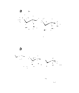
Fig. 1. Lipid A architecture. A, E. coli lipid A consists of a
bisphosphorylated
glucosamine disaccharide substituted with four R-3-hydroxymyristoyl moieties,
of
which the 2' and 3' fatty-acyl chains are esterified with laurate and
myristate,
respectively. B, Regulated modifications of Salmonella lipid A. Substitution
of the
phosphate moieties with L-Ara4N or pEtN is mediated by ArnT and PmrC,
respectively, the formation of a 2-hydroxymyristate-modified lipid A by Lpx0,
the
addition of a secondary palmitoyl chain at the 2-position by PagP, and the
removal of
the 3-hydroxymyristoyl moiety at the 3-position by PagL.
Fig. 2. Multiple sequence alignment of the PagL proteins. Sequences were
aligned
using ClustalW (http://www.ch.embnet.org/software/ClustalW.html). Hyphens
indicate gaps introduced for optimal alignment. Absolutely conserved residues
are
marked with asterisks. Indicated by colons and dots are strongly and weakly
conserved
residues, respectively. The pagL ORF in B. pertussis is disrupted by a frame
shift,
which was restored for this alignment by adding two nucleotides in codon 33.
The
GenBank protein accession numbers for the PagL homologs are: S. Typhimurium
AAL21147, B. bronchiseptica NP_890306, B. parapertussis NP_885487, B.
pertussis
BX470248 , P. aeruginosa NP_253350, P. fluorescens NZ_AAAT03000006 , P.
putida NC_002947 , P. syringae ZP_00125465, B. fungorum NZ_AAAJ03000003 , B.
mallet NC 002970 , B. pseudomallei NC 002930 , R. metallidurans ZP 00274744,
R.
solanacearum NP 522762, and A. vinelandii ZP 00089534. The symbol indicates
GenBank Accession Numbers of whole (unfinished) genomes, in which the PagL
homologs were manually identified.
Fig. 3. Expression and membrane localization of PagL in E. coli BL21 StarTM
(DE3). Membranes from E. coli BL21 StarTM (DE3) containing empty pET-1 la or
the
CA 02590906 2007-06-15
WO 2006/065139 PCT/NL2005/050081
pPagL plasmids were isolated and analyzed by SDS-PAGE. Proteins were stained
with
Coomassie Brilliant Blue. Asterisks indicate the bands that were subjected to
microsequencing and were found to correspond to the mature PagL proteins. The
band
indicated by the double asterisk corresponds to the PagL(3b) precursor
protein.
5 Molecular weight standard proteins are present on the left side.
Fig. 4. Analysis by Tricine-SDS-PAGE of LPS modification in vivo.
Exponentially
growing E. coli BL21 StarTM (DE3) cells containing pET-1 la or the pPagL
constructs
were induced with IPTG for the indicated time, after which 1 0D600 unit
culture
10 samples were collected and analyzed by Tricine-SDS-PAGE.
Fig. 5. GC/MS analysis of wild-type and PagL-modified E. coli BL21 StarTM
(DE3)
LPS. GC/MS analysis of purified E. coli BL21 StarTM (DE3) wild-type LPS (WT),
PagL(s)-modified LPS (L(St)), PagLo3brmodified LPS (L(Bb)), and
PagLwarmodified
15 LPS (L(Pa)) (t= time after induction). Indicated are the normalized
C14/C14-30TI
ratios with wild-type LPS set at 100 (values shown above bars).
Fig. 6. Structural analysis by ESI-MS of wild-type and PagL-modified E. coli
BL21 StarTM (DE3) LPS. Lipid A species from wild-type E. coli BL21 StarTM
(DE3)
containing empty pET-1 la (A), and lipid A species modified by PagL(s) (B),
PagLwo
(C), and PagL(3b) (D) were analyzed by ESI-MS. Major peaks at m/z 1797, 1928,
1622,
and 1490 were interpreted as the characteristic hexa-acylated bis-phosphate
species that
is typically found in E. coli, a hexa-acylated bis-phosphate species
substituted with an
L-Ara4N moiety, a 3-0-deacylated mono-phosphate species substituted with an L-
Ara4N moiety, and a 3-0-deacylated mono-phosphate species, respectively. The
major
peaks at m/z 1716 and 1847 probably represent fragment ions of the species at
m/z 1797
and 1928.
Fig. 7. In vivo re-modification of deacylated LPS and the role of endogenous
PagP.
A, Exponentially growing E. coli BL21 StarTM (DE3) cells containing the empty
pET-
1 la vector or the pPagL(Bb) plasmid were induced with IPTG for the indicated
time
period. Samples corresponding to 1 0D600 unit were collected and analyzed by
Tricine-
SDS-PAGE. B and C, The fatty acid content of purified E. coli BL21 StarTM
(DE3)
CA 02590906 2007-06-15
WO 2006/065139 PCT/NL2005/050081
16
wild-type LPS (WT) and PagLo3brmodified LPS (L(Bb)), isolated at the indicated
time
after induction of pagL expression, was analyzed by GC/MS. Indicated are the
normalised C14/C14-30Tl (B) and C16/C14 (C) ratios with wild-type LPS set at
100
(values shown above bars). D, Exponentially growing wild-type E. coli BL21
StarTM
(DE3) or E. coli BL21 StarTM (DE3) and its pagP mutant derivative JG101,
containing
pPagL(po, were induced with IPTG for the indicated time period, after which 1
0D600
unit culture samples were collected and analyzed on Tricine-SDS-PAGE gel.
Fig. 8. Topology model for PagL from P. aeruginosa. A model for the topology
of
PagL(pa) was constructed using the general rules of outer membrane protein
architecture
as described in (44). The proposed model consists of an eight-stranded 13-
barrel with
four loops (L1-4) extending into the external environment. Residues in the
postulated
I3-strands are shown in diamonds, which are shaded for residues that are
exposed to the
lipid bilayers. His149 and Scrim (marked in red; position in the PagL(pa)
precursor) are
absolutely conserved (Fig. 2) and are suggested to be part of a 'classical'
catalytic triad
of a serine hydrolase. Potential candidates for the acidic residue of the
catalytic triad
are indicated in yellow. Numbers refer to the position of the residues in the
precursor
sequence.
Fig. 9. Identification of PagL(pa) active-site residues by amino acid
substitution.
Exponentially growing E. coli BL21 StarTM (DE3) cells containing the empty pET-
1 la
vector, the pPagLwo plasmid, or the mutant pPagL(po plasmids were induced with
IPTG for 75 min, after which 1 0D600 unit culture samples were collected and
analyzed
by SDS-PAGE followed by immunoblotting with primary antibodies against
PagL(pa)
(A) and by Tricine-SDS-PAGE to visualize LPS (B).
Fig. 10. In vivo modification of B. pertussis LPS. A, LPS from wild-type B.
pertussis
strain Tohama or B. pertussis strain Tohama carrying the pMMB67EH-PagL(3b)
plasmid was isolated and analyzed by Tricine-SDS-PAGE. B, The fatty acid
content of
purified B. pertussis strain Tohama wild-type LPS (WT), and PagL(3b)-modified
LPS
(PagL) was analyzed by GC/MS. Indicated is the normalised C14-30H/C10-301-1
ratio
with wild-type LPS set at 100 (values shown above bars).
CA 02590906 2007-06-15
WO 2006/065139 PCT/NL2005/050081
17
Fig. 11. Biological activity of isolated LPS. IL-6 (A) or IL-10 (B) induction
in MM6
cells by purified LPS. The horizontal axes give the LPS concentration in mg/ml
and the
vertical axes give the ELISA-read out at 450 nm.
Fig. 12. Heat-modifiability of purified, refolded PagL(pa)(-) analysed by semi-
native
SDS-PAGE. Coomassie Brilliant Blue stained semi-native SDS-PAGE gel showing
the heath-modifiability of purified, refolded PagLwo(-). Samples were treated
in sample
buffer containing 0.1% SDS at room temperature (RT) or 2% SDS at 100 C (15
min),
prior to electrophoresis. Molecular weight standard proteins are present on
the left side.
Fig. 13. In vitro LPS modification by membrane-bound or in vitro refolded
PagL.
Silver-stained Tricine-SDS-PAGE gels showing in vitro PagL activity. A,
Purified N
meningitidis L3-LPS was incubated in a detergent-containing buffer for 18 h at
37 C
with or without cell envelopes prepared from E. coli BL21 StarTM (DE3)
containing
empty pET-11a, or the pPagL plasmids. B, Purified N meningitidis L3-LPS was
incubated in a detergent-containing buffer in the absence or presence of 5 mM
EDTA
for 18 h at 37 C with or without 4 mg in vitro refolded PagL(pa) without its
signal
sequence (PagLwo(-)). Similar amounts of assay mixes were loaded in all lanes.
Fig 14. Analysis by Tricine-SDS-PAGE of in vivo LPS modification. LPS was
isolated from wild-type and PagP/PagL-expressing B. pertussis strain Tohama by
hot
phenol/water extraction and analysed by Tricine-SDS-PAGE.
Fig. 15. Structural analysis by ESI-MS of wild-type and PagL/PagP-modified B.
pertussis LPS. Lipid A species from wild-type B. pertussis strain Tohama (A),
and
lipid A species modified by PagL(Bb) (B), PagP(E) (C), and Pag13030 (D) were
analysed
by ESI-MS. Major peaks at m/z 1557, 1477, 1387, 1307, 1251, and 1081 were
interpreted as the characteristic penta-acylated bis-phosphate species that is
typically
found in B. pertussis, the corresponding penta-acylated mono-phosphate
species, the
deacylated lipid A species of the molecular ion at m/z 1557 missing the
primary 3-
hydroxydecanoic acid residue at the 3 position, the deacylated lipid A species
of the
molecular ion at m/z 1477 missing the primary 3-hydroxydecanoic acid residue
at the 3
position, the deacylated lipid A species of the molecular ion at m/z 1477
missing a
CA 02590906 2007-06-15
WO 2006/065139 PCT/NL2005/050081
18
primary 3-hydroxytetradecanoic acid residue, and the deacylated lipid A
species of the
molecular ion at m/z 1477, missing both the primary 3-hydroxydecanoic acid
residue at
the 3 position and a primary 3-hydroxytetradecanoic acid residue,
respectively. The
peaks at m/z 1320, 1490, 1545, 1625, 1715, and 1796 correspond to the PagP-
mediated
palmitoylation of the molecular ions present at m/z 1081, 1251, 1307, 1387,
1477, and
1557, respectively.
Examples
Experimental procedures
Bacterial Strains and Growth Conditions
All bacterial strains used in this study are described in Table I. Typically,
the E.
coli and P. aeruginosa strains were grown at 37 C on modified Luria-Bertani
broth
agar, designated LB agar (26), or in LB broth, while shaking at 200 rpm. For
E. coli,
the medium was supplemented with 0.2% glucose. When appropriate, bacteria were
grown in the presence of 100 ig/m1 ampicillin, 50
kanamycin, 50 ig/m1
naladixic acid, or 100 ig/m1 streptomycin, for plasmid maintenance or strain
selection.
S. Typhimurium SR11 was grown on LB agar plates at 37 C. B. bronchiseptica and
B.
pertussis strains were grown at 35 C on Borduet-Gengou agar (Difco)
supplemented
with 15% defibrinated sheep blood. To induce the expression of the pagL(3b)
gene in B.
pertussis, the bacteria were grown in synthetic Thijs medium (48) supplemented
with 1
mM isopropyl-1-thio-13-D-galactopyranoside (IPTG) (end concentration) at 35 C,
while
shaking (180 rpm).
TABLE 1: Bacterial strains and pla,smids used in this study
Strain or plasmid Genotype or description Source or reference
Strains
B. bronchiseptica
B505 Wild-type strain N.V.I.a
B. pertussis
B509 Dutch vaccine strain N.V.I.a
B134 Dutch vaccine strain N.V.I.a
Tohama Wild-type strain NalR StrepR 36
P. aeruginosa
PA025 PA01 leu arg 45
S. Typhimurium
CA 02590906 2007-06-15
WO 2006/065139 PCT/NL2005/050081
19
SR11 Wild-type strain 46
E. con
TOP1 OF ' Fylaclq Tn10 (TetR)} mcrA A(mrr-hsdRMS-mcrBC) 0801acZAM15
AlacX74
deoR recAl araD139 (ara-leu)7697 galU galK rpsL endAl nupG Invitrogen
DH5 a F A(1acZYA-algF)U169 thi-1 hsdR17 gyrA96 recAl endAl supE44 relAl
phoA 080 dlacZAM15 47
BL21 Stele's" (DE3) F ompT hsdS B (r13- m13) gal dcm rne131 (DE3)
Invitrogen
SK2257 F crcA280::Tn10' thyA6 rpsL120(StrR) deoC1 CGSCb
JG101 BL21 Stele's" (DE3) crcA280: :Tn10' This study
SM10 RP4-2-Tc::Mu recA Km' 50
Plasmids
pCR1I-TOPO E. coli cloning vector AmpR Kan' Invitrogen
pET-11 a E. coli high-copy expression vector, AmpR, T7 promotor
Novagen
pMMI367EH Broad-host-range expression vector, AmpR, tac promotor 51
pMMI367EH Broad-host-range expression vector, AmpR, tac promotor
51
pMMI367-PagL(Bb) pMMB67 derivative harboring B. bronchiseptica pagL This
study
pPagL(po pET-11a derivative harboring P. aeruginosa pagL This
study
pPagLa3b) pET-11a derivative harboring B. bronchiseptica pagL This
study
pPagL(s) pET-11a derivative harboring S. TyphimuriumpagL This study
pPagL(p0(-) pET-11a derivative encoding P. aeruginosa pagL without
signal sequence This study
pPagL(po (H81A) pPagL(po encoding PagL(po with H81A
substitution This study
pPagL(poalsm) pPagL(po encoding PagL(po with H81N
substitution This study
pPagL(po (S84A) pPagL(po encoding PagL(po with S84A
substitution This study
ppagi,a) (S84C) pPagL(po encoding PagL(po with S84C
substitution This study
pPagL(po (H149A) pPagL(po encoding PagL(po with H149A
substitution This study
pPagLa,o(H149N) pPagL(po encoding PagL(po with H149N
substitution This study
pPagLa,o(sisim pPagL(po encoding PagL(po with S151A
substitution This study
pPagLa,o(sism) pPagL(po encoding PagL(po with S151C
substitution This study
aNetherlands Vaccine Institute, Bilthoven, The Netherlands
b E. coli genetic stock center, Yale university, New Haven (CT) 'pagP is
also known as crcA
Recombinant DNA Techniques
Plasmid DNA was isolated using the Promega Wizard P/us SV Minipreps
system. Calf-intestine alkaline phosphatase and restriction endonucleases were
used
according to the instructions of the manufacturer (Fermentas). DNA fragments
were
isolated from agarose gels using the Qiagen quick gel extraction kit.
Ligations were
performed by using the rapid DNA ligation kit (Roche).
The pagL genes from S. Typhimurium SR11 (pagL(s0), B. bronchiseptica B505
(pag1,030, and the pagL gene, with or without its signal sequence-encoding
part, from
P. aeruginosa PA025 (pagLwo, pagL(Pa)(-)) were cloned into pET-11a (Novagen)
behind the T7 promoter. The genes were amplified by PCR using chromosomal DNA
as template. Template DNA was prepared by resuspending ¨109 bacteria in 50 il
CA 02590906 2007-06-15
WO 2006/065139 PCT/NL2005/050081
distilled water, after which the suspension was heated for 15 min at 95 C. The
suspension was then centrifuged for 1 min at 16,100x g, after which the
supernatant
was used as template DNA. The sequences of the forward primers, which
contained an
NdeI site (underlined), including an ATG start codon, were 5'-
5 AACATATGAAGAGAATATTTATATATC-3' (pagL(s0), 5'-
AACATATGAAGAAACTACTTCCGCTGG-3' (pagLwo), 5'-
AACATATGGCGGACGTCTCGGCCGCCG-3' (pagLwo(-)), and 5'-
AACATATGCAATTTCTCAAGAAAAACA-3' (pag1,030. The sequences of the
reverse primers, which contained an BamHI site (underlined) and included a
stop
10 codon, were 5'-AAGGATCCTCAGAAATTATAACTAATT-3' (pagL(s0), 5'-
AAGGATCCCTAGATCGGGATCTTGTAG-3' (pagL(po, Pag4p0(-)), and 5'-
AAGGATCCTCAGAACTGGTACGTATAG-3' (pagL(3b)). The PCRs were done
under the following conditions: 50 tl total reaction volume, 25 pmol of each
primer,
0.2 mM dNTPs, 3 tl template DNA solution, 1.5% dimethylsulfoxide, 1.75 units
of
15 Expand High Fidelity enzyme mix with buffer supplied by the
manufacturer (Roche).
The temperature program was as follows: 95 C for 3 min, a cycle of 1 min at 95
C, 1
min at 60 C, and 1 min 30 s at 72 C repeated 30 times, followed by 10 min at
72 C and
subsequent cooling to 4 C. The PCR products were purified from agarose gel and
subsequently cloned into pCRII-TOPO. Plasmid DNA from correct clones was
digested
20 with NdeI and BamHI, and the PagL-encoding fragments were ligated into
NdeI/BamHI¨digested pET-1 la. The ligation-mixture was used to transform E.
coli
DH5sx using the CaC12 method (27). Plasmid DNA from transformants was checked
for
presence of the correct PagL-encoding insert by digestion with NdeI and BamHI.
Plasmids that gave a correct digestion profile were designated pPagLwo,
pPagL(po(-),
pPagL(3b), and pPagL(s) (Table I). The correct coding sequences of the cloned
pagL
genes were confirmed by nucleotide sequencing in both directions. To subclone
the
pagL(3b) gene into the broad-host-range, low-copy pMMB67EH vector, pPagL(3b)
plasmid DNA was digested with XbaI and HinDIII, and the PagLo3brencoding
fragment was ligated into XbaI/HinDIII-digested pMMB67EH. The ligation mixture
was used to transform E. coli DH5a. Plasmid DNA from transformants was checked
for presence of the correct PagL-encoding insert by digestion with XbaI and
HinDIII. A
plasmid that gave a correct digestion profile was designated pMMB67EH-PagL(3b)
(Table I). The latter plasmid was used to transform transform E. coli SM10,
which
CA 02590906 2007-06-15
WO 2006/065139 PCT/NL2005/050081
21
allowed subsequent transfer of pMMB67EH-PagLo3b) to B. pertussis by
conjugation on
solid medium as described by Stibitz et al (52). Mutations were introduced in
pagL by
using the QuikChange Site-Directed Mutagenesis Kit (Stratagene) and the
primers
listed in Table II. Plasmid pPagivo was used as the template in which the
mutations
were created. The presence of the correct mutations was confirmed by
nucleotide
sequencing in both directions.
TABLE II
Primers used for site-directed mutagenesis
Namea Sequence (5' -3 ')b
H81A FW GAAGGCGCCGGCAAGGCGTCGCTGTCGTTCGCT
H81A REV AGCGAACGACAGCGACGCCTTGCCGGCGCCTTC
H81N FW GAAGGCGCCGGCAAGAACTCGCTGTCGTTCGCT
H8 1N REV AGCGAACGACAGCGAGTTCTTGCCGGCGCCTTC
S84A FW GGCAAGCATTCGCTGGCGTTCGCTCCGGTATTC
S 84A REV GAATACCGGAGCGAACGCCAGCGAATGCTTGCC
584C FW GGCAAGCATTCGCTGTGCTTCGCTCCGGTATTC
S 84C REV GAATACCGGAGCGAAGCACAGCGAATGCTTGCC
H14 9A FW GGCGTTCGGGCGATCGCGTATTCCAACGCCGGC
H14 9A REV GCCGGCGTTGGAATACGCGATCGCCCGAACGCC
H14 9N FW GGCGTTCGGGCGATCAACTATTCCAACGCCGGC
H14 9N REV GCCGGCGTTGGAATAGTTGATCGCCCGAACGCC
S151A FW CGGGCGATCCACTATGCGAACGCCGGCCTGAAA
S151A REV TTTCAGGCCGGCGTTCGCATAGTGGATCGCCCG
S151C FW CGGGCGATCCACTATTGCAACGCCGGCCTGAAA
Si 51C REV TTTCAGGCCGGCGTTGCAATAGTGGATCGCCCG
a The primer name gives the amino acid substitution, e.g. H81A_FW indicates
that the oligonucleotide shown was
used as the forward primer in a site-directed mutagenesis procedure to
substitute the histidine at position 81 of the
precursor FagL(poby an alanine.
b Introduced mutations are underlined.
SDS-PAGE and Immunoblotting
Proteins were analyzed by sodium dodecyl sulfate-polyacrylamide gel
electrophoresis (SDS-PAGE) (28), with 0.2% SDS in the running gel, by using
the Bio-
Rad Mini-PROTEAN 3 apparatus. Samples were applied to a 13% polyacrylamide gel
with a 4% stacking gel and subjected to electrophoresis at 150 V. Proteins
were stained
with Coomassie Brilliant Blue. Prestained or unstained Precision Plus
ProteinThil
CA 02590906 2013-01-16
WO 2006/065139 PCT/NL2005/050081
22
Standard from Bio-Rad was used to determine the relative molecular mass (Mr).
For
Western blotting, proteins were transferred from SDS-PAGE gels onto
nitrocellulose
membranes. The membranes were blocked overnight in phosphate-buffered saline
(PBS) (pH 7.6), 0.5% non-fat dried milk, 0.1% Tweertivii-20 and incubated with
primary
antibodies directed against PagL(po in blocking buffer, followed by an
incubation with
horse-radish peroxidase-conjugated rabbit anti-guinea pig IgG antibodies
(Sigma) in
blocking buffer. Blots were developed using SuperSignal WestPico
Chemiluminescent
Substrate (Pierce).
Semi-Native SDS-PAGE
Proteins were analysed by sodium dodecyl sulfate-polyacrylamide gel
electrophoresis (SDS-PAGE) (28), with 0.2% SDS in the running gel, by using
the Bio-
Rad Mini-PROTEAN 3 apparatus. For semi-native SDS-PAGE, no SDS was added to
the running and stacking gel, and the samples were not heated prior to
electrophoresis.
Samples were applied to a 13% polyacrylamide gel with a 4% stacking gel and
subjected to electrophoresis at 150 V. For semi-native SDS-PAGE,
electrophoresis was
performed at a constant current of 15 inA on ice. Proteins were stained with
Coomassie
Brilliant Blue. Prestained or unstained Precision Plus Protein Standard from
Bio-Rad
was used to determine the relative molecular mass (Mr).
Tricine-SDS-PAGE
To LPS-containing samples 0.5 mg/ml proteinase K (end concentration) was
added to the sample buffer (28). The samples were incubated for 60 min at 55
C,
followed by 10 min at 95 C to inactivate proteinase K. The samples was then
diluted 10
fold by adding sample buffer, after which 2 1.1.1 of the sample were applied
to a Tricine-
SDS-PAGE gel (30). The bromophenol blue was allowed to run into the separating
gel
at 35 V, after which the voltage was increased to 105 V. After the front
reached the
bottom of the gel, the samples were left running for another 45 min. The gels
were
fixed overnight in water/ethanol/acetic acid 11:8:1 (v/v/v) and subsequently
stained
with silver as described (31).
CA 02590906 2007-06-15
WO 2006/065139 PCT/NL2005/050081
23
Polyclonal Antibodies
For antibody production, pPagL0)0(-), was used to transform E. coli BL21
StarTM (DE3) to allow for expression of the truncated pagL gene. The PagLwo
protein,
accumulating in inclusion bodies, was isolated (29), purified from a
preparative SDS-
PAGE gel, and used for immunization of guinea pigs at Eurogentec.
Microsequencing
Proteins were transferred from SDS-PAGE gels to an Immobilonmil-P
polyvinylidene difluoride membrane (Millipore Corp.) in 192 mM glycine, 25 mM
Tris
(pH 8.3), 10% methanol (v/v) at 100 V for 1 h using the Bio-Rad Mini-PROTEAN 2
blotting apparatus. After transfer, the membrane was washed 3 times for 15 min
with
distilled water. Transferred proteins were stained with Coomassie Brilliant
Blue. The
membrane was dried in the air, and the putative PagL bands were excised and
subjected
to microsequencing at the Sequencing Center Facility, Utrecht University, the
Netherlands.
Isolation of LPS and analysis by Gas Chromatography-Mass Spectrometry (GC/MS)
LPS was isolated using the hot phenol/water extraction method (3). In short,
B.
pertussis strain Tohama, with or without plasmid pMMB67EH-PagL(3b), was grown
in
3 liters Thijs medium (48) in the presence of 1 mM IPTG (end concentration).
Cells
were harvested by centrifugation and resuspended in 40 mM sodiumphosphate
buffer
(pH 7.0) containing 5 mM EDTA. The cells were treated over night with lysozyme
at
4 C, after which an equal volume of phenol was added. The suspension was
heated to
70 C and incubated for 30 minutes while shaking. The suspension was cooled to
10 C,
after which phases were separated by centrifugation. The upper phase was
collected
and the extraction was repeated by adding an equal volume of distilled water
to the
lower phase. After subsequent incubation at 70 C, cooling, and centrifugation,
the two
upper phases were mixed and dialysed against tap water until the phenol odour
disappeared. After freeze-drying the dialysed fractions, LPS was dissolved in
phosphate-buffered saline (pH 7.2) at a concentration of 1 mg/ml. For fatty
acid
analysis by GC/MS, a five-fold (v/v) excess of acetone was added to an aliquot
of the
isolated LPS, after which the solution was dried at 60 C under a nitrogen
flow.
Subsequently, 10 mg of C12:0(2011) (1 mg/ml in ethanol) was added as an
internal
CA 02590906 2007-06-15
WO 2006/065139 PCT/NL2005/050081
24
standard, as well as 100 tl of acetylchloride/ethanol 1:9 (v/v), after which
the samples
were derivatized for 1 h at 90 C. After cooling, the reaction was stopped by
adding 200
tl of 1 M K2111304 (pH 8.0), followed by extraction of the acyl-ethyl esters
with 200 tl
ethyl acetate. A 1- 1 volume of the upper phase was used for analysis by GC/MS
on a
Finnigan MAT SSQ in the electron-impact mode.
Biological activity of LPS
IL-6 and IL-10 induction by wild type and PagL-modified B. pertussis Tohama
LPS was tested with the human macrophage cell line MM6 (49). MM6 cells were
seeded in microtiter plates (2.105/well) in 400 tl of IMDM (Gibco BRL)
supplemented
with 10% fetal calf serum (Gibco BRL) and stimulated with 200 tl of serial
dilutions
of the LPS stock solution, for 16-18 h at 37 C in a humid atmosphere
containing 5%
CO2. IL-6 and IL-10 levels in the culture supernatants were quantified with an
ELISA
against human IL-6 or IL-10 according to the instructions of the manufacturer
(PeliPairTM reagent set, Sanquin Reagents, Amsterdam, The Netherlands).
Isolation of Cell Envelopes
Cells were harvested by centrifugation for 10 min at 1,500x g, and washed once
in 50 ml of cold 0.9% sodium chloride solution. The cell pellets were frozen
for at least
15 min at ¨80 C, and then suspended in 20 ml of 3 mM EDTA, 10 mM Tris-HC1 (pH
8.0) containing Complete Protease inhibitor cocktail (Roche). The cells were
disrupted
by sonication, after which unbroken cells were removed by centrifugation for
10 min at
1,500x g. The cell envelopes were pelleted from the supernatant by
centrifugation for
1.5 h at 150,000x g and resuspended in 2 mM Tris-HC1 (pH 7.4). The cell
envelopes
were stored at ¨80 C in aliquots.
Isolation of inclusion bodies
For inclusion body isolation, PagL(pa)(-) was expressed in E. coli BL21 StarTM
(DE3) from pPagL0)0(-) (Table 1). A Two-liter culture was grown at 37 C in LB
medium supplemented with ampicillin till an 0D600 between 0.4 and 0.6. Then, 1
mM
IPTG (end concentration) was added to the culture to induce expression of the
recombinant gene, after which the culture was incubated further at 37 C, while
shaking.
CA 02590906 2013-01-16
=
WO 2006/065139 PCT/N1,2005/050081
After approximately 4 hours, cells were harvested by centrifugation (15 min at
4,000
rpm (4 C)). Harvested cells were washed once in 400 ml 0.9% NaC1 and then
resuspended in 80 ml TE 50:40 (50 mM Tris-HC1 (pH 8.0), 40 mM EDTA). Sucrose
(0.25 g/m1 (end concentration)) and lysozyme (0.2 mg/m1 (end concentration))
were
5 added, after which the suspension was incubated for 30 min at RT, while
shaking. The
suspension was sonicated three times on ice (1.5 min, with 2 min pauses in-
between)
using a Branson 250 Sonfier with macrotip (output 9, duty cycle 50%).
Following
TM
sonication, 0.13% (w/v) Brij-35P (Fluka) was added, and the suspension was
sonicated
for an additional 2 min. Dense material (inclusion bodies) was collected by
10 centrifugation for 2 his at 4,000 rpm (4 C), after which the pellet was
washed once in
40 ml TE 50:40, followed by another washing step using 40 ml 10 mM Tris-HC1
(pH
8.3). The obtained inclusion bodies were solubilized in 8 M urea supplemented
with 10
mM glycine (pH 8.3) and precipitated with TCA. Finally, the obtained proteins
were
solubilized in 8 M urea supplemented with 10 mM glycine (pH 8.3) at a protein
15 concentration of 10 mg/ml. This mixture was centrifugated for 2 his at
13,000 rpm to
remove residual insoluble material and membranes.
Refolding and purification of PagLmak)
Pagl4p0(-) was refolded in vitro by two-fold dilution of the 10 mg/m1 protein
20 solution (see above) in 10% (w/v) lauryldimethylamine oxide (LDAO) and
subsequent
sonication for 10 min. Refolded PagL(pa)(-) was purified by Fast Protein
Liquid
Chromatography (FPLC) using a 1 ml MonoQ (Amersham Biosiences) ion-exchange
column. The protein solution was diluted 4 times in buffer A (20 mM Tris-HC1
(pH
8.0), 0.08 % (w/v) C10E5). The solution was loaded onto the column, which was
pre-
25 equilibrated with buffer A, and washed once with buffer A, and the
proteins were
eluted with a linear gradient of 0-1 M NaC1 in buffer A. Fractions were
analysed by
SDS-PAGE for the presence of the refolded PaglApo(-) protein. Those containing
the
protein were pooled and concentrated to a protein concentration of 10 mg/ml
using
TM
Centricon concentrators with a molecular mass cut-off of 3 kDa (Araicon). The
protein
solution was then dialyzed three times overnight against 10 ml 2 mM Tris-HC1
(pH
8.0), 0.06% (w/v) CI 0E5 using a membrane with a molecular mass cut-off of 3.5
kDa.
CA 02590906 2013-01-16
WO 2006/065139 PCT/NL2005/050081
26
In vitro Modification Assay
Refolded PagL0)0(-) (10 mg/nil) or cell envelopes isolated from E. colt BL21
StarTm (DE3) containing the empty vector pET-1 la or the pPagL plasmids were
diluted
fold in double distilled water. 4 Ill of the diluted refolded protein or cell
envelope
5 solution was incubated in 50 mM Hepes (pH 8.0), 0.1% TritoTX-100, 0.5 M
NaC1, and
0.75 nmol N meningitidis L3-LPS in a final volume of 10 Id at 37 C for 16 h.
To test
whether the reaction was dependent on divalent cations, 5 mM EDTA was added
into
the reaction with the refoled PagLo*(-). The reactions were terminated by
boiling in
sample buffer (28), after which the samples were treated with 0.5 mg/ml
proteinase K
10 for 1 hour at 55 C, followed by 10 min incubation at 95 C. The samples
were diluted
25 fold by adding sample buffer, after which 2 ill of the samples were
analysed by
Tricine-SDS-PAGE (see above).
Isolation of LPS and analysis by Electrospray Ionisation-Mass Spectrometry
(ESI-MS)
LPS was isolated using the hot phenol/water extraction method (Westphal and
Jann, Methods Carbohydr. Chem. 5; 83-91,1965) with slight modifications. In
short,
bacteria were grown in TIHJS medium in the presence of 1 mM IPTG (end
concentration) for 64 h. Cells were harvested by centrifugation and
resuspended in 40
mM sodium phosphate buffer (pH 7.0) containing 5 mM EDTA. The cells were
treated
overnight with lysozyme at 4 C, after which an equal volume of phenol was
added. The
suspension was heated to 70 C, incubated for 30 min while shaking, and
subsequently
cooled to 10 C, after which phases were separated by centrifugation for 10 min
at 8,000
x g. The upper phase was collected and the extraction was repeated after
adding an
equal volume of distilled water to the lower phase. The two upper phases were
combined, dialysed against tap water until the phenol odour disappeared,
freeze-dried,
and subsequently taken up in distilled water. The LPS was subsequently
pelleted by
centrifugation for 3 h at 150,000 x g and dissolved in distilled water, after
which the
LPS concentration was determined by analysing the 3-hydroxytetradecanoic acid
content, using a 6890 Agilent gas chromatograph, as described (Welch, Clin.
Microbiol. Rev. 1991). For ESI-MS, a 200 IA aliquot of isolated LPS (50
nmol/ml) was
freeze-dried and taken up in 0.1 ml 2% acetic acid. The mixture was heated for
2 h at
95 C to hydrolyse the LPS and release the lipid A moiety. Subsequently, the
mixture
was cooled to room temperature and centrifuged for 10 min at 16,100 x g. The
pellet
CA 02590906 2007-06-15
WO 2006/065139 PCT/NL2005/050081
27
was washed twice in 0.1 ml double-distilled water, taken up in 0.1 ml double-
distilled
water, and 0.3 ml chloroform/methanol (2:1, v/v) was added. After vigorous
vortexing,
phases were separated by centrifugation for 10 min at 16,100 x g. The upper
phase
was then used for structural analysis of purified lipid A by nanoelectrospray
tandem
MS on a Finnigan LCQ in the negative ion mode (Wilm and Mann, Anal. Chem.
1996).
Example 1: Identification of PagL Homologs in various Gram-negative Bacteria
The 187-amino acid sequence of the S. Typhimurium PagL precursor protein
(GenBank Accession Number AAL21147, SEQ ID No. 17) was used as a lead to
identify putative PagL homologs in other Gram-negative bacteria, by searching
all
completed and unfinished genomes of Gram-negative bacteria present in the NCBI
database (http://www.ncbi.nlm.nih.gov/sutils/genom_table.cgi). BLAST search
(34)
revealed the presence of putative homologs in the Bordetella spp. B.
pertussis, B.
bronchiseptica, and B. parapertussis (Fig. 2). The PagL homologs of B.
bronchiseptica
and B. parapertussis are two mutually identical 178-amino acid polypeptides
(Fig. 2)
with, as predicted by the signalP server (35), a 25-amino acid N-terminal
signal
peptide. A gene for a PagL homo log was also found in the genome of the B.
pertussis
Tohama I strain (36), but this open reading frame (ORF) was disrupted by a
frame shift
(SEQ ID No. 4), which could be restored as in SEQ ID No. 5 to encode a protein
as in
SEQ ID No. 1. Nucleotide sequencing of the PagL ORFs from B. pertussis strains
B509
and B134 also showed the presence of the same frame shift2, which indicates
that
disruption of the PagL ORF might be a common feature in B. pertussis strains.
By
using the newly identified B. bronchiseptica PagL homolog as a probe for
further
BLAST analysis, additional putative pagL homologs could be identified in the
genomes
of P. aeruginosa (SEQ ID No 6, 30% identity), Pseudomonas fluorescens (SEQ ID
No
7, 29% identity), Pseudomonas syringae (SEQ ID No 8, 31% identity),
Pseudomonas
putida, 2x (SEQ ID No 9 + 10, 32/33%), Ralstonia metallidurans (SEQ ID No 15,
28%), Ralstonia solanacearum (SEQ ID No 16, 29%), Burkholderia mallei (SEQ ID
No 12, 28%), Burkholderia pseudomallei (SEQ ID No 13, 28%), Burkholderia
fungorum (SEQ ID No 11, 29%), and Azotobacter vinelandii (SEQ ID No 14, 27%)
Alignments are shown in Fig. 2. Together, all PagL homologs exhibited a low
overall
mutual sequence identity, albeit higher than with S. typhimurium (24%
identity), but
contained a clear homologous domain near the C terminus. Our finding of this
conserved motif allows identification of PagL homologs in other (bacterial)
species and
CA 02590906 2007-06-15
WO 2006/065139 PCT/NL2005/050081
28
allows the use of a suitable PagL homolog for any host bacterium and/or any
LPS to be
3 -0-deacylated.
Example 2: Cloning of pagL and Heterologous Expression in E. coli
To verify their putative lipid A-deacylase activity, we cloned the pagL
homologs of P. aeruginosa (pagLwo) and B. bronchiseptica (pagL(3b)). We
included in
these studies pagL(S) as a reference. These pagL genes were amplified from the
chromosomes by PCR and eventually cloned in pET-1 la under the control of the
T7
promoter, resulting in plasmids, pPagL(po, pPagL(3b), and pPagL(s).
To investigate expression and membrane localization of PagL in E. coli, E.
coli
BL21 StarTM (DE3) containing the empty vector pET-1 la or the pPagL plasmids
were
grown overnight in LB, after which cell envelopes were isolated. Analysis by
SDS-
PAGE revealed the presence of prominent additional bands with Mrs of 15000-
18000 in
the cell envelopes of the cells expressing PagL (Fig. 3). This was consistent
with the
expected molecular masses of the mature PagL proteins, i.e. PagL(pa) 16.1 kDa,
PagL(Bb) 17.2 kDa, and PagL(S) 18.2 kDa. To identify the additional protein
bands, they
were subjected to microsequencing. The sequences of the first 5 amino acid
residues of
PagLwo, PagL(3b), and PagL(S) were ADVSA, QPTQG, and NDNVF, respectively,
indicating that cleavage of the signal peptide by leader peptidase occurs
between amino
acid residues 23 and 24 (AQA-ADV), 25 and 26 (AQA-QPT), and between 20 and 21
(CSA-NDN), respectively. Particularly in the case of expression of PagL(3b),
an
additional band with a higher Mr was visible on the gel (Fig. 2). The N-
terminal
sequence of this band, MQFLK, corresponded with that of the precursor of
PagL(3b).
Example 3: In vivo Modification of E. coli LPS by PagL
To study whether the cloned PagL homologs were active on E. coli LPS, IPTG
was added to exponentially growing E. coli BL21 StarTM (DE3) cells containing
the
empty vector pET-1 la or the pPagL plasmids, and after various incubation
periods,
samples equivalent to one 0D600 unit were collected and their LPS content was
analyzed by Tricine-SDS-PAGE. In accordance with the expected hydrolysis of
the R-
3-hydroxymyristate at the 3 position of lipid A, expression of any of the
three pagL
homologs converted the LPS into a form with a higher electrophoretic mobility
(Fig. 4).
CA 02590906 2007-06-15
WO 2006/065139 PCT/NL2005/050081
29
The conversion was almost complete within 75 min after PagL(pa) or PagL(Bb)
were
induced, but took somewhat longer in the case of PagL(s)=
Structural Analysis of PagL-Modified LPS: to determine its fatty acid content,
LPS was isolated from bacteria that were grown in the presence of 10 mM MgC12
to
suppress PhoP/PhoQ-regulated modifications of lipid A and analyzed by GC/MS.
The
C14:0/C14:0(30H) ratio in the PagL-modified LPS samples was increased as
compared with that in the wild-type LPS (Fig. 5), consistent with the expected
removal
of a C14-30H from lipid A. To confirm these data, the lipid A moieties were
isolated
and analyzed by ESI-MS in the positive ion mode, which revealed the presence
of four
major lipid A species in wild-type LPS (Fig. 6A). The peak at m/z 1797
represents the
characteristic hexa-acylated bis-phosphate species that is typically found in
E. coli,
whereas the peak at m/z 1928 corresponds to a hexa-acylated bis-phosphate
species
substituted with an L-Ara4N moiety. The two remaining peaks at m/z 1716 and
m/z
1847 most likely represent fragment ions of the two former species missing a
phosphate
group. Upon expression of PagL(S) (Fig. 6B), PagL(pa) (Fig. 6C), or PagL(Bb)
(Fig. 6D),
the major lipid A species were present at m/z 1622 and m/z 1490, which
correspond to
the loss of one I3-hydroxymyristate residue and one phosphate group from the
major
species at m/z 1928 and m/z 1797 present in the empty vector control,
respectively.
Also here, the loss of the phosphate group is probably an artefact of the
ionisation
procedure. Based upon the GC/MS and ESI-MS data, it can be concluded that the
identified PagL homologs of P. aeruginosa and B. bronchiseptica, like that of
S.
Typhimurium, are active lipid A deacylases. Furthermore, the data suggest that
the
deacylation is not dependent upon the absence or presence of an L-Ara4N
moiety, since
both species were deacylated efficiently.
Example 4: Subsequent In Vivo Modification of PagL-deacylated LPS
In the course of these experiments, it was observed that after prolonged PagL
expression, PagL-modified LPS was no longer detectable on Tricine-SDS-PAGE
gels,
and that the LPS migrated again at the position of wild-type LPS, as
illustrated for the
strain expressing PagL(Bb) (Fig. 7A). The PagL protein was still abundantly
present at
this time point, as revealed on SDS-PAGE gels (data not shown). Furthermore,
analysis
by GC/MS revealed that the C14:0/C14:0(301-I) ratio was not decreased again
for the
LPS isolated after 5 h induction of PagL(Bb) (Fig. 7B). Thus, the secondary
modification
CA 02590906 2007-06-15
WO 2006/065139 PCT/NL2005/050081
observed on the Tricine-SDS-PAGE gel (Fig. 7A) was not the consequence of
restoration of the PagL modification, but the result of (an) additional
modification(s)
that restored the electrophoretic mobility to that of wild-type LPS.
Therefore, other
fatty acid ratios were compared. A striking increase in the C16:0/C14:0 ratio
was found
5 in the LPS of cells induced 5 h for PagL production (Fig. 7C), suggesting
that the
PagL-deacylated LPS was subsequently palmitoylated.
A protein that adds palmitate to lipid A is the outer membrane protein PagP
(19)
(Fig. 1). Therefore, we hypothesized that the secondary modification of PagL-
modified
LPS might have been the result of endogenous PagP activity. To investigate
this
10 possibility, we transformed wild-type E. coli BL21 StarTM (DE3) and its
pagP mutant
derivative JG101 with the pPagLwo plasmid. The secondary modification of PagL-
modified LPS was again observed in the case of the wild-type strain, but not
in that of
the mutant strain (Fig. 7D). This result strongly suggests that the secondary
modification of PagL-modified LPS (Fig. 7A) was indeed the consequence of
15 endogenous PagP activity.
Example 5: Identification of PagL Active-Site Residues
The mutual sequence identity between the identified PagL homologs is very low
(Fig. 2). Among the few totally conserved residues are a histidine and a
serine, which,
20 we hypothesize, might be part of a 'classical' Asp/Glu-His-Ser catalytic
triad of serine
hydrolases. These putative active-site residues are located at the lipid-
exposed side near
the top of a 13-strand in a topology model we propose (Fig. 8). Interestingly,
in the outer
membrane phospholipase A, the active-site His and Ser are located in a similar
position
(37). To test whether these residues, located at positions 149 and 151 of the
PagL(pa)
25 precursor protein, respectively, are indeed important for catalytic
activity, they were
substituted by alanine or asparagine, and by alanine or cysteine,
respectively. As a
control, the same substitutions were made for a non-conserved histidine and
serine
residue, located at positions 81 and 84 of the PagL(pa) precursor,
respectively. The
protein and LPS profiles of E. coli BL21 StarTM (DE3) cells carrying the
relevant
30 plasmids and induced for 75 min with IPTG were analyzed by
immunoblotting (Fig.
9A) and Tricine-SDS-PAGE (Fig. 9B), respectively. Whereas substitution of the
non-
conserved His81 and Ser84 did not affect LPS deacylation, deacylation of LPS
was no
longer observed when the conserved His149 and Ser151 were substituted (Fig.
9B),
CA 02590906 2007-06-15
WO 2006/065139 PCT/NL2005/050081
31
even though the expression of these mutant proteins was not affected (Fig.
9A). These
results strongly support the hypothesis that the conserved histidine at
position 149 and
serine at position 151 of the precursor PagL(pa) protein are active-site
residues and that
PagL mechanistically functions as a serine hydrolase.
Example 6: Cloning of pagL(Bb) and Heterologous Expression in B. pertussis
To modify B. pertussis LPS in vivo, we cloned the pagL gene of B.
bronchiseptica (pagL(3b)). The pagL gene was amplified from the chromosome by
PCR and eventually cloned in pMMB67EH under the control of the Toe promoter,
resulting in plasmid, pMMB67EH-PagL(3b), which was transferred to B. pertussis
strain
Tohama by conjugation.
To address the modification of B. pertussis LPS in vivo, wild-type B.
pertussis
strain Tohama, or B. pertussis strain Tohama containing the pMMB67EH-PagL(3b)
plasmid were grown in Thijs medium supplemented with 1 mM IPTG (end
concentration). LPS was isolated by the hot phenol-water extraction method and
analysed by Tricine-SDS-PAGE (Fig. 10A) and GC-MS (Fig. 10B). Analysis on
Tricine-SDS-PAGE gel showed that the LPS isolated from the PagL(Bb) expressing
strain migrated slightly faster as compared to the wild type B. pertussis LPS.
Strikingly, the LPS isolated from the PagL(Bb) expressing strain showed two
distinct
LPS populations. One population migrating around the height of the wild type
Tohama
LPS, and one population migrating faster. This latter LPS population can also
be seen
in the wild type LPS preparation, however the abundance of it is much lower.
To verify
that the LPS from the PagL expressing strain was indeed deacylated at its 3
position, it
was analysed by GC-MS. The C14:0(30H)/C10:0(30H) ratio in the PagL-modified
LPS sample was increased as compared with that in the wild-type LPS (Fig.
10B),
consistent with the expected removal of a CIO-30H from the 3 position of B.
pertussis
lipid A.
Example 7: Biological activity of PagL-modified LPS
To assess the endotoxic activity of the PagL-modified and wild-type B.
pertussis
LPS, their ability to stimulate the production of IL-6 and IL-10 in the human
macrophage cell line MM6 was measured. As can be seen in figure 2, for wild-
type
LPS, the production of both IL-6 (Fig. 11A) and IL-10 (Fig. 11B) by the MM6
cells is
increased as compared to when the cells were stimulated with an equal amount
of
CA 02590906 2007-06-15
WO 2006/065139 PCT/NL2005/050081
32
PagL-modified LPS. Thus, it can be concluded that the in vivo deacylation of
B.
pertussis LPS by PagL results in a reduction in endotoxic activity of this
LPS.
Example 8: Cloning, Expression, Purification, and Refolding ofPagL(pa)
The pagL gene from P. aeruginosa PA025 without its signal sequence-
encoding part was cloned into pET-1 la, resulting in plasmid pPagL0)0(-). To
obtain
inclusion bodies, PagL without its signal sequence was expressed in E. coli
BL21
StarTM (DE3). Inclusion bodies were isolated and solubilized in urea, after
which the
protein was refolded by diluting two-fold in 10% lauryldimethylamine oxide
(LDAO)
and further purified by Fast Protein Liquid Chromatography (FPLC). Correct
refolding
was confirmed by SDS-PAGE (Fig. 12) and circular dichroism (CD) measurements
(data not shown). On SDS-PAGE gel, the refolded protein had a lower
electrophoretic
mobility as compared to the denatured form, whereas CD measurements showed
that
the refolded protein predominantly had a 13-sheet conformation.
Example 9: In vitro LPS Modification by Membrane-Localized and Refolded PagL
To test whether membrane-localized or in vitro refolded PagL was capable of
modifying externally added LPS in vitro, we incubated refolded PagL0)0(-), or
isolated
cell envelopes from E. coli BL21 StarTM (DE3) containing the empty vector pET-
11 a, or
the pPagL plasmids, together with purified LPS of N meningitidis. Modification
of
LPS was assessed by Tricine-SDS-PAGE (Fig. 13). In accordance with the
expected
hydrolysis of the R-3-hydroxymyristate at the 3 position of lipid A, LPS was
converted
into a form with a higher electrophoretic mobility when membrane-localized
PagL (Fig
13A) or refolded Pagl4p0(-) (Fig 13B) was present. The reaction with the
refolded
Pagl4p0(-) was independent on the presence of divalent cations, as deacylation
of LPS
was still observed in the presence of 5 mM EDTA (Fig. 13B).
Example 10: Altered lipid A structure after expression of PagP and PagL in
B.pertussis
To express PagP and PagL in B. pertussis strain Tohama, the pagL gene of B.
bronchiseptica (pagL(3b)) and the pagP gene of B. pertussis (pagP030) were
expressed
from the broad-host range low-copy number expression vector pMMB67EH. As a
control, a strain expressing the pagP gene of E. coli (pagP(E)) was also
constructed.
LPS was isolated from wild-type, PagP-expressing, or PagL-expressing B.
pertussis
CA 02590906 2007-06-15
WO 2006/065139 PCT/NL2005/050081
33
strain Tohama and analysed by Tricine-SDS-PAGE. LPS isolated from the PagL(3b)-
expressing strain appeared unaffected on the gel, whereas that from the PagP-
expressing appeared potentially modified, since a band with a lower
electrophoretic
mobility than that of wild-type B. pertussis LPS was detected (Fig. 14).
Furthermore, as
compared to the Pag13030-expressing strain, the modification-efficiency
appeared higher
in the PagP(Ec)-expressing strain (Fig. 14). To evaluate the possible LPS
modifications
in further detail, the lipid A moieties of the strains were analysed by ESI-MS
in the
negative-ion mode. This analysis revealed the presence of four major lipid A
species in
wild-type LPS (Fig. 15A). The peak at m/z 1557 represents the characteristic
penta-
acylated bis-phosphate species that is typically found in B. pertussis (Caroff
et al.,
Microbes. Infect., 1994), whereas the peak at m/z 1477 corresponds to a penta-
acylated
mono-phosphate species. The two remaining peaks at m/z 1307 and 1251 represent
deacylated lipid A species of the molecular ion at m/z 1477, which miss the
primary 3-
hydroxydecanoic acid residue at the 3 position or a primary 3-
hydroxytetradecanoic
acid residue (either at the 2 or the 3' position), respectively. These results
indicate a
high heterogeneity among the lipid A species in wild-type B. pertussis, which
was
apparently not resolved in the gel analysis (Fig. 14). Interestingly,
calculation of the
relative amounts of the individual lipid A species from the corresponding peak
heights
revealed that in wild-type B. pertussis LPS, a large quantity of lipid A
species (-50%)
consists of tetra-acylated forms. Furthermore, the large majority of lipid A
species are
mono-phosphate forms (-80%). To exclude the possibility that the high
abundancy of
under-acylated and hypo-phosphorylated lipid A species was an artefact of the
hydrolysation procedure used to isolate lipid A, we tested whether shorter or
longer
periods of hydrolysation (varying between 1 and 4 h) influenced the relative
abundance
of the lipid A species, which was, however, not the case (data not shown).
Furthermore,
the total phosphate content of a solution with a known concentration of
purified wild-
type B. pertussis LPS was determined. Consistent with the high prevalence of
mono-
phosphate lipid A species detected by ESI-MS, only slightly more than half of
the
phosphate content expected, when LPS would have been fully phosphorylated, was
detected (data not shown).
Upon expression of PagL(3b) (Fig. 15B), three lipid A species, at m/z 1081,
1307, and 1387, respectively, were present. The major peak at m/z 1307
corresponds to
the mono-phosphate deacylated form missing the 3-hydroxydecanoic acid residue
at the
CA 02590906 2007-06-15
WO 2006/065139 PCT/NL2005/050081
34
3 position, whereas the peak at m/z 1387 corresponds to the bis-phosphorylated
form of
the molecular ion at m/z 1307. The peak at m/z 1081 corresponds to a mono-
phosphate
form missing both a 3-hydroxydecanoic and a 3-hydroxytetradecanoic acid
residue.
The relative content of lipid A species that miss the 3-hydroxydecanoic acid
residue at
their 3 position was increased from about 37 percent in wild-type B. pertussis
LPS to
more than 92 percent in the strain expressing PagL(Bb). Thus, even though the
electrophoretic mobility of the LPS was not affected (Fig. 14, lane 2), the
pagLmbr
encoded lipid A 3-0-deacylase was active in B. pertussis.
Upon expression of PagP(E) (Fig. 15C) and PagP(Bp) (Fig. 15D), several new
lipid A species were detected (Table III). The peaks at m/z 1320, 1490, 1545,
1625,
1715, and 1796 correspond to the expected PagP-mediated palmitoylation of the
molecular ions present at m/z 1081, 1251, 1307, 1387, 1477, and 1557,
respectively.
The difference in modification-efficiency between E. coli and B. pertussis
PagP, which
was seen after analysis by Tricine-SDS-PAGE (Fig. 14), was also revealed in
the mass
spectrometrical analysis. In the strain expressing E. coli PagP, ¨47% of the
total lipid A
population was palmitoylated, in contrast to only ¨9% in the strain expressing
PagP(Bp).
Interestingly, in the strain expressing PagP(Bp), in contrast to that
expressing PagP(E),
lipid A species missing a 3-hydroxytetradecanoic acid residue were not found
to be
palmitoylated. A possible explanation for this discrepancy is the difference
in
specificity of the two PagP enzymes. Whereas E. coli PagP adds an acyl chain
at the 2
position of lipid A, B. pertussis PagP adds a palmitate at the 3' position
(Bishop et al.,
EMBO J., 2000; Preston et al., Mol. Microbiol., 2003). Thus, the complete
absence of
palmitoylated lipid A species that miss one 3-hydroxytetradecanoic acid
residue in the
strain expressing B. pertussis PagP suggests that the lipid A molecules
missing a 3-
hydroxytetradecanoic acid residue miss it specifically at their 3' position.
This could
then partially explain the difference in modification efficiency that was
observed
between the two PagP enzymes, as the substrate pool for E. coli PagP would be
larger
than that for B. pertussis PagP. Furthermore, this hypothesis is consistent
with the
presence of hypo-acylated lipid A species in vivo.
35
0
t..)
=
Table III: Relative abundance of lipid A molecular ions as determined by ESI-
MS =
cA
-E:-5
cA
u,
1081 1251 1307 1320 1331 1387 1477 1490
1545 1557 1625 1715 1796 palmitoylatei`z
-C14-30H -C14-30H -C10-30H -C14-30H -C14-30H -C10-30H -PO4 -C14-30H -C10-30H
-C10-30H -PO4 +C16
-C10-30H -PO4 -PO4 -C10-30H -PO4
-PO4 +C16 +C16
-PO4 -PO4 +C16
+C16
+C16
...............................................................................
.................................................. 0
0
3.0 15.6 29.9 0.0 4.5 3.9 29.0 0.0
0.0 14.1 0.0 0.0 0.0 0.0 iv
in
q3.
0
l0
Wild-type
0
c7,
tv
0
0
PagL(eb) 8.5 2.1 70.9 0.0 0.0 12.8 3.5 0.0
0.0 2.1 0.0 0.0 0.0 0.0
1
0
01
I
H
PagP(E0 4.5 2.3 25.0 5.0 2.3 10.3 2.0 3.0
14.8 6.0 6.3 5.3 12.3 46.5 in
PagP(13) 8.3 5.0 27.6 0.0 6.1 21.3 3.0 0.0
3.9 17.1 2.5 0.0 2.9 9.1
00
n
,-i
z
r
t..)
=
=
u,
-E:-5
u,
=
=
oe
CA 02590906 2007-06-15
WO 2006/065139 PCT/NL2005/050081
36
References
1. Raetz, C. R. II., and Whitfield, C. (2002) Annu. Rev. Biochem. 71, 635-
700
2. Loppnow, II., Brade, II., Durrbaum, I., Dinarello, C. A., Kusumoto, S.,
Rietschel, E. T., and Flad, II. D. (1989) J. ImmunoL 142, 3229-3238
3. Steeghs, L., Berns, M., ten Hove, J., de Jong, A., Roholl, P., van
Alphen, L.,
Tommassen, J., and van der Ley, P. (2002) Cell. MicrobioL 4, 599-611
4. Nikaido, II., and Vaara, M. (1987) in Escherichia coli and Salmonella:
Cellular
and Molecular Biology (Neidhardt, F. C., ed) Vol. 1, pp. 7-22, American
Society for Microbiology, Washington, D. C.
5. Caroff, M., Karibian, D., Cavaillon, J-M., and Haeffner-Cavaillon, N.
(2002)
Microbes Infect. 4, 915-926
6. Guo, L., Lim, K. B., Gunn, J. S., Bainbridge, B., Darveau, R. P.,
Hackett, M.,
and Miller, S. I. (1997) Science 276, 250-253
7. Guo, L., Lim, K. B., Poduje, C. M., Daniel, M., Gunn, J. S., Hackett,
M., and
Miller, S. I. (1998) Cell 95, 189-198
8. Gunn, J. S., Belden, W. J., Miller, S. I. (1998) Microb. Pathog. 25, 77-
90
9. Gunn, J. S., Lim, K. B., Krueger, J., Kim, K., Guo, L., Hackett, M., and
Miller,
S. I. (1998) MoL MicrobioL 27, 1171-1182
10. Gunn, J. S., Ryan, S. S., Van Velkinburgh, J. C., Ernst, R. K., and
Miller, S. I.
(2000) Infect. Immun. 68, 6139-6146
11. Miller, S. I., Kukral, A. M., and Mekalanos, J. J. (1989) Proc. Natl.
Acad. Sci.
U.S.A. 86, 5054-5058
12. Gunn, J. S., and Miller, S. I. (1996) J. BacterioL 178, 6857-6864
13. Ernst, R. K., Guina, T., and Miller, S. I. (1999) J. Infect. Dis. 179,
Suppl. 2,
326-330
14. Ernst, R. K., Yi, E. C., Guo, L., Lim, K. B., Burns, J. L., Hackett,
M., and
Miller, S. I. (1999) Science 286, 1561-1565
15. Trent, M. S., Ribeiro, A. A., Lin, S., Cotter, R. J., and Raetz, C. R.
H. (2001) J.
Biol. Chem. 276, 43122-43131
16. Lee, H., Hsu, F. F., Turk, J., and Groisman, E. A. (2004) J. BacterioL
186,
4124-4133
CA 02590906 2007-06-15
WO 2006/065139 PCT/NL2005/050081
37
17. Gibbons, II. S., Lin, S., Cotter, R. J., and Raetz C. R. II. (2000) J.
Biol. Chem.
275, 32940-32949
18. Karbarz, M. J., Kalb, S. R., Cotter, R. J., and Raetz, C. R. II. (2003)
J. Biol.
Chem. 278, 39269-39279
19. Bishop, R. E., Gibbons, II. S., Guina, T., Trent, M. S., Miller, S. I.,
and Raetz,
C. R. II. (2000) EMBO J. 19, 5071-5080
20. Tanamoto, K., and Azumi, S. (2000) J. ImmunoL 164, 3149-3156
21. Robey, M., O'Connell, W., and Cianciotto, N. P. (2001) Infect. Immun.
69,
4276-4286
22. Trent, M. S., Pabich, W., Raetz, C. R. II., and Miller, S. I. (2001) J.
Biol. Chem.
276, 9083-9092
23. Bhat, U. R., Forsberg, L. S., and Carlson, R. W. (1994) J. Biol. Chem.
269,
14402-14410
24. Moran, A. P., Lindner, B., and Walsh, E. J. (1997) J. BacterioL 179,
6453-6463
25. Kumada, II., Haishima, Y., Umemoto, T., and Tanamoto, K. (1995) J.
BacterioL 177, 2098-2106
26. Tommassen, J., van Tol, II., and Lugtenberg, B. (1983) EMBO J. 2, 1275-
1279
27. Sambrook, J., Fritsch, E. F., and Maniatis, T. (1989) in Molecular
Cloning: A
Laboratory Manual. Cold Spring Harbour Laboratory Press, Cold Spring
Harbour (NY)
28. Laemmli, U. K. (1970) Nature 227, 680-685
29. Dekker, N., Merck, K., Tommassen, J., and Verheij, H. M. (1995) Eur.
Biochem. 232, 214-219
30. Lesse, A. J., Campagnari, A. A., Bittner, W. E., and Apicella, M. A.
(1990) J.
ImmunoL Methods 126, 109-117
31. Tsai, C. M., and Frasch, C. E. (1982) Anal. Biochem. 119, 115-119
32. Westphal, 0., and Jann, J.K. (1965) Methods Carbohydr. Chem. 5, 83-91
33. Wilm, M., and Mann, M. (1996) Anal. Chem. 68, 1-8
34. Altschul, S. F., Gish, W., Miller, W., Myers, E. W., and Lipman, D. J.
(1990) J.
MoL BioL 215,403-410
35. Nielsen, H., Brunak, S., and von Heijne, G. (1999) Protein Eng. 12, 3-9
36. Parkhill, J., Sebaihia, M., Preston, A., Murphy, L. D., Thomson, N.,
Harris, D.
E., Holden, M. T., Churcher, C. M., Bentley, S. D., Mungall, K. L., Cerdeno-
CA 02590906 2007-06-15
WO 2006/065139 PCT/NL2005/050081
38
Tarraga, A. M., Temple, L., James, K., Harris, B., Quail, M. A., Achtman, M.,
Atkin, R., Baker, S., Basham, D., Bason, N., Cherevach, I., Chillingworth, T.,
Collins, M., Cronin, A., Davis, P., Doggett, J., Feltwell, T., Goble, A.,
Hamlin,
N., Hauser, H., Holroyd, S., Jagels, K., Leather, S., Moule, S., Norberczak,
H.,
O'Neil, S., Ormond, D., Price, C., Rabbinowitsch, E., Rutter, S., Sanders, M.,
Saunders, D., Seeger, K., Sharp, S., Simmonds, M., Skelton, J., Squares, R.,
Squares, S., Stevens, K., Unwin, L., Whitehead, S., Barre11, B. G., and
Maskell,
D. J. (2003) Nat. Genet. 35, 32-40
37. Snijder, H. J., Ubarretxena-Belandia, I., Blaauw, M., Kalk, K. H.,
Verheij, H.
M., Egmond, M. R., Dekker, N., and Dijkstra, B. W. (1999) Nature 401, 717-
721
38. McClelland, M., Sanderson, K. E., Spieth, J., Clifton, S. W.,
Latreille, P.,
Courtney, L., Porwollik, S., Ali, J., Dante, M., Du, F., Hou, S., Layman, D.,
Leonard, S., Nguyen, C., Scott, K., Holmes, A., Grewal, N., Mulvaney, E.,
Ryan, E., Sun, H., Florea, L., Miller, W., Stoneking, T., Nhan, M., Waterston,
R., and Wilson, R. K. (2001) Nature 413, 852-856
39. Basu, S. S., White, K. A., Que, N. L., and Raetz, C. R. H. (1999) J.
Biol. Chem.
274, 11150-11158
40. Kulshin, V. A., Zahringer, U., Lindner, B., Jager, K. E., Dmitriev, B.
A., and
Rietschel, E. T. (1991) Eur. J. Biochem. 198, 697-704
41. Hwang, P. M., Choy, W. Y., Lo, E. I., Chen, L., Forman-Kay, J. D.,
Raetz, C.
R., Prive, G. G., Bishop, R. E., and Kay, L. E. (2002) Proc. Natl. Acad. Sci.
USA. 99, 13560-13565
42. Kol, M. A., van Dalen, A., de Kroon, A. I., and de Kruijff, B. (2003)
J. Biol.
Chem. 278, 24586-24593
43. von Heijne, G. (1983) Eur. J. Biochem. 133, 17-21
44. Tommassen, J. (1988) in Membrane Biogenesis (Op den Kamp, J.A.F., ed)
NATO ASI series, Vol. H16, pp. 351-373. Springer Verlag, Berlin, Heidelberg,
New York.
45. Haas, D., and Holloway, B.W. (1976) Mol. Gen. Genet. 144, 243-251
46. Pace, J., Hayman, M. J., and Galan, J. E. (1993) Cell 72, 505-514
47. Hanahan, D. (1983) J. Mol. Biol. 166, 557-580
48. Thalen, M., van den Ussel, J., Jiskoot, W., Zomer, B., Roholl, P., de
Gooijer,
CA 02590906 2007-06-15
WO 2006/065139 PCT/NL2005/050081
39
C., Beuvery, C., and Trampen, J. (1999) J.Biotechnol. 75, 147-159.
49. Ziegler-Heitbrock, II. W., Thiel, E., Futterer, A., Herzog, V., Wirtz,
A., and
Riethmuller, G. (1988) J. Cancer 41, 456-461
50. Simon, R., Priefer, U., and Puhler, A. A. (1983) Bio/Technol. 1, 784-
791
51. Furste, J. P., Pansegrau, W., Frank, R., Blocker, II., Scholz, P.,
Bagdasarian,
M., and Lanka, E. (1986) Gene 48, 119-131
52. Stibitz, S., Black, W., and Falkow, S. (1986) Gene 50, 133-140