Note: Descriptions are shown in the official language in which they were submitted.
CA 02591303 2007-06-07
WO 2006/061565 PCT/GB2005/004529
- 1 -
RAMAN SPECTRAL ANALYSIS OF SUB-SURFACE TISSUES
AND FLUIDS
Field of the invention
The present invention relates to methods and
apparatus for determining characteristics of in-vivo
sub-surface tissues by detecting Raman features of
diffusely scattered light. For example, the described
methods and apparatus are suitable for detecting Raman
spectral features of in-vivo bone through skin, nail
and other surface tissues, without requiring exposure
of the bone tissue by incision or puncture.
Discussion of the Prior Art
The field of investigative studies of bone and
other biological and living tissues encompasses a
myriad of analytical techniques. Such techniques have
been developed in response to the many situations in
which it has been found to be important to assess the
bone quality or tissue composition of a particular
patient, human or animal. For instance, a person with
osteoporosis has a significantly increased risk of bone
fracture in comparison to the normal population.
Diagnosis of degenerative skeletal diseases, for
example osteoporosis, is important in allowing a
sufferer to adapt their lifestyle or to seek
intervention to mitigate the significant risk of
fracture. A further example is found in
musculoskeletal tissue research. An important aspect
of this field is comparison of tissue composition and
CA 02591303 2007-06-07
WO 2006/061565 PCT/GB2005/004529
- 2 -
molecular structure with function using carefully
selected populations of normal and, in many cases,
transgenic animals.
To date however there are few non-invasive or
minimally-invasive methods for examining details of
bone or composition of other tissues. In studies of
animal models of genetic or metabolic diseases, the
standard procedure'is to sacrifice animals and harvest
tissue specimens for study. It would clearly be
preferable to study living animals and with methods
that cause minimal discomfort or harm. This need is
most felt in studies that follow tissue changes over an
extended period.
Sacrificial procedures are clearly not an option
in examining human patients. The currently available
methods of assessing bone quality are based primarily
on radiography and, in particular, dual energy X-ray
absorptiometry (DEXA). This technique however is only
able to measure the inorganic phase of bone
(hydroxyapatite) and the organic phase (primarily
collagen I) is largely invisible. It is known that the
material strength of bone is dependent on both the
collagen and hydroxyapatite compositions. A crucial
piece of data needed to assess bone quality is
therefore neglected by the DEXA technique. To date,
the only known procedures for obtaining organic
(collagen) data involve analysis either of physically
exposed bone or of a sample removed by biopsy. Both of
these can often cause discomfort or pain to the
patient.
CA 02591303 2007-06-07
WO 2006/061565 PCT/GB2005/004529
- 3 -
Infrared and Raman spectroscopies provide a wealth
of information on the physio-chemical state of a wide
range of tissues and fluids (see, for example
US 6,681,133 or WO 01/52739). In bone analysis, for
example, these techniques can provide information on
mineral / matrix ratio, mineral crystallinity, matrix
cross-linking and reversible and irreversible changes
caused by mechanical loading. Unfortunately analysis
by these methods has been limited to surface studies
i.e. on exposed bone. Infrared radiation does not
generally penetrate more than a few um into tissue
before being completely absorbed by water. The near-
infrared range (700 - 850 nm) penetrates further, but
multiple scattering causes loss of detailed spatial
information. Confocal microscopy, a standard technique
used to probe sample depths, is precluded on living
tissue as a tight focal spot causes local heating and
tissue damage. Moreover this technique is
substantially less effective in diffusely scattering
media, such as biological tissue, in which it is only
practicable to depths of around ten times the mean free
path of photons in the medium.
Elastically scattered photons have been used to
probe beneath a scattering surface for compositional
information. For example,B.B.Das, et al. in Rep.
Prog. Phys. 60, 227 (1997) describes an approach using
temporal gating. This technique relies on the fact
that it takes a finite time for light to penetrate a
diffusely scattering medium. Scattering events will
therefore occur later at lower depths, and so
CA 02591303 2007-06-07
WO 2006/061565 PCT/GB2005/004529
- 4 -
monitoring a scattered signal over ti-me should,
theoretically, provide information as to the nature of
the scattering centres at progressively greater depths.
The wide application of Raman spectroscopy to the
extraction of information that can be critical to
medical diagnosis has driven the search for a system
that is capable of measuring sub-surface Raman
scattering. Such a system should theoretically be
capable of compositional analysis of bone and cartilage
beneath the skin; of intravertebral disc tissue within
a cartilage sack; of tendon and ligaments with
differing material and functional properties; of gut
wall or oesophageal tissue, which again are protected
by a membranous coating in vivo. The elastic
scattering technique of Das et al. is however not
directly extendable to Raman spectroscopy. Inelastic
scattering of photons is a much weaker process due to
far smaller cross-section for generating Raman light.
This results in a much weaker signal. Furthermore the
Raman signal is far more susceptible to interference
from luminescence or sample damage.
One approach that has been used to obtain depth
information from Raman scattering is described in
"Three dimensional imaging of objects embedded in
turbid media with fluorescence and Raman spectroscopy"
by Jun Wu, et al., Appl. Optics 34(18), 3425 (1995).
This paper describes a technique that exploits the fast
rise-time of fluorescent decay and Raman scattering to
infer depth information from the time delay between
surface illumination and earliest detection of a
CA 02591303 2007-06-07
WO 2006/061565 PCT/GB2005/004529
- 5 -
scattered photon. A single-photon detection system is
set to monitor back-scattered photons from a sample
surface from the time it is illuminated by a 1 MHz
pulsed laser beam. The spread in photon arrival times
over a number of pulsed illuminations is shown to have
an onset time delay that is characteristic of the depth
of the scattering object.
The benefits that could be derived using non-
invasive Raman probing of bones, where signal quality
from bone can be crucial in arriving at an accurate and
correct diagnosis as to whether disease is present, are
apparent from A. Carden and M.D. Morris, J. Biomed.
Optics 5, 259 (2000). However, conventional Raman
signatures of bone collagen are masked by undesired
Raman signals from overlaying tissue and so data on
chemical composition is generally obtained by means of
a biopsy.
There is a perceived need for an alternative non-
invasive or minimally-invasive method of performing
sub-surface Raman spectroscopy. Such a method should
be capable of providing the basis for a more flexible
in vivo analytical technique that overcomes the
limitations of DEXA. In particular, DEXA is restricted
to obtaining partial information relating to bone
composition, whereas the more wide-ranging
applicability of a Raman-based technique is desired.
Summary of the Invention
It is an object of this invention, therefore, to
provide an analysis technique based on Raman
spectroscopy, that is capable of extracting sub-surface
CA 02591303 2007-06-07
WO 2006/061565 PCT/GB2005/004529
- 6 -
chemical compositional information of in-vivo and
living tissues such as bone tissue. It is also an
object of the invention to enable such analysis without
requiring any incision, puncture or other surgical
intervention to expose the target sub-surface tissue.
Accordingly, the invention provides a method of
carrying out a sub-cutaneous inspection of sub-surface
tissue or fluid, comprising: irradiating a surface
tissue with a probe light beam; collecting light
scattered beneath the surface, from one or more
collection regions or locations on the surface, the
collection regions being spaced from the probe beam;
and detecting one or more Raman spectral features from
the collected light. Biomedical or chemical
characteristics may then be derived from the Raman
spectral features.
More generally, the invention provides a method of
determining one or more characteristics of a sub-
surface tissue or fluid, through a diffusely scattering
overlying tissue, comprising: supplying incident light
at an entry region on a surface of the overlying
tissue; collecting light scattered within the overlying
tissue, from a collection region on the surface, the
collection region being spaced from the entry region;
and detecting, in the collected light, one or more
Raman features, spectrally related to the incident
light, which originate from the sub-surface tissue or
f luid .
The overlying tissue may be skin or nail, and the
sub-surface tissue or fluid may be bone, cartilage,
breast tissue or blood, but there are many other
applications, some of which may require surgical
CA 02591303 2007-06-07
WO 2006/061565 PCT/GB2005/004529
- 7 -
intervention to expose the overlying layer such as
membranes or mucus covering and protecting an
underlying layer, layers or organ to be studied. The
tissue to be studied may be fluid in form, such as
blood, lymph or fluid within a joint, an eye or between
membranes.
Single entry and collection regions are sufficient
in many cases to d_erive useable Raman spectral data. In
other cases one or more entry and one or more
collection regions of various physical spacings may be
used, and the spectral data so obtained combined to
yield a more accurate determination of the required
characteristics of the sub-surface tissue, for example
by using the data from multiple spacings to
preferentially select the Raman signal of the sub-
surface tissue.
A single or multiple collection regions
surrounding or distributed about a central entry region
are advantageous since this provides an increased
collection area over a simple displaced collection
region. Alternatively, a single or multiple entry
regions surrounding or distributed about a single
collection region may be used. Concentric annular or
other shaped entry and collection regions may not be
fully utilised. For example, a ring of closely packed
terminating optical fibres may fill about 60% of the
associated annulus. Preferably, at least 10% of the
collection or entry annulus is optically utilised.
Preferably, associated entry and collection
regions do not overlap.
The invention also provides associated methods of
diagnosis of human or animal medical conditions, by
CA 02591303 2007-06-07
WO 2006/061565 PCT/GB2005/004529
- 8 -
interpretation of the determined characteristics of
sub-surface tissue.
The invention provides corresponding apparatus for
determining, one or more characteristics of a sub-
surface tissue.
The present invention also provides a method of
measuring, a sub-surface Raman spectrum of a diffusely-
scattering tissue,.the method comprising the steps of:
a) irradiating the tissue with a light probe;
b) collecting light scattered by the tissue; and
c) spectrally separating at least a portion of the
collected light to detect one or more Raman spectral
features,
wherein light scattered by the sample is collected from
a plurality of spatial locations on the surface of the
sample, each spatial location being at a different
distance from the point of irradiation, at least a
portion of the light collected at each spatial location
being separately spectrally dispersed to form a
plurality of Raman spectra and wherein the method
further includes the step of:
d) analysing the plurality of Raman spectra to
extract information on the Raman spectrum of a sub-
surface region of the tissue.
Thus, with this method spectroscopic information
is obtained non-destructively that can be interpreted
to establish the nature and composition of a diffusely
scattering tissue below a surface layer. The
present invention effectively implements a form of
spatial gating of the Raman signal obtained from the
sample to isolate the Raman signal from a sub-surface
CA 02591303 2007-06-07
WO 2006/061565 PCT/GB2005/004529
- 9 -
layer which has a different composition to that of the
surface layer. This method is referred to herein as
Spatially Offset Raman Spectroscopy (SORS).
With the present invention for tissues having one
or more different chemical compositions at differing
depths within the sample, the collection of Raman
spectra from regions spatially offset, by different
amounts, from the point of incidence of the probe laser
beam results in a series of spectra (two or more
spectra) each spectra including Raman signals emanating
from different depths within the tissue. The series of
spectra taken contain different relative contributions
of the Raman signals generated from the tissue surface
layer and the tissue sub-surface layers. In collecting
the data series, as the signal collection point is
moved away from the point of incidence of the probe
laser beam, the contribution of the surface layer
signal diminishes much faster than for signals
generated by different compositions at deeper layers
within the bulk of the tissue. This enables the
contribution of deeper, sub-surface tissues to be
extracted either directly or by applying numerical
processing to the collected spectral set for a higher
degree of separation (e.g. multivariate data analysis
or scaled subtraction of spectra from each other).
In a preferred embodiment two or more Raman
spectra are collected and are analysed using a scaled
subtraction, the Raman spectrum collected from or at a
distance closest to the point of irradiation being
CA 02591303 2007-06-07
WO 2006/061565 PCT/GB2005/004529
- 10 -
subtracted from the Raman spectrum collected further
from the point of irradiation, whereby features of the
Raman spectrum for a sub-layer of the tissue are
identified.
In a further alternative, where the Raman spectrum
for the chemical composition of the surface of the
tissue is known, the collected Raman spectra are
analysed by scaled_subtraction of the known Raman
spectrum from the Raman spectra of the collected light.
In an alternative preferred embodiment at least
twenty Raman spectra are collected at different
distances from the point of irradiation and the
plurality of Raman spectra are analysed using
multivariate data analysis. Principal component
analysis may be used as the multivariate data analysis.
A preferred feature of the present invention is
irradiation of the tissue at two or more different
wavelengths, where the collected light is a combination
of a Raman spectrum and fluorescence, so that the Raman
spectrum can be extracted from the collected light.
At least one of the tissue, the collection optics
and the point of irradiation may be moved relative to
the others to enable the collection of Raman spectra at
different distances from the point of irradiation. For
example, a movable stage could be provided on which a
subject limb or head is mounted and the probe beam
arranged to track the movement of the limb or head
whereby the subject tissue is moved relative to fixed
collection optics for the collection of scattered light
at different distances from the point of irradiation.
CA 02591303 2007-06-07
WO 2006/061565 PCT/GB2005/004529
- 11 -
The scattered light may be collected from point
regions at different distances from the point of
irradiation or the scattered light may be collected
from a plurality of substantially parallel lines
substantially transverse to the distance as measured
from the point of irradiation.
Alternatively, the probe beam is supplied using
optical fibres ancT.,the scattered light may be collected
using optical fibres arranged in a plurality of
concentric circles around the probe beam optical fibres
whereby the scattered light is collected in concentric
rings at differing radii from the point of irradiation.
Ideally, the light probe is at >200 nm and <2000
nm and may be generated by one or more quasi-
monochromatic lasers or a diode laser which is tunable,
for example with respect to temperature. To avoid
haemoglobin absorption, the light probe is preferably
>600 nm, and to avoid melanin absorption, a wavelength
>800 nm is preferred.
In an alternative aspect the present invention
provides apparatus for selective measurement of Raman
spectra generated at different depths within a
diffusely-scattering tissue, the apparatus comprising:
a light source for irradiating a tissue with a probe
beam; collection optics for collecting light scattered
by the tissue and passing it to a spectrometer;
detection means for detecting light dispersed by the
spectrometer, wherein the apparatus is adapted for
scattered light to be collected at a plurality of
spatial locations on the surface of the tissue, each
CA 02591303 2007-06-07
WO 2006/061565 PCT/GB2005/004529
- 12 -
spatial location being at a different distance from the
point of irradiation and at least a portion of the
light collected at each spatial location being
separately spectrally dispersed by the spectrometer to
form a plurality of.Raman spectra and wherein the
apparatus further includes an analyser for identifying
features specific to the Raman spectrum of a sub-layer
of the tissue from'ethe plurality of Raman spectra.
The light source may consist of one or more quasi-
monochromatic lasers or a diode laser which are
tunable, for example with respect to temperature.
In a further alternative aspect the present
invention provides a method of diagnosis comprising
collecting from a tissue, consisting of a surface
region of an overlying tissue and a sub-layer region of
a deep tissue which is different to the overlying
tissue, one or more Raman spectra using the method as
described above.
Preferably one or more features specific to the
Raman spectrum of the sub-layer region of the tissue
are identified in the one or more collected Raman
spectra and are compared with those obtained from a
healthy control specimen.
The methods and apparatus set out above may
particularly be applied to determining characteristics
of in-vivo tissues, in the human or animal body, and to
determining biomedical characteristics of tissues.
The invention also provides apparatus as set out
above incorporating an endoscope, to enable
CA 02591303 2007-06-07
WO 2006/061565 PCT/GB2005/004529
- 13 -
subcutaneous and internal tissues or fluids to be
studied using the described Raman techniques.
Embodiments of the invention will now be described
by way of example only and with reference to the
accompanying drawings.
Brief Description of the Drawings
Figure 1 illustrates principles of the invention,
in which illumination by source 10 leads to Raman
scattering 16 at in-vivo bone tissue;
Figures 2a to 2c illustrate some different tissue
configurations with which the invention can be used;
Figures 3a to 3c illustrate various entry and
collection region arrangements;
Figure 4 illustrates an arrangement for varying
the diameter of an annular collection region using an
optical arrangement 50, 54;
Figure 5 illustrates the use of mirrors 60 to
enhance the collection of Raman photons;
Figure 6a shows an optical head for sub-surface
in-vivo tissue analysis, for coupling to spectral
detector 22;
Figure 6b shows an endoscope incorporating the
invention;
Figures 7a and 7b show plan details of the optical
head and connector of Figure 6;
Figure 8 illustrates schematically analysis
apparatus in accordance with the present invention set
up to extract Raman spectra generated beneath a surface
layer of a sample representative of in-vivo tissue;
CA 02591303 2007-06-07
WO 2006/061565 PCT/GB2005/004529
- 14 -
Figure 9 illustrates a point collection geometry
for collection of spatially offset Raman spectra in
accordance with the present invention;
Figure 10 illustrates a concentric circle
collection geometry.for collection of spatially offset
Raman spectra in accordance with the present
invention;
Figure 11 shows a series of Raman spectra for a
two layer sample generated at different offsets using
the analysis apparatus of the present invention;
Figure 12 illustrates the dependence on offset
distance of the absolute intensities of the Raman
spectra for the sample of Figure 11;
Figure 13 illustrates the ratio of the Raman
spectra of Figure 12 with respect to offset distance;
Figure 14 shows a series of Raman spectra for the
same two layer sample scaled to the same height of
trans-stilbene bands;
Figure 15 illustrates the PMMA contributions
within the individual spectra of Figure 14;
Figure 16 shows, for the same sample, the relative
ratio of a trans-stilbene Raman signal in comparison
with fluorescence originating from the PMMA layer as a
function of the spatial collection offset;
Figure 17 shows the results of a PCA analysis of a
series of Raman spectra for the same sample obtained
using the analysis apparatus in accordance with the
present invention; and
Figure 18 shows the results of a simple
subtraction process with respect to the same sample
CA 02591303 2007-06-07
WO 2006/061565 PCT/GB2005/004529
- 15 -
using Raman spectra obtained by the analysis method in
accordance with the present invention.
Detailed Description of Preferred Embodiments
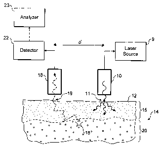
Referring now to Figure 1 an embodiment of the
invention is shown in operation, in schematic cross
section. A light source 10, incorporating or supplied
by laser 9, is used to irradiate localised entry region
of a surface 12 of in-vivo tissue 14, which in the
present example is made up of a skin layer 15 and
underlying bone tissue 20. The incident radiation from
the light source is scattered diffusely through the
sample, especially the upper skin layer. Some of the
radiation may be absorbed by the tissue, some may give
rise to optical emissions for example by fluorescence,
and some re-emerges unchanged through the tissue
surface 12.
A small proportion of the photons of the incident
radiation are inelastically scattered giving rise to
Raman photons, for example as illustrated by Raman
event 16.. The Raman photons in turn are diffusively
scattered through the tissue. Some may be absorbed, for
example giving rise to fluorescence, but some emerge
unchanged through the surface 12 to be collected at
collector 18.
The likelihood of a Raman photon undergoing a
second Raman event is very small.
The collected light is analysed, for example using
filters or a spectrometer, and a suitable sensor, in
detector 22, and the determined Raman spectra or
spectral features are used further in analyser 23. The
CA 02591303 2007-06-07
WO 2006/061565 PCT/GB2005/004529
- 16 -
detector may use a fourier transform rather than a
dispersive spectrometry technique.
Typically, most Raman photons will be generated
close to the light source 10, where the incident
radiation is most intense. These Raman photons may
best be detected by collecting light at the light
source 10, for example by using optics common with the
light source. As ~listance from the light source
increases, however, the intensity of Raman photons
originating near the light source falls away more
quickly than the intensity of Raman photons originating
further away from the light source, especially from
deeper within the tissue. Preferential sampling of
Raman photons from deeper within the tissue can
therefore be achieved by spacing the location at which
light in collected from the location at which the
tissue is illuminated.
In Figure 1 Raman event 16 occurs in or at the top
of the sub-surface bone layer 20. The spacing d between
the light source 10 and the collector 18, or
equivalently between an entry region 11 and a
collection region 19 can be adjusted to select for a
particular depth. In preferred embodiments, however,
light is collected at a range of spacings d, and
analyser 23 is used to infer depth dependent
characteristics of the tissue from the Raman features
of the collected light for different values of d.
The Raman signal for a particular layer can be
preferentially selected by numerical processing of the
Raman signal at several spacings. Equally, the Raman
signal for one or more layers can be preferentially
rejected by similar numerical processing. Such
CA 02591303 2007-06-07
WO 2006/061565 PCT/GB2005/004529
- 17 -
numerical processing may be by simple weighted
comparison or subtraction of signals from different
spacings, or a more complex PCA technique could be
used.
In Figure 1 the in-vivo tissue 14 displays an
abrupt boundary between the surface skin tissue 15 and
bone tissue 20. In Figures 2a to 2c some other tissue
configurations are.shown. In Figure 2a there is a
gradual change from surface layer 30 to deep layer 32,
and deep layer 32 may be diffusely scattering, or
partly or completely opaque with Raman photons
representative of layer 32 being generated in the
interface between the layers. In Figure 2b the surface
layer 30 and the deep layer 32 are separated by a
further transparent or semi transparent layer 34 which
may be, for example, a space filled with a body fluid.
In Figure 2c a more complex tissue structure is shown,
in which graduated or abrupt sublayers 36 and 38 are
embedded beneath or within the surface layer 30.
The Raman techniques and apparatus used herein may
be.applied, for example, to non-invasive investigations
for cancer tissue, either as a stand-alone technique to
locate cancer affected tissue or in conjunction with
existing techniques, such as mammography in the case of
breast cancer. In a conjunction approach to breast
cancer, mammography would be used to identify suspect
regions within breast, and the Raman techniques would
then be used to probe the suspect regions and identify
their nature. This may alleviate the need for biopsy
and provide immediate results, thus dramatically
reducing patient trauma. The Raman techniques
described are particularly suitable for deep-layer
CA 02591303 2007-06-07
WO 2006/061565 PCT/GB2005/004529
- 18 -
probing of breast, because breast tissue exhibits long
photon scattering path lengths and low absorption
coefficients compared to many other types of tissue.
Where appropriate, the Raman apparatus may be
combined into an endoscope arrangement. In this
manner, body cavities may be probed, or partially
invasive techniques used to probe body areas that
should preferably ziot be disrupted, but where Raman
probing through surrounding tissue or membrane into the
areas is desired. Example areas are brain (for example
to identify Alzheimer's and Hutchinson's diseases and
CJD), liver, heart, kidney, prostate gland, veins,
nervous system, spinal cord and kneecap. Other target
physiological conditions include probing for the nature
of stones in kidney and bladder.
The described Raman methods and apparatus may also
be used for the non-invasive detection of blood
characteristics, especially through skin. In this
application, the invention is used to reject the
overwhelming Raman and fluorescence signal originating
from the skin to reveal the underlying signal from
blood contained in blood vessels. In this way, glucose
levels, oxygenation, micro organisms, types and amounts
of cholesterol, and other blood components such as
urea, total protein and albumin may be detected and
measured. Other fluids such as lymph and eye fluids
may be studied.
To improve coupling between the tissue and the
light source and/or collector and index matching fluid
may be used. An index matching fluid, such as
glycerol, may also be deposited into tissue to locally
and temporarily reduce photon scattering. This
CA 02591303 2007-06-07
WO 2006/061565 PCT/GB2005/004529
- 19 -
increases the workable depth of the described
techniques. The index matching fluid may be referred
to as a "contrasting agent" or "image enhancing agent".
The incident irradiation and collection of light
at a single, at mult,iple or at a variable spacing can
be achieved using a variety of geometries. In Figure 3a
there is a single illumination or entry region 40 on
the sample surface. Spaced from this illumination
region is either a single collection point or region
42, or multiple regions as indicated by the broken
lines. Alternatively the single collection region, or
equivalently the illumination region may be moved to
provide a variable spacing.
In Figure 3b the single illumination region 40 is
surrounded by an annular collection region 44, or by
multiple or a variable radius annular collection region
as indicated by the broken lines. Instead of an annular
collection region, a broken annulus or multiple
separate regions at similar distances from the point of
illumination could be used.
In Figure 3c an annular illumination region 46 and
central collection point 48 are used, thereby reducing
the localised intensity of incident radiation required
to generate a given number of Raman photons. The
annulus may be varied in radius or be provided as
multiple annuli having a range of radii. A broken
annulus of multiple separate illumination regions
distributed at similar distances from the central point
of collection could also be used.
Generally, it is beneficial to collect light, or
to provide incident radiation at as large a proportion
of an entry or collection region as possible. However,
CA 02591303 2007-06-07
WO 2006/061565 PCT/GB2005/004529
- 20 -
in practical embodiments the coverage may be limited.
For example, in arranging cylindrical optical fibres in
an annulus a coverage of 10% may be adequate, but 25%
would be preferred and 60% or more may be possible.
In simplistic embodiments a single entry region
may be provided by a single optical fibre brought close
to the sample surface, and multiple collection regions
may be provided by,a linear array of collection fibres.
Optical fibres may be similarly used to provide annular
and other configurations of single and multiple fixed
spacings and various mechanical arrangements may be
used to provide variable spacings.
To provide a variable radius entry region or
collection region an optical arrangement such as that
illustrated in figure 4 may also be used. Optics 50
located between the tissue and the collector, and/or
sample-to-detector distance, is adjustable to direct
light from different parts of the tissue surface onto
collector 18 which is concentric with the light source
10. A lens arrangement(and/or the illumination source
and Raman collector/detector) which can be translated
in an axial direction 52 by an optics drive 54 directs
light from an annular region of varying radius onto the
collector, but other configurations are also envisaged.
A further aspect, which may be used with any of
the arrangements discussed above, is illustrated in
figure 5. One or more mirror elements 60 are presented
to the sample surface. When either incident or Raman
radiation emerges from the tissue away from the
collector 18, these mirror elements redirect the
emerging radiation back into the tissue. This
increases the intensity of incident radiation and so
CA 02591303 2007-06-07
WO 2006/061565 PCT/GB2005/004529
- 21 -
the generation of Ramarn photons within the tissue, and
also increases the proportion of Raman photons received
at the collector 18. The mirror elements are preferably
absent from the surface adjacent to the light source 10
or entry region, and adjacent to the collection
regions.
In alternative embodiments non-imaging optics,
such as those descxibed in Applied Optics vol 35 p 758,
may be used to achieve higher collection efficiency by
use of a mask placed directly onto the tissue surface,
or placed in an image plane if other imaging optics are
also used. The mask blocks appropriate areas of the
tissue to collect signal from a desired spatial offset
only. The masking is preferably synchronised with a
detector such as a charge coupled device such that
sequential readings from the detector relate to masks
providing light collected from correspondingly
sequential spacings between the illumination and
collection regions. The masking could be mechanical and
could also be performed between imaging optics and a
non-imaging type detector.
Figure 6a illustrates a practical embodiment of
the invention comprising an optical head 70 coupled by
an optical fibre bundle 72 to detector 22. The results
of the optical detection are fed to a laptop or other
computer 23 which analyses the Raman features to infer
characteristics of the tissue 14. Detail of the
optical head 70 is shown in the plan schematic view of
figure 7a (which is not to scale). A bundle of light
source optical fibres 74 terminate in the central
region of the head. These light source fibres are
embedded in a filler 76 such as epoxy, and surrounded
CA 02591303 2007-06-07
WO 2006/061565 PCT/GB2005/004529
- 22 -
by an annular spacer element 78. Collection optical
fibres 80 terminate in an annular region surrounding
the spacer element, again embedded in a filler, and
surrounded by an external casing. This arrangement may
be adapted to included the various mirror and optical
arrangements discussed above.
In this particular embodiment each optical fibre
has a core of 200'~im diameter and a cladding bringing
the fibre thickness to 230 um. The inner bundle
consists of seven light source optical fibres 74, and
the outer bundle consists of 26 collection optical
fibres 80. The spacer 78 is sized to space the
collection fibres 80 about 3mm from the centre of the
head, and the terminations of the collection fibres are
distributed approximately evenly in an annulus of
constant radius about this centre. The collection
fibres should be suitable for carrying out optical or
near infra red Raman work, and may be made of silica.
The illumination and collection optical fibres
terminate, about 100cm distant from the optical head,
in a connector illustrated schematically in figure 7b.
The connector presents the six illumination and twenty
six collection fibres for coupling into the detector 22
of figure 6, which incorporates a light source
illumination quasi-monochromatic laser operating at
827nm and a Kaiser Holospec optical analyser.
In figure 6b the invention is implemented as an
endoscope. An insertion tube 90 is used to enter a
human or animal through a natural or surgically formed
orifice 92. The illumination and collection fibres
terminate in a detection head (not shown) and pass back
through the insertion tube to a control handle 94.
CA 02591303 2007-06-07
WO 2006/061565 PCT/GB2005/004529
- 23 -
Optical detection may be carried out in the control
handle or in a connected detector unit 22.
A schematic diagram of another spatial gating
analysis apparatus for identifying depth specific Raman
spectra from in-vivo sub-surface tissues is shown in
Figure 8. Features and variations described below may
be applied to the more general embodiments already
discussed, as appropriate. The apparatus generally
comprises a laser 101, Raman detection apparatus 102,
103 and an analyser 104. The probe beam 105 of the
apparatus is generated using a quasi-monochromatic
laser such as a diode laser, preferably operating at
827nm in the case of tissue analysis, with 12 mW power
which is directed using conventional optics at a
sample. The sample has a surface layer 106 and a deeper
layer 107 of a different chemical composition to that
of the surface layer.
The actual set up illustrated in Figure 8 is
experimental. The layers of the sample constitute a
mock-up of actual in-vivo tissue, and for convenience
are mounted on a stage. However, it is straightforward
to substitute real in-vivo tissue with little
modification. In the present demonstration an argon
ion laser operating at 514nm was used.
With this apparatus the laser plasma lines were
blocked using a Pellin-Broca prism (not illustrated).
The apparatus includes a 1 m focal length lens 108 for
weakly focusing the laser beam onto the sample to a
spot diameter of 300 pm and at normal incidence. Raman
light produced as a result of the irradiation of the
sample is collected in backscattering geometry using a
CA 02591303 2007-06-07
WO 2006/061565 PCT/GB2005/004529
- 24 -
2" diameter collection lens 109 with f-number -1 and is
imaged with the lens 109 onto the slit of a
spectrometer 2, which is part of the Raman detection
apparatus, with a magnification of 2.5. A conventional
imaging spectrometer 102 (for example a Spex
TriplemateTM with f-number 6.3) is preferably used to
disperse the Raman light and image the Raman light onto
a CCD camera 103. The camera 103 is preferably a liquid
nitrogen cooled back-illuminated deep depletion CCD
camera (for example Andor, DU420-BU2 (250 nm) 1024x255
active pixels). The CCD quantum efficiency of such a
camera in the region of Raman spectra is around 65% and
has a pixel size of 26x26 pm. The final stage slit
width of the spectrometer 102 was set to 120 um. The
CCD was binned vertically across 20 pixels to maintain
the spatial selectivity on the collection side.
The sample 106, 107 was mounted on an x-y-z micro-
positioning stage 110 which includes a controlled drive
(not illustrated) which moves the stage (vertically in
Figure 8) together with the final optics to keep the
incidence point of the laser beam fixed on the sample
with respect to the sample. In this configuration, the
Raman detection apparatus 102, 103 always collects back
scattered Raman shifted photons from a fixed imaging
zone in space and the sample is scanned across this
imaging zone whilst the pump beam incidence point
remains fixed in its position on the surface of the
sample. A filter (not illustrated) may also be used to
block any residual elastically scattered probe laser
light from reaching the spectrometer 102. The SORS
CA 02591303 2007-06-07
WO 2006/061565 PCT/GB2005/004529
- 25 -
apparatus described above may be deployed using a point
collection laterally offset from the point of probe
beam incidence (Figure 9). Alternatively, a movable
stage or other movement control means may be used for
achieving relative movement between one or more of the
sample, point of irradiation and the Raman detection
apparatus.
Raman spectra'using apparatus sirriilar to that
described above were collected for a test sample
similar in optical behaviour to a layered diffusive in-
vivo tissue, in which the first layer 106 consisted of
a 1 mm optical path cuvette of 1 cm width and - 4 cm
height, with 300 pm custom made fused silica front and
back windows, filled with PMMA (poly(methyl
methacrylate)) spheres of -20 pm diameter. The spheres
were loosely packed in the cell using mechanical
tapping on the cell during filling to eliminate any
larger voids. This first layer was followed by a second
layer 107 consisting of another cell of 2 mm optical
path filled with trans-stilbene fine powder ground
using a mortar and pestle. The cuvettes 20 were
employed in order to provide a simple method of sample
handling and are not an essential feature of the
apparatus.
With the probe laser beam incident on the sample
positioned with the first layer 106 uppermost,
spatially offset Raman spectra using the SORS
method described herein were collected using a basic
point collection geometry in which collection is from
points laterally displaced from the probe beam's
CA 02591303 2007-06-07
WO 2006/061565 PCT/GB2005/004529
- 26 -
incidence point (Figure 9). The point of collection
geometry as illustrated in Figure 9 represents the
simplest implementation of the method of the present
invention. On the other hand, the concentric circle
geometry illustrated in Figure 10, which does not
require the use of an x-y positioning stage,
advantageously yields much higher collection efficiency
but involves the u',ge of optical fibres to image the
individual circles at different heights on the
spectrometer slit enabling their imaging after
dispersion on the CCD 103 onto separate horizontal
strips with the vertical position of the spectra on the
CCD corresponding to a given offset collection distance
on the sample surface with respect to the probe beam's
incidence point. The use of a fiber optic bundle for
the collection of Raman spectra is described in an
article by Jiaying Ma and Dor Ben-Amotz entitled "Rapid
Micro-Raman Imaging using Fiber-Bundle Image
Compression" Applied Spectroscopy Vol. 51, No. 12, 1997
the contents of which is incorporated herein by
reference.
It will, of course, be apparent that further
alternative collection geometries could be employed
whilst still achieving spatially offset Raman
spectra collection in accordance with the present
invention.
Additionally, with no sample illumination, an
"above the sample" Raman spectrum may be collected
which represents background and apparatus noise. This
"above the sample" Raman spectrum can then be
CA 02591303 2007-06-07
WO 2006/061565 PCT/GB2005/004529
- 27 -
subtracted from the set of Raman spectra to remove
noise from the spectra.
When taking Raman spectra using the resonance
Raman technique, whereby the wavelength of the incident
probe beam is tuned-to match chromophores of the
material or materials being investigated, the Raman
signatures may be swamped by fluorescence
(luminescence) generated from electronic excitation.
For example, fluorescence will be stimulated in
room temperature or in-vivo studies of bone, but
phosphorescence is more likely in colder samples.
Similarly, Raman probing of metallic systems will often
stimulate room temperature phosphorescence.
In such cases the Raman spectra can be recovered
using the SORS method at two or more laser wavelengths.
This relies upon the fact that the spectral profile of
a fluorescent background is not normally dependent on
the excitation wavelength whereas the Raman spectrum is
dependent on the excitation wavelength. Hence, spectra
collected at the same spatial distance from the point
of illumination at two or more different wavelengths of
irradiation may be subtracted from each other to give a
derivative type plot of where the Raman bands are and
this can be mathematically processed to give a truer
looking Raman spectrum. This technique for identifying
Raman bands is described in an article by S. E. J.
Bell, E. S. O. Bourguignon and A. C. Dennis entitled
"Subtracted shifted Raman spectroscopy (SSRS) method"
Analyst, 1998, 123, 1729-1734. This technique is also
referred to as the Shifted Excitation Raman Difference
CA 02591303 2007-06-07
WO 2006/061565 PCT/GB2005/004529
- 28 -
technique (SERD) as described in a paper of the same
name by P. Matousek, M. Towrie and A. W. Parker IJ.
Raman Spec., 33, 128-242 (2002) the contents of which
is incorporated herein by reference.
The two or more wavelengths of incident
irradiation may be generated by means of separate
lasers or by means of a single laser, such as a diode
laser, the output of which is varied for example
through temperature tuning. The difference in
wavelength required is generally about half the width
of the Raman bands, typically approximately 5-10 cml.
A set of Raman spectra for the test sample
described above, measured with a varying degree of
spatial offset with respect to the Raman collection
point and the point of laser incidence on sample
surface is shown in Figure 11. For comparison, the
Raman spectra of pure layers measured in separate
measurements are also displayed. The top spectrum in
Figure 11 is that of pure trans-stilbene and the bottom
spectra that of pure PMMA. The spectrum measured with
the zero offset (0 mm) represents the Raman spectrum
one would typically obtain using a conventional Raman
instrument. It is evident that it contains an
appreciable contribution from both the top and bottom
layers of the sample and that the contribution of the
top layer gradually decreases with offset distance in
the spatially offset spectra. For real applications,
where a pure spectrum of the bottom layer needs to be
recovered, the top layer signal might represent an
unacceptable distortion to the Raman signal of a lower
CA 02591303 2007-06-07
WO 2006/061565 PCT/GB2005/004529
- 29 -
layer. The gradual separation between the two signals
is clearly accomplished using the SORS approach as the
lateral offset between the Raman collection point and
the point of probe beam incidence is increased and is
clearly observable from the illustrated data set. At a
distance of >2 mm (third spectra down in Figure 11) an
order of magnitude improvement in the ratio of the
lower.over the top;layers Raman signals is achieved.
Figure 12 shows the dependence of the absolute
Raman intensities of the individual spectra on the
spatial offset. The data was obtained by numerical
fitting of two intense trans-stilbene bands at 1575,
1595, 1632 and 1641 cm-1 and bands at around 809, 1455,
and 1728 cm-1 for PMMA. The plot clearly demonstrate
that as the Raman collection point is moved sideways
from the probe illumination zone, i.e. the lateral
offset is increased, the Raman signal from the bottom
layer diminishes much more slowly than that from the
top layer. This results in the overall relative Raman
intensity ratio of the bot-tom over the top layer
improving with increasing spatial offset as shown in
Figure 13.
To quantify the contrast improvement achieved
using the method and apparatus of the present invention
with respect to the test sample described above, a
Raman spectrum with a longer acquisition time (1000 s)
at an offset of 3.5 mm was acquired. Figure 14 shows
this spectrum along with a Raman spectrum acquired with
zero offset scaled to the same height of trans-stilbene
bands. By subtracting the pure trans-stilbene spectrum
CA 02591303 2007-06-07
WO 2006/061565 PCT/GB2005/004529
- 30 -
from these spectra we obtained the PMMA contributions
within the individual spectra (see Fig. 15). By fitting
these we established that the contrast of the lower
layer had been improved by a factor of 15 by rejecting
the top layer spectral component. Another striking
observation is that the signal-to-noise obtained using
this spatial gating approach is good in comparison to
alternative approdches.
The total attenuation of the Raman trans-stilbene
signal by the 1 mm PMMA layer was measured with the
zero offset to be around 80. This loss of signal
through the diffusion process, inevitably present also
in conventional Raman spectroscopy, can be, however,
effectively offset through further refinements in the
collection efficiency: for example by adopting the
circular collection geometry shown in Figure 10 or by
using a lower f-number and a higher throughput
spectrograph.
Figure 16 demonstrates another useful feature of
the spatial gating analysis apparatus and method of the
present invention. The analysis apparatus is capable of
suppressing fluorescence in the lower layer Raman
spectrum if it originates from the top layer. The plot
shown in Figure 16 gives the relative ratio of the
trans-stilbene Raman signal in comparison with the
fluorescence originating from the PMMA layer as well as
the fluorescence absolute intensity as a function of
the spatial collection offset. The trans-stilbene
Raman intensity relative to fluorescence intensity is
CA 02591303 2007-06-07
WO 2006/061565 PCT/GB2005/004529
- 31 -
improved by a factor of approx. 2 with the introduction
of a 2.5 mm displacement.
In a situation where a larger separation of the
data obtained from surface and sub-surface layers is
required than that achievable directly within the raw
spectra, by offsetting the collection and probe launch
points a multivariate data analysis procedure may be
deployed using the"analyser 104 of Figure 8. The data
collected by SORS is particularly amenable to
multivariate data analysis because for this approach to
be applicable, the set of Raman spectra measured at
various offsets is still required. To achieve an
effective numerical decomposition the number of spectra
within the set should ideally be at least an order of
magnitude higher than the number of layers present in
the sample. To demonstrate this a multivariate analysis
of the form of principal component analysis (PCA) was
employed.
Approximately twenty Raman spectra acquired on the
PMMA and trans-stilbene two-layer system represented in
Figure 8 and produced using the SORS method and
apparatus described herein were imported into MatlabTM
R11 (The Mathworks Inc., Natick, MA) and processed with
both built in and locally written scripts. The ten
largest eigenvectors generated after performing a
singular value decomposition on the original
data set were included in the PCA rotation. The pure
spectra of PMMA and trans-stilbene were not included in
this dataset and no baseline correction was performed.
CA 02591303 2007-06-07
WO 2006/061565 PCT/GB2005/004529
- 32 -
Multivariate data reduction techniques are
advantageous when a complete separation of the spectral
features of the surface and sub-surface layers is
required. These data reduction techniques also provide
a means of separating spectral features from layers
that may have a moderate to high degree of spectral
overlap or where contributions of individual components
to spectral bands'envelopes may not be known because
spectra of the pure components may not be obtainable or
known.
The recovered factors from the multivariate
analysis are shown in Figure 17. The procedure cleanly
decomposed the Raman spectra collected in this way into
the pure spectra of the two individual layers, i.e. a
PMMA (top layer) and a trans-stilbene (bottom layer). A
factor for pure trans-stilbene was recovered by
targeting the ca. 1595 cm-' band (pixel
730) and a factor for pure PMMA was recovered by
targeting the ca. 809 cm-1 band (pixel 80). The
luminescence background factor was constructed from one
of the original input spectra. This factor was
generated using an iterative polynomial fitting
algorithm (Lieber CA and Mahadevan-Jansen A 2003)
typically used for baseline correction. In this case
100 fitting cycles using a third order polynomial were
used to generate the baseline. This baseline was used
as a factor representing the luminescence background.
These three factors were then used to reconstruct the
dataset with less than 3% error.
CA 02591303 2007-06-07
WO 2006/061565 PCT/GB2005/004529
- 33 -
Although in the above example twenty separate
Raman spectra were collected, where a scaled
subtraction of individual Raman spectra is possible, as
few as two or three spectra are required. Even with
multivariate data analysis, although it is preferred to
perform the analysis on at least a factor more than the
number of components to be identified, such analysis
can often be successfully performed using smaller data
sets of, for example, around ten spectra.
The following is the inventors' current theory for
explaining the efficacy of the analysis method and
apparatus described herein. This theory is supported by
Monte Carlo scattering modelling studies carried out by
the inventors, which yield results in very good
agreement with experiment. The variation in the
relative content of Raman signals from different layers
as the collection point is spatial offset originates
from the random properties of the photon migration
effect. The migrating photons in essence undergo a
'random walk' within the medium and the photon
direction is randomised every transport length along
the propagation distance. When a Raman signal is
collected from the surface of a sample at the point
where the probe beam is incident, the spectrum contains
a relatively large signal contribution from the top
layer due to the probe photon density being highest at
the point of sample exposure. With increasing sample
depth the probe intensity fast diminishes as the photon
intensity is progressively diluted through the photon
diffusion process. Moreover, Raman light generated at
CA 02591303 2007-06-07
WO 2006/061565 PCT/GB2005/004529
- 34 -
deeper layers of the sample is scattered as it
propagates back to the surface and is subject to the
same diffusion. This therefore leads to further
dilution of the intensity of Raman spectra generated at
deeper sample layers. This effect results in a
substantially larger proportion of Raman photons
generated at the sample surface being collected than
those generated at;c3.eeper sample layers when a signal
is collected from the surface of a sample at the point
where the probe beam is incident, in comparison to the
signal that would be collected for an optically
transparent media probed in the same geometry.
However, when Raman light is collected from a
point laterally offset from the point of probe beam
incidence, the probe light intensity within the sample
is becoming more equally distributed along its depth.
This is because the incident light first had to
propagate sideways through the sample from the probe
incidence point to the collection area and was on its
way randomised through photon diffusion. Consequently,
the scattered Raman signal collected at a position
offset from the probe incident point contains a higher
proportion of the deeper layer signal than that in the
spectrum collected from the probe beam incidence point.
The described spatial gating analysis apparatus
and method thus offers an extremely powerful, yet
simple means for extracting pure Raman signals from
individual layers within diffusely scattering media.
The probed sample depths can be well in excess of the
transport length, which sets a depth limit on the
CA 02591303 2007-06-07
WO 2006/061565 PCT/GB2005/004529
- 35 -
conventional confocal Raman microscopy. In the above
example, the transport length of the medium was
estimated to be 200 pm. Importantly, the apparatus and
method can be used 'blind', i.e. without any prior
knowledge of the chemical constituents of individual
.layers. The technique has thus ideal prerequisites for
sensitive sub-surface, non-destructive probing of
diffusely scatteri'ng materials in both industrial and
medical applications.
In situations where a sample is known to consist
of only two layers of different composition, such as
in-vivo skin and bone layers, (if this is not known
then this information can be obtained directly from
pure PCA) the method and apparatus can be used to
extract the pure signals of individual layers without
the involvement of multivariate data analysis
techniques. This is possible where the two spectra of
the two layers each include an identifiable band or
bands that do not overlap. In this situation a simple
scaled subtraction can be used to separate the spectra
of each of the individual-layers from each other. In
this process one Raman component is eliminated by a
scaled subtraction of two spectra measured with two
different spatial offsets cancelling out one or other
spectral component in the process. The results of this
simple extraction procedure are shown in Figure 18. The
spectra used in the analysis were measured with a zero
and a 2 mm offset. The result is clearly satisfactory,
although the applicability requires the above
conditions to be satis,fied. In contrast, the PCA
CA 02591303 2007-06-07
WO 2006/061565 PCT/GB2005/004529
- 36 -
analysis described above can be used in circumstances
where there is no knowledge of the compositions of the
different layers of a sample.
Thus, it will be apparent that it is not in all
cases essential for.a complete Raman spectrum to be
generated with the present invention. Where there is
some knowledge of the materials involved or the
compositions to bd detected, detection of individual
Raman spectral features using, for example, one or more
band pass filters is also encompassed by the SORS
method and apparatus described herein.
The exact degree of the 'suppression' or
separation of two layers in general situations depends
on a variety of parameters. These parameters include
the thickness of the top layer, that of the underlying
matrix, the probe beam diameter, the exact collection
geometry, the wavelength of the probe light used and
the transport length of the medium. For non-invasive
sub-surface probing, as a rule of thumb, it is believed
that the ideal offset should be on the scale of the
thickness or several thicknesses of the overlying
medium. Also, for the technique to be effective the
beam diameter should be smaller than the thickness of
the top layer. In general terms the thinner the top
layer is and the thicker the underlying matrix is
favours a better spectral separation of the two
components.
For this reason the present invention is
particularly suited for use in non-invasive medical
diagnosis applications. However, any degree of
CA 02591303 2007-06-07
WO 2006/061565 PCT/GB2005/004529
- 37 -
absorption of the probe or Raman photons will result in
the overall yield of Raman signals from the sample
surface being diminished. Therefore, for SORS analysis
to be effective, it is important that the measurements
be performed at wavelengths substantially free
from any absorption. In the case of living tissue, this
condition is well satisfied outside the haemoglobin
absorption region '(>600 nm) in the NIR (-800 nm). Thus,
for living tissues the preferred laser source generates
light at wavelengths of at least 600 nm. Laser sources
generating light above 800 nm are also desirable as
this reduces absorption of the incident light by
melanin. Moreover, at this wavelength, bone tissue has
relatively low fluorescence.
However, the use of light of wavelength 514 nm or
>600 nm is not critical to this invention. The choice
of probe wavelength is essentially a trade off between
depth penetration, which improves with longer
wavelength, and detector quantum efficiency, which is
higher at shorter wavelengths. As mentioned earlier,
the detector 103 used herein is a backilluminated deep
depletion CCD detector based on silicon technology.
This detector is selected as it the best .sensitivity
and signal-to-noise ratio of those that are currently
available, but alternatives can be used. Longer
wavelengths avoid exciting H20 modes in Raman spectra,
but the cut-off limit for Si detection is 1.1 pm.
InGaAs detectors can be used at longer wavelengths, but
these have presently reduced sensitivity.
CA 02591303 2007-06-07
WO 2006/061565 PCT/GB2005/004529
- 38 -
As an example of the potential medical
applications of the SORS analysis, it is known that a
Raman spectrum measured from bone tissue is indicative
of its physio-chemical state. The peaks in the spectrum
are indicative both.of mineral components, such as
phosphates, carbonates, hydroxides as well as
interstitial and residual water molecules, and of
organic material,'primarily the collagen matrix.
Relative intensities of mineral peaks and of collagen
peaks should therefore be expected to differ from
normal if there is an abnormality in bone structure.
The technology of generating chemically specific
signatures buried within diffusely scattering matrices
is applicable to many medical applications. In fact, it
is envisaged that the non-destructive extraction of
sub-surface information will have medical applications
ranging across detection of embedded lesions and
assessment of tumour, skin and blood compositions.
With the method and apparatus of the present
invention, substantially pure Raman spectra can be
retrieved from depths well in excess of those
accessible with conventional confocal microscopy.
Moreover, the present invention has the advantage that
it is compatible with the use of cw lasers beams and is
suited to remote monitoring in both industrial and
medical applications.