Note: Descriptions are shown in the official language in which they were submitted.
CA 02591511 2012-08-20
ROTATIONAL ALIGNMENT FEMORAL SIZING GUIDE
FIELD OF THE INVENTION
The present invention relates to a sizing and rotational alignment apparatus
for determining the anterior-posterior size of a distal end of a femur in
order to
determine the proper implant size, and to methods for using such an apparatus.
BACKGROUND
In total knee joint replacement surgery or arthroplasty, the knee joint is
replaced with an artificial implant. The implant typically includes a
tibial
component, a femoral component, and a mobile bearing insert (a patella
component).
Most femoral components have an inner surface with intersecting flat surfaces
that interface with a surgically prepared distal femoral surface. The outer
surface
typically has a curvature that faces a corresponding tibial component attached
to the
patient's tibia. Two curved condylar regions on the femoral component
replicate the
natural femoral condyles.
Determining the proper anterior/posterior (A/P) size of the distal femur is a
difficult procedure. The surgeon must measure the correct A/P size of the
distal
femur, while setting rotational alignment to anatomic landmarks (e.g.,
epicondylar
axis and the A/P axis). The A/P femoral size is usually derived from the
distance
between the posterior condyles and the anterior cortex of the distal femur.
The
rotational alignment (rotation on a transverse plane at the distal face of the
femur)
is usually derived from anatomic landmarks. This rotational alignment,
however,
is typically not in perpendicular/parallel relation or at a set angle to the
A/P
measurement plane needed to properly size the distal femur. Accordingly, it is
not as easy as measuring from the back (posterior) to the front (anterior) of
the
bone and choosing an implant.
1
CA 02591511 2007-06-20
WO 2006/069336
PCT/US2005/046863
Instruments used for this sizing in the past have included two stationary
paddles that are placed vertically against the posterior surface of a resected
distal femur. These instruments do not account for or measure rotational
alignment between two boundaries (for alignment with anatomic landmarks),
while still referencing both posterior condyles to determine the proper NP
size of
the distal femur. Stationary paddles also do not account for condyles of
different
sizes and shapes (e.g., one condyle may extend further than another due to
patient irregularity or diseased tissue).
However, instruments that address each variable (rotation and size)
independently increase procedural time and, more importantly, can result in
less
than optimal NP sizing. For example, the use of two different instruments
prevents
the sizing from being measured in relation to the rotational location being
used.
Specifically, the sizing is not measured perpendicular to the rotational
location being
used.
Some instruments do measure both rotational alignment and A/P sizing
simultaneously. However, these instruments lack adjustment, so that when they
are
rotated with respect to the anatomic landmarks, they only reference one
posterior
condyle. (Again, a precise measurement is not obtainable because one condyle
may extend further than another due to patient irregularity or diseased tissue
or
any other reason.) In other cases, the instruments have additional components
that
can be added to the instrument to substitute for condylar contact, but these
additional components are in increments that limit adjustability and can add
to
surgical procedure time because they need to be interchanged to determine
"best fit"
scenarios.
Accordingly, there is a need to provide a femoral sizing guide that measures
and correlates both (1) rotational alignment (the rotation at the distal face
of the
femur) and A/P sizing.
SUMMARY
Embodiments of the present invention allow unlimited rotational alignment
between two boundaries for alignment with anatomic landmarks while still
referencing both posterior condyles for A/P sizing of the distal femur. In
particular
2
CA 02591511 2007-06-20
WO 2006/069336
PCT/US2005/046863
embodiments, the system provides at least one movable paddle that provides a
reference point from the condyles so that the measuring assembly can be
aligned
to be parallel with the epicondylar axis. Once the measuring assembly is
properly angled, the NP length of the bone can then be measured from a proper
reference point.
BRIEF DESCRIPTION OF THE DRAWINGS
FIG. 1 shows an exploded perspective view of an assembly according to one
embodiment of the invention.
FIG. 2 shows the embodiment of FIG. 1 in an assembled position.
FIG. 3 shows a top plan view of an assembly according to one embodiment of
the invention in place on a patient's femur, prior to activation of the cam
assembly.
FIG. 4 shows the assembly of FIG. 3 in place, once the cam has been
activated and in the process of measurement.
FIG. 5 shows the assembly of FIG. 3 being used to measure the A/P length of
the femur.
FIG. 6 shows a side perspective view of the assembly of FIGS. 3-5.
DETAILED DESCRIPTION OF THE DRAWINGS
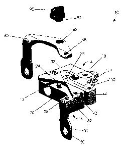
Fig. 1 shows a body 12, a movable paddle 60, and a rotational actuator 90,
collectively referred to as assembly 10. The body 12 is a block with an upper
surface 14, a lower surface 16, an anterior portion 18, and a posterior
portion 20.
(These anatomical directional components are intended to refer to body 12 when
it is
in place on a distal resected femur.) Extending from posterior portion is a
first
paddle 22. This paddle 22 is intended to provide a stable condylar reference
point
for body 12. Paddle 22 is also shown having a fixation portion 30, which is
intended
to receive a fixation pin, screw, or any other securing mechanism to secure
paddle
22 in place. (Although this first paddle 22 is provided in preferred
embodiments of
the invention, it is not essential for the assembly 10 to function as
desired.)
Upper surface 14 of body 12 has an actuator member 24. Actuator member
24 is located toward the posterior portion of the body and substantially
midway
between either side 32 of body 12. Although member 24 is shown as a protruding
pin, it should be understood that it could be an indentation, dimple, or any
other
3
CA 02591511 2007-06-20
WO 2006/069336
PCT/US2005/046863
feature of any shape and size that will allow body 12 to cooperate with a
second
paddle, described more below.
Body 12 also features a pivot member 28. As with actuator member 24, pivot
member 28 is shown as a protruding pin, but it should be understood that it
could be
an indentation, dimple, or any other feature of any shape and size that will
allow a
pivoting motion to occur between a second paddle and body 12.
Body 12 is also shown having sizing indicia 34. In a preferred embodiment,
the indicia are at 1 mm intervals, although other intervals are possible.
Body 12 further has a shiftable measuring member 110. In some
embodiments, member 110 is on a track 38 that cooperates with stylus 40, as
shown
in FIG. 6, although it may cooperate with body in any other way. In a
particularly
preferred embodiment, measuring member 110 is a separate portion that is
adjusted
by member 120, as shown in FIG. 5. Member 120 may be a cam portion that is
rotated, it may be an indentation that receives a hex driver, it may be a
raised pin
that is turned, it may be a threaded or lead screw, or any other type of
adjustment
appropriate for the measuring described below. As shown in more detail by FIG.
2,
member 110 preferably has a mark 114 on its upper surface 112 indicating the
epicondylar axis 140, shown in Figure 3. Mark 114 is preferably aligned with a
zero-
mark 36 on the sizing indicia 34 of the body 12.
As will be described in more detail below, once the proper rotation is
determined using paddles to align body 12 to be parallel or at least
substantially
parallel with the epicondylar axis, the measuring member 110 is used to
determine
the proper NP size of the distal femur. Member 110 is movable in the anterior
and
posterior direction (shown by the A/P axis 142, in FIG. 3) with respect to
body 12.
Referring back to FIG. 1, second paddle 60 is shown as an L-shaped member
that is adapted to cooperate with body 12 via an actuator member 62. In use,
actuator member 62 is intended to associate with actuator member 24. Although
cooperator 62 is shown as an opening, hole, or slot, it should be understood
that it
could be a protruding pin or any other feature of any shape and size that will
allow
member 60 to cooperate with body 12.
Second paddle 60 also has a pivot member 68. Pivot member 68 works with
pivot member 28 of body 12. Although pivot member 68 is shown as an opening,
4
CA 02591511 2007-06-20
WO 2006/069336
PCT/US2005/046863
hole, or slot, it should be understood that it could be a protruding pin or
any other
feature of any shape and size that will allow member 68 to cooperate with body
12.
Rotational actuator 90 is has a cam member 92 that cooperates with actuator
member 24 and actuator member 62 to measure the rotational alignment of the
condyles (or any other desired anatomical reference point, in this example and
description, the condyles are the reference points being used, but it should
be
understood that it is possible to use other anatomical reference points).
In use, the second paddle 60 is assembled onto the body 12 by aligning the
pivot members 68 and 28 together and the actuator member 24 and actuator
member 62 together. The rotational actuator 90 is then assembled to cooperate
with
the member 24 and/or cooperator 62. A screw may be used to make this
connection
if these portions are both openings or indentations. If the actuator. member
24 is a
pin, as shown, then the pin may cooperate with an opening in rotational
actuator 90,
if desired. The completed device is assembly 10, shown in FIG. 2.
It is worth noting here that two paddles are not required, and that the one or
more paddles do not necessarily have to extend to the posterior condyles. In
other
words, one paddle may be used while the other side is left off, although it is
preferable for the paddle that is provided to be rotatable. If one of the
paddles is left
off, the surgeon usually prefers a visual posterior reference, and any type of
reference may be provided. In some instances, a paddle may be machined off of
the
body member with a visual reference created by the posterior edge of what is
left. In
other embodiments, there may be an arrow where the stationary paddle would be.
In sum, two posterior paddles are not a necessity as long as both posterior
condyles
(or epicondyles, or two other planer points) are referenced via contact or
visual
reference.
Device 10 also has degree or angle markings 50. These markings 50 may be
provided on paddle 60, as shown in FIG. 2, or they may be provided on body 12,
rotational actuator 90, an anywhere else that allows markings 50 to be easily
read
when the actuator 90 is activated. Markings 50 are intended to show how many
degrees the body 12 needed to be rotated in order to be parallel to the
epicondylar
axis 140.
In use, assembly 10 is placed on the resected distal femur (which is the
preferable method, although it may be possible to use assembly 10 without
5
CA 02591511 2007-06-20
WO 2006/069336
PCT/US2005/046863
resecting the femur). Body 12 is slid or rotated anteriorly until both
posterior
paddles 22, 60 are in contact with the posterior condyles of the distal femur.
While maintaining contact of the paddles 22, 60 with the femoral-posterior
condyles, the rotational actuator 90 is turned until the desired rotational
alignment
is achieved. When turned, the rotational actuator 90 causes cam 92 to move
second paddle 60 radially about a pivot axis defined by pivot members 28, 68.
Alternatively, actuator 90 could move body with response to paddle 60.
A specific example will now be described for clarity, but it is not intended
to
be a limiting description of the uses or structures defined herein. FIG. 3
shows
assembly 10 in place on a patient's resected femur. Paddles 22, 60 are in
contact with the posterior portion of the patient's condyles. Paddle 60
cooperates
with body 12 via pivot members 28, 68. Rotational actuator 90 is secured in
place. In FIG. 3, the assembly is shown as not being oriented at any angle,
because marking reads "0."
As rotational actuator 90 is turned (in this example, it is turned clockwise,
but it should be understood that any rotational method or direction may be
used),
the body 12 is rotated slightly, as shown by arrow X in FIG. 4. FIG. 4 shows
an
example where body 12 is rotated 3 , as shown by marking 50 and indicated by
the space 52 created between paddle 60 and body 12. It can also be seen that,
in this example, the mark 114 is even with the epicondylar axis 140, so proper
rotational alignment has been reached. In the example, shown in FIG. 2, proper
rotational alignment is reached at 6 . Mark 114 is just one example of an
indicia
that may be used to determine when rotational alignment has been reached, and
it should be understood that other options are possible.
It should also be noted that markings 50 are shown in 3 intervals for the
sake of example only. They could be provided in any increment desired, such as
degree by degree, half degrees, and so forth. It should also be understood
that if
millimeters or some other measurement was desired instead of degrees, that
option is within the scope of this invention.
As described, this rotational adjustment allows the body 12 to be
rotationally aligned to the patient's anatomy. An optional rotational fixation
hole
42 can provided to receive a fixation pin to aid in maintaining posterior
paddle
6
CA 02591511 2012-08-20
22/condylar contact. Once rotational alignment is achieved, fixation pins can
be
inserted through the fixation holes 44 to fixate the assembly 10 to the distal
femur
in the proper rotational alignment position.
Then, the A/P sizing is performed using the measuring member 110. As
shown in FIG. 5, measuring member 110 is adapted to expand assembly 10
anteriorly or contract it posteriorly (shown by arrows "A" and "P"). One way
such
measuring movement may be achieved is via member 120 on measuring
member 110, although many other options are possible and considered within the
scope of various embodiments of this invention. If provided, drill guide holes
116
in the measuring member 110 are used to place the distal-femoral resection
guides (various forms of which are known in the art and used as cutting blocks
to
prepare the patient's femur to receive an implant.) The drill guide holes 116
provide a scaffold through which a drill can extend and prepare holes in the
distal
femur at the appropriate location in order to provide location reference marks
for
the cutting block to be used.
In an alternate embodiment, both first and second paddles may be
movable and/or rotatable. They may both be separate pieces and communicate
with a mating pivot post or they may have separate mating pivot posts.
Additionally, although the paddle(s) have been described as preferably
actuated via a cam mechanism, the paddles could be actuated in any number of
ways, for example, they may be gear-driven, driven via a radial slot with a
lock,
threaded or lead screw actuated, variable angled inserts or modules, or via
any
other appropriate method.
The scope of the claims should not be limited by particular embodiments set
forth herein, but should be construed in a manner consistent with the
description as
a whole.
7