Note: Descriptions are shown in the official language in which they were submitted.
CA 02594994 2007-07-17
WO 2006/079211 PCT/CA2006/000107
COMPUTER-ASSISTED HIP JOINT
RESURFACING METHOD AND SYSTEM
CROSS-REFERENCE TO RELATED APPLICATION
The present patent application claims priority
On United States Provisional Patent Application
No. 60/646,603, filed on January 26, 2005, by the
present applicants.
FIELD OF THE INVENTION
The present invention generally relates to hip
joint resurfacing surgery and, more precisely, to a
method for assisting hip joint resurfacing surgery with
computer-assisted surgery systems.
BACKGROUND OF THE INVENTION
Hip joint resurfacing surgery involves the
introduction of hip joint components in a patient. The
acetabulum and the femoral head are resurfaced so as to
receive an acetabular cup implant and a femoral head
implant, respectively. The
femoral head implant
consists of a ball head received at an end of the
resurfaced femoral head.
Therefore, the implanted
femoral head and the cup (i.e., acetabular or pelvic
implant) coact to create the artificial hip joint. In
comparison with total hip joint implanting surgery, the
hip joint resurfacing surgery removes a relatively small
amount of bone while providing high levels of joint
stability.
Different output values are of concern in hip
replacement surgery. In
order to reproduce a natural
and/or improved gait and range of motion to a patient,
the position and orientation of the implants, the offset
-1-
CA 02594994 2007-07-17
WO 2006/079211 PCT/CA2006/000107
of the femur and the limb length must be considered
during surgery. The
work of the surgeon during hip
replacement surgery will have a direct effect on these
output values.
Known hip joint resurfacing surgery techniques
presently involve specific tools so as to obtain precise
position and orientation for the implants. As various
types of reamers are used to resurface the femoral head,
a plurality of alignment steps are performed to align
the tools with the cuts to be made. It is,
for
instance, of nonnegligible importance that the femoral
neck not be damaged (i.e., notched) by the reamers, to
prevent fracture-prone weakness in the femoral head.
Moreover, the resurfacing must be as precise as
possible, for instance, to reduce the amount of cement
required for implanting the ball head implant to the
resurfaced ball head.
SUMMARY OF THE INVENTION
It is an aim of the present invention to
provide a novel method for guiding an operator in
inserting implants in hip joint resurfacing surgery.
= It is a further aim of the present invention
to provide a method of performing hip joint resurfacing
surgery with computer assistance.
- It is a still further aim of the present
invention to provide a computer-assisted surgery system
for guiding an operator in resurfacing bone surfaces in
hip joint resurfacing surgery.
Therefore, in accordance with the present
invention, there is provided a hip resurfacing CAS
system for guiding an operator in altering a femoral
head in computer-assisted surgery for subsequent
implanting of a femoral head implant, comprising: a
- 2 -
CA 02594994 2007-07-17
WO 2006/079211 PCT/CA2006/000107
trackable reference on the femur, the trackable
reference being trackable to form a femoral frame of
reference of the femur; a registration tool being
trackable; at least one bone-altering tool associated
with a resurfacing of the femoral head, the at least one
bone-altering tool being trackable; a tracking apparatus
for tracking the trackable reference, the registration
tool and the at least one bone-altering tool; and a
resurfacing processing unit connected to the tracking
apparatus so as to receive tracking data for the
trackable reference, the registration tool and th at
least one bone-altering tool, the resurfacing processing
unit having a position/orientation calculator to
.calculate from the tracking data a position and
orientation of the ,trackable reference to track the
femoral frame of reference, and of the registration tool
and the at least one bone-altering tool; a model
generator receiving position and orientation data of the
registration tool to produce a model of the femoral head
and neck with respect to the femoral frame of reference;
and a resurfacing evaluator determining an evaluated
bone resurfacing alteration as a function of a position
and/or orientation of the at least one bone-altering
tool with respect to the bone model of the femoral head
and neck, and a tool geometry model, at least prior to
resurfacing being performed.
Further in accordance with the present
invention, there is provided a method of doing surgical
treatment with a tracking apparatus in computer-assisted
surgery for guiding an operator in resurfacing a femoral
head for a subsequent implanting of a femoral head
implant, comprising the steps of: defining a frame of
reference of the femur, the frame of reference being
trackable in space for position and orientation;
producing a model of a femoral head and neck with
- 3 -
CA 02594994 2007-07-17
WO 2006/079211 PCT/CA2006/000107
respect to the frame of reference; selecting an
orientation of a bone-altering tool with respect to the
model of the femoral head and neck as a function of an
evaluated bone resurfacing alteration varying with said
orientation of the bone-altering tool; and creating a
guide channel in the femoral head with the bone-altering
tool in the selected orientation, for subsequent
resurfacing of the femoral head.
BRIEF DESCRIPTION OF THE DRAWINGS
These and other features, aspects and
advantages of the present invention will become better
understood with regard to the following description and
accompanying drawings wherein:
Fig. 1 is a front elevation view of leg bones
involved in a hip replacement method in accordance with
the present invention;
Fig. 2 is a flowchart of a method for hip
joint resurfacing surgery in accordance with a preferred
embodiment of the present invention; and
Fig. 3 is a view of a user interface
illustrating the Step 120 of selecting a guide
orientation; and
Fig. 4 is a block diagram of a hip resurfacing
CAS system in accordance with another preferred
embodiment of the present invention.
DESCRIPTION OF THE PREFERRED EMBODIMENT
According to the drawings, and more
particularly to Fig. 1, bones of the leg that will be
involved in the hip replacement surgery of the present
invention are generally shown at 1. Fig. 1 is provided
as reference for the description of the steps of the hip
-4-
CA 02594994 2007-07-17
WO 2006/079211 PCT/CA2006/000107
replacement surgery method described herein. The bones
are the pelvis 10, the femur 20, the tibia 30 and the
fibula 40. Hereinafter, parts of these bones will each
be referenced by numerals from the same numeric decade.
For instance, parts of the pelvis (e.g., the acetabulum
11) will bear reference numerals between 11 and 19. .
Referring to Fig. 2, a method for hip joint
resurfacing surgery in accordance with the present
invention is generally shown at 100.
Although the
method 100 is referred to in the singular, various
choices of procedure will be given to the surgeon, as
will be set forth in the forthcoming description,
according to the preferences of the surgeon. A
plurality of methods can be derived from the method 100
according to the decisions of the surgeon.
In Step 102, preparative steps for surgery are
effected.
Namely, general patient information can be
entered into a CAS system for opening a patient file.
For instance, a general patient profile can be entered,
consisting of the name, birth date, identification
number, sex and the like, the side to be operated, as
well as more specific data pertaining to the surgery,
such as leg length discrepancy (with the identification
of the longer leg), if applicable, and parameters to
define the flow of the application and the display. For
instance, the leg length discrepancy is measured using
X-rays of the hip joint. More precisely, the leg length
discrepancy is measured from the vertical comparison
between the lesser trochanters. These
X-rays are
typically taken during the diagnostic stages leading to
surgery, so they are usually available for hip joint
surgery. Alternatively, X-rays may be taken as part of
Step 102. It is also contemplated to import DICOM files
or digital X-rays.
- 5 -
CA 02594994 2007-07-17
WO 2006/079211 PCT/CA2006/000107
It is pointed out that the general patient
information can be entered preoperatively.
Moreover,
the entering of the general patient information is
straightforward such that the surgeon need not be
involved. However,
in order to minimize the
preoperative procedures, actions of Step 102 can be
performed at the beginning of the surgical session,
during the short time span preceding the surgery.
Other values that will potentially be
considered in the method 100 are inclination and
anteversion for the pelvic implant, CCD and anteversion
for the femoral implant
The calibration of the various surgical tools
to be used is done. For instance, a calibration base
and method, as set forth in International Publication
No. WO 01/67979 Al by Jutras et al., can be used for the
calibration. Also, correspondence between the tracking
of the tools and the display on a CAS system can be
verified in further calibration steps included in Step
102. A permanent calibration system can also be used,
as set forth in International Publication No.
WO 2005/102202.
Surgery is initiated between Step 102 and
subsequent Step 104, by the surgeon exposing the hip
joint. No computer assistance is required thereat.
In Step 104, the trackable references are
secured to the pelvis with a pelvic modular reference,
and to the femur with a femoral modular reference. The
pelvic modular reference can be inserted in a cranial or
lateral position. Alternatively, the trackable
references may be secured prior to exposing the hip
joint.
It is pointed out that the pelvic modular
reference, in a preferred embodiment, must be positioned
while the patient is in supine decubitus. Moreover, as
-6-
CA 02594994 2007-07-17
WO 2006/079211 PCT/CA2006/000107
will be described hereinafter, the pelvic coordinate
system and table reference must also be digitized in
supine decubitus. After
those manipulations, the
patient can be repositioned in lateral decubitus.
The femoral modular reference can be inserted
at the proximal third from the femoral head of the femur
or at the distal third from the femoral head. These
insertion points are examples, as any other suitable
point on the femur is considered.
Positions of the
trackable references are, for example, (1)
looking
posterior and towards the head, prior to dislocation,
and (2) a longer trackable reference, looking posterior,
for the dislocated position. It is contemplated to use
a single modular base.
In Step 106, it is contemplated to digitize
the coordinate system in lateral decubitus. It is also
contemplated to collect posture information, as
described 1 in International
Publication
No. WO 2004/030559 Al, by Jansen et al. Criteria may be
used to validate the points taken and the computed
surface.
In Step 106, a pelvic coordinate system and a
femoral coordinate system are digitized. The
pelvic
coordinate system is digitized with a registration
pointer. In an
embodiment, three points are taken on
the pelvis 10 to create the frontal plane of the
acetabular coordinate system.
Referring to Fig. 1,
there is one point on the iliac crest 12 of the operated
side, one point on the contra lateral iliac crest 13,
and one point on one of the two pubic tubercles 14 of
the pelvis 10. To be
generally aligned, the points
digitized on the iliac crests 12 and 13 are taken at the
outermost anterior point of the iliac crests 12 and 13.
The points digitized on the iliac crests 12 and 13 are
preferably taken directly on the soft tissue covering
-7-
CA 02594994 2012-11-30
the bone pelvis on the iliac crests, as the soft tissue
is relatively thin thereon. The point
on the pubic
tubercle 14 completes a first plane, the frontal plane.
A second plane, the transverse plane, is perpendicular
s to the frontal plane and includes the points on the
iliac crests. A third
plane, the sagittal plane, is
perpendicular to the frontal and transverse planes.
Supplemental information regarding the frontal
plane can be obtained for various postures of a patient.
For instance, trackable references can be used to gather
information about sitting, standing and walking
postures. This
information can be used to adjust the
orientation of the frontal plane, as these postures can
provide information not available from the typical lying
posture in which a patient is during surgery. This
information can influence the anteversion positioning of
the implants.
It is possible to obtain anteversion and/or
inclination values of the acetabulum of the patient, to
be used as a reference (e.g., comparison basis) later in
the surgery. To do so,
points are digitized using a
registration pointer on the generally circular edge of
the acetabulum 11 and a plane is defined from these
points. A normal to this plane and the pelvic frontal
plane give the anteversion angle. The normal
to this
plane is projected onto the acetabular frontal plane to
give an inclination angle with a cranial-caudal axis.
For the digitization of the femoral coordinate
system, it is contemplated to collect five points of
reference on the leg to the computer assisted surgery
system, which is equipped with software that will create
the femoral coordinate system.
Referring to Fig. 1, a first point is taken on
the tip of the greater trochanter of the femur 20, and
will be defined as a starting point of an anatomical
- 8 -
CA 02594994 2007-07-17
WO 2006/079211 PCT/CA2006/000107
axis of the femur 20. Thereafter, points are taken on
the medial and lateral epicondyles 24 and 25 of the
femur 20, respectively. A midpoint between the medial
epicondyle and lateral epicondyle points, in alignment
therewith, is defined as an endpoint of the anatomical
axis of the femur. The
fourth and fifth points are
taken on the medial malleolus 31 of the tibia 30 and on
the lateral malleolus 41 of the fibula 40, with the leg
being bent at the knee. By having the leg bent at the
knee, the tibia 30 stands on the posterior condyles 26
of the femur 20.
Therefore, an assumption is made
wherein an aligned midpoint of the medial and lateral
malleoli points is said to define a plane (i.e.,
sagittal plane) with the anatomical axis, with an axis
of the knee being normal to the sagittal plane. The
frontal plane is perpendicular to the sagittal plane,
with the anatomical axis lying therein. The transverse
plane is perpendicular to the sagittal and frontal
planes, and can be positioned at any height. With the
anatomical axis and the midpoint of the malleolus region
digitized, the femoral coordinate system, i.e., the
femoral frame of reference, is complete. It is
noted
that it is not required to measure two points to obtain
a midpoint of the malleolus region. As
this latter
point will be in the sagittal plane, the only
requirement is that a point is taken at a midpoint of
the malleolus region, and may thus be placed
approximately by the operator.
It is pointed out that the projection values
described herein (e.g., inclination, anteversion, etc.)
are based on the acetabular and the femoral coordinate
systems. As it
is contemplated to use alternative
methods of digitizing the acetabular and the femoral
coordinate systems, in addition to the preferred methods
of Step 116, the projection values would be related to
- 9
CA 02594994 2007-07-17
WO 2006/079211 PCT/CA2006/000107
the alternative acetabular and femoral coordinate
system.
Other methods to gather information pertaining
to surgical parameters are as follows.
(1) The user
digitizes a point on the greater trochanter before
dislocation and retakes the same point, with the leg
aligned in the same orientation, after reduction.
(2) The user digitizes a point on the greater trochanter
before dislocation and the system helps the user to
replace the leg in the same orientation after reduction.
The leg length and the offset are automatically computed
when the leg is positioned in range of the initial
position before dislocation.
(3) The user digitizes
many points near the greater trochanter before
dislocation, the center of rotation of the acetabulum as
described in Step 112 and the same points after
= reduction. The system aligns these points and computes
the leg length and the offset. Also, in each case, the
CAS system may help the operator in placing the leg in a
required initial position.
In optional Step 108, a relative position
between the pelvis and the femur is registered with
respect to the trackable references. The leg is simply
left in a straight position, to align with a
longitudinal axis of the body, and a relative position
is acquired between tracking references secured to their
respective bones.
In Step 110, the femur is dislocated from the
pelvis, so as to expose the acetabulum 11 and the
femoral head 21 and neck 22.
In Step 112, a center of rotation is digitized
for the acetabulum, by taking reference points on the
surface of the acetabulum 11.
A center calculator
(e.g., sphere fitter algorithm) is used to find the
acetabular center of rotation, and will be described
-W-
CA 02594994 2007-07-17
WO 2006/079211 PCT/CA2006/000107
hereinafter with the description of a hip resurfacing
CAS system. The
acetabular center of rotation is
therefore known as a function of the tracking reference
on the pelvis 10. In order to ensure precise results,
it may be required that a predefined number of points be
taken until validation criteria are met. Visual
validation of the sphere found by the algorithm can also
be performed. The center of rotation and the diameter
found may be displayed. Points
are digitized in the
fossa (depth of the acetabulum). Points
are displayed
by small spheres or disks (many colors possible). If
the center of rotation of the acetabulum is known, it is
not necessary to digitize the center of rotation of the
femoral head. However, it can be done without departing
from the spirit of the present embodiment.
The registration of points in the acetabulum
can also be taken by a real-time tracing of the
acetabulum surface (i.e., painting the acetabulum
surface). In this case, points can be used to build a
mesh. The mesh
can be constructed while points are
acquired so the user may take more points when needed to
have a more precise reconstruction. Criteria may then be
used to validate the points taken and the computed
center of rotation and diameter.
In Step 114, the acetabulum is altered in view
of accommodating the acetabular cup implant. In order
to guide the operator in altering the acetabulum, reamer
position and orientation information is preferably
provided, such, that an axis of actuation of the reamer
is for instance visually displayed. The
previous
acetabular center of rotation is known as a function of
the tracking reference secured to the pelvis 10, as it
was acquired in previous Step 112.
Preferably, the
reamer is tracked for position and orientation.
-11-
CA 02594994 2007-07-17
WO 2006/079211 PCT/CA2006/000107
Examples of information that can be provided
to the operator are as follows:
generic 2D images,
mosaic or mesh in 3D viewers along with drive
shaft/reamer assembly in real time and/or display
targeting views to help the user to align with target
values, frontal and lateral views, inclination,
inclination adjusted with the pelvic tilt, anteversion,
anteversion adjusted with the pelvic tilt angles in real
time, 3D position of the reamer center of rotation
relatively to the acetabulum center of rotation, the
distance between the reamer pole and acetabular wall.
The diameter of the pelvic implant chosen by
the surgeon can be used to display a position of the new
acetabular center of rotation in comparison to the
digitized acetabular center of rotation (Step 112). For
instance, the distance between the centers of rotation
can be displayed numerically (e.g., in mm) as a function
of the acetabular coordinate system digitized in
previous Step 106. Also,
the anteversion and
inclination of the actuation axis of the reamer, both as
a function of the acetabular coordinate system, can be
given numerically (e.g., in degrees) to guide the
surgeon in the reaming. More precisely, the anteversion
is calculated as the angle between the axis of the
reamer and the pelvic frontal plane, and the inclination
is the angle between the reamer axis projected onto the
acetabular frontal plane and a cranial-caudal axis
(Step 106).
Step 116 consists in the insertion of the
pelvic implant in the acetabulum 11, but it is pointed
out that this step can also be performed once the
femoral head implant has been secured to the femur,
according to the preference of the operator. A tracked
impactor is preferably used. As the pelvic implant size
is known, the diameter thereof and the known relation
- 12 -
CA 02594994 2007-07-17
WO 2006/079211
PCT/CA2006/000107
between the impactor and the pelvic implant is used with
the tracking of the impactor to give the anteversion and
the inclination of the pelvic implant. Also,
the
distances between the current and the digitized centers
of rotation can be displayed. Therefore, the surgeon is
guided during the use of the impactor so as to position
the pelvic implant to a given position of the center of
rotation thereof, and to a given orientation [with
respect to anteversion and inclination] to provide a
maximal range of motion and stability of the leg.
Although the pelvic implant is secured at this
point to the pelvis 10, it is possible to adjust the
position and orientation of the pelvic implant.
Firtly, the tracked impactor, handle or like tool may
be reconnected to the pelvic implant to serve as a lever
in manipulating the pelvic implant with the tracked
impactor, allowing position and orientation information
(e.g., anteversion and inclination) to be calculated
from the tracking of the impactor.
Alternatively,
points on the circular edge of the pelvic implant may be
digitized to define a plane, with the normal to this
plane being used to calculate the anteversion and the
inclination, as suggested previously to obtain this
information for the acetabulum.
Information typically provided with the use of
the impactor includes:
Display of generic 2D images,
thosaic or mesh in 3D viewers along with impactor/cup
assembly in real time and/or display targeting views to
help the user to align with target values, frontal and
lateral views, navigation of the impactor and cup,
display of inclination, inclination adjusted with the
pelvic tilt, anteversion, anteversion adjusted with the
pelvic tilt angles in real time, display of the 3D
position of the cup center of rotation relatively to the
acetabulum center of rotation.
- 13 -
CA 02594994 2007-07-17
WO 2006/079211
PCT/CA2006/000107
In Step 118, a bone model is digitized for the
femoral head 21 and neck 22. A
registration pointer
having its tip tracked in space is used to register
points on the surface of the femoral head 21 and neck
22. Therefore, points of contact between the tip and a
given surface can be registered as a function of the
tracking reference (Step 104). As tracking references
have been secured to the femur 20 and the pelvis 10 in
Step 104, the points on the surface of the femoral head
21 are known as a function of the tracking of the
respective tracking reference of the femur 20. As will
be described hereinafter, a digital model of the femoral
head and neck is produced, and may be displayed visually
by the hip resurfacing CAS system.
It is pointed out that the neck/head
connection is identified in the digital model of the
femoral head and neck. Information preferably obtained
includes the lateral aspect of femur at the greater
trochanter and the following 10 cm distally (as far as
possible), internal aspect of femur at the lesser
trochanter and the following distal region, and femoral
neck itself (varus/valgus, anteversion). The head-neck
junction is digitized or computed based on the points
taken. If points are acquired automatically, collection
of points can be taken by painting the femur. If points
are acquired to build a mesh, points are taken on all
the surface of the femur and not only on the frontal and
transverse plane. The mesh can be constructed while
points are acquired so users may take more points to
have a more precise reconstruction.
The center of rotation of the femoral head may
also be calculated from the digital model, for instance
using a sphere fitter algorithm. If the
center of
rotation of the acetabulum is known, it may not be
CA 02594994 2007-07-17
WO 2006/079211 PCT/CA2006/000107
necessary to digitize the center of rotation of the
femoral head.
In Step 120,* the desired guide orientation is
determined. More specifically, the resurfacing of the
femoral head will be dependent on the orientation of the
guide wire. Therefore, computer assistance is provided
to the operator so as to orient the guide wire in view
of the subsequent resurfacing of the femoral head.
In order to plan the orientation of the guide
wire, various views are provided such as the frontal and
top views of the reconstructed femur. A template of the
femoral implant over the femur model is also provided,
as well as the following information: the initial CCD
and anteversion angles, an initial template position,
orientation and size with respect to the femoral center
of rotation. The CCD is calculated as the angle between
the projection of the guide wire on the femoral frontal
plane and the longitudinal axis of the femur. Widgets
are provided on screen to translate and rotate the
template in each view.
Selectors are provided to set
the size of the implant and the neck diameter. The neck
diameter is found by two moving lines parallel to the
template axis. When the lines are on the contour of the
neck, the diameter is determined. The
CCD and
anteversion angles are computed and displayed while the
user is positioning the template. It is
also
contemplated to provide means to rotate the model so it
' can be viewed in 360 degrees.
Implant position,
orientation and size are computed and suggested to the
operator as information to consider.
Information that
is preferably computed and displayed includes: The
estimated range of motion, the estimated final leg
length and offset, a graphical representation of the
femoral preparation (final result).
Potential
dislocation and/or impingement is identified based on
- 15 -
CA 02594994 2007-07-17
WO 2006/079211 PCT/CA2006/000107
the cup position and orientation and the planned
position and orientation of the femoral implant. If the
femur is reconstructed with a mesh, the percentage of
coverage may be provided. Indications of where notching
may happen should also be provided.
In Step 122, the femur is altered for the
insertion of the guide wire. In
order to guide the
operator in positioning and orienting the guide wire as
planned, various information is provided, such as:
Generic 2D images, mosaic or mesh in 3D viewers along
with guide wire/drill guide in real time and/or display
targeting views to help the user to align with planned
values, frontal and top views of the reconstructed
femur, navigation of the guide wire with a drill guide,
the CCD and anteversion angles, alignment views of the
guide wire tracked with the drill guide on the CCD and
anteversion axis found during the planning phase
(aligning "bull's-eyes" or axes), the CCD and
anteversion angles of the guide wire, audio and/or
visual cues to let the operator know he/she is "in
range" near the targeted angles by the means, the depth
of the guide wire so the operator will be able to
determine when the tip of the guide wire is near the
lateral cortex of the proximal femur, potential notching
with audio and/or visual feedback, and where this
notching could potentially occur. An
example is
provided in Fig. 3, in which a drill guide axis is
oriented as a function of the bone model.
The same information can be provided for the
insertion of a cannulated drill guide, with a display of
the depth of drilling so the user will be able to
determine when to stop drilling according to the chosen
implant size.
- 16 -
CA 02594994 2007-07-17
WO 2006/079211
PCT/CA2006/000107
Haptic devices can be used to ensure that the
drilling only occurs when the orientation of the guide
wire is as planned.
In Step 124, the femoral head 21 is
resurfaced, by way of a reamer. It is contemplated to
provide visual information to the operator at this step.
However, the guides inserted in the femur ensure that
the reaming follows planning. It is preferred that the
operator keeps inspecting the actual femur especially
during the cylindrical reaming, so as to avoid notching
of the femoral neck 22.
Information that can be
provided is as follows: Tracking for position and
orientation of the cylindrical reamer, generic 2D
images, mosaic or mesh in 3D viewers along with
cylindrical reamer in real time, frontal and top views
of the reconstructed femur, navigation of the
cylindrical reamer to track the reamed depth,
orientation and position, the CCD and anteversion
angles, a graphical representation of the result of the
reaming, a pre-notching warning system based on
probability to notch the cortex when the instrument is
close to it.
For the planar reaming, information that can
be provided is as follows: Generic 2D images, mosaic or
mesh in 3D viewers along with planar reamer in real
time, frontal and top views of the reconstructed femur,
tracking of the planar reamer to track the reamed depth,
orientation and position, the CCD and anteversion
angles, the distance between the head-neck junction and
the plane surface of the planar reamer, indications to
the operator to stop reaming based on the selected
implant size, how much bone has been removed, the leg
length and the offset based on the position of the
planar reamer, a graphical representation of the result
of the reaming, pre-notching warning system based on
-17-
CA 02594994 2007-07-17
WO 2006/079211 PCT/CA2006/000107
probability to notch the cortex when the instrument is
close to it.
In Step 126, the femoral implant is secured to
the resurfaced femoral head.
Information that can be
provided is as follows: position and orientation of the
femoral component, generic 2D images, mosaic or mesh in
3D viewers, frontal and top views of the reconstructed
femur, navigation of the cement mantel to track the
position and the orientation of the implant, the
distance between the implant and the plane surface of
the femur, the leg length and the offset. It is
contemplated to provide the possibility to attach the
femoral implant while in place.
Although not illustrated in the method, there
is provided the possibility to ream again the acetabulum
after the placement of the femoral component if the
initial reaming is not adequate, following the options
provided in Step 114. Also, Step 116 could be performed
at this point.
Information that can be provided
includes: the leg length and the offset based on the
position of the reamer relatively to the acetabulum
center of rotation and the position and orientation of
the femoral implant with respect to the femur.
In the event that the acetabular cup is
implanted at this point, the information that can be
provided is as follows: Tracking of the cup impactor,
generic 2D images, mosaic or mesh in 3D viewers along
with impactor/cup assembly in real time and/or display
targeting views to help the user to align with target
values, frontal and lateral views, display inclination,
inclination adjusted with the pelvic tilt, anteversion,
anteversion adjusted with the pelvic tilt angles in real
time, 3D position of the cup center of rotation
relatively to the acetabulum center of rotation, the leg
length and the offset based on the position of the
- 18 -
CA 02594994 2007-07-17
WO 2006/079211 PCT/CA2006/000107
impactor relatively to the acetabulum center of rotation
and location of the femoral component on the femur.
In Step 128, an analysis of range of motion is
performed. Information is calculated, such as the range
of motion of the joint after reduction, inclination,
rotation and flexion/extension, possible dislocation
(i.e., detect if the center of rotation has moved)
and/or impingement.
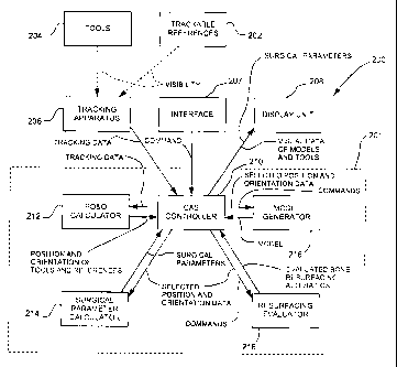
Referring to Fig. 4, a hip resurfacing CAS
system is generally shown at 200. The CAS
system 200
has a resurfacing processing unit 201. The resurfacing
processing unit 201 is typically a computer or like
device having a processor.
Peripherals are provided in association with
the resurfacing processing unit 201. In view
of the
trackable references 202 that will be secured to the
femur and pelvis to define frames of reference (Steps
104 and 106) and to the tracked tools 204 used
throughout the method 100, tracking apparatus 206 is
connected to the processing unit 201. The
tracking
apparatus 206 is provided to track the trackable
references 202 and the tools 204 in the selected
surgical environment. The tracking apparatus 206 may be
any of optical sensors, RF sensors, magnetic sensors and
the like used in CAS systems.
Interface 207 is connected to the processing
unit 201. The
interface 207 enables data entry and
communications from the operator/surgeon of the system
200 to the processing unit 201. For
instance, the
interface 207 may be a keyboard, mouse and/or touch
screen or the like.
A display unit 208 is connected to the
processing unit 201. The
display unit 208 provides
information to the operator/surgeon throughout the steps
of the method 100. The data
may be in the form of
- 19 -
CA 02594994 2007-07-17
WO 2006/079211 PCT/CA2006/000107
numerical values, as well as virtual representations of
bone models along with simulations of tools.
Further
detail about the data displayed by the display unit 208
will be given hereinafter.
The resurfacing processing unit 201 has a
computer-assisted surgery controller 210. The
CAS
controller 210 is connected to the tracking apparatus
206 and to the interface 207, so as to receive
information therefrom. More
specifically, the CAS
.10 controller 210 receives tracking data from the tracking
apparatus 206, which tracking data will be interpreted
by the processing unit 201. The
CAS controller 210
receives user commands given by the operator of the
system 200 using the interface 207, and essentially
controls the flow of information between the peripherals
= 206 to 208, and between the other components 212, 214,
216, and 218 of the resurfacing processing unit 201.
The CAS controller 210 performs certain tasks as well,
such as calibration of tools.
The CAS controller 210 is also connected to
the display unit 208. The CAS controller 210 provides
display data, in the form of numerical values and visual
representations, to the display unit 208. The display
unit 208 displays this information.
A position/orientation calculator 212 is
connected to the CAS controller 210. The
position/orientation calculator 212 receives the
tracking data of the tracking apparatus 206 from the CAS
controller 210. The
information provided to the CAS
controller 210 by the position/orientation calculator
212 is in the form of the position/orientation of a
selected item of the trackable references 202 or tools
204. For instance, following the method 100, the data
provided by the calculator 212 may be the pelvic and
femoral coordinate systems from the trackable references
CA 02594994 2007-07-17
WO 2006/079211
PCT/CA2006/000107
202. As another example, the data takes the form of a
real-time orientation of the operating axis of one of
the tools 204, such as the axis of a reamer, or a real-
time position of a tip of one of the tools 204, such as
a registration pointer.
A center calculator 214 is associated with the
CAS controller 210. The
center calculator 214 is
provided to digitize the center of rotation of the
pelvis (as described for Step 112) and the center of
rotation of the femoral head (optionally in Step 118).
The center calculation is performed using the
position/orientation data calculated by the
position/orientation calculator 212, as well as commands
from the CAS controller 210.
For instance, the center calculation is
performed using the tracked position of a registration
pointer/tool from the tools 204, pointing or brushing
the surface of the acetabulum (Step 112) or of the
femoral head (Step 118). An indication that the center
calculation is to be performed by the center calculator
214 is commanded by the CAS controller 210, for instance
as a response to a command from the operator using the
interface 207. The position of the centers is therefore
calculated with respect to the coordinate systems (Step
106), and the information is updated in real-time by the
CAS controller 210.
A model generator 216 is associated with the
CAS controller 210. The
model generator 216 receives
position/orientation data in combination with commands
from the CAS controller 210, following Steps 112 and
118. For instance, points registered by a registration
pointer/tool or the like from amongst the tools 204 are
used to construct a bone surface model. As discussed
previously, a bone model may be generated prior to
surgery, whereby the model generator 216 is provided to
-21-
CA 02594994 2007-07-17
WO 2006/079211 PCT/CA2006/000107
calibrate the bone model with the femoral frame of
reference. For instance, in Step 118, a surface model
of the femoral head and neck is obtained. The surface
model is associated with the coordinate systems obtained
from the tracking of the trackable references 202.
A resurfacing evaluator 218 is provided in
association with the CAS controller 210. The
resurfacing evaluator 218 is provided to determine the
evaluated bone resurfacing alteration, which is the
effect of a resurfacing tool (from the tools 204) on the
bone model. Accordingly, bone model data is provided by
the model generator 216, along with the position and
orientation of a reaming tool as determined by the CAS
controller 210 from tool geometry data and an
orientation of a bone-altering tool (such as a drill)
from the tools 204.
In the case of femoral head resurfacing, as
the precision of the reaming must be respected, it has
been described previously that a guide wire is provided,
in order to drill a guiding bore in the femoral head
prior to reaming.
Therefore, the evaluated bone
resurfacing alteration is indicated as a function of the
orientation of the axis of the drill guide. Therefore,
information associated with a potential wrongful reaming
is provided to the operator, such that the operator is
guided into drilling the drill guide in a suitable
orientation in view of the effects on resurfacing. The
resurfacing evaluator 218 may also be used to calculate
the effect of acetabulum reaming on associated data
(pelvic center of rotation, anteversion, etc.)
Throughout surgery, the display unit 208
provides the data discussed above. For
instance, the
output of the model generator 210 is converted by the
CAS controller 210 to a virtual model of the bone
surface to be altered, for instance with virtual real-
-22-
CA 02594994 2007-07-17
WO 2006/079211 PCT/CA2006/000107
time representations of the tools with respect to the
bone models. Accordingly, warning can be signaled to
the operator/surgeon if the effects of resurfacing are
outside acceptable standards. Again,
in femoral head
resurfacing, the femoral neck must not be nicked,
whereby drill guide axis data can be associated with a
warning signal to guide the operator/surgeon in
adjusting the orientation of the drill.
Moreover, numerical information is also
provided to the operator, which numerical information is
described previously for the steps of the method 100.
Various instruments can be used, such as blunt
tracked pointers (straight or curved), adapted to fit on
a rotational tracker or a universal handle to paint
bones (acetabulum, femur, etc.). The drill
guide or
guides can be designed to fit on a universal handle or a
rotational tracker. A
mechanism may be used to
block/hold the position and the orientation of the drill
guide. Planar
reamer is modified to be used in
conjunction with the rotational tracker. Technology to
have appropriate drilling instrument if the user wants
to navigate the drill bit only.
In other contemplated options there are the
possibility to navigate the guide wire, the guide wire
and the cannulated drill bit or only the drill bit, the
possibility to rotate, translate and zoom the viewers,
the animation or illustration to describe to the
operator the upcoming tasks, the possibility to take
snapshots, menus allowing selection of options and
parameters during the procedure, allowing navigating
through the surgical steps in the application, step-
driven (wizardlike sequence of pages), status icons to
display tracking state of an instrument, volume view/aim
camera to display in space the location of the trackers
seen by the camera, give information pn the tracked
CA 02594994 2007-07-17
WO 2006/079211 PCT/CA2006/000107
state of a tracker (out of volume, missing sphere, IR
interference, etc).
A calculation that can be performed and
provided as information to the operator is the femoral
target height. The target height is a desired position
for the femoral center of rotation, and is calculated as
follows:
(target height) = (ApELlacam) - (initial ALL),
where (ApaLlacam) is the deviation of the
implant center of rotation with respect to the digitized
acetabular center of rotation, in cranial-caudal (y)
direction (with a cranial deviation having a positive
value), and (initial ALL) is the initially acquired limb
length discrepancy.
- 24 -