Note: Descriptions are shown in the official language in which they were submitted.
CA 02596078 2007-07-26
WO 2006/083728 PCT/US2006/003029
-1-
END EFFECTOR FOR SURGICAL INSTRUMENT, SURGICAL INSTRUMENT, AND
METHOD FOR FORMING THE END EFFECTOR
Technical Field
The present invention relates to an end effector for a
surgical instrument, a surgical instrument having the end
effector, and a method for forming the end effector.
The end effector of the present invention is for a
surgical instrument and is made from two pieces. The pieces are
joined to form the end effector. The pieces can be made by
powder metal processes, machining, stamping, fine blanking,
deep drawing, casting etc. The pieces can be joined by
riveting, welding, soldering, brazing, folded tabs, pressing,
etc. The pieces can be made from the same materials, or
different materials.
Also disclosed is an end effector made from one piece of
flat sheet material, preferably metal, and, in particular, heat
treatable stainless steel. The end effector is specifically
designed to take advantage of this low cost process.
Further disclosed is an end effector for a surgical
instrument having teeth that are formed so that they curve
toward the inside of the end effector and create an undercut
along their inner surface
Finally, a self-centering spike for an end effector is
disclosed for a surgical instrument. The spike has a piercing
blade. As the jaws of the end effector are opened, the spike
moves from a floating position to a secured centered position
between the jaws of the end effector.
CA 02596078 2007-07-26
WO 2006/083728 PCT/US2006/003029
-2-
Background Art
A number of different types of biopsy forceps are in common
use, typically, in conjunction with endoscopic assistance.
Ordinarily, these devices are of complicated construction,
requiring the manufacturing and machining of precise
miniaturized components, which are, therefore, generally quite
expensive.
One early example of flexible forceps is shown in U.S. Pat.
No, 3,895,636 to Schmidt, wherein a pair of cup shaped jaws
having an annular rim mate with a hub and a sharpened trocar.
The Schmidt jaws are of a nature that requires machining for
the edge, each jaw being different from the other jaw.
United States Patent No. 4,887,612 to Esser et al. discloses
a similar biopsy forceps that utilizes a cam linkage to effect
movement of the cup-shaped jaws toward and away from one
another. The Esser et al. jaws are made from stainless steel
and, likewise, require expensive machining.
United States Patent No. 4,763,668 to Macek et al. discloses
a biopsy forceps whose cup-shaped forceps are driven by a
linkage. Each pivot point in the linkage establishes a new
place for stress, wear and breakage. This is similar to the
linkage assembly shown in U.S. Pat. No. 4,721,116 to Schintgen
et al. A needle between the forceps is retractable as the
forceps close.
United States Patent No. 3,921,640 to Freeborn shows a
surgical instrument manufactured from a single piece of molded
plastic. The instrument can have any of various forms of jaws
including a configuration of teeth for holding towels or
surgical dressings.
United States Patent No. 4,200,111 to Harris describes a
pair of spring-biased jaws slidably disposed within the end of
CA 02596078 2007-07-26
WO 2006/083728 PCT/US2006/003029
-3-
a trocar. The jaws are moved inwardly and outwardly from the
trocar by movement from a twisted wire.
United States Patent No. 4,669,471 to Hayashi shows a biopsy
forceps device having a pair of cups attached by a pivot pin.
There are several linkages between the cups and the operating
wire, which are, likewise, connected by pivot pins. The pins
are welded or fused to their components by laser welding.
United States Patent No. 4,815,460 to Porat et al. describes
a medical device for gripping. The Porat et al. device has a
pair of jaws identical to one another. The jaws have an array
of teeth disposed completely thereacross. The teeth are
divided longitudinally across each jaw and are out of phase
from one another. A further device is shown in U.S. Patent No.
825,829 to Heath. The Heath appliance utilizes two different
sets of engaging jaws to accomplish its cutting purpose.
United States Patent Nos. 5,507,296, 5,666,965, 6,024,707,
6,264,617 to Bales et al. describe a biological forceps device
for the taking of tissue samples from a body. The forceps
device has a flexible main coil and, at a distal end thereof, a
pair of homologous cast jaws. The jaws have radially disposed
teeth on their distalmost end. The jaws are opened and closed
by attachment to a pair of pull wires that extend through the
main coil, into a handle at a proximal end of the coil. The
handle has a spool that slides about a central shaft attached
to the main coil. The spool is attached to the pull wires so
that movement of the spool with respect to the central shaft
effectuates a force on the proximal ends of the levered jaws to
open and close them.
What is needed is an improved jaw assembly that grasps
tissue better, travels inside a working channel of an endoscope
CA 02596078 2007-07-26
WO 2006/083728 PCT/US2006/003029
(or other scope), and is easier to manufacture with decreased
cost.
In flexible endoscopy, long, flexible instruments are
inserted into and withdrawn from the working channel of a
flexible endoscope frequently during a given surgical
procedure. The endoscopist does this quickly to help move the
procedure along. Sometimes, upon rapid withdrawal, the
instrument accidentally exits the scope and whips into the air,
flinging body fluids around the room and potentially in the
endoscopist's or nurse's face. To help prevent this
undesirable situation, indicator marks have been added to the
distal portion of the sheath to give the physician a visual cue
that the tip of the instrument is approaching. The outer
sheath is, typically, an opaque color, the color indicating the
length of the device. Marks are also added to the proximal
portion of the sheath to indicate, upon insertion, that the
instrument is almost deep enough to exit the distal end of the
endoscope. These marks are printed on the outside of the
polymer sheath. Currently, such printing is performed using
hot stamping or pad printing. The polymer outer sheath is
delicate and can be damaged during printing. It would be
desirable to eliminate any possibility of damaging the outer
sheath by adding such markers.
Flexible endoscopes are expensive reusable devices, and
the cost of repairing a worn out working channel is
considerable. Because biopsy forceps are passed so frequently
through the working channel of a flexible endoscope, the
channel is subject to wear. It is, therefore, advantageous to
design the end effector of the biopsy forceps such that it
minimizes scope channel wear.
CA 02596078 2007-07-26
WO 2006/083728 PCT/US2006/003029
-5-
Disclosure of Invention
It is accordingly an object of the present invention to
provide an end effector for a surgical instrument, a surgical
instrument having the end effector, and a method for forming
the end effector, that overcome the hereinafore-mentioned
disadvantages of the heretofore-known devices and methods of
this general type and that has teeth providing an improved
grasping ability because of an inward curve that forms an
undercut on their inner surface, that has a smooth outer
surface, and that can be manufactured efficiently and
inexpensively.
The forceps teeth are recurved to curve inwards and create
an undercut on their inner surface. The undercut traps the
tissue that is being grasped and holds it better (like a hook).
The angle formed by the recurve cams the teeth deeper into the
tissue as the end effectors are being closed.
The tang of the end effector of the present invention can
be used for many different style end effectors.
It is beneficial if the end effector is formed from two
pieces that are connected together. Therefore, the tang and
the end effector can be made by different processes to allow
for flexibility in constructing each piece of the end effector
and to take advantage of differing manufacturing processes.
Alternatively, the end effector can be made from a single
sheet of thin material configured for inherent strength.
Preferably, the material is a heat treatable stainless steel
that can be easily formed in its annealed condition and heat
treated after forming to improve its strength and hardness. In
the one-piece embodiment of the jaw, the tang is formed from
two separate tangs that both include axle and actuation holes.
CA 02596078 2007-07-26
WO 2006/083728 PCT/US2006/003029
-6-
Even though the end effector is shown as a biopsy forceps
jaw in the drawings, the same tang configuration could be used
for many different kinds of end effectors such as: graspers,
dissectors, clamps, etc.
With the foregoing and other objects in view, there is
provided, in accordance with the invention, a n end effector
jaw for an endoscopic surgical instrument, including a tang
portion, a hollow body portion having a lateral side with
substantially straight opposing edges and a proximal portion
connecting the lateral side to the tang portion, and a hollow
nose portion connected to the lateral side and having
substantially linear edges at an angle to the edges of the
lateral side. The hollow body and nose portions define a
biopsy cup for receiving a tissue sample therein and the
opposing and linear edges form a pinching surface for
contacting extremities of the tissue sample.
In accordance with another feature of the invention, the
edges of the nose portion are each connected at an angle to a
respective one of the opposing edges of the lateral side.
In accordance with a further feature of the invention, the
linear edges of the nose portion are two linear edges, the
opposing edges of the lateral side are two opposing edges, and
the two edges of the nose portion are each connected at an
angle to a respective one of the two opposing edges.
In accordance with an added feature of the invention, the
linear edges of the nose portion are two linear edges.
In accordance with an additional feature of the invention,
the two linear edges oppose one another, in particular, the two
linear edges oppose one another at an angle.
In accordance with yet another feature of the invention,
the nose portion forms one half of a frustoconical body having
CA 02596078 2007-07-26
WO 2006/083728 PCT/US2006/003029
-7-
two substantially linear side edges, a substantially linear
front edge, and a substantially linear front face.
In accordance with yet a further feature of the invention,
the nose portion has a side portion connected to the lateral
side and having the linear edges at the angle to the lateral
side and a front portion having a substantially linear front
face and a linear edge connected at an angle to each of the
edges of the side portion.
In accordance with yet an added feature of the invention,
the side portion and the front portion form one half of a
frustoconical body.
In accordance with yet an additional feature of the
invention, the side portion is a set of angled side portions.
In accordance with again another feature of the invention,
the set of angled side portions forms one half of a bi-
frustoconical body.
In accordance with again a further feature of the
invention, at least one of the opposing edges and the linear
edges has at least one tooth. In one embodiment, the opposing
edges and the linear edges each have teeth.
In accordance with again an added feature of the
invention, the linear edge of the front portion has at least
one tooth.
In accordance with again an additional feature of the
invention, the tang portion, the body portion, and the nose
portion are integral.
In accordance with still another feature of the invention,
the tang portion, the body portion, and the nose portion are
stamped from a single piece of material.
With the objects of the invention in view, there is also
provided an end effector for an endoscopic surgical instrument
CA 02596078 2007-07-26
WO 2006/083728 PCT/US2006/003029
-8-
having a longitudinal body with proximal and distal ends and an
actuator at the proximal end of the body, the end effector
including a clevis to be connected to the distal end of the
body and two jaws, each of the jaws having a tang portion
pivotally connected to the clevis and to be connected to the
actuator for pivoting the jaw, a hollow body portion having a
lateral side with substantially straight opposing edges and a
proximal portion connecting the lateral side to the tang
portion, and a hollow nose portion connected to the lateral
side and having substantially linear edges at an angle to the
edges of the lateral side. The hollow body and nose portions
define a biopsy cup for receiving a tissue sample therein and
the opposing and linear edges form a pinching surface for
contacting extremities of the tissue sample.
In accordance with still a further feature of the
invention, for each of the jaws, the edges of the nose portion
are each connected at an angle to a respective one of the
opposing edges of the lateral side.
In accordance with still an added feature of the
invention, the jaws have a closed orientation and the nose
portion of each of the jaws forms one half of a frustoconical
body that is fully formed by both of the jaws when the jaws are
in the closed orientation.
In accordance with still an additional feature of the
invention, each of the opposing edges of the lateral side and
the linear edges of the nose portion define an abutting
periphery for each of the jaws, a junction of the opposing
edges of the lateral side and the linear edges of the side
portion at the abutting periphery of each of the jaws defines a
first intersection, and the abutting periphery has areas
CA 02596078 2007-07-26
WO 2006/083728 PCT/US2006/003029
-9-
containing teeth and tooth-free areas at the first
intersection.
In accordance with another feature of the invention, the
nose portion has a front face with a front edge and a proximal
portion having the linear edges, a junction of the front edge
and the linear edges at the abutting periphery of each of the
jaws defines a second intersection, and the abutting periphery
contains tooth-free areas at the first and second
intersections.
In accordance with a further feature of the invention, the
the front face is substantially planar and the front edge is
substantially linear and has at least one tooth.
In accordance with an added feature of the invention, the
end effector is to be inserted into a working channel of an
endoscope and contact between the end effector and the working
channel is to occur substantially at the first and second
intersections.
With the objects of the invention in view, there is also
provided an endoscopic surgical instrument, including a hollow
body having a distal end and a proximal end, the end effector
of the present invention, the clevis connected to the distal
end of the body to attach the end effector to the distal end of
the hollow body, and an actuator disposed at the proximal end
of the hollow body and connected through the hollow body to the
tang portion of at least one of the jaws to pivot at least one
of the jaws relative to the other of the jaws when actuated.
The actuator pivots the at least one jaw to engage the opposing
edges of the lateral side and the linear edges of the nose
portion of one of the jaws with the opposing edges and the
linear edges of the other of the jaws when actuated.
CA 02596078 2007-07-26
WO 2006/083728 -10 PCT/US2006/003029
-
In accordance with an additional feature of the invention,
the jaws have a closed orientation, and the nose portion of
each of the jaws forms one half of a frustoconical body that is
fully formed by both of the jaws when the jaws are in the
5' closed orientation.
With the objects of the invention in view, there is also
provided an endoscopic surgical instrument, including a hollow
body having a distal end and a proximal end, an end effector
having a clevis connected to the distal end of the body and two
jaws. Each of the jaws have a tang portion pivotally connected
to the clevis, a hollow body portion having a lateral side with
substantially linear opposing edges, and a proximate portion
connecting the lateral side to the tang portion, and a hollow
nose portion connected to the lateral side and having
substantially linear edges at an angle to the edges of the
lateral side. The hollow body and nose portions define a
biopsy cup for receiving a tissue sample therein and the
opposing and linear edges form a pinching surface for
contacting extremities of the tissue sample. An actuator is
disposed at the proximal end of the body and connected through
the body to the tang portion of the jaws to pivot at least one
of the jaws relative to the other of the jaws when actuated.
In accordance with yet another feature of the invention,
for each of the jaws, the edges of the nose portion are each
connected at an angle to a respective one of the opposing edges
of the lateral side.
In accordance with yet a further feature of the invention,
each of the opposing edges of the lateral side and the linear
edges of the nose portion define an abutting periphery for each
jaw, a junction of the opposing edges of the lateral side and
the linear edges of the side portion at the abutting periphery
CA 02596078 2007-07-26
WO 2006/083728 PCT/US2006/003029
-11-
of each of the jaws defines a first intersection, and the
abutting periphery has areas containing teeth and tooth-free
areas at the first intersection.
In accordance with yet an added feature of the invention,
the nose portion has a front face with a front edge and a
proximal portion having the linear edges, a junction of the
front edge and the linear edges at the abutting periphery of
each of the jaws defines a second intersection, and the
abutting periphery contains tooth-free areas at the first and
second intersections.
In accordance with yet an additional feature of the
invention, the front face is substantially planar and the front
edge is substantially linear and has at least one tooth.
In accordance with again another feature of the invention,
the end effector is to be inserted into a working channel of an
endoscope and contact between the end effector and the working
channel is to occur substantially at the first and second
intersections.
With the objects of the invention in view, there is also
provided an end effector jaw for an endoscopic surgical
instrument, including a hollow body portion having a proximal
tang and a distal body with lateral sides and a hollow nose
portion connected to the body portion and having substantially
linear edges at an angle to the lateral side. The hollow body
and nose portions define a biopsy cup for receiving a tissue
sample therein and the opposing and linear edges form a
pinching surface for contacting extremities of the tissue
sample.
In accordance with again a further feature of the
invention, the nose portion has a proximal portion connected to
the body portion and having the substantially linear edges at
CA 02596078 2007-07-26
WO 2006/083728 PCT/US2006/003029
-12-
an angle to the lateral side and a front portion having a
substantially linear front face with a substantially linear
edge connected to the linear edges of the proximal portion.
With the objects of the invention in view, there is also
provided an end effector jaw for an endoscopic surgical
instrument, including a body portion having a proximal tang and
a distal body with lateral sides and a half-frustoconical nose
portion connected to the distal body.
With the objects of the invention in view, there is also
provided an end effector for an endoscopic surgical instrument
having a longitudinal body with proximal and distal ends and an
actuator at the proximal end of the body, the end effector
including a clevis to be connected to the distal end of the
body and two jaws, at least one of the two jaws being pivotally
connected to the clevis, each of the jaws having a body portion
having a proximal tang and a distal body with lateral sides,
and a half-frustoconical nose portion connected to the distal
body.
In accordance with again an added feature of the
invention, the half-frustoconical nose portion has two
substantially linear edges and a substantially linear front
face having a substantially linear edge connected at an angle
to the two linear edges.
With the objects of the invention in view, there is also
provided an end effector jaw assembly for a clevis of an
endoscopic surgical instrument, including two opposing jaws to
be connected to the clevis, the jaws having a closed
orientation, each of the jaws having a body portion having a
proximal tang and a distal body with lateral sides, and a half-
frustoconical nose portion connected to the distal body and
having at least two substantially linear edge segments.
CA 02596078 2007-07-26
WO 2006/083728 PCT/US2006/003029
-13-
In accordance with again an additional feature of the
invention, the jaws define a central longitudinal axis and each
of the at least two edge segments has at least one tooth at
least partially curved about the longitudinal axis.
In accordance with still another feature of the invention,
the at least two substantially linear edge segments of
different ones of the jaws are appositioned and the at least
one tooth of each of the jaws is interdigitated when the two
jaws are in the closed orientation.
With the objects of the invention in view, there is also
provided an end effector jaw for an endoscopic surgical
instrument, including a tang portion, a hollow body portion
having a lateral side with substantially linear opposing edges
and a proximal portion connecting the lateral side to the tang
portion, and a hollow nose portion having a side portion having
substantially linear edges connected at an angle to the
opposing edges of the lateral side and a front portion having a
substantially linear front face with a substantially linear
edge connected at an angle to the linear edges of the side
portion. The hollow body and nose portions define a biopsy cup
for receiving a tissue sample therein and the opposing and
linear edges form a pinching surface for contacting extremities
of the tissue sample.
Other features that are considered as characteristic for
the invention are set forth in the appended claims.
Although the invention is illustrated and described herein
as embodied in an end effector for a surgical instrument, a
surgical instrument having the end effector, and a method for
forming the end effector, it is, nevertheless, not intended to
be limited to the details shown because various modifications
and structural changes may be made therein without departing
CA 02596078 2007-07-26
WO 2006/083728 -14 PCT/US2006/003029
-
from the spirit of the invention and within the scope and range
of equivalents of the claims.
The construction and method of operation of the invention,
however, together with additional objects and advantages
thereof, will be best understood from the following description
of specific embodiments when read in connection with the
accompanying drawings.
Brief Description of Drawings
FIG. 1 is a fragmentary, perspective view of a distal end
of an embodiment of a surgical instrument according to the
invention;
FIG. 2 is an exploded, perspective view of a first
embodiment of a jaw assembly of the instrument of FIG. 1;
FIG. 3 is a perspective view of the jaw assembly of FIG. 2
in a connected position;
FIG. 4 is an exploded, perspective view of another
embodiment of the jaw assembly of the instrument of FIG. 1;
FIG. 5 is a perspective view of the jaw assembly of FIG. 4
in a connected position;
FIG. 6 is a fragmentary, perspective view of a distal end
of an end effector of the surgical instrument according to the
invention with a second embodiment of a jaw assembly with the
jaws in an opened position;
FIG. 7 is a fragmentary, perspective view of the surgical
instrument of FIG. 6 with the jaws in a closed position;
FIG. 8 is a fragmentary, side elevational view of the
surgical instrument of FIG. 6;
FIG. 9 is a fragmentary, side elevational view of the
surgical instrument of FIG. 7;
CA 02596078 2007-07-26
WO 2006/083728 PCT/US2006/003029
-15-
FIG. 10 is a perspective view from a right side of one jaw
of the instrument of FIG. 6;
FIG. 11 is a perspective view from a left side of the jaw
of FIG. 10;
FIG. 12 is an elevational view from the left side of the
jaw of FIG. 10;
FIG. 13 is an elevational view from the right side of the
jaw of FIG. 10;
FIG. 14 is an enlarged, fragmentary, cross-sectional view
of the distal portion of the jaw of FIG. 10 viewed from the
right side;
FIG. 15 is a plan view of the jaw of FIG. 10;
FIG. 16 is an elevational view from the distal end of the
jaw of FIG. 10;
FIG. 17 is an enlarged, fragmentary, elevational view of
the jaw of FIG. 9 from the left side of the jaw;
FIG. 18 is an enlarged, elevational view from the distal
end of the jaw of FIG. 9;
FIG. 19 is a perspective view of a clevis of the
instrument of FIGS. 1, 6, 7, 8, and 9;
FIG. 20 is a side elevational view of the clevis of FIG.
19;
FIG. 21 is a plan view of the clevis of FIG. 19;
FIG. 22 is a plan view of the clevis of FIG. 19 after
being stamped but before being shaped;
FIG. 23 is an enlarged, perspective view of a blade
according to the invention;
FIG. 24 is a perspective view of a third embodiment of a
jaw assembly according to the invention with the blade of FIG.
23 viewed from a distal end of the jaw assembly;
CA 02596078 2007-07-26
WO 2006/083728 -16 PCT/US2006/003029
-
FIG. 25 is a perspective view the jaw assembly of FIG. 24
from a right-side thereof;
FIG. 26 is a fragmentary, enlarged cross-sectional view of
a central portion of a pair of an alternative embodiment of the
jaws of FIGS 10 to 18 mounted upon an axle;
FIG. 27 is a fragmentary, perspective view of a distal end
of a fourth embodiment of the end effector according to the
invention viewed from distal end thereof with the jaws in an
open position;
FIG. 28 is a fragmentary, perspective view of the end
effector of FIG. 27 rotated approximately 25 degrees with the
jaws in a closed position;
FIG. 29 is a fragmentary, perspective view of the end
effector of FIG. 27 viewed from a proximal side thereof and
rotated approximately 90 degrees;
FIG. 30 is a fragmentary, plan view of the end effector of
FIG. 28 without the control rods;
FIG. 31 is a fragmentary, side elevational view of the end
effector of FIG. 30;
FIG. 32 is a fragmentary, cross-sectional view of the end
effector of FIG. 31 with a spike in a centered position;
FIG. 33 is a fragmentary, cross-sectional view of the end
effector of FIG. 31 with the spike in a non-centered position
FIG. 34 is a fragmentary, cross-sectional view of the end
effector of FIG. 31 with the jaws in an open position and the
spike in the centered position;
FIG. 35 is a fragmentary, side elevational view of the end
effector of FIG. 34;
FIG. 36 is a fragmentary, perspective view of the end
effector of FIG. 30;
CA 02596078 2007-07-26
WO 2006/083728 PCT/US2006/003029
-17-
FIG. 37 is a fragmentary, perspective view of the end
effector of FIG. 30 with the jaws in the opened position;
FIG. 38 is a fragmentary, exploded, perspective view of
the end effector of FIG. 30;
FIG. 39 is a fragmentary, perspective view of a jaw of the
end effector of FIG. 27 viewed from distal of a distal end
thereof;
FIG. 40 is a side elevational view of the jaw of FIG. 39
viewed from a right side thereof;
FIG. 41 is a side elevational view of the jaw of FIG. 39
viewed from a left side thereof;
FIG. 42 is a plan view of the jaw of FIG. 39;
FIG. 43 is a perspective view of the jaw of FIG. 27
axially rotated approximately 180 degrees;
FIG. 44 is a perspective view of the jaw of FIG. 43
centrally rotated approximately 180 degrees;
FIG. 45 is a plan view of the bottom of the jaw of FIG.
39;
FIG. 46 is a fragmentary, perspective view of the end
effector of FIG. 27 viewed from distal of a distal end thereof
and off-axis thereof with shading;
FIG. 47 is a fragmentary, enlarged plan view of the end
effector of FIG. 30;
FIG. 48 is a fragmentary, enlarged plan view of the end
effector of FIG. 30 with shading;
FIG. 49 is a fragmentary, enlarged front elevational view
of the end effector of FIG. 27 viewed along the longitudinal
axis thereof;
FIG. 50 is a fragmentary, enlarged front elevational view
of the end effector of FIG. 27 viewed along the longitudinal
axis thereof and with shading;
CA 02596078 2007-07-26
WO 2006/083728 PCT/US2006/003029
-18-
FIG. 51 is a fragmentary, perspective view of a fifth
embodiment of an end effector according to the invention viewed
from distal of a distal end thereof and having a bi-frusto-
conical distal end;
FIG. 52 is a perspective view of an alternative embodiment
of a jaw of the end effector according to the invention with a
single tang;
FIG. 53 is an alternative perspective view of the jaw of
FIG 52;
FIG. 54 is a side elevational view of the jaw of FIG. 52;
FIG. 55 is a plan view of the jaw of FIG. 52 viewed from
an interior side of the jaw;
FIG. 56 is a cross-sectional view of the jaw of FIG. 52;
FIG. 57 is an alternative cross-sectional view of the jaw
of FIG. 52;
FIG. 58 is a side elevational view of a sixth embodiment
of the end effector according to the invention with a pair of
the jaws of FIG. 52;
FIG. 59 is a plan view of the end effector of FIG. 58
viewed from an exterior side of one of the jaws;
FIG. 60 is an enlarged, fragmentary, perspective view of a
tang portion of the end effector of FIG. 58;
FIG. 61 is an alternative, enlarged, fragmentary,
perspective view of the tang portion of FIG. 60;
FIG. 62 is a perspective view of the end effector of FIG.
58 viewed from the distal end thereof with the jaws in an
opened position;
FIG. 63 is an alternative, perspective view of the end
effector of FIG. 62;
CA 02596078 2007-07-26
WO 2006/083728 PCT/US2006/003029
-19-
FIG. 64 is a perspective view of the end effector of FIG.
58 viewed from a side of the proximal end thereof with the jaws
in an opened position;
FIG. 65 is an enlarged perspective view of a further
embodiment of the jaw assembly according to the invention;
FIG. 66 is an enlarged, exploded perspective view of the
jaw assembly of FIG. 65;
FIG. 67 is a perspective view of an alternative embodiment
of a jaw of the end effector of FIG. 27;
FIG. 68 is a perspective view of an enlarged portion of
the end effector of FIG. 28 having a pair of the jaws shown in
FIG. 67 and rotated with respect thereto;
FIG. 69 is a perspective view of an enlarged portion of
the end effector of FIG. 27 having a pair of the jaws shown in
FIG. 67;
FIG. 70 is a perspective view of the jaw of FIG. 67
rotated approximately 180 degrees about its longitudinal axis;
FIG. 71 is a fragmentary, partially cross-sectional and
partially hidden view of an end effector according to FIGS. 27
et seq. about to traverse a curve of a working channel of a
flexible endoscope before rotating therein;
FIG. 72 is a fragmentary, partially cross-sectional and
partially hidden view of the end effector of FIG. 71 traversing
the curve and before rotating therein;
FIG. 73 is a fragmentary, partially cross-sectional and
partially hidden view of the end effector of FIG. 71 further
traversing the curve and after rotating therein;
FIG. 74 is a plan view of an alternative embodiment of the
jaw of FIG. 67 viewed from a bottom thereof and illustrating a
second embodiment of the jaw control wire;
FIG. 75 is a perspective view of the jaw of FIG. 74;
CA 02596078 2007-07-26
WO 2006/083728 PCT/US2006/003029
-20-
FIG. 76 is an enlarged perspective view of a portion of
the jaw of FIG. 75;
FIG. 77 is a plan view of the jaw of FIG. 74 illustrating
a third embodiment of the jaw control wire;
FIG. 78 is a perspective view of the jaw of FIG. 77;
FIG. 79 is an enlarged perspective view of a portion of
the jaw of FIG. 78;
FIG. 80 is a fragmentary, side elevational view of the end
effector of FIG. 34 approaching a tissue surface at an angle;
FIG. 81 is a fragmentary, side elevational view of the end
effector of FIG. 80 after the end effector has been pressed
against the tissue surface to rotate and align with the tissue
surface;
FIG. 82 is an exploded view of an exemplary embodiment of
a handle for the forceps medical instrument according to the
invention;
FIG. 83 is a fragmentary, enlarged, cross-sectional view
of a distal portion of the handle of FIG. 82 and an exploded
view of the distal portion, a shaft retainer, and a shaft;
FIG. 84 is a fragmentary, further enlarged, cross-
sectional view of the distal portion of the handle of FIG. 82
and an exploded view of the distal portion, the shaft retainer,
and the shaft;
FIG. 85 is a fragmentary, cross-sectional view of the
distal portion of the handle, the shaft retainer, and the shaft
of FIG. 84;
FIG. 86 is an enlarged, perspective view of a first
exemplary embodiment of the shaft retainer of FIGS. 82 to 85;
FIG. 87 is an enlarged, perspective view of a second
exemplary embodiment of the shaft retainer of FIGS. 82 to 85;
CA 02596078 2007-07-26
WO 2006/083728 PCT/US2006/003029
-21-
FIG. 88 is a side elevational view of the handle of FIG.
82 and an exploded view of the handle and the shaft assembly
according to the invention, including an actuation rod, the
shaft retainer, and the shaft;
FIG. 89 is a diagrammatic illustration of the medical
instrument according to the invention with a forceps, a handle,
and an exemplary embodiment of markers; and
FIG. 90 is a fragmentary, enlarged, broken away side
elevational view of the shaft of the instrument of FIG. 82 with
markers on a coil of the shaft.
Best Mode for Carrying Out the Invention
While the specification concludes with claims defining the
features of the invention that are regarded as novel, it is
believed that the invention will be better understood from'a
consideration of the following description in conjunction with
the drawing figures, in which like reference numerals are
carried forward.
Before the present invention is disclosed and described,
it is to be understood that the terminology used herein is for
the purpose of describing particular embodiments only and is
not intended to be limiting. It must be noted that, as used in
the specification and the appended claims, the singular forms
"a, "an," and "the" include plural references unless the
context clearly dictates otherwise.
Referring now to the figures of the drawings in detail and
first, particularly to FIG. 1 thereof, there is shown a
diagrammatic illustration of an embodiment of a end effector 1
for a surgical instrument, in particular, the end effector 1 is
a jaw actuation assembly for a biopsy forceps. The end
effector includes jaws 10, a clevis 20, and a mounting axis 30.
CA 02596078 2007-07-26
WO 2006/083728 -22 PCT/US2006/003029
-
The surgical instrument includes a flexible coil 2 in which
actuating wires or rods 3 (described in further detail below
with regard to FIGS. 26 to 29 and only illustrated in FIG. 1
diagrammatically with dashed lines) are disposed slidably.
While a covering for the coil 2 is not necessary, in a
preferred embodiment, a protective material 4 can cover the
coil 2. The material 4 is in the form of a watertight, shrink-
sealed tubing, shown only diagrammatically in FIG. 1.
A proximal end of the actuating wires 3 is connected to a
proximal actuator 60 (see FIGS. 63 to 70), which is, typically,
in the form of a plunger that moves the wires 3 relative to the
coil 2. A movement in a first direction closes the jaws 10 and
a movement in a second direction opens the jaws. If desired,
the actuator can include a locking mechanism that secures the
jaws 10 from opening again once the locking mechanism is
locked. Thus, in a biopsy forceps configuration, after the
tissue sample is obtained inside the jaws 10, the lock will
prevent the jaws from opening and, therefore, drop the tissue
sample. Further description of the actuator 60 is set forth
below with respect to FIGS. 63 to 70.
The jaws 10 include a distal mouth 12, a central mounting
frame 14, and a proximal tang 16. The mouth 12, in a biopsy
forceps configuration, is used to obtain the tissue specimen.
The mounting frame 14 pivotally connects the jaw 10 to the
clevis 20 so that the jaw 10 can be pivoted between the open
and closed positions. See FIGS. 6 and 7, respectively. The
tang 16 has a connection area, preferably, in the form of a
bore for receiving a wire or rod. Accordingly, to connect the
actuation assembly to the jaws 10, the distal end of each of
the wires 3 is connected to the tang 16 of a respective one of
the jaws 10. Thus, when actuated, the wires 3 move proximally
CA 02596078 2007-07-26
WO 2006/083728 PCT/US2006/003029
-23-
or distally to pivot both jaws 10 and, thereby, close or open
the jaws 10.
FIGS. 2 to 5 illustrate an embodiment of a two-piece jaw
of the present invention. The distal mouth 12 forms a first
5 of the two pieces of the jaw 10 and the frame 14 and tang 16
form the second of the two pieces of the jaw 10. The two
pieces can be connected in various ways.
FIGS. 2 and 3 illustrate a first connection assembly. The
proximal face of the mouth 12 has two features, a connection
10 slot 122 and a groove 124 to provide clearance for a central
anchoring spike or needle. The distal face of the frame 14 has
a tab 142 projecting in a distal direction. The shape of the
slot 122 substantially corresponds to the outer shape of the
tab 142. FIG. 2 illustrates the two pieces separated from one
another and FIG. 3 illustrates the tab 142 inside the slot 122.
A preferred method of connecting the two pieces is to make the
longitudinal extent of the tab 142 be longer than the depth of
the slot 122 (which is defined, primarily, by the thickness of
the material making up the mouth 12). Such a configuration is
shown in FIG. 3. After the tab 142 is in this position, the
two pieces are held together and the distal end of the tab 142
is crushed (in the manner of a rivet) to connect permanently
the two pieces together.
FIGS. 4 and 5 illustrate a second connection assembly of
the jaw 10. The proximal face of the mouth 12 has two
features, a connection groove 126 and a groove 124 to provide
clearance for a central anchoring spike or needle. The distal
face of the frame 14 has a T-shaped tab 144 having a base
projecting in a distal direction and a top projecting on either
side of the base in a direction orthogonal to the base. The
shape of the connection groove 126 substantially corresponds to
CA 02596078 2007-07-26
WO 2006/083728 -24 PCT/US2006/003029
-
the outer shape of the base of the tab 144. FIG. 4 illustrates
the two pieces separated from one another and FIG. 5
illustrates the tab 144 inside the connection groove 126.
Connecting the two pieces together simply requires sliding the
base of the tab 144 through the connection groove 126 to the
bottom thereof. If the longitudinal extent of the base of the
tab 144 substantially corresponds to the depth of the groove
126 (corresponding to the thickness of the material making up
the mouth 12), then the T-shape of the tab 144 is enough to
connect the two pieces together. For additional connection
strength, the top of the T-shape can be pressed and deformed
(in the manner of a rivet) to connect permanently the two
pieces together. Alternatively, they can be welded, brazed, or
soldered together, for example.
Preferably, the two pieces are formed by stamping in one
step. All of the features shown in FIGS. 2 to 5 can be formed
by the single-step stamping of each of the two pieces.
FIGS. 6 to 18 illustrate another embodiment of the jaw 100
of the present invention. The jaw 100 is connected pivotally
to the clevis 20, which is, in turn, connected to the coil 2.
The jaw 100 of FIGS. 6 to 8 is not in two pieces. Instead,
this jaw 100 is a one-piece part and is formed by stamping a
sheet of material into a flat stamped part and, then, shaping
the stamped part into the form shown, in particular, in FIGS.
10 to 18. Thus, the jaw 100 is formed in two-steps: stamping a
flat sheet of the material and shaping the stamped part. If
desired, a third step can be added to connect permanently the
two proximal ends of the tang 160 to one another, for example,
by welding, brazing, interlocking a tab in a groove, or any
other similar process.
CA 02596078 2007-07-26
WO 2006/083728 PCT/US2006/003029
-25-
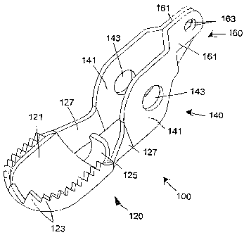
FIG. 10 clearly shows the features of the one-piece jaw
100. The mouth 120 has a cup-shaped distal portion 121 having
radial teeth 123, a proximal tab 125, and two proximal arms
127. The radial teeth 124 project in the direction of the
other jaw 100 and are formed to intermesh with teeth in the
other jaw 100 as shown, in particular, in FIGS. 17 and 18. The
proximal tab 125 is used to retain the tissue sample without
limiting the cup volume. The two proximal arms 127 are
connected to the distal end of the frame 14 and are integral
with the frame 14.
The frame 14 is made of two substantially parallel plates
141 each defining a pivot port 143. The frame 14 is connected
at its distal end to the proximal arms 127 and at its proximal
end to the tang 16. Like the frame 14, the tang 16 is formed
from two parallel plates 161 each defining a control rod port
163. The connection between the plates 141 of the frame 14 and
the plates 161 of the tang 16 are formed by inwardly tapered
proximal portions 145 of the two plates 141, which can be seen
clearly in FIGS. 15 and 16.
Both the frame 14 and tang 16 have contours to make way
for various features of the jaws 10, control wires 3, and/or
clevis 20. For example, the proximal arm 127 closer to the
viewer of FIG. 10 is shorter than the proximal arm 127 further
to the viewer. Also, the plate 161 of the tang 16 closer to
the viewer of FIG. 11 is narrower than the opposing plate 161.
Compare plates 161 in FIGS. 12 and 13.
There are significant advantages that are provided by
having a dual-part tang 16 as compared to a single tang as
shown in FIGS. 1 to 5. Dual tangs are advantageous
particularly for a stamped part because the two tangs share
loads during use. Accordingly, each tang needs to only, be half
CA 02596078 2007-07-26
WO 2006/083728 -26 PCT/US2006/003029
-
as strong as compared to a single tang; this also means that
thinner materials can be used -- which facilitates the stamping
process. The presence of two thin tangs 161 per jaw 100 allows
the tangs 161 to be spread wide from one another and interlaced
when assembled together. See, e.g., FIGS. 26, 28, 30, and 46
to 50. Because the tangs of a dual tang end effector are not
displaced toward the centerline of the part as much as is
required by a single tang end effector, there is less plastic
deformation of the material during the stamping process, which
improves stamping consistency and tool life. Another advantage
achieved by spreading the two tangs 161 from one another is
that it adds stability to the distal assembly. In the dual
tang system, the end effector is supported by two contact
points on the axle, spread apart as far as possible from each
other. The single tang system supports the end effector by one
contact point on the axle. Because of the added support, the
dual tang end effector is less prone to cocking (being
angularly displaced with respect to the longitudinal axis of
the device) than the single tang end effector. This added
stability helps maintain end effector, and in the case of a
biopsy forceps, tooth, alignment.
Another important advantage of a dual-part tang 16 lies in
the clearance that is formed in between the tangs 161.
Positioning the outer plates 141 of the jaws 100 against the
clevis and the inner tangs of the jaws 100 against the inner
surface of the outer tangs to align the jaws 100 to one another
creates a natural gap in the center of the jaw assembly. This
gap is best seen in FIGS. 24, 28, and 47 to 50. Therefore, a
component, such as the stabilization needle 40, can be added
between the jaws 100 within the gap if desired. Single tang
configurations need spacers to fill in gaps between the jaws to
CA 02596078 2007-07-26
WO 2006/083728 PCT/US2006/003029
-27-
prevent jaw movement. The dual-part tang 16 of the present
invention eliminates the need to fill in the center gap with a
component.
The stability afforded by the dual-part tang allows use of
thinner and, thus, more flexible materials. This is
advantageous for assembly purposes because flexible material
tends to give and flex as the device is assembled instead of
interfering rigidly with the assembly, as occurs with thicker
parts. This benefit allows for more open tolerances during
manufacture.
A significant different feature of the one-piece jaw 100
lies in the teeth 123. The distal-most teeth 1231 are larger
than the other teeth 1233. The distal-most teeth 1231 are the
primary teeth for grabbing and cutting the tissue sample to be
obtained. FIG. 16 shows the teeth 1231, 1233 viewed from the
distal end of the jaws 100. FIGS. 17 and 18 illustrate the
mated nature of the teeth 1231, 1233 and, in particular, show
the backward curve of the distal-most teeth 1231. The curve of
these teeth 1231 is beneficial because the rearward angle helps
them to hook into the tissue better and prevent the teeth from
sliding on the tissue while closing the jaws. It also shields
the sharp tips of the teeth from the outer surface of the jaws
when the jaws are closed, thus making the outer surface
smoother and less traumatic to the working channel of the
endoscope while advancing the device therethrough.
FIGS. 19 to 22 illustrate the preferred embodiment of the
clevis 20 of the present invention. To best understand the
features of the clevis 20, reference will be made to FIG. 22,
in particular, which is the stamped form that, after
processing, is shaped into the final clevis 20 illustrated in
FIGS. 19 to 21. The clevis 20 has a number of different
CA 02596078 2007-07-26
WO 2006/083728 PCT/US2006/003029
-28-
portions, all of which are integral because the clevis is
formed, originally, from the stamped part shown in FIG. 22.
The distal-most portion of the clevis 20 is the pivot portion
22. A control portion 24 is adjacent the pivot portion 22 in a
proximal direction. A coil-holding portion 26 is adjacent the
control portion 24 in a proximal direction. Finally, the coil-
retaining portion 28 is adjacent the coil-holding portion 26 at
the proximal end of the clevis 20.
Preferably, all of distal components (including jaws 100,
clevis 20, and needle 40 (see, e.g., FIG. 23)) are of stainless
steel, either work hardened or hardenable by heat treatment to
improve the mechanical properties. Use of thin, heat-treatable
stainless steel in the surgical end effector has several
important advantages. First, the stamping process can be
performed when the material is in its pre-heat-treated or in an
intermediate, ductile, heat-treat condition. In this formable
condition, the material is easily shaped by conventional
stamping and forming processes. Once formed to its final
shape, the part can be heat treated to improve mechanical
properties such as yield strength, ultimate strength, and
hardness. By selecting the alloy properly, such as 17-7
precipitation hardening stainless steel, or Carpenter
Technology Corporation's Carpenter Custom 455TM or Custom 465TM
,
and controlling the heat-treating process (time, temperature,
ramp-soak-cool cycles), a combination of high strength and
elongation before fracture can be achieved. The preferred
materials for the distal components are precipitation
hardenable stainless steels such as UNS S17700 (17-7), UNS
S17400 (17-4), and age-hardenable stainless steels such as UNS
S45500 (Carpenter 455) and UNS S46500 (Carpenter 465). These
materials offer the best corrosion resistance of the hardenable
CA 02596078 2007-07-26
WO 2006/083728 PCT/US2006/003029
-29-
stainless steels. The precipitation hardenable alloy 17-7 is of
particular interest because, unlike some of the other alloys
listed above, 17-7 is austenitic in structure in its annealed
state. Thus, there is an improvement in its ability to be
formed. Martensitic stainless steels such as 420 or 440 can
also be used, but they are not as desirable because their
corrosion resistance is not as good as the other alloys
mentioned herein.
A second advantage to using such materials is that the
distal parts can each be constructed from a thinner material
because strength will be added through the heat-treating
process. Using thinner material allows for sharper cutting
edges in the case of biopsy forceps jaws, for example.
Finally, the ability to use a thinner material affords the
stamping die designer more latitude in construction of the die
and also improves process capability. There is less strain and
subsequent material flow in a thinner section part when bent or
formed into the same shape as a thicker section part. This
characteristic improves consistency of forming and puts less
strain and wear on the die.
With regard to the clevis 20, when the material is of the
kind of steel mentioned and when the arms 222, 242 of the
control portion 24 are bent outward to load the jaws 10, 100
therein, the arms 222, 242 spring back to their original,
parallel orientation such that the tapered ends of each axle
half 224 meet without substantial gap therebetween.
The pivot portion 22 has two substantially parallel axle
plates 222. After stamping the clevis part shown in FIG. 22,
these axle plates 222 are further stamped on the dashed lines
shown in FIG. 22 to produce an axle half 224 in each of the
axle plates 222. The stamp to produce the halves 224 is a
CA 02596078 2007-07-26
WO 2006/083728 PCT/US2006/003029
-30-
three-sided cut that forms a tab, which after formation by
bending inward, rests in a position orthogonal to the plane
defined by the respective axle plate 222. Thus, when the
stamped part of FIG. 22 is shaped to place the axle plates 222
parallel to one another, the axle halves 224 will lie in a
straight line and, therefore, form a bearing upon which the
jaws 10, 100 pivot. The ends of the axle halves 224 each have
a chamfer so that the axle 50 (see FIG. 24) passing through the
pivot port 143 can pass easier and with less outward bending of
the control portion 24 when the jaws 10, 100 are installed.
The control portion 24 includes two control plates 242,
which have a length sufficient to allow the control parts (the
frame 14, 140, the tang 16, 160, and the wires 3) to move
without hindrance as they are used to control the opening and
closing of the jaws 10, 100. The control plates 242 transition
from a substantially flat and planar distal portion to a
rounded proximal portion that is adjacent the coil-holding
portion 26. When the axle 50 and the jaws 10, 100 are
installed on the axle halves 224, the control plates 242
receive most of the outward bending forces. But, because the
material of the clevis 20 is sufficiently bendable, the
stretching of the two control plates 242 away from one another
does not stress the control plates 242 sufficient to
plastically deform them or only plastically deforms them to a
minimal, non-damaging extent. Thus, the control plates 242
spring back into the position shown in FIGS. 19 to 21 when the
jaws 10, 100 are installed.
The coil-holding portion 26 is shaped in an important way.
The coil 2 is formed of a rod that is in the form of a spiral
where each turn rests on the previous one and the next one.
Because of this construction, the last turn does not define a
CA 02596078 2007-07-26
WO 2006/083728 -31 PCT/US2006/003029
-
plane. In fact, the longitudinal distal end of the last 360-
degrees of the coil is constantly changing. It is important to
have the coil be very secure. If the distal stop for receiving
the distal end of the coil 2 was planar, then only a portion of
the last coil turn would contact the distal stop. To make sure
that the last coil turn is contacted properly and securely, the
stamped part of the clevis 20 is formed with four coil-holding
tabs 262, 264, 266, 268. Each of the tabs is created to be at
a different longitudinal distance that follows the pitch of the
wire forming the coil 2. Accordingly, when the stamped clevis
of FIG. 22 is formed into the part shown in FIGS. 19 to 21,
the four tabs 262, 264, 266, 268 are not orthogonal to the
longitudinal axis of the clevis 20. Instead, the tabs 262,
264, 266, 268 travel along the spiral path of the coil 2, as
15 shown most clearly in FIGS. 21 and 32 to 34. In order from
proximal to distal, the tabs include the first tab 262, the
second tab 264, the third tab 266, and the fourth tab 268.
These tabs 262, 264, 266, 268 are shown in FIG. 22 as beginning
on the far right of the stamped clevis 20. The order
20 illustrated is not required. The first tab 262 can begin on
the far left, for example. However, to insure that the tabs
262, 264, 266, 268 follow the shape of the coil, the tabs 262,
264, 266, 268 must be in order (first to fourth) that
corresponds to the turn of the coil (left to right if clockwise
and right to left if counter-clockwise).
The coil-retaining portion 28 is the tube in which the
distal end of the coil 2 is clamped, preferably, by crimping,
and secured. If the coil-retaining portion 28 is sized to fit
the outer circumference of the coil 2 very closely, then no
additional securing device is needed. However, it is important
for the clevis 20 to not fall off of the coil 2 and for the
CA 02596078 2007-07-26
WO 2006/083728 PCT/US2006/003029
-32-
clevis 20 to not deform from the final shape shown in FIGS. 19
to 21. Therefore, the stamped clevis 20 of FIG. 22 is formed
with a connection device 282 that prevents the circular coil-
retaining portion 28 from moving out of its circular shape. It
is preferable if the clevis 20 can be formed into the part
shown in FIGS. 19 to 21 without connecting the ends together
with some external measures. To do so, the connection device
282 uses mechanical measures to connect the right and left ends
shown in FIG. 22. The preferred embodiment of the connection
device 282 is the dovetail connection shown in FIGS. 19 to 22.
With the dovetail, the connection of the right and left ends of
the coil-retaining portion 28 are secured to one another merely
by mechanical measures.
When the jaws 10, 100 are opened and pressed against
tissue to be sampled, it has been found to be beneficial to
have a central pin or spike inside the area of the jaws 10, 100
that will house the tissue sample after closing of the jaws 10,
100. The present invention includes a special self-centering
blade 40 that is to be located between the jaws 10, 100 inside
the cavity formed by the mouth 12, 120 of the jaws 10, 100. As
shown in FIG. 23, the blade 40 has a distal cutting area 42, an
intermediate pivot area 44 having a pivot bore 442, and a
proximal centering device 46 with two centering control
surfaces 462. FIGS. 24 and 25 illustrate how the blade 40 is
installed between and in the jaws 10, 100.
The preferred cutting area 42 is shown in FIG. 23 and is
in the shape of a triangular, double-edged spike. Thus, when
tissue is captured in the cavity of the mouth 12, 120, the
blade 40 bisects the tissue along the length of the blade 40.
FIGS. 24 and 25 show the blade 40 within the jaws 10, 100 and
mounted coaxially with the jaws 10, 100 on the axle 50. It is
CA 02596078 2007-07-26
WO 2006/083728 PCT/US2006/003029
-33-
noted that the axle 50 does not extend past the right side of
the frame 14, 140 when inserted between the axle plates 222 of
the clevis 20 as shown in FIGS. 24 and 25. The axle 50 is
shown longer in FIGS. 24 and 25 for clarity purposes only. In
the preferred embodiment, the axle 50 is approximately flush
with the outer planar surfaces of the frame 14, 140 or
extending just a little bit out from that plane so that the
frame 14, 140 does not rub against the inner surfaces of the
axle plates 222 of the clevis 20 when pivoting thereon.
As set forth above with regard to the tangs 160 and the
wires 3, the tangs 16, 160 are shaped to not interfere with the
pivoting of the one-piece jaw 100. FIG. 26 illustrates an
embodiment of the connection between the control wire 3 and the
tang 150 of each jaw 100. The embodiment shown in FIG. 26 does
not include the centering blade 40, however, with appropriate
shaping of the transition between the frames 140 and the tangs
160, a space can be left in the center of the two jaws 100 so
that the blade 40 can fit on the axle 50 and in the center of
the jaws 100 as indicated, for example, along the dashed line
52 on the axle 50. The wires 3 extend longitudinally up to the
control rod port 163, bend ninety degrees to enter into the
control rod port 163, extend through both the control rod ports
163 of each of the two parallel plates 161 along an orthogonal
portion 32, exit the control rod port 163 of the second
parallel plate 161, and bend ninety degrees to, thereby, engage
the tang 160 without the possibility of falling out of the
ports 163. It is noted that the fit between the ports 163 and
wires 3 is sufficiently loose to permit the tangs 160 to rotate
freely about the orthogonal portion 32.
FIGS. 27 to 29 illustrate the preferred embodiment of the
end effector 200 of the present invention. The clevis 20 and
CA 02596078 2007-07-26
WO 2006/083728 -34 PCT/US2006/003029
-
coil 2 are the same as in the other embodiment but the jaws 220
are different and will be explained in further detail with
regard to FIGS. 39 to 50. The clevis 20 of FIGS. 27 et seq.,
however, illustrates the coil-retaining portion 28 without the
dovetail connection device 282.
As can be seen, the control wires 3 emerge from the distal
end of the coil 2 and from the coil-holding portion 26 of the
clevis 20 at the control portion 24 of the clevis 20. The
wires 3 bend ninety degrees to enter into the control rod port
263, extend through both the control rod ports 263 of each of
the two parallel plates 261 along an orthogonal portion 32 (see
FIG. 28), exit the control rod port 263 of the second parallel
plate 261, and bend ninety degrees to, thereby, engage the tang
260 without the possibility of falling out of the ports 263.
As seen, in particular, in FIGS. 27 and 29, the fit between the
ports 263 and wires 3 is sufficiently loose to permit the tangs
260 to rotate freely about the orthogonal portion 32.
One of the two centering control surfaces 462 of the
proximal centering device 46 of the blade 40 is shown clearly
in FIGS. 28 and 30 to 34. The control surface 462 interacts
with a blade control tab 241 on the frame 240. The cross-
sectional view of FIGS. 32 to 34 shows the interaction between
control surface 462 and the blade control tab 241. When the
jaws 220 are closed, the control surfaces 462 do not interact
with the blade control tabs 241. The movement of the blade 40
shown in FIGS. 32 and 33 illustrates the free pivot of the
blade 40 when the jaws 220 are closed. In contrast, when the
jaws 220 are opened past a given extent, the control surfaces
462 interact with the blade control tabs 241 and, as shown in
FIG. 34, rest directly thereupon so that the control surface
462 lies on and parallel to the respective contacting surface
CA 02596078 2007-07-26
WO 2006/083728 PCT/US2006/003029
-35-
of the blade control tab 241. Because of this configuration,
the blade control tab 241 also acts as a jaw limiter, which
defines the greatest extent that the jaws 220 can be opened.
Simply put, the angle a defined by the two control surfaces 462
(see FIG. 34) defines the maximum opening angle of the jaws
220. If the angle a increases from that illustrated in FIG.
34, then the jaws 220 can open more and if the angle a
decreases, then the jaws 220 can open less. FIG. 35
illustrates the elevational view of the jaws 220 of FIG. 34.
FIGS. 36 and 37 show views of the preferred embodiment
similar to FIGS. 27 and 28 but without the control wires 3, and
FIG. 38 illustrates, in an exploded view, the various parts of
the end effector of FIGS. 27 to 50. To assembly the end
effector of the present invention, one set of the parallel
plates 261 of one tang 26 is pulled apart and the two jaws 220
are nested. See, e.g., FIGS. 46 to 50. The blade 40 is placed
in the center of-the two jaws 220 and the axle is passed
through the pivot ports 243 of each and the pivot bore 44 of
the blade 40. Then, the two axle halves 224 are pulled apart
so that the axle 50 can fit over one of the axle halves 224.
One opening of the axle 50 is placed all the way on one axle
half 224 and the other axle half 224 is allows to spring back
to, with the aid of the chamfer on the distal end of the other
axle half 224, enter into the second, opposite opening of the
axle 50. The control wires 3 are, then, inserted through the
control rod ports 263 to, thereby, fix the jaws 220 in position
in the clevis 20.
FIGS. 27 to 50 illustrate the preferred embodiment of the
jaws 220 of the present invention. As can be seen in all of
FIGS. 27 to 50 and, especially, in FIGS. 39 to 50, the mouth
portion 221 is angular in its configuration. The two lateral
CA 02596078 2007-07-26
WO 2006/083728 -36 PCT/US2006/003029
-
sides 223 are substantially parallel to one another and are
substantially linear and the front face 225 is substantially
orthogonal to the lateral sides 223 and is also substantially
linear. Respective angled sides 229 connect the lateral sides
223 to the front face 225. Like the lateral sides 223 and the
front face 225, the angled sides 229 are substantially linear.
The proximal arms 227 connect the lateral sides 223 to the
frame 240.
In FIGS. 27 to 50, the lateral sides 223 are shown with
teeth 2232, the front face 225 is shown with teeth 2252, and
the angled sides 229 are shown with teeth 2292. However, any
of these teeth 2232, 2252, 2292 can be removed. In one
preferred embodiment, the teeth 2232, 2292 of the respective
sides 223, 229 can be configured to form a space therebetween
to create gaps at the junctions of the lateral-angled sides and
the angled side-front face. Such a configuration allows the
captured tissue in the mouth portion 221 to flow or bulge out
of the resulting spaces and, therefore, makes room for more
tissue to be captured as the biopsy sample. Gaps at these
junctions also provides the second benefit of minimizing edges
that can scrape or otherwise damage the working channel of an
endoscope as the end effector passes therethrough. These gaps
will be discussed in further detail below.
Each outer surface of each frame 240 is formed with a
protruding ring 245 surrounding the pivot port 243 for
receiving the axle 50. This ring 245 permits the nested parts
-- needle 40, jaw 220, jaw 220 -- to pivot freely with respect
to one another about the axle 50.
The jaw configuration of FIGS. 27 to 50 is not the only
variation for the frusto-conical distal end 229, 225 of the
jaws 220. As shown in FIG. 51, for example, the distal end can
CA 02596078 2007-07-26
WO 2006/083728 PCT/US2006/003029
-37-
be bi-frusto-conical and have two angled sides portions, a
proximal portion 229' and a distal portion 229''. Also, as
shown in FIGS. 65 and 66, the frusto-conical portion 229 can
have a curved or frusto-hemispherical shape.
As set forth above, the two tang embodiment is a preferred
configuration. However, like FIGS. 1 to 5, the embodiment of
FIGS. 27 to 50 can also be constructed with a single tang.
Such a variation is illustrated in FIGS. 52 to 64.
FIGS. 52 to 57 illustrate the alternative embodiment of
the jaw 320 of the end effector 300 having a single tang 360.
Teeth 3232, 3252, 3292 can be present or absent from any of the
edges. In the illustrated embodiment, the two lateral sides
323 are shown with four teeth 3232 each and the frusto-conical
portion 329 has one or two teeth 3292 on the two edges,
respectively. Finally, the front face 325 is shown with two
teeth 3252. The opposing jaw 320, shown particularly in FIG.
58 (and also in FIGS. 60 to 64) can be configured without teeth
or with teeth that interdigitate with the teeth of the jaw 320
shown in FIG. 52.
As can be seen in all of FIGS. 52 to 64 and, especially,
in FIGS. 52 and 58, the mouth portion 321 is angular in its
configuration. The two lateral sides 323 are substantially
parallel to one another and are substantially linear and the
front face 325 is substantially orthogonal to the lateral sides
323 and is also substantially linear. Respective angled sides
329 connect the lateral sides 323 to the front face 325. Like
the lateral sides 323 and the front face 325, the angled sides
329 are substantially linear (but can be curved as shown in
FIGS. 65 and 66). The proximal arms 327 connect the lateral
sides 323 to the frame 340.
CA 02596078 2007-07-26
WO 2006/083728 - 38- PCT/US2006/003029
In FIGS. 52 to 64, the lateral sides 323 are shown with
teeth 3232, the front face 325 is shown with teeth 3252, and
the angled sides 329 are shown with teeth 3292. However, any
of these teeth 3232, 3252, 3292 can be removed. In one
preferred embodiment, the teeth 3232, 3292 of the respective
sides 323, 329 can be configured to form a space therebetween
to create gaps at the junctions of the lateral-angled sides 323
and the angled side-front face 325. Such a configuration
allows the captured tissue in the mouth portion 321 to flow or
bulge out of the resulting spaces and, therefore, makes room
for more tissue to be captured as the biopsy sample. Gaps at
these junctions also provides the second benefit of minimizing
edges that can scrape or otherwise damage the working channel
of an endoscope as the end effector passes therethrough. These
gaps will be discussed in further detail below.
The features of the tang 360 and the tang interface to the
blade 40 are similar to the dual-tang configuration of the
stamped jaw shown in FIGS. 10 to 18 and 27 to 51 and,
therefore, will not be repeated herein.
It is noted that each of the distal end portions of the
present invention is illustrated as being frusto-conical.
This, however, is not the only configuration for the distal-
most end of the end effector. In an alternative embodiment,
the distal-most end (i.e., 225, 325) can come to a point, can
be curved, or can be entirely non-existent. As such, in one
alternative embodiment of the front face 225, 2252, 325, 3252,
either the teeth 2252, 3252 or both the teeth 2252, 3252 and
the front face 225, 325 can be removed to provide additional
spaces in which captured tissue in the mouth portion 221, 321
can flow or bulge out.
CA 02596078 2007-07-26
WO 2006/083728 PCT/US2006/003029
-39-
Like the jaw 220, each outer surface of each frame 340 can
be formed with a protruding ring surrounding the pivot port 343
for receiving the axle 50, which permits the nested parts --
needle 40, jaw 320, jaw 320 -- to pivot freely with respect to
one another about the axle 50.
It is advantageous to use annealed, thin sheet metal when
forming the end effectors of the present invention by stamping.
Such thin sheet metal allows tighter bends and finer detail to
be produced during the stamping process. Annealed material is
easier to form and cut and produces less tool wear. Once
formed from the thin stock, the end effector jaw may not have
the required mechanical properties without secondary
processing. Secondary processing can greatly enhance the
mechanical properties of such sheet metal and examples of such
processing include: heat treatment, age hardening, case
hardening, ion implantation, carbon-nitriding, cold working or
combinations of these. In the case of biopsy forceps jaws
formed from 17-7 precipitation hardenable stainless steel, post
processing (precipitation hardening) to increase the mechanical
properties of the material is required as the ratio of the
diameter of the end effector to the thickness of the material
approaches 11:1. Preferably, the ratio for a flexible,
endoscopic instrument (such as a biopsy forceps) is between
approximately 30:1 and approximately 11:1, in particular,
between approximately 20:1 and 11:1, and specifically, at
approximately 17:1 or at approximately 11.4:1.
FIGS. 65 and 66 show a further embodiment of the jaw
assembly of the present invention. The jaws 300 have a curved
intermediate section 329 flanked by linear sides 323 and a
linear front face 325. In this embodiment, the axle 50 has an
outer rectangular shape with rounded ends. The outer shape
CA 02596078 2007-07-26
WO 2006/083728 - 4 0- PCT/US2006/003029
corresponds to the shape of the axle port in the spike 40 so
that the spike remains in a position corresponding to the
orientation of the axle 50.
FIGS. 67 to 70 show yet another embodiment of the jaw
assembly of the present invention. This embodiment expands
upon the ability of the jaws 220 to ensure that the maximum
volume of tissue is captured in the mouth portion 221 of the
jaws 220. Many of the features are similar to FIGS. 39 to 45,
for example, and, therefore, explanation of similar features is
not repeated. In contrast to the embodiment illustrated in
FIGS. 27 to 50, the mouth portion 221 and two lateral sides 223
define fenestrations 226. Such a configuration allows the
captured tissue in the mouth portion 221 to flow or bulge out
of the resulting spaces and, therefore, makes room for more
tissue to be captured as the biopsy sample.
The fenestrations 226 need not merely be cut at right
angles to the plane of the stamped jaw 220. The edges of each
fenestration 226 can be formed towards the inside in a stamped
forceps cup to effectively round the edges thereof and,
therefore, decrease the possibility of scope channel wear
caused by movement of the forceps inside the endoscope, for
example. Two teardrop-shaped or egg-shaped fenestration holes
226 are better than one hole 226 for scope wear because the web
2262 between the holes 226 will be presented to the scope
channel as the forceps pass therethrough instead of the edge of
the hole being presented to the scope channel.
Biopsy forceps cups are typically fenestrated. However,
the prior art designs do not reduce chances for endoscope wear,
rather, they increase the probability. As set forth above, the
exposed edges of the fenestrations 226 (see FIGS. 67 to 72 and
74 to 75) are shaped to minimize such contact. By separating
CA 02596078 2007-07-26
WO 2006/083728 -41 PCT/US2006/003029
-
the fenestrations 226 with a center web 2262, only the smooth
surface of the web 2262 is in contact with the scope channel as
the end effector passes through the channel. See, i.e., FIG.
73. Accordingly, as the end effector 200 impinges upon a curve
in the working channel, the intersection of the angled nose
edge and the lateral straight edge of the jaw can contact the
scope channel. If this intersection includes a tooth edge, the
edge can be forced against the wall of the scope channel
potentially damaging it. Therefore, it is advantageous to
interrupt the teeth in this transition area to minimize the
potential of scope channel wear.
When passing through such a curve, end effectors are
forced against the surface of the working channel 1000 as they
track around the curve. Similarly, as an end effector of a
typical biopsy forceps passes through a working channel, it,
too, slides along the interior surface of the working channel.
This increased force of sliding can cause endoscope channel
wear if the surface of the end effector is rough, or if it has
exposed edges -- such as the exposed edges of sides of forceps
teeth. Thus, the present invention provides various aspects
that decrease the possibility of damaging the working channel
of a flexible endoscope.
FIGS. 71 to 73 illustrate the end effector 200 of the
present invention passing through a curve in a working channel
1000 of a flexible endoscope. FIG. 71 illustrates a "worst-
case" scenario with respect to contact between the end effector
200 and the channel 1000. Specifically, the bending axis of
the end effector 200 - corresponding to the axle 50 (see, i.e.,
FIG. 24) - is parallel to and lies in the plane defined by the
curve of the working channel 1000 (which is also, in this
example, the plane in which all of FIGS. 71 to 73 lie). In
CA 02596078 2007-07-26
WO 2006/083728 PCT/US2006/003029
-42-
such a configuration, the end effector 200 substantially cannot
bend when it first hits the curve -- shown by first contact
point 1010 in FIG. 71 - because the jaws 220 only pivot about
the axle 50. Therefore, to traverse the curve, the axle 50
needs to be at least at an angle to the plane of the curve or,
ideally, perpendicular to the plane of the curve.
The first contact point 1010 is located at a first tooth
gap 2254 on either side of the teeth 2252 on the front face
225. See, i.e., FIG. 31. If a tooth 2252 were located at the
gap 2254, then the potentially sharp outer surfaces of the
tooth 2252 could damage the inner surface of the working
channel 1000. Thus, the first gap 2254 is provided by the
present invention. Because the end effector 200 is being
pushed further into the curve, as the end effector 200 is
traversing the curve, the outside of the coil 2 can bend
slightly to place a second contact point 1020 against the
interior surface of the working channel 1000. FIG. 72
indicates the second contact point 1020 touching the working
channel 1000 and the first contact point 1010 not touching the
working channel 1000. Of course, depending on the curve of the
working channel 1000, both contact points 1010, 1020 can touch
the interior surface of the working channel 1000.
This second contact point 1020 is located at a second
tooth gap 2256 between the teeth 2292 and the side teeth 2232
on the angled side 229 and the lateral side 223, respectively.
See, i.e., FIG. 31. If a tooth 2232 or 2292 were located at
the gap 2256, then the potentially sharp outer surfaces of the
tooth could damage the inner surface of the working channel
1000. Thus, the second gap 2256 is provided.
The forceps of the present invention has a relatively long
coil 2 that can be rotated somewhat about its longitudinal axis
CA 02596078 2007-07-26
WO 2006/083728 PCT/US2006/003029
-'~3-
without irreparable deformation. As such, when the end
effector 200 traverses through the curve, the rotational
resistance is much less than the lateral bending forces imposed
in the plane of the axis 50 to, thereby, naturally rotate the
end effector 200 inside the working channel 1000 to an
orientation shown, for example, in FIG. 73. In such an
orientation, the end effector 200 can bend about the axle 50
and, ideally in this example, rotate ninety degrees to place
the web 2262 against the inner surface of the curve at third
contact point 1030. Because the web 2262 is smooth and curved,
it passes through the curve of the working channel 1000 with
minimal wear.
Next, the force of passage can be mitigated by decreasing
the rigid (non-bendable) length of the end effector so that it
tracks around and conforms better to the curves.
Prior art devices include a centering spike that is
significantly longer than the spike 40 of the present
invention. These prior art centering spikes can even extend
through the end effector in a proximal direction well past the
clevis. The centering spike of the Radial JawTM biopsy forceps
manufactured by Boston Scientific is one example where the
rigid length is approximately two times longer than the spike
40 of the present invention. The presence of such a long
centering spike prohibits the prior art end effector from
pivoting when the jaws are closed, thus effectively increasing
the rigid length of the prior art end effector.
In contrast, the present invention allows the centering
spike 40 to freely pivot within and with the jaws 220 to,
thereby, effectively decrease the rigid length of the end
effector 200 and make passage through the working channel much
easier than the prior art end effectors.
CA 02596078 2007-07-26
WO 2006/083728 PCT/US2006/003029
-44-
FIGS. 27 to 29, 68, and 69 show a first embodiment of the
actuating wires or rods 3. In that embodiment, the wires 3
extend longitudinally up to the control rod port 163, 263, bend
ninety degrees to enter into the control rod port 163, 263,
extend through both the control rod ports 163, 263 of each of
the two parallel plates 161, 261 along an orthogonal portion
32, exit the control rod port 163, 263 of the second parallel
plate 161, 261, and bend ninety degrees to, thereby, engage the
tang 160, 260 without the possibility of falling out of the
ports 163, 263. To actuate the forceps, a force is exerted on
the rod 3. The S-bend of the wire 3 significantly reduces any
possibility that the wire 3 becomes dislodged from the tang
160, 260.
FIGS. 74 to 79 illustrate another embodiment 360 of the
tangs 160, 260 and second and third embodiments of the wires 3
shown in the previous figures.
As set forth above, the wire 3 is attached to the jaw 10
to allow the wire 3 to freely rotate and to allow each jaw 10
of the end effector 1 to pivot. The wire 3 must be secured so
that it can both push and pull on the tang 160, 260, 360 of the
jaw 10 but not become dislodged therefrom. The primary
transmission of force occurs during closing of the end effector
1, which is accomplished by pulling on the wire 3 in a distal
direction. As tension is applied to the wire 3, the bends in
the distal end of the wire 3 that connect the wire 3 to the
tang 160, 260, 360 are stressed and can begin to straighten --
allowing the wire 3 to pull though the control rod port 163,
263, 363 and slip free from the tang 160, 260, 360. This
possibility only exists, however, if no measures are taken to
prevent such straightening.
CA 02596078 2007-07-26
WO 2006/083728 PCT/US2006/003029
-45-
In particular, FIGS. 74 to 76 illustrate the second
embodiment of the wires 3. In these figures, only one jaw 10
is shown. However, each jaw 10 has its respective wire 3 and,
therefore, the description herein need not be repeated. To
attach to the jaw 10, the wire 3 extends longitudinally up to
the control rod port 363 (from the proximal end of the forceps
device) along a first portion 31, bends ninety degrees to enter
into the control rod port 363, extends through both the control
rod ports 363 of each of the two parallel plates 361 along an
orthogonal portion 32, exits the control rod port 363 of the
second parallel plate 361, and bends another ninety degrees to
extend back in the proximal direction along an third portion 33
and, thereby, engage the tang 360. This second embodiment of
the wire 3 significantly reduces any possibility that the wire
3 becomes dislodged from the tang 360.
To effect the reduction of unintended wire 3 removal, one
plate 361 of the two parallel plates 161, 261 has a hooded wire
support 365. The hooded wire support 365 is configured to
provide support to the wire 3 and, thereby prevent removal of
the wire 3 from the tang 360. The hooded wire support 365 is
substantially U-shaped in cross-section and, therefore, has a
vertical first leg 366, a horizontal portion 367, and a
vertical second leg 368 opposite the first leg 366. The
vertical first leg 366 is integral with the plate 361 and
extends therefrom at one edge of a proximal portion of the
plate 361(of course it can extend from the other edge or from
both edges). The support 365 surrounds the third portion 33 of
the wire 3 on three sides. Importantly, the support 365 makes
contact or is close to a side of the third portion 33 that is
opposite the first portion 31. When a force F is imparted on
the wire (see FIG. 74), the third portion 33 is urged to
CA 02596078 2007-07-26
WO 2006/083728 PCT/US2006/003029
-46-
straighten in a direction G. That straightening force is
counteracted with the presence of the vertical second leg 368
acting as a stopping surface. In this embodiment, the wire 3
approaches the tangs 360 from the plate 361 opposite the
support 365 and bends around to end inside the support 365.
FIGS. 77 to 79 illustrate the third embodiment of the
wires 3. Like FIGS. 74 to 76, only one jaw 10 is shown. Each
jaw 10 has its respective wire 3 and, therefore, the
description herein is not repeated. Unlike FIGS. 74 to 76, in
the third embodiment, the wire 3 approaches the tangs 360
through the support 365 and bends around to end inside the tang
360.
To attach the wire 3 to the jaw 10, the wire 3 extends
longitudinally from the proximal end of the forceps device up
to the control rod port 363 along a first portion 31, bends
ninety degrees to enter into the control rod port 363, extends
through both the control rod ports 363 of each of the two
parallel plates 361 along an orthogonal portion 32, exits the
control rod port 363 of the second parallel plate 361, and
ends. This third embodiment of the wire 3 similarly reduces
any possibility that the wire 3 becomes dislodged from the tang
360.
To effect the reduction of unintended wire 3 removal, one
plate 361 of the two parallel plates 161, 261 has a hooded wire
support 365. The hooded wire support 365 is configured provide
support to the wire 3 and, thereby prevent removal of the wire
3 from the tang 360. The hooded wire support 365 is
substantially U-shaped in cross-section and, therefore, has a
vertical first leg 366, a horizontal portion 367, and a
vertical second leg 368 opposite the first leg 366. The
vertical first leg 366 is integral with the plate 361 and
CA 02596078 2007-07-26
WO 2006/083728 PCT/US2006/003029
-47-
extends therefrom at one edge of a proximal portion of the
plate 361 (of course it can extend from the other edge or from
both edges). The support 365 surrounds the first portion 31 of
the wire 3 on three sides. Importantly, the support 365 makes
contact or is close to a side of the first portion 31 that is
opposite the plate 161, 261. When a force F is imparted on the
wire (see FIG. 77), the horizontal portion 32 is urged to
straighten. That straightening force is counteracted with the
presence of the vertical second leg 368 acting as a stopping
surface.
It is noted that the two actuating wires 3 are not
illustrated as extending through the entirety of the body 2, 4
and into the handle 62 because such illustration is deemed to
be unnecessary. Nonetheless, the preferred embodiment of the
present invention provides the two actuating wires 3 with a
length that extends all the way into the handle 62 and through
most (if not all) of the rod 5 and, in particular, through the
three sections 511, 513, 515 of the proximal connecting portion
51 of the rod 5, because it is the bends of the three sections
511, 513, 515 that form a structure that longitudinally holds
the two wires 3 within the rod 5.
The configuration of the wires 3 are mentioned here to
assist in the explanation of a significant feature of the
present invention that is particularly illustrated in FIGS. 80
and 81.
When biopsy instruments present themselves to a tissue to
be sampled, it is a rare occurrence for the front face of the
biopsy jaws to be exactly orthogonal to the tissue to be
sampled. This non-orthogonal angle-of-attack means that, when
the jaws are pressed into the tissue 1100 to be sampled, the
tissue 1100 will not entirely fill up the mouth of the jaws
CA 02596078 2007-07-26
WO 2006/083728 z U_ PCT/US2006/003029
(the rear corner of the mouth portion further away from the
tissue will be empty). To insure that there is a maximum
volume of tissue sampled, the jaws 220 are formed with the
linear front face 225 and angled sides 229. The edge 228
(formed between the front face 225 and each side 229) forms a
gripping corner that, when pressed against tissue 1100 to be
sampled, plants itself in the tissue 1100 and, naturally,
creates a pivoting force that attempts to orient the front face
225 in a direction parallel to the surface of the tissue 1100.
This movement of the jaws 220 from an off-axis approach to an
orthogonal approach ensures that there is a maximum volume of
tissue captured in the mouth portion 221 of the jaws 220 when
the jaws 220 are closed.
Prior art biopsy forceps, such as the Radial JawT'" forceps
manufactured by Boston Scientific, contained a center spike
between the jaws. However, the spike of this prior art forceps
remains longitudinally aligned with the clevis (as the
orientation of the needle 40 shown in FIG. 80) even if the jaws
are pivoted into an orientation shown in FIG. 81. The needle
40 of the present invention, in contrast, pivots with the two
jaws 220 to remain centered therebetween when the jaws 220 are
fully opened anywhere between the position shown in FIG. 80 and
the offset position shown in FIG. 81 (of course the jaws 220
can pivot in the opposite direction than that illustrated in
FIG. 81). This pivoting of the needle 40 ensures that the
problem of the prior art does not occur -- the needle no longer
crushes the biopsy sample that is contained in the close side
jaw.
The length of each of the actuating rods 3 inside the coil
2 (which is flexible and, therefore, constructed to bend) are
sufficient to compensate for the differential lengths that are
CA 02596078 2007-07-26
WO 2006/083728 - 4 9- PCT/US2006/003029
needed to place the jaws 220 and the needle 40 in the position
illustrated in FIG. 81. Of course, there is the possibility of
an alternative embodiment where the two wires 3 are
significantly shorter and, instead, connect to a distal end of
a third non-illustrated connector that extends through the body
2, 4 to longitudinally connect the rod 5 in the handle 60 to
the two wires 3. However, in such an embodiment, if the wires
3 are too short, then there will not be enough play for the
wires 3 to permit the offset centering of the end effector 220
against tissue that is presented at an angle to the distal end
of the end effector 220 as described above with respect to
FIGS. 80 and 81.
Actuation of the end effector 1 of the present invention
occurs by applying a force on the wires 3 as shown by the
comparison of FIGS. 28 and 29 or 36 and 37. To close the jaws
10, the wires 3 are drawn in a proximal direction and to open
the jaws 10, the wires 3 are extended in a distal direction.
The embodiments of the wires 3 illustrated herein all show two
separate wires 3 each respectively connected to one of the jaws
10 at a distal end thereof. The proximal end of the wires 3
are not illustrated but are each connected to the distal end of
a single actuating rod 5. The proximal end of the actuating
rod 5 is illustrated in FIG. 82 with the ellipses indicating
that the rod 5 and/or the wires 3 therein extend in the distal
direction to the end effector.
FIG. 82 illustrates all of the parts making up the
proximal actuator or handle 60 of the present invention. The
proximal-most portion of the jacketed coil (including the outer
coating 4 and the inner coil 2) is shown separated from the
actuating rod 5 (which, in the assembled condition, extends
through the coil 2). The actuating rod 5 has, at the proximal
CA 02596078 2007-07-26
WO 2006/083728 _ 5 0 - PCT/US2006/003029
end, a connection portion 51 for connecting the rod 5 to a
handle center 62 in a manner that will be explained in greater
detail below.
The actuating portions of the handle 60 are composed of
the handle center 62, which, in the preferred embodiment, has a
thumb ring 622 for receiving therein a thumb of a user, and a
two-part spool 70, 72 for receiving at least one finger of a
user, preferably, at least the index and middle fingers. The
first part 70 of the spool contains features for applying
cauterizing energy to the metallic jaws 10 and for securing the
two spool parts together about the handle center 62.
Cauterizing is not a required function of the forceps of the
present invention but is preferred when tissue samples being
taken will be better obtained when the patient's sample injury
area is sealed by cauterization. Therefore, the present
invention will be described with a cauterization feature. The
second spool part 72 also can contain features for securing the
two spool parts together about the handle center 62.
The cautery spool part 70 houses an electrically
conducting cautery plug 80 for connecting to a cauterizing
energy supply device 82 (illustrated only diagrammatically in
FIG. 82). The supply device 82 engages a cautery supply
connector (e.g., in the form of a plug receiver 74) disposed on
the cautery spool part 70 for supplying cauterizing energy to
the cautery plug 80. The cautery plug 80 is made of metal, for
electrical conductivity, such as stainless steel but,
preferably, copper, or copper alloys such as phosphor bronze,
or beryllium copper.
The preferred embodiment of the cautery plug 80 is formed
with a supply connector 81 having a shape that can removably
connect to the supply device 82. This supply connector 81
CA 02596078 2007-07-26
WO 2006/083728 -51 PCT/US2006/003029
-
extends through a plug receiver 74 that is only illustrated in
part in FIG. 82. Preferably, this receiver 74 is shaped to
receive a standard cautery supply plug in conventional cautery
supply devices.' In particular, the orifice of the receiver 74
and the supply connector 81 are shaped to removably connect to
a cautery supply device manufactured by Olympus and/or
Microvasive. A plug orifice is formed in the cautery spool
part 70 for receiving the cautery plug 80 in a form-fitting
manner and can be seen partially in FIG. 83 by the outline of
the cautery plug 80. A form-locking or form-fitting connection
is one that connects two elements together due to the shape of
the elements themselves, as opposed to a force-locking
connection, which locks the elements together by force external
to the elements.
On the cautery plug 80, a spool connector 83 is disposed
on a side opposite the supply connector 81 and form-fittingly
inserts into a non-illustrated orifice located on the interior
of the second spool part 72. Thus, when the two spool parts
70, 72 are connected together to surround the handle center 62
(and slide thereon as will be explained below), the cautery
plug 80 is held in place.
The cautery plug 80 also needs to contact the rod 5 so
that the cauterizing energy can be transmitted from the supply
device 82 through the plug 80, then through the rod 5 and wires
3, to the jaws 10 of the end effector 1. To make such a
contact, the cautery plug 80 has a rod connection device 85,
which, in a preferred embodiment, is in the form of a
downwardly extending flange 85 defining a hole 87 for receiving
therethrough the proximal connecting portion 51 of the rod 5.
To attach the connecting portion 51 of the rod 5 to the flange
85, the proximal end 511 of the rod 5 is inserted into the hole
CA 02596078 2007-07-26
WO 2006/083728 PCT/US2006/003029
-52-
87 in a position orthogonal to the position shown in FIG. 82.
Then, the rod is rotated in a direction towards the
longitudinal axis of the flange 85 so that a curved portion
having a horizontal section 513 rests within the hole 87. At
this point, the extent of the rod 5 is parallel to the
longitudinal axis of the flange 85 as shown in particular in
FIG. 83. A distal portion 515 of the connecting portion slants
towards the longitudinal axis so that when the rod bends away
from the distal portion 515, the longitudinal axis of the rod 5
is substantially aligned with the longitudinal axis of the
flange 85 as shown in FIG. 83.
The handle center 62 has the thumb ring 622 on a proximal
end, a cable receiver 624 on a distal end, and a slide portion
626 disposed between the ring 622 and the cable receiver 624.
The slide portion 626 is shaped to receive the two spool parts
70, 72 thereat so that the spool 70, 72 can slide between a
distal position and a proximal position. The slide portion 626
is also shaped to receive the cautery plug 80 therein between
the two spool parts 70, 72 so that the cautery plug 80 travels
with the spool 70, 72 when moved and so that the rod 5 moves
with the plug 80 and spool 70, 72.
To better explain the connectivity between the moving and
stationary parts of the handle 60, reference is made, now, to
FIG. 83. Sliding movement of the spool 70, 72 is created by
forming the outer circumference of the handle center 62 in a
curved manner, preferably, in a circle. Similarly, inside
bearing and sliding surfaces 722, 702 are respectively formed
on the interior of the spool part 72 and the cautery spool part
74.
Free movement of the cautery plug 80 (disposed between the
spool parts 70, 72) is permitted by forming a longitudinal slot
CA 02596078 2007-07-26
WO 2006/083728 -53 PCT/US2006/003029
-
6262 inside the handle center 62. As such, when the spool 70,
72 moves up and down along the slide portion 626, the cautery
plug 80 moves as well. Such movement, therefore, causes the
rod 5 to move with and dependent upon movement of the cautery
plug 80. So that the connecting portion 51 of the rod 5 does
not rotate, swivel, or rock, each of the spool parts 70, 72
contains bearing surfaces. Two of these bearing surfaces 704
are shown in FIG. 83 on the cautery spool part 70.
To prevent the spool 70, 72 from spinning around the
longitudinal axis of the handle center 62, each of the spool
parts 70, 72 contains a portion of an anti-spinning device 706,
726 in the form of a flange having a distance slightly less
than a width of the slot 6262. Of course, one or the other of
the spool parts 70, 72 can contain this device 706, 726.
However, in the embodiment shown, each spool part contains half
of the device 706, 726.
There are many ways to connect the two spool parts 70, 72
together, whether removably or permanently. FIGS. 82 and 83
illustrate one preferred way to connect the spool parts 70, 72
together. Each spool part 70, 72 contains two pegs 708, 728
and two connector receiving ports 710, 720. The pegs 708, 728
can be press fit into the ports 710, 720 in a form-fitting
and/or a force-fitting manner. In the embodiment shown in
FIGS. 82 and 83, the pegs 708, 728 are hollow and can have a
smooth or threaded interior. Non-illustrated screws or bolts
are inserted through the ports 710, 720 from a side opposite
the pegs 708, 728 and screw or thread into the interior of the
pegs 708, 728.
Connection of the shaft 2, 4 to the handle center 62
occurs at the cable receiver 624 as shown in FIGS. 83 to 85.
The cable receiver 624 has an interior bore 6242 from its
CA 02596078 2007-07-26
WO 2006/083728 - 5 4- PCT/US2006/003029
proximal end to its distal end. The bore 6242 has a constant
diameter. However, an integral spacer 6244 is disposed inside
the proximal end of the bore 6242 in a co-axial orientation.
The spacer 6244 functions in two ways. First, it centers the
rod 5 after the rod 5 has been threaded through the bore 6242
from the distal end through the proximal end to, thereafter, be
threaded through the hole 87 of the flange 85 of the cautery
plug 80. The spacer 6244 is shallow enough to not prevent the
bent connecting portion 51 from passing therethrough. Second,
the spacer 6244 acts as a proximal stop for the shaft 2, 4 to
prevent further proximal threading of the shaft 2, 4 through
the bore 6242.
However, if the shaft 2, 4 is merely inserted into the
bore 6242, it will not be held therein. It is desirable to
have a single component that accomplishes the task of
connecting the shaft 2, 4 to the handle center 62 without the
use of adhesives and without any additional crimping or forming
operations. Accordingly, a shaft retainer 90 is provided.
The shaft retainer 90 is a device that grasps onto both
the cable receiver 624 and the polymer coated material 4 around
the spring coil shaft 2 to join the two parts together. The
shaft retainer 90 simply slides over the proximal end of the
shaft 2, 4. To control the depth of insertion, in a first
embodiment shown in FIG. 86, the shaft retainer 90 has a stop
92 formed by folded over tabs. In a second embodiment shown in
FIG. 68, the stop 92 can be formed with dimples, coffee spouts,
or any other shape that protrudes into the internal diameter of
the shaft retainer 90 sufficient to stop entry of the shaft 2,
4. The shaft 2, 4, with the retainer 90 attached, is, then,
inserted into a hole 6246 at the distal end of the handle until
it hits the spacer 6244. This fully inserted and connected
CA 02596078 2007-07-26
WO 2006/083728 _ nn_ PCT/US2006/003029
position is illustrated in FIG. 85 (the rod 5 is removed for
clarity).
Merely providing the shaft retainer 90 on the proximal end
of shaft 2, 4 may not be sufficient to prevent removal of the
shaft 2, 4 if a distal force is exerted upon the shaft 2, 4.
Therefore, the shaft retainer 90 is formed with outwardly
projecting tines 94 and inwardly projecting tines 96. The
outwardly projecting tines 94 are flattened as the shaft
retainer 90 is pressed into the bore 6242. But, when such
movement stops, the outward bias of the tines 94 press against
the interior wall of the bore 6242. The sharpness and hardness
of the tines 94 combined with the relative softness of the
interior of the bore 6242 (which is, preferably, a plastic)
digs into the bore 6242 and prevents withdrawal of the shaft
retainer 90 from the bore 6242. If the bore 6242 is provided
with interior roughness (whether random, threaded, or
periodic), then the tines 94 may be assisted in firmly
contacting the interior wall of the bore 6242 by providing
spaces for the tines 94 to spring. In such a configuration,
the shaft retainer 90 is locked in the bore 6242. The locking
can also be accomplished with holes 6248 that penetrate through
the body of the cable receiver 624 (illustrated only
diagrammatically with dashed lines in FIG. 85.
The above connection securely connects the shaft retainer
90 to the cable receiver 624. The shaft retainer 90 is further
provided with inwardly projecting tines 96. These tines 96
protrude toward the inside of the shaft retainer and, after the
shaft 2, 4, is inserted therein, dig into the softer plastic
coating of the protective material 4 around the spring coil 2
and may even penetrate enough to grab onto the ridges of the
coil 2 itself as shown in FIG. 85. Thus, without the use of
CA 02596078 2007-07-26
WO 2006/083728 -56 PCT/US2006/003029
-
adhesives and without any additional crimping or forming
operations, the shaft retainer 90 fixedly connects the shaft 2,
4 to the handle center 62 in a very simple manufacturing step.
FIG. 88 illustrates the assembly containing the rod 5, the
shaft 4, 2, and the shaft retainer 90 separate from the
assembly containing the handle center 62 and the spool 70, 72
holding the cautery plug 80. To conserve material when
manufacturing the handle center 62, cutouts 628 may be provided
in any manner or shape on the exterior of the handle center 62.
Because a forceps user desires to capture a sample within
the closed jaws 10, it is desirable to have the end effector 1
biased in a closed position. Thus, a bias device can be
provided between the handle center 62 and the spool 70, 72.
For example, a spring 92 can be disposed inside the
longitudinal slot 6262 and around the rod 5. In such a
configuration, the spool 70, 72 can be provided with a surface
similar to the anti-spinning device 706 but on the lower set of
bearing and sliding surfaces 722, 702-or the anti-spinning
device 706 can be disposed on the lower side of the spool 70,
72 and, thereby, provide the upper bearing surface of the
spring 92. The opposed lower bearing surface of the spring 92
is provided by the lower surface 6264 of the longitudinal slot
6262 (see FIG. 88).
As set forth herein, it would be beneficial to improve the
prior art indicator marks that have been added to the distal
portion of the sheath to give the physician a visual cue that
the tip of the instrument is approaching, for example.
Improvement of printing such marks was also discussed and it
was observed that it would be desirable to eliminate any
possibility of damaging the delicate outer sheath by adding
better indicating markers.
CA 02596078 2007-07-26
WO 2006/083728 -57 PCT/US2006/003029
-
FIGS. 89 and 90 diagrammatically illustrate a medical
instrument according to the present invention having an end
effector 1 (e.g., a forceps 10, 20, 30) and a handle 60
connected to the end effector 1 through a shaft. The shaft
includes an interior coil 2 and an outer sheath 4 covering the
coil 2.
Indicating markers 6 for the, in particular, endoscopic
instrument are applied to the coil 2 of the shaft and are
covered by a clear sheath 4. As indicated above, prior art
outer sheaths are opaque, and color coded to indicate overall
length (orange=240 cm, yellow=160 cm, blue=100 cm for
instance). The markers of the present embodiment, 6 can be
colored to indicate the length of the device. The sheathing
operation, therefore, is not unique to the length of the device
and, in such a configuration, streamlines production. All
sizes are covered by the same clear sheath material and can be
run on one extrusion line without having to change materials.
Also, because the markers 6 are applied directly to the coil 2,
there is no chance for the delicate polymer of the outer sheath
4 to be damaged during printing. Because the markers 6 are
covered by the outer sheath 4, they can include inks that might
not be useable otherwise on the outside surface of such a
medical device, e.g., glow-in-the-dark inks. Additionally,
markers 6 according to the present invention are more durable
than the printed exterior marks of the prior art.
The markers 6 are applied, in particular, to the coil 2 of
the shaft during manufacture while the coil 2 is being wound
and cut to length. The markers 6 can be of a specific color
corresponding industry standards. The markers 6 can correspond
to length of the shaft 2, 4 from the end effector 1 or
correspond to the length of the shaft 2, 4, from the handle 60.
CA 02596078 2007-07-26
WO 2006/083728 - 5$ - PCT/US2006/003029
In the former embodiment, for example, a red band can indicate
cm from the end effector 1, orange can indicate 10 cm
therefrom, yellow can indicate 15 cm therefrom, green can
indicate 20 cm therefrom, blue can indicate 25 cm therefrom,
S and violet can indicate 30 cm therefrom. Alternatively, or
additionally, a non-illustrated scale, such as a metric ruler,
can be added to the length of the coil 2 and, then, covered by
the transparent outer sheath 4.
One method of creating such a coil 2 with markers 6 can be
implemented as the coil 2 is wound on a continuous coil winder.
One end of the spinning coil 2, as it increases in length from
the beginning of winding, passes through an optical sensor.
This sensor can be configured to trigger a color spray head or
a print head or a pad print head to mark the coil 2 until the
end of the spinning coil 2 trips another sensor that stops the
printing. The so-labeled coil 2, then, passes through a forced
air and/or high temperature curing chamber as it is further
wound. The coil 2 is, then, cut to length for discrete length
overextrusion, or taken up on a spool in a long continuous
length for subsequent continuous overextrusion. In the case of
a continuous long length of coil for subsequent overextrusion,
the printing sensors on the coiling machine can be controlled
either by time or by the revolutions of the rollers feeding the
wire into the coiling machine. In this way, the marks are
printed in the correct locations on the coil.
The markers 6 can also be thin rings of material such as
those shown in the partially cut-away view of FIG. 90.
Use of the jaws and clevis has been explained in the
description of the present invention for a biopsy forceps. It
is to be noted, however, that the present invention is not so
limited. The device and method according to the invention can
CA 02596078 2007-07-26
WO 2006/083728 -59 PCT/US2006/003029
-
be used with any need. For example, the same tang
configuration could be used for many different kinds of end
effectors such as: graspers, dissectors, clamps, etc. in many
areas of surgery, such as laparoscopic, general, arthroscopic,
etc., both for rigid and flexible instruments.