Note: Descriptions are shown in the official language in which they were submitted.
CA 02596436 2007-06-29
WO 2005/065306 PCT/US2004/043625
TAPERED BONE FUSION CAGES OR BLOCKS, IMPLANTATION
MEANS AND METHOD
Cross-Reference to Related Applications
This application claims the benefit of U.S. Provisional Patent Application
Serial No. 60/533,622, filed December 31, 2003 and entitled "Tapered Bone;
Fusion Cages or Blocks, Implantation Means and Method", the teachings of
which are incorporated herein in its entirety.
Background
The present invention relates to fusions of the spine, more specifically to
intradiscal or interbody fusions utilizing hollow, formed, perforated,
threaded
cages in severe, disabling discogenic back pain problems, with or without a
herniation (protrusion) of the disc.
Degenerative changes of the human spinal column often are accompanied
by severe, disabling back pain; one method of success in eliminating such pain
originating from within the disc, called discogenic pain, is to surgically
eliminate
the disc and create a fusion or bony union between adjacent vertebrae,
eliminating the offending, painful disc. Several successful surgical devices
and
methods are now available to obtain the desired bone or suitable substitute
fusion. One such valid method utilizes fusion cages that are hollow, usually
threaded devices, to contain and protect the bone graft material; the cage is
driven or screwed into the prepared disc space to facilitate the development
of a
solid bony fusion. Into these cages morcelized bone graft or substitute fusion
inducing material is placed with the fusion developing by a growth of the
contained material from one vertebra, through the multiple perforations in the
cage walls, into the adjacent vertebra. Such devices are nearly all
cylindrical
with parallel walls; however, at some disc spaces having a forward-opening
taper, it is preferable to use inserts having the same taper so they will more
closely conform to that disc space taper and provide an improved distributed
attachment of the insert along essentially all of the tapering disc space.
Further,
tightness of the laminated, circumferential collagen fibers of the annulus,
the
CA 02596436 2007-06-29
WO 2005/065306 PCT/US2004/043625
outer part of the flexible disc structure, is essential for early stability of
the
movable spinal segment. Thus, the goal is to immediately stabilize the segment
by the implant while the fusion slowly develops. The device or material used
to
facilitate a fusion formation must initially be able to support the vertical
forces,
roughly up to 1.8 times the body weight and to induce or conduct the fusion
formation.
The threaded fusion cage system was designed to simplify the surgery for
spine fusion. Each cage of the pair ordinarily used in the procedure is
inserted
into the surgically prepared and tapped or threaded hole formed between the
two
adjacent vertebrae, penetrating into the bone of each. Although shaped bone
grafts or substitute material may be used instead of the cages to accomplish
the
fusion in many cases, the cages permit the use of disorganized bone chips
being
held into position by the cage structure. The optimal penetration, called
purchase, of each cage into each of the opposing vertebral bodies to be united
by
bone growth, known as a fusion process, has led to substantial success in
fusing
the spine for over 14 years and cages of various manufactures have been
implanted in perhaps 500,000 patients worldwide. Cages are hollow threaded
titanium devices nearly always formed as straight non-tapering cylinders and
are
appropriate for most applications since the end plates of the vertebral bodies
are
generally quite parallel.
Anatomically, however, some of the disc spaces are not parallel,
particularly at the lowest lumbar space which adjoins the top of the sacrum
bone.
This space, called Lumbar 5-Sacral 1 (or L5-Sl) is commonly involved in the
disa'oiing, degei7eraiive discogenic pain process. The L5-S1 disc space
normaiiy
has a taper, with a larger opening at the front. When parallel-walled fusion
cages are inserted into the L5-S1 disc space, more commonly from a frontal or
anterior approach through the abdomen, sections of the usual parallel walled
cages maybe too deeply purchased towards the posterior portion of the disc
space and essentially have little or no purchase into the more anterior or
frontal
portion of the tapering disc space. One solution to this problem to obtain
good
purchase along the majority of the disc space is to use a tapered fusion cage
whose angle of taper is chosen to more closely match the forwardly widening
2
CA 02596436 2007-06-29
WO 2005/065306 PCT/US2004/043625
angle of the disc. To suit a variety of anatomical variations, a range of
tapering
angles of cages is needed, usually 6 , 9 and 12 , larger towards the front.
In
addition, with this normally greater forward opening of the taper, it is
extremely
unlikely that it would be practical or safe (relative to posterior nerve
issues) to
utilize a posterior approach for implantation. Various tapered cage designs
and
methods of implant are described in Ray CD, Dickhudt EA: V-threaded fusion
cage and method of fusing a joint. U.S. Patent No. 4,961,740; and, Ray CD,
Dickhudt EA: Surgical method and apparatus for fusing adjacent bone
structures.
U.S. Patent No. 5,026,373; and Ray CD: Surgically implanting threaded fusion
cages between adjacent low-back vertebrae by an anterior approach. U.S. Patent
No. 5,05.5,X04; and Ray CD: Instrumentation and method for facilitating
insertion of spinal implant. U.S. Patent No. 6,042,582; and Winslow CJ,
Mitchell ST, Jayne K, Ray CD: Open posterior lumbar fusion cage insertion set
and method. U.S. Patent No. 6,083,225 Systems presently manufactured by
Stryker Spine, Inc., of Allendale, NJ, as the Ray Threaded Fusion Cage and
associate instruments are also instructive. Additionally, other tapered cage
systems have been allowed US patent coverage by other inventors.
Related Art - in the past various instruments and methods have been
developed for anterior insertion of various appropriately shaped supportive
materials that can induce or conduct the formation of a solid fusion. Such
materials have included solid bone autografts (the patient's own shaped bone)
or
allografts (shaped cadaver bone), shaped artificial bone substitutes
(bioceramics
or ocean coral) or a variety of appropriately shaped cage-like devices, each
of
wiiicii is cut or foriiied to iiiatch tiie desired angie of forward taper. The
greatest
problems associated with the instrumentation used for the implantation of
these
materials or devices to be implanted have been: (1) rigidly attaching a
guiding
assembly (usually tubular) onto or within the disc space of adjacent vertebrae
for
subsequent preparation of the bed and subsequent insertion of the
appropriately
tapered devices or cages while maintaining the proper spacing (for a plurality
of
implants) and angulation of the devices to be used, (2) reaming (tapered
drilling)
the recipient bed while rigidly maintaining the direction and depth of this
process, creating the appropriate recipient bed, (3) tapping the recipient
bed, or
3
CA 02596436 2007-06-29
WO 2005/065306 PCT/US2004/043625
utilizing self-tapping cages, while maintaining the same initial angulation
and
spacing used in the above earlier stages, and (4) appropriate tightening of
the
circumferential fibers of the annulus with stabilization of the operated
segment
through a means to expand the disc. For such procedures, most commonly
utilized is an essentially tubular guiding means through which the preparation
and steps of implantation are performed. Such tubular means are temporarily
attached across or within the disc space. This tubular means, usually a
singular
or double-barreled device, is forcefully driven onto or into the disc space.
Through this stabile tubular device, the bed for the paired cage is formed by
boring or reaming, then the tapping, and followed by cage or device insertion.
Subsequently or prior to insertion, the utilized cages are filled with
appropriate
bone chips or substitute. Importantly, all such procedures place the tapered
cages or materials with their long axes parallel to each other. Since the disc
space and matching angulated cages are larger in front, and after insertion
may
even touch, the placement of ancillary bone outside and between the implants,
as
many surgeons prefer, is inhibited. Further, because the overall widtli of the
two
adjacent implants is twice their diameters, the pair of implants may be
excessively wide for that disc space.
Summary
The novel system in accordance with one embodiment utilizes a variety
of related instruments. Following adequate anterior lumbar transabdominal
surgical approach and preparation of the affected disc space, including the
scrapiiYg of end plate cariiiages away irom the adjacent vercebrai bodies, a
rod
stabilizing/guiding instrument is driven into the disc space using a hammer
striking against a removable plate. The guide has means to expand vertically
to
appropriately tighten its grip inside the tapering disc space while minimally
changing the disc taper angle, by its expanding/lifting upper and lower
knurled
surfaces engaging the end plates along the majority length of their angulated
central disc space surfaces. Each desired angulation, preferably 6 , 9 or 12
has
a set of reaming or boring, threading or tapping instruments coded for ease in
matching their various angulated components. The stabilizer/guide of this
novel
4
CA 02596436 2007-06-29
WO 2005/065306 PCT/US2004/043625
set of instruments is adjustable to accommodate various disc taper angles.
Once
the guide device is driven and stabilized inside the disc space and tightened,
appropriately guided ancillary rod-mounted instruments are used to ream and
tap
for the later insertion of suitable tapered cages with great precision. These
rod-
mounted components pass through lateral guiding members, fixably removable
and changeable as the procedure progresses. These rod guiding members are
appropriately marked as to depth of penetration into the disc space. At
conclusion of the prepared threaded recipient bed, the stabilizing/guiding
unit is
unlocked and removed from the vertebrae. The tapered insert is attached to the
insertion rod and following the visible tapped recipient bed, screwed into
position freehand under direct observation and by intraoperative fluoroscopy,
a
technique familiar to skilled surgeons such as is performed routinely in the
insertion of pedicle screws and the like. When a prepared cage with arcs cut
into
the outer sides is used, it is implanted as first of the pair where the second
may
or may not have such side cuts. The pair of cages will this be nestled closer
together, with a narrower total width, than if cages with no such side cut
arcs
were used. Since the insert is tapered, the disc space does not require a
support
to keep it open after the guide unit is removed; the taper will reopen the
disc
according to the depth of insertion of the tapered insert. Additional bone may
be
packed posterior and lateral to the cages.
Brief -vescription of the Drawings
FIG. 1 is a simplified, top plan view of a system for implanting tapered
spinal fusion cages or similar devices in accordance with the present
invention in
conjunction with a spinal disc space;
FIG. 2 is a perspective, exploded view of a portion of the system of FIG.
1;
FIG. 3 is a cross-sectional view of the system of FIG. 2;
FIG. 4 is a perspective view of various rods useful with the system of
FIG. 1;
5
CA 02596436 2007-06-29
WO 2005/065306 PCT/US2004/043625
FIG. 5 is a simplified, top plan view of an alternative embodiment system
in accordance with the present invention; and
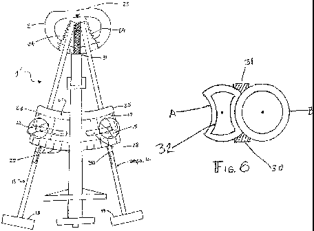
FIG. 6 is an enlarged, cross-sectional view of two tapered fusion cages
following implant using the system and method of the present invention.
DETAILED DESCRIPTION OF THE PREFERRED
EMBODIMENTS
In the following Detailed Description, reference is made to the
accompanying drawings, which form a part hereof, and in which is shown by
way of illustration specific embodiments in which the invention may be
practiced. In this regard, directional terminology, such as "top," "bottom,"
"front," "back," "leading," "trailing," etc., is used with reference to the
orientation of the Figure(s) being described. Because components of
embodiments of the present invention can be positioned in a number of
different
orientations, the directional terminology is used for purposes of illustration
and
is in no way limiting. It is to be understood that other embodiments may be
utilized and structural or logical changes may be made without departing from
the scope of the present invention. The following detailed description,
therefore,
is not to be taken in a limiting sense, and the scope of the present invention
is
defined by the appended claims.
Figure 1 is a composite, diagrammatic plan view from above showing
one embodiment of a stabilizing/guiding system 1 in accordance with the
present
invention engaged against the anterior aspect of a vertebra 2 and a portion
extending inside a vertebral disc space 3 with tabs 4 firmiy against the
anterior
aspect of the vertebrae 2, along with a generic representation of a tapered
cage
14. In general terms, and with additional reference to FIG. 2, the system 1
includes a guide body 50 defining a proximal surface 6 and a distal end
forming
tabs 4 and stabilizer tips 9 that are pivotable about a corresponding hinge
point
10 via interface with a wedge or similar device 8. Movement of the wedge 8 is
dictated by a central drive body 52 (e.g., a shaft) otherwise proximally
accessible
via a knob 54. In addition, the system 1 includes opposing lateral guide or
rod
retaining members 11 attached to the guide body 50. The lateral guide members
6
CA 02596436 2007-06-29
WO 2005/065306 PCT/US2004/043625
11 are, in one embodiment, releasably secured to the guide body 50 by a plate
17. Further, each of the lateral guide members 11. defines a selectively
accessible passage 56 adapted to selectively receive and maintain a rod unit
12
via, for example, a winged knob 15 secured within a socket 18. The rod unit 12
is described in greater detail below with reference to FIG. 4, but generally
includes a distal, working end and a proximal knob 13 or similar structure.
During use, the system 1 is driven into the surgically prepared disc space 3
using
a hammer (not shown) struck against a removable striking block 5 which is
temporarily positioned against the proximal surface 6 by indexing pins (7).
After driving the system 1 against the vertebrae 2, the striking block 5 is
removed and the tightening knob 54 is rotated which pulls the wedge or other
means 8 inside the knurled stabilizer tips (9) causing their knurled surface
to be
temporarily imbedded into the opposing walls of -the vertebral end plates (not
shown). The knurled stabilizer tips 9 are hinged 10 so they may spread apart
inside the disc space 3. The lateral guide or rod retaining members 11 for
guidance of rod units 12 having suitable knobs 13 for hand drilling or
reaming,
tapping or threading of the bed for later free hand cage 14 insertion is thus
under
firm and repeatable control. The lateral guide members 11 are opened for
changing the rod units 12 then firmly held in proper position using winged
knobs
15, or similar means, as needed to drill or ream the recipient bone bed,
thread or
tap the bed and provide the suitable bed for subsequent freehand implantation
of
the suitable tapered cages 14. The angulation 16 of the lateral guide members
11
relative to the midline is selected and fixably alterable to be the same as
the
angulation of ihe tapered cages 14. i ne lateral guide members 11 are
removably
attached to the guide body 50. A hinged screw 15 on the lateral guide member
11 fits into the rounded socket 18 and held firmly by means such as winged
knobs 15. The adjoining surfaces of the subsequently implanted, paired cages
14
are closely paralleled to each other.
Figure 2 is a diagrammatic perspective view seen laterally of the
stabilizing/ guiding system or unit 1 and associated components. As shown in
Figure 1, the long axis of the system 1 is driven into the surgically prepared
disc
space 3 until the tabs 4 engage the anterior vertebral margins, utilizing the
block
7
CA 02596436 2007-06-29
WO 2005/065306 PCT/US2004/043625
removably attached to the end 6 by index pins 7. After firmly seating the
stabilizing/guiding system 1, the striking block 15 is removed and the
fixation
knob 54 is rotated pulling the wedge 8 or suitable means inside the knurled
stabilizer tips 9, hinged 10 on the stabilization/guiding system 1, thus
firmly
5 anchoring the deep end of the unit tips 9 inside the disc space 3 and
maximally
opening and tightening the fibers of the circumferential, ligamentous
vertebral
annulus (unnumbered). The angulated lateral guide members 11 are opened
laterally for placement of the rod units 12 for drilling (reaming) and tapping
(threading) to prepare the recipient bed for subsequent insertion of cages 14.
The lateral guide members 11 are removably attached to the
stabilization/guiding
unit 1 by suitable means such as screws (not shown). The tapered half-sockets
18 are shown where the bases of the screws with winged knobs are seated when
tightened.
Figure 3 is a diagrammatic cross-section showing the wedge 8 inside the
knurled tips 9 of the stabilizing/guiding unit 1 and the cages (not shown),
one
with arcuate lateral cuts, are closely approximated.
Figure 4 is a composite diagram of the two rod units 12 for drilling
(reaming) A, tapping (threading) B, and the separate rod later used for
subsequent free hand cage insertion C. Each rod unit 12 is hand torqued using
its knob 13. Each rod 12 unit is calibrated in millimeter/centimeter
increments
20 as referenced to the outer margin of the lateral guide member 11 (FIG. 2)
to
indicate the depth of reaming or tapping. Each rod unit 12 is changeably
removable from the lateral guiding member 11 as needed without disturbing the
= 1 1 1 l1 l Y Y Y=,
stability or gulaance of LneY main unit. i ne LLL~ rod unit 1 G l!-1 has an
oaa plurallLy
of sharp flat cutting (reaming) vanes 19, usually three, where the odd number
provides a more uniform torque on reaming between the parallel vertebral end
plate surfaces than would be obtained if the reamer used an even number of
vanes. The vanes 19 may also be slightly spiraled for even more uniform torque
and spontaneous removal of debris. The tapping rod unit 12B has an odd fluted
spiraled tap 21 for even torque on tapping the reamed hole. The subsequently
free-hand cage insertion rod unit 12C has a suitable engaging means 22 to
removably connect a cage mounted on its tip. A ball detent or similar means
8
CA 02596436 2007-06-29
WO 2005/065306 PCT/US2004/043625
serves to removably retain the cage (not shown) until it is firmly installed.
The
handle of this rod unit 12C is oval witli directional markings to indicate the
position of the potentially arcuate side cuts of cages, if they are provided,
that
must face the full cage. The insertion rod unit 12C, or similar insertion
device,
can be used apart from the system 1 (e.g., freehand, with the system 1 removed
from the disc space 3), or with the system 1 still in place.
Figure 5 is a composite, diagrammatic plan view of an alternative
embodiment stabilizinglguiding system 1' closely reflecting major details of
Figure 1. The system 1' is adapted to selectively retain rod units 12 (FIG.
1),
such as rod units for reaming 12A (FIG. 4), tapping 12B (FIG. 4), and
subsequent free-hand insertion 12C (FIG. 4) of the tapered cages 14, 24. To
this
end, the system 1' is adapted to direct the rods 12 towards a virtual
confluent or
hinge point 23 near the distal margin of the vertebral bodies 2. Maintenance
of
correct angulation for implantation of tapered cages, determined by the
angular
opening of the disc space between the adjacent vertebral bodies, matched with
tapered cage angulation, is determined by rod attachment member 60 having a
sector plate 25 and adjustable components having the same virtual hinge point
23.
The sector plate 25 has a flat upper surface and a keystone mortised
under surface 26 into which a sliding guide 27 closely and fixably fits. The
sliding guide 27 is adapted to receive one of the rods units 12. The upper
surface
of the sector plate 25 has a curving slot 28 through which a screw 62 provided
with the rod attachment member 60 passes to be movably fixated at a desired
position by tightening a wing nut 15. T his nxation simuitaneousiy nxates the
sliding guide 27, and thus the associated rod unit. Markings 29 on the sector
plate 25 denote the chosen angulation. Markings 30 on the rod units 12
indicate
the depth of penetration into the disc space by their tip structures.
Figure 6 is a diagrammatic cross-sectional view of two, implanted
Stryker Spine, Inc. Ray 'Unite' TM tapered fusion cages. The elevating and
fixating spreader tips 31 having knurled outer surfaces associated with the
stabilizing/guiding unit of the present invention are adjusted to accommodate
the
contour of the appropriate tapered cages. In the Unite cage implantation
version,
9
CA 02596436 2007-06-29
WO 2005/065306 PCT/US2004/043625
the first implanted of the pair, cage A has relieved medial walls 32 one on
each
side, 180 apart, against either of which the second cage B with a standard
wall
closely fits. Two 'Unite' or similar cages (having arcuate side cuts) may also
be
used. This configuration permits a closer proximity of the centers of the two
cages and a reduced overall width of the combined implants than would be
permitted by two fully cylindrical cages.
The structure of the principal stabilizing/guiding unit or system of the
present invention may be constructed essentially oval in cross section as well
as
essentially flat as shown. Depending on the width of the surgical approach and
the depth to the target vertebrae, varying with the obesity of the patient's
abdomen, various implantation/guidance units may be of different size or may
be
constructed with length telescoping/expanding capability. Other variations may
include torque knob design, internal low friction bearing surfaces of the
lateral
guiding members or methods of attachment of these members to the main unit as
well as variations in means of closure of the guiding surface halves of these
members. Design of the cages may be simple tapered adaptations of the present
Ray threaded fusion cages, similar to considerably smaller, tapered ones now
manufactured by Stryker Spine for fusions of the cervical spine (neck).
EXAMPLE AND METHOD OF USE
A suitable patient having discogenic, painful degenerative disc disease is
examined using x ray techniques and on finding that the angulation between the
particular vertebral end plates is 6 degrees or greater, the surgeon may
decide-to
use tapered rather than parallel cylindrical cages for his patient. if the
segment
to be operated is at L5-S 1 (the usual one), the surgeon then notes the
anatomical
position of the top of the symphysis pubis, where the pelvis joins at the
front of
the body. He then draws a line through the middle of the disc to be operated
extending it in the direction of the symphysis. If this line extends below the
top
of the symphysis, it indicates that the stabilizing/guiding unit may be too
large,
vertically, and therefore not usable on this patient. This means that the
angle of
taper as well as the segment tilt angle through the disc centrum relative to
the
symphysis are both important in patient selection. In some patients the tilt
angle
CA 02596436 2007-06-29
WO 2005/065306 PCT/US2004/043625
is so vertically severe that an anterior approach to the L5-S 1 disc space may
not
be possible. Fortunately, this situation is quite uncommon.
11
CA 02596436 2007-06-29
WO 2005/065306 PCT/US2004/043625
The appropriate tapered cages or suitable insert and associated
instruments are chosen and made ready. The patient is anesthetized and
appropriately positioned, the abdomen is prepared with an exposure usually via
a
retroperitoneal dissection (moving the abdominal organs from the patient's
left to
the right side, along with the intact peritoneal sac). The abdominal exposure
must be wide enough to accommodate the angulation of the rods used in the
procedure to ream, tap and then insert. The major anterior vessels and other
important structures are mobilized and handled as for any routine anterior
retroperitoneal fusion approach, a common technique during anterior spinal
fusion procedures. The anterior annulus is removed sufficient only to
accommodate the width of the pair of cages or inserts; the entire cartilage of
the
end plates is scraped away down to bleeding bone but not to penetrate the bone
of the end plates. The stabilizing/guiding unit is driven into position
against the
anterior aspect of the vertebra and its locking tip is expanded to fully
stabilize
the unit and this stability is evaluated by moving the unit in several planes,
showing that the two vertebrae and the unit move essentially as though a
single
structure. The reaming and tapping into the disc space as indicated above. The
guide unit is removed and the tapered cage is inserted free hand using its
rod,
carefully inserting the first arcuate side cut cage then the fully round one.
The
final positioning of the cage pair is demonstrated by intraoperative x-ray
fluoroscopy then the cages are filled with morsels of bone or substitute and
the
procedure finished by routine closure of the tissue layers and the skin
incision.
In that the procedure closely parallels common anterior fusions, patients
should
1 1
respona quite well to the procedure and post-operative care. Patients
ordinariiy
wear a corset but in some cases a rigid brace may be needed for a few months.
Subsequent office visits should include repeat x-rays to determine the
progress
of the fusion and if any displacements or other problems have arisen before
the
fusion becomes fully solid (in about 3 - 5 months).
ADVANTAGES
The invention has the novel ability to utilize a pair of fusion cages or
suitable material blocks having The selected angulation of taper to, match the
12
CA 02596436 2007-06-29
WO 2005/065306 PCT/US2004/043625
tapering angles found in several patients having disabling discogenic pain and
disc degeneration, particularly at the L5-S 1 space. The divergent angulation
of
the approach to insert the cages or blocks of material being the same as the
actual taper angle of the disc space permits the facing or medial edges of the
implants inside the disc space to be parallel along their lengths, uniquely
improving the availability of surrounding disc space for a narrower overall
width
of the implant pair plus additional bone graft placement. The angulation of
the
set of appropriate instruments and the cage pair to be used is determined
preoperatively and the overall procedure is uniquely well controlled by the
means of central stabilization of the disc space and the ancillary guidance
components. The stabilizing/ guiding unit firmly controls the approach angle
bilaterally throughout the procedure without slipping or dislocation assuring
excellent matching of the steps of the procedure and therefore the overall
fusion
rate and success. Further, the vertical opening or expansion of the knurled
fixation member is placed at or close to its virtual disc center of
flexion/extension motion of the disc and therefore on distraction, elevation
and
tightening of the circumferential annuls fibers, the taper angle is largely
unchanged. If the disc space is tilted laterally, as in cases with localized
scoliosis, the taper angle is generally the same but the height of the disc
space is
different on the two sides. By inserting the tapered cage of block more or
less
into the depths of the disc space, thus difference in height can be adjusted
or
even corrected; not possible when using straight cylindrical cage implants.
Therefore, the require accuracy of the implantation is controlled throughout
the
reaming and fhreading steps of the procedure in preparation for the final
direct
visual free-hand insertion of the tapered cages, all of which expectedly will
improve on the overall results where occasional surgical problems have arisen
from other instrument methods not so well controlled. Since the entire
procedure is as well under direct vision by the surgeon, the steps are better
controlled than with implantation techniques utilizing essentially blinded
tubular
guides for each stage from the reaming to the insertion. Lastly, the
angulation
for the final position of the implants has a confluent and not parallel
insertion
13
CA 02596436 2007-06-29
WO 2005/065306 PCT/US2004/043625
path, there should be an improvement in the rotary and lateral translocation
stability of the final result.
The novel system places the tapered cages or fusion inducing devices
material with their long axes convergent posteriorly where the convergent
angle
is the same as the taper angle of the disc space. Therefore, the cages come
close
together at a constant distance between them, throughout their lengths. Cages
machined with arcs cut from one or more outer surfaces equal to their
circumferential contour permit them to be brought into close contact at a
width
less than the combined diameters, throughout their lengths. At the deep
converging tip of the implants, sufficient width within the vertebrae remains
so
additional graft material can be placed, as may be desired by the surgeon.
Additionally, with the cage's long axes nested together, the disc is more
stable
against lateral translocation or "side roll". The depth of insertion of the
tapered
implants is useful to wedge tighten the annular fibers, promoting immediate
stability over parallel walled implants. The novel system stably achieves the
boring or reaming and tapping in preparation for cage insertion quite
accurately,
utilizing a rod alignment/guiding instrument and attachments. A tubular guide
unit is not employed for any of these steps and the common rod guidance is
utilized only for the boring or reaming and threading or tapping steps. That
is,
once the reaming or boring and tapping are performed, the guiding assembly is
removed and the insertion of cages installed by hand into the provided
appropriately oriented and tapered cavities. The angulation and orientation
for
boring and tapping provided by the adjustable long rod/guide assembly are
selected and fixed according to the required taper angle of the disc space and
selected cage implant. Further, prior to attachment of the guiding/stabilizing
assembly, the deep central disc space may be prepared laterally into the
vertebral
bone for ancillary bone placement outside the anticipated confluence of the
cage
tips. The fixed (static) and opening up or spinal extension position (dynamic)
tapering angles which the implants should closely match are determined in
advance of the surgery utilizing lateral x-ray views of the lumbar spine in
neutral, forward flexion and reverse extension positions. In addition to
determining the taper angle of the disc space, this maneuver aids in assessing
the
14
CA 02596436 2007-06-29
WO 2005/065306 PCT/US2004/043625
flexibility of the space. That is, the more flexible the disc the more it will
open
on extension and the greater the cage taper angle that may be needed following
the attachment of the stabilizing/guiding assembly. The novel
stabilizing/guiding instrument is adjustable to the same angular taper as the
implants to be used, primarily 6 , 9 or 12 or other suitable angle as
indicated.
Those skilled in these arts may provide other means for adjustment of the
guiding instrument to suit various taper angles, in addition to the rod
assembly
herewith disclosed without departing from the intent and novelty of this
document system.
Although specific embodiments have been illustrated and described
herein, it will be appreciated by those of ordinary skill in the art that a
variety of
alternate and/or equivalent implementations may be substituted for the
specific
embodiments shown and described without departing from the scope of the
present invention. This application is intended to cover any adaptations or
variations of the specific embodiments discussed herein. Therefore, it is
intended that this invention be limited only by the claims and the equivalents
thereof.