Note: Descriptions are shown in the official language in which they were submitted.
CA 02596595 2007-08-01
WO 2006/084382
PCT/CA2006/000207
- 1 -
COMPOSITIONS AND METHODS FOR MULTIMODAL IMAGING
FIELD OF THE INVENTION
This invention relates to the field of medical imaging and more
specifically to the use of signal modifying agents in medical imaging.
BACKGROUND OF THE INVENTION
In recent years significant effort has been devoted to the
development of multimodality imaging. Since each medical imaging
modality has unique strengths and limitations, it is often through the
compound use of multiple modalities that the complete assessment of a
patient is achieved. Interest in the area of multimodality imaging has also
been prompted by the realization that such techniques offer much more
sophisticated characterization of the morphology and physiology of tissues
and organs, and that confidence gained in the accurate correspondence or
registration of different modalities greatly enhances their value (Barillot C,
Lemoine D, Le Briquer L, et aL. Eur J Radio! 1993;17:22-27.).
Consequently, this improved value of imaging will ultimately allow for
advances in diagnosis and evaluation of disease, image-guided therapeutic
interventions, and assessment of treatment outcomes. The recent
integration of computed tomography (CT) and positron-emission
tomography (PET) systems is a good example of the advantages of the
multimodal approach (Townsend DW. Mo/ Imaging Biol 2004;6:275-290;
Townsend DW, Carney JP, Yap JT, et al. J Nucl Med 2004;45 Suppl 1:4S-
14S; Townsend DW, Beyer T. Br J Radio! 2002;75 Spec No:S24-30). The
CT-PET combination has revolutionized the utilization of PET and served
to increase the specificity of PET-based assessment. In the context of
radiation therapy, there is a need to merge CT and magnetic resonance
(MR) imaging with CT employed for 3D volumetric dose calculation
(Rosenman JG, Miller EP, Tracton G, et al. Int J Radiat Oncol Biol Phys
1998;40:197-205.) and MR for accurate delineation of the target and
normal structures as it provides exceptional soft tissue definition. For
CA 02596595 2007-08-01
WO 2006/084382
PCT/CA2006/000207
- 2 -
=
example, accurate delineation and targeting of the prostate gland in
radiation therapy of prostate cancer necessitates parallel use of CT and
MR imaging (Rasch C, Barillot I, Remeijer P, et al. Int J Radiat Oncol Biol
Phys 1999;43:57-66.). Furthermore, CT technology in the form of
conventional and cone-beam systems is employed on a daily basis to
guide the delivery of radiation therapy on treatment machines (Uematsu M,
Sonderegger M, Shioda A, et al. Radiother Oncol 1999;50: 337-339;
Jaffray DA, Siewerdsen JH, Wong JW, et al. Int J Radiat Oncol Biol Phys
2002;53:1337-1349.).
Clinical imaging in all modalities requires an adequate level of
differential contrast relative to noise be achieved in order to identify the
structures or phenomena under observation. Although imaging on CT and
MR can be performed without the administration of signal modifying agents
there are numerous instances in both disease diagnosis and treatment, in
which procedures benefit from the improved contrast and dynamics that
are added by the use of these agents (Krause W. Adv Drug Deliv Rev
1999;37: 159-173; Saeed M, Wendland MF, Higgins CB. J Magn Reson
Imaging 2000;12:890-898).
To date, although a multitude of signal modifying agents are
commercially available for single modality imaging, few attempts have been
made to develop signal modifying agents that can be used across multiple
imaging modalities (McDonald MA, Watkin BS, Watkin KL. Small Invest
Radio/ 2003;38:305-310; Mem JL, Wondergem J. Radiology
1989;171:578-579; Gierda DS, Bae KT.. Radiology 1999; 210: 829-834;
Quinn AD, O'Hare NJ, Wallis FJ, et al. J Comput Assist Tomogr 1994;18:
634-636; Pena CS, Kaufman JA, Geller SC, et al. J Comput Assist Tomogr
1999;23:23-24.). The lack of development in this area is likely due to
challenges presented by the fact that the distinct imaging modalities have
different sensitivities for different signal modifying agents (Krause W. Adv
Drug Deliv Rev 1999;37: 159-173.). A simple approach for realizing a
multimodal signal modifying agent for CT and MR has been to exploit
CA 02596595 2007-08-01
WO 2006/084382
PCT/CA2006/000207
- 3 -
commercially available extracellular gadolinium-based signal modifying
agents for enhancement in both of these modalities. In this case, the
properties of gadolinium that allow for use in both CT and MR include its
relatively high atomic number and paramagnetic characteristics (McDonald
MA, Watkin BS, Watkin KL. Small Invest Radio! 2003; 38: 305-310; Bloem
JL, Wondergem J. Radiology 1989;171:578-579; Gierda DS, Bae KT..
Radiology 1999; 210: 829-834; Quinn AD, O'Hare NJ, Wallis FJ, et al. J
Comput Assist Tomogr 1994;18:634-636; Pena CS, Kaufman JA, Geller
SC, et al. J Comput Assist Tomogr 1999;23:23-24.). However, due to their
low molecular weight, these agents only remain in the vascular system for
a short period of time, exhibit rapid dynamic distribution changes in
different organs and are excreted quickly. The use of these agents for
cross-modality imaging would therefore require both multiple
administrations and fast imaging sequences. Also, the low gadolinium
payload per molecule, relative to conventional iodinated signal modifying
agents, would necessitate the administration of higher doses for adequate
CT enhancement which may have implications in terms of both cost and
toxicity (McDonald MA, Watkin BS, Watkin KL. Small Invest Radio!
2003;38:305-310; Bloem JL, Wondergem J. Radiology 1989;171:578-579;
Gierda DS, Bae KT.. Radiology 1999;210:829-834; Quinn AD, O'Hare NJ,
Wallis FJ, et al. J Comput Assist Tomogr 1994;18:634-636; Pena CS,
Kaufman JA, Geller SC, et al. J Comput Assist Tomogr 1999;23:23-24.).
Furthermore, the short in vivo residence time of these agents would impose
limitations on the size of the anatomic region that could be imaged
optimally and would exclude them from being used in image-guidance
applications due to their inability to provide prolonged contrast
enhancement for the entire course of treatment (Saeed M, Wendland MF,
Higgins CB. J Magn Reson Imaging 2000;12:890-898).
A viable way to effectively deliver the required amount of contrast
in each imaging modality and to prolong the presence of the agents in vivo
is to employ carriers such as liposomes. Specifically, liposonne-based
CA 02596595 2007-08-01
WO 2006/084382
PCT/CA2006/000207
- 4 -
systems have been evaluated for either encapsulating (Kao CY, Hoffman
EA, Beck KC, et al. Acad Radio! 2003;10:475-483; Leike JU, Sachse A,
Rupp K. Invest Radio! 2001;36:303-308; Leander P, Hoglund P, Borseth A,
et al. Eur Radio! 2001;11:698-704; Schmiedl UP, Krause W, Leike J, et al.
Acad Radio! 1999;6:164-169; Spinazzi A, Ceriati S, Pianezzola P, et al.
Invest Radio! 2000;35:1-7; Petersein J, Franke B, Fouillet X, et al. Invest
Radio! 1999;34:401-409; Leander P, Hoglund P, Kloster Y, et al. Acad
Radio! 1998;5 Suppl 1:S6-8; discussion 528-30; Krause W, Leike J,
Schuhmann-Giampieri G, et al. Acad Radio! 1996;3 Suppl 2:S235-237;
Dick A, Adam G, Tacke J, et al. Invest Radio! 1996;31:194-203; Revel D,
Corot C, Carrillon Y, et al. Invest Radio! 1990;25 Suppl 1:S95-97; Musu C,
Felder E, Lamy B, et al. Invest Radio! 1988;23 Suppl 1:S126-129; Zalutsky
MR, Noska MA, Seltzer SE.. Invest Radio! 1987;22:141-147; Seltzer SE,
Shulkin PM, Adams DF, et al. AJR Am J Roentgenol 1984;143:575-579;
Jendrasiak GL, Frey GD, Heim RC, Jr. Invest Radio! 1985;20:995-1002;
Torchilin VP. Curr Pharm Biotechnol 2000;1:183-215; Schneider T, Sachse
A, Robling G, Brandl M. Int J Pharm 1995;117:1-12; Pauser S, Reszka R,
Wagner S, et al. Anticancer Drug Des 1997;12:125-135.) or chelating
(Weissig W, Babich J, Torchilin W. Colloids Surf B Biointerfaces
2000;18:293-299; Misselwitz B, Sachse A. Acta Radio! Suppl 1997;412:51-
55; Unger E, Needleman P, Cullis P, etal. Invest Radio! 1988;23:928-932;
Kabalka G, Buonocore E, Hubner K, et al. Radiology 1987;163:255-258;
Grant CW, Karlik S, Florio E. Magn Reson Med 1989;11:236-243) single
CT or MR signal modifying agents. Most of these liposome-based signal
modifying agents have been explored for blood pool imaging due to the
long in vivo circulation lifetimes that may be achieved for these carriers.
Yet, liposomes have also been identified as suitable carriers for the
delivery of agents to the lymphatic system since they have been shown to
avoid aggregation at the site of injection and localize in lymph nodes
(Nishioka Y, Yoshino H. Adv Drug Deliv Rev. 2001;47:55-64; Moghimi SM,
Rajabi-Siahboomi AR. Prog Biphys Molec Biol. 1996;65:221-249;
CA 02596595 2007-08-01
WO 2006/084382
PCT/CA2006/000207
- 5 -
Oussoren C, Storm G. Adv Drug Deliv Rev 2001;50:143-156). The
potential use of liposome-based signal modifying agents for lymphatic
imaging is worth noting as it is well-known that the lymph nodes are the
primary site for the metastases of many cancers (Swartz MA. Adv Drug
Deliv Rev. 2001;50:3-20; Swartz MA, Skobe M. Microsc Res Tech
2001;55:92-99.). Until
recently, there were no available non-invasive
methods for distinguishing between lymph nodes enlarged due to the
presence of metastatic cancer cells and nodes enlarged due to
inflammation, or for identifying cancerous nodes of normal size. With the
advent of Combidex (Advanced Magnetics, Inc. USA), lymph nodes can
now be enhanced in MR, and metastatic nodes can be differentiated from
normal or inflamed nodes based on morphology and changes in signal
intensity between scans performed before and after signal modifying agent
injection (Xiang Y, Wang J, Hussain SM, Krestin GP. Eur Radio!.
2001;11:2319-2331). However no delivery system has been developed for
prolonged co-localization in vivo of two or more signal modifying agent for
multiple medical imaging.
SUMMARY OF THE INVENTION
In a broad aspect of the invention there is provided signal
modifying compositions for medical imaging comprising a carrier and signal
modifying agents specific for two or more imaging modalities. In a preferred
embodiment the compositions are characterized by retention efficiency,
with respect of the signal modifying agents, that enables prolonged
contrast imaging without depletion of the signal modifying agent from the
carrier. The carriers of the present invention are lipid based or polymer
based the physico-chemical properties of which can be modified to entrap
or chelate different signal modifying agents and mixtures thereof and to
target specific organs or tumors within a mammal.
The co-localization of imaging modalities specific signal
modifying agents in a carrier advantageously enables the registration of
images obtained from different imaging modalities. The registration can be
CA 02596595 2007-08-02
Oft' 0416,4W9 a or
= Os APRIL 2007 05 =.0 4 07
- 6 -
exploited to refine diagnosis, design of therapeutic regimen, follow the
progress of therapy such as radiation therapy and optimize contrast
enhancement.
Thus, in one aspect, there is provided an image signal modifier
composition for imaging of a biological tissue, the composition comprising:
two or more signal modifying agents, each of the agent being specific for at
least one imaging modality; and a carrier comprising the two or more signal
modifying agents and wherein the carrier is capable of retaining a sufficient
amount of the agents for a time sufficient to acquire imaging data using the
composition.
The signal modifying agents are specific for imaging modalities
selected from but not limited to magnetic resonance imaging (MRI), X-ray,
ultrasound (US), positron emission tomography (PET), computed
tomography (CT), autoradiography, single-photon emission computed
tomography (SPECT), fluoroscopy, optical imaging, fluorescence imaging
and bioluminescence imaging.
In a further aspect, the carrier is a lipid-based carrier such as a
liposome or a micelle.
In an embodiment of the invention the composition can be
targeted to a desired location within a subject or within a tissue. This can
be achieved through control of the carrier physico-chemical properties or
by inserting one or more recognition molecules such as antibodies,
receptors/ligands, carbohydrates, proteins and peptide fragments.
In. another embodiment the composition may comprise a
therapeutic agent such as anticancer; antimicrobial, antifungal and antiviral
agents.
In yet another aspect of the invention there is also provided a
method for imaging one or more region of interest in a mammal the method
comprising: administering to the mammal a signal modifier composition
AMENDED SHEET
CA 02596595 2007-08-02
046 .600207
Nag
05 APRIL 2007 0
5 0 4 = 07
- 7 -
waiting for a time sufficient for. the composition to reach the region of
interest; and obtaining an image of the one or more region of interest.
There is also provided a method for registering images obtained
from two or more imaging modalities the method comprising: administering
to a mammal a signal modifier composition, each agent being specific for at
least one of the at least two or more imaging modalities; obtaining an
image of one or more region of interest in the mammal using each of the at
least two or more imaging modalities; and comparing the images obtained
to derive complementary information from the one or more region of
interest.
In the present description by signal modifier or signal modifying it
is meant that the signal obtained with a particular imaging modality is
modified by an agent. Typically the agent is a signal enhancing agent
(contrast agent) but the agent may also provide for signal attenuation or
any other form of signal modification so as to provide a desired effect on
the image.
By biological tissue or tissue it is meant any part of an animal,
such as a mammal, including but not limited to organs, vessels, blood,
breast tissue, muscular ,tissue, bones and the like.
By retaining or retention efficiency it is meant the capacity of a
carrier to prevent leakage of a signal modifier agent out of the carrier.
= By targeting it is meant the preferential accumulation of the
compositions of the present invention .in a given organ or anatomical
structure or tissue, including cell populations, By active targeting it is
meant
that a target binding molecule, specific for a molecule in the target, is
incorporated in (or associated with) the composition. Examples comprise
antibodies and receptor/ligand pairs. Passive targeting refers to preferential
distribution of the composition due to its physico-chemical properties.
AMENDED SHEET
CA 02596595 2007-08-01
WO 2006/084382
PCT/CA2006/000207
- 8 -
BRIEF DESCRIPTION OF THE DRAWINGS
Further features and advantages of the present invention will
become apparent from the following detailed description, taken in
combination with the appended drawings, in which:
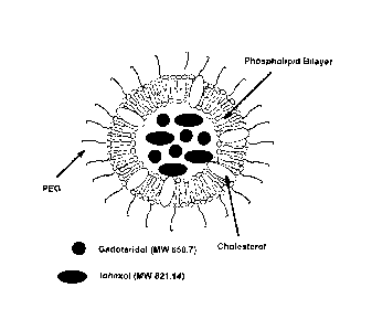
Figure 1 is a schematic representation of the liposome-based
signal modifying agent system;
Figure 2 is a transmission electron micrograph of the negatively
stained dual-agent containing liposomes at (a) 40000 magnification and (b)
80000 magnification;
Figure 3 is an in vitro release profile for lohexol and Gadoteridol
from DPPC/cholesterol/DSPE-PEG (55/40/5 mol %) liposomes dialyzed
under sink conditions (250-fold volume excess) against HBS (a) at 4 C
(n=3) and (b) at 37 C (n=4);
Figure 4 is a plot of the size of the dual agent-containing
liposomes during dialysis under sink conditions (250-fold volume excess)
against HBS at 37 C (n=3);
Figure 5 is an image showing in vitro imaging efficacy of the
liposome-based signal modifying agent system (a) in CT (2.5mm slice
thickness, 120kV, 300mA and 15.2 cm FOV) and (b) in MRI (450ms TR,
9ms TE, 3mm slice thickness, 19.9cm FOV and 256 x 192 image carrier)
[iodine] (mg/mL) [gadolinium] (mg/mL)
A 16.98 3.55
B 8.49 1.77
C 1.70 0.35
D 0.17 0.04
E 0.07 0.02
CA 02596595 2007-08-01
WO 2006/084382
PCT/CA2006/000207
- 9 -
Figure 6 (a) CT (2.5mm slice thickness, 120kV, 300mA and
15.2 cm FOV) attenuation in HU as a function of signal modifying agent
concentration in mmol/L; although gadolinium has CT attenuation
properties, iodine provides more effective CT enhancement. (b) Differential
signal intensity (with respect to water) in MRI (400ms TR, 9ms TE, 3mm
slice thickness, 19.9 cm FOV and 256 x 192 image carrier) as a function of
increasing gadolinium and iodine concentrations; symbols represent
liposome encapsulated agents (o), free lohexol and Gadoteridol (A), free
Gadoteridol (.) and free lohexol (V);
Figure 7 (a) 1/T1 relaxation rate and (b) 1/T2 relaxation rate as a
function of gadolinium (Gd) and iodine (I) concentration obtained at 20 C
with a 1.5T, 20-cm-bore superconducting magnet controlled by an SMIS
spectroscopy console; Encapsulation of Gadoteridol greatly reduces both
the ri and r2 of the gadolinium atoms;
r2 (s mmol-
r1 (s-immoriL)
(0) Free Gadoteridol 5.14 0.06 6.21
0.08
(.) Free Gadoteridol and lohexol (1:29 mole ratio
6.38 0.16 7.83 0.20
of Gd to I)
(A) Free lohexol (x-axis = [I] in mmol/L) 0.00 0.00 0.01
0.01
(7) Liposome encapsulated agents 1.23 0.02 1.46
0.02
Figure 8 is a liver cross-section images from a rabbit, before and
after injection of signal modifying agent, in CT and MR;
Figure 9 is a confocal microscopy image of a liposome
formulation containing DPPC/Cholesterol/DSPEPEG/DPPE-NDB 1
(54.5/40/5/0.5 mole ratios) encapsulating iohexol and gadoteridol; 1,2-
CA 02596595 2007-08-01
WO 2006/084382
PCT/CA2006/000207
- 10 -
dipalmitoyl-sn-glycero-3-phosphoethanolamine-N-(7-nitro-2-1,3-
benzoxadiaxol- 4-y1), the excitation wavelength is 460 nm and the
emission wavelength is 534 nm; this liposome formulation is suitable for
CT, MR and optical imaging;.
Figure 10 is a graphic of a relative signal enhancement of blood
(aorta), liver (parenchyma) and kidney (medulla and cortex) up to 200
minutes following intravenous administration of the liposome-based signal
modifying agent in (a) CT and (b) MR;
Figure 11 is (a) CT and MR liver cross section scans of a 2.1 kg
white New Zealand rabbit obtained before (0 min.) and after (10, 30, 90
and 200 minutes) signal modifying agent injection. Note the visual contrast
enhancement obtained in the aorta, the hepatic vessels, and the liver
parenchyma up to 3 hours and 20 minutes in both imaging modalities. (b)
CT and MR cross section scans of the left kidney obtained before (0 min.)
and after (10, 60, 120 and 200 minutes) signal modifying agent injection.
Note the visual contrast enhancement obtained in the kidney. The same
window level was used for pre-and post-injection images;
Figure 12 are 3D maximum intensity projection images (anterior
view) of the rabbit in CT (120 kV, 200 mA) and MR (3D FSPGR,
TRiTE=9.8/4.3) before the injection of the contrast agent modified
liposomes (0 minutes) and 48 hours and 168 hours post-injection (300
mg/kg of iodine and 16 mg/kg of gadolinium encapsulated in liposomes),
the parallel visual enhancement seen in both CT and MR obtained in the
major blood vessels, liver, spleen and intestines represents the liposome
distribution over a 7-day period, the spine and part of the ribs of the rabbit
have been masked in the CT image set for better soft tissue visualization;
Figure 13 is a graphic of the percentage of the total injected CT
(iohexol, detected with HPLC at 245 nm wavelength) or MR agent
(gadoteridol, detected with ICP-AES) remaining in mouse plasma (female
Balb-C, 18-23g, one mouse per time point) and rabbit plasma (female New
CA 02596595 2007-08-02
4100006,11e0a 0 ?
05 APRIL 2007 0;414:07
-11 -
Zealand White, 2 kg, same rabbit used for all time points) at specific time
points following administration, the ratio of ibdine to gadolinium is 13.9 .
3.0 in mice and 11.9 0.5 in the rabbit at all time points; =
Figure 14 is a liposome distribution estimated from the
percentage of the injected gadolinium encapsulated in liposomes per gram
of tissue (kidney, liver, spleen, heart and lung) over a 8-day period in
female Balb-C mice; and
Figure 15 are relative signal differences measured in the rabbit
aorta using CT and MR correlate linearly (R2=0.9) with the iodine and
gadoteridol concentrations detected in the rabbit plasma using HPLC and
ICP-AES assays, respectively, the relative HU (AHUrei) was calculated as a
function of the HU value found at the same anatomic location prior to the
injection of the liposome sample (AMA) as described in equation (1),
similarly, the relative MR signal intensity (ASIrei) was calculated as a
function of the pre-injection signal intensity value (AS10) as described in
equation (2).
kRuo)
AflUrei (1 ) Asird (AS/õ, ¨ AS/0) (2)
mitio As/0
DETAILED DESCRIPTION OF THE INVENTION =
= A novel approach is provided, in which image signal modifier
compositions are designed to provide long-lasting image signals for
accurate spatial registration over the course of therapy or diagnosis and
between imaging-modalities used in the design and guidance of the
therapy. Such a composition provides a unique platform for accurate
design, image-guided delivery, and assessment of therapy,
Thus, there is provided compositions and methods for signal
modification such as contrast enhancement in imaging modalities. In one
AMENDED SHEET,
CA 02596595 2007-08-01
WO 2006/084382
PCT/CA2006/000207
- 12 -
aspect there is provided rnultimodal signal modifier compositions that
comprise at least two signal modifying agents and a carrier, each signal
modifying agent being specific for at least one imaging modality. The
combination of the signal modifying agents enables the co-localization,
within specific anatomical structures as part of *biological tissues (organs,
tumors and the like) of mammals (including humans), of the signal
modifying agents which, in turn, allows acquisition of the images obtained
by two (or more) imaging modalities and also allows for registration of the
images. Such compositions may be used for imaging various organs and
tissues as well as any tubule and vessel system in the body (i.e. blood
vessels, hepatic vessels, renal vessels, and the lymphatics).
The multimodal signal modifier compositions of the present
invention may be used with imaging modalities that are based on magnetic
resonance, ultrasound, X-ray, optical, positron-emission, single-photon
emission, radioactivity and the like provided that the signal modifying
agents possess the required signal modifying properties as would be
known to a person skilled in the art. For example in the case of magnetic
resonance imaging (MRI) the signal modifying agent should possess
magnetic properties (high relaxivity) capable of modifying the relaxation
time of bulk water molecules. As another example, signal modifying agents
for X-ray imaging should exhibit bulk attenuation characteristics. Signal
modifying agents can possess properties that render them suitable for
signal modification of more than one imaging modality. A carrier may
comprise any combination of signal modifying agent. Non-limiting examples
include: signal modifying agents for MRI/X-ray, MRI/optical, MRI/X-
ray/optical, optical/PET, MRI/CT/optical, etc.
Signal modifying agents specific for each imaging modalities
(CT, MR' radionuclide, optical) are well known in the art. Non-limiting
examples of signal modifying agents include gadolinium, manganese and
iron based agents (MRI), iodine based agent (CT), alpha, beta and positron
CA 02596595 2007-08-02
00110100610011211te
=
-13- n 5 APRIL 2007
. -S = 0 4 ""
4,07
emitting radiotracers (autoradiography, PET and SPECT), fluorophores
(optical),
and perfluorocarbons.
The multimodal signal modifier compositions of the present invention
comprise a carrier having physico-chemical properties compatible with the
retention of the signal modifying agents. Retention of the signal modifying
agent
molecules is desirable to prevent dispersion of the agent within the body and
to
prevent the depletion of the signal modifying agents from the carrier, which
would
reduce the signal intensity. Thus, effective retention results in prolonged in
vivo
contrast enhancement thereby avoiding the need for multiple administration
over
the course of image acquisition and allowing registration of images obtained
over
a period of time. In a preferred embodiment the carrier can retain between
about
10 and 100% of the signal modifying agent over the course of imaging. In a
more
preferred embodiment this retention is of the order of about 80 to 100% and in
an
even more preferred embodiment the retention is above 90%. Thus the carrier
should be sufficiently stable with respect to agents' retention so as to allow
sufficient time for the composition to reach a region of interest an enable
acquisition of imaging data. Furthermore the carrier should also remain in the
tissue of interest for a time sufficient to allow acquisition of imaging data
over a
desired period of time. This period of time may depend on the information that
is
required, the nature of therapeutic regimens being applied, the progression of
a
disease and the like. The period of time may extend from a few minutes to
several
days. In an embodiment of the invention the period of time is between about 1
minute and about 14 days, preferably between about 5 minutes and about 7 days.
In one embodiment, the carrier is used to entrap (encapsulate) the
signal modifying agents and in a preferred embodiment the carrier consists of
a
lipid based carrier such as lipid micelles, unilamellar (see figure 1) and
multilamellar vesicles such as liposomes.
Lipid micelles have small diameters: 8nm-50nm and are made of a
single lipid layer and are therefore suitable for encapsulating hydrophobic
signal
modifying agents, such as Perfiuorooctyl bromide (perflubron).
AMENDED SHEET.
CA 02596595 2007-08-02
11MaDo6,01/0 a up
== 05
APRIL 2001 41 5 -- = (14 .07
- 14 -
The composition of the carrier may be adjusted as required in order to
optimize the loading capacity, release kinetic profiles for each agent, and
the stability
of the overall system. For example, for a lipid-based carrier such as
liposome, it is
well known that the membrane fluidity may affect the permeability of certain
compounds. The molecular characteristics of the membrane that are known to
affect
fluidity include, but are not limited to, lipid saturation, fatty acid chain
length, charge of
the polar head of the lipids, cholesterol content and the like. It will be
appreciated that
encapsulation of the signal modifying agents should not substantially affect
their
signal modifying (for example contrast enhancing) properties. In this respect,
the
composition of the carrier preferably minimizes the leakage of the
encapsulated
agents and optimizes the contrast enhancement abilities of the encapsulated
agents.
For example, bulk water accessibility to signal modifying agents should be
considered
when designing a carrier composition for MRI. It will also be appreciated that
the
signal modifying agents may be chosen to be compatible with a given carrier
composition. For example, while a signal modifying agent may be prone to leak
out of
a liposome having a given lipid composition, a different signal modifying
agent may be
less so for the same lipid composition.
In an embodiment, the lipid base carrier is a liposome comprising one or
more lipids, cholesterol (CHOL) and one or more PEGylated lipids. In another
embodiment, the one or more lipids is a neutral lipid, for example
phosphatidylcholine.
In a preferred embodiment the lipid composition of the lipid based carrier
comprises a neutral lipid, cholesterol and polyethylene glycol (PEG2000)-
phosphatidylethanolamine (PE). In a preferred embodiment, the PC: CHOL: PEG-PE
are in a molar ratio of about 55:40:5.
A second approach to couple the signal modifying agent(s) to the carrier
involves chelation or covalent linking of at least one of the signal modifying
agents to
the outer surface of the carrier (such as a liposome). This approach can, for
example,
increase the access to bulk water thereby enhancing the efficiency of MR
signal
modifying agents. This strategy also maximizes the entire internal aqueous
volume of
the carrier as cargo space for the other or several other signal modifying
agents. For
example, radionuclides can be chelated on derivatized lipids. Hydrophilic
agents can
be chelated (see below) onto their outer surface along with Poly-ethylene
tal) birigET,
CA 02596595 2007-08-01
WO 2006/084382
PCT/CA2006/000207
- 15 -
glycol (PEG) groups. Chelators may comprise EDTA, DTPA, TETA, HYNIC
and other structurally related analogues. It will be appreciated that coupling
of signal modifying agents may comprise high affinity linker molecules such
as avidin-biotin. The signal modifying agent may also be covalently linked
to the carrier. For example fluorophore can be thus linked to lipid molecules
that can in turn be incorporated in a lipid carrier.
The encapsulation (or chelation) of small molecular weight signal
modifying agents into a macromolecule carrier (i.e. liposome) significantly
reduces their in vivo volume distribution, prolongs their in vivo circulation
time and increases their ability to accumulate in specific locations within
the
body such as in tumors. It will be appreciated that accumulation may take
place through passive or active targeting mechanisms. With respect to
active targeting mechanisms, techniques such as antibody coating or
attachment of specific cellular receptors/ligands (such as Epidermal Growth
Factor, EGF and its receptor, EGFR) onto the surface of the carrier or in
association with polymeric matrices may be used as would be known to
those skilled in the art. Non-limiting examples also include small molecules
(Saul JM, Annapragada A, Natarajan JV, et al. J Control Release
2003;92:49-67; Lee RJ, Low PS. Biochim Biophys Acta 1995;1233:134-
144; Lee RJ, Low PS. J Biol Chem 1994;269:3198-3204.), sugar
(carbohydrates) molecules (Spanjer HH, Scherphof GL. Biochim Biophys
Acta 1983;734:40-47; Spanjer HH, MorseIt H, Scherphof GL. Biochim
Biophys Acta 1984;774:49-55; Banerjee G, Nandi G, Mahato SB, et al. J
Antimicrob Chemother 1996;38:145-150; Luciani A, Olivier JC, Clement 0,
et al. Radiology 2004;231:135-142.), serum proteins (Afzelius P, Demant
EJ, Hansen GH, et al. Biochim Biophys Acta 1989;979:231-238; Brown
PM, Silvius JR. Biochim Biophys Acta 1990;1023:341-351; Lundberg B,
Hong K, Papahadjopoulos D. Biochim Biophys Acta 1993;1149:305-312.)
and antibodies (Heath TD, Montgomery JA, Piper JR, et al. Proc Natl Acad
Sci U S A 1983;80:1377-1381; Debs RJ, Heath TD, Papahadjopoulos D.
Biochim Biophys Acta 1987;901:183-190; Matthay KK, Abai AM, Cobb S,
CA 02596595 2007-08-01
WO 2006/084382 - -
PCT/CA2006/000207
- 16 -
et al. Cancer Res 1989;49:4879-4886; Maruyama K, Holmberg E, Kennel
SJ, et at. J Pharm Sci 1990;79:978-984; Allen TM, Ahmad I, Lopes de
Menezes DE, et al.Biochem Soc Trans 1995;23:1073-1079) or antibody
fragments (Kirpotin D, Park JW, Hong K, et al. Biochemistry 1997;36:66-
75; Park JW, Hong K, Carter P, et at.. Proc Natl Acad Sci U S A
1995;92:1327-1331.). Consequently, nonspecific toxicity can be greatly
reduced (i.e. renal-toxicity often associated with iodine-based signal
modifying agents) and specific imaging efficacy increased.
It will be appreciated that active targeting can be tested for
example by injecting a signal modifier composition comprising a target
binding molecule for which the target is known and measuring the amount
of the composition reaching the target. The target may be an extrinsic
target, that is to say, the target can be incorporated in an animal at a
predetermined location such as a tumor expressing a particular receptor for
which the ligand is known and introduced in the composition.
The in vivo behavior of carrier such as distribution and clearance
kinetics is highly dependent on the their size, composition, surface
characteristics and route of administration. The size distribution of the
carrier used in the present invention is between 30 and 1000 nm,
preferably between 30 and 500 nm and most preferably between 50 and
150 nm.
Preferably the composition of the present invention will remain in
circulation or in an organ for an extended period of time. Preferably the
composition will remain for several hours and more preferably for several
days.
It will be appreciated that the signal modifying agents may be
separately encapsulated in or associated with carriers of the same sizes,
membrane compositions and surface characteristics, conferring similar
pharmacokinetic properties enabling the co-localization within tissues.
However, the carriers may also differ in their properties and their
CA 02596595 2007-08-02
1211142006,0402
-17- 05
APRIL 2007 =04 = 0 4 .07
pharmacokinetics properties may therefore be different. Insofar as the
differences in the pharmacokinetics are known or measured, they may be
exploited for differential localization within regions of interests in the
body.
It will be appreciated that the carriers of the invention may
comprise polymer-based material.
The contrast enhancing compositions of the present invention
may also comprise therapeutic agents for delivery in organs/tissues/cells
targeted by the carriers. The combination of the signal modifying agents
and therapeutic agents advantageously allows the monitoring of the in-vivo
distribution of therapeutic agent at least at the stage of agent delivery and
the biological effects of the therapeutic agent (such as tumor shrinkage,
etc.). Examples of therapeutic agents include anticancer drugs such as
anthracyclines (i.e. doxorubicin, daunorubicin), vinca alkaloids (i.e.
vincristine, navelbine) and other drugs such as 5-FU, ara-C, camptothecin
analogues (i.e. lurtotecan, topotecan), platinum-based compounds (i.e.
cisplatin, carboplatin), anti-fungal agents such as amphotericin B, anti-
bacterial agents such as antibiotics (minocycline, doxycycline and the like),
anti-viral agents and other therapeutic agents as would be know to those
skilled in the art.
In another aspect of the invention, there is provided a method for
imaging biological tissue using the image signal modifier composition of the
invention. The image signal modifier composition is administered to a
subject and one or more images can be obtained with one or more imaging
modality for which the composition provides signal modification such as
contrast enhancement. It will be appreciated that a time sufficient to allow
distribution of the signal modifier composition within the subject may be
allowed prior to acquisition of the image.
The kinetics of distribution of the composition may depend on
several factors such as the nature of the composition itself, the mode of
injection and the like. Determination of the kinetics can be achieved, for
AMENDED SHEET_
CA 02596595 2007-08-02
24i
11.0' aonotion
9 5 APRIL
an oc .0 4 47
-18-
example, by acquiring images at different times after administration of the
composition.
The properties of the signal modifying agents can also influence
the duration of the signal modification. Thus it will be appreciated that the
-- stability of the signal modifying agent may influence the quality of the
image as well as the available window of time to acquire imaging data. The
half-life of radionuclides and lifetime of fluorophores are examples of
stability characteristics that should be taken in consideration. It will be
further appreciated that the optimal concentration of the signal modifying
-- agents within the carrier depends on the type of imaging being performed,
the region of interest being imaged, the duration of the imaging protocol,
the stability of the agent, the characteristics of the agents such as specific
activity, quantum efficiency and the like, and any other factor as would be
known to the person skilled in the art. =
Image acquisition using the signal modifier composition of the
invention may be used for the detection of abnormalities within biological
tissues. By abnormalities it is meant anatomical structures not normally
present in a tissue such a tumors for example.
In another aspect of the invention there is provided a method for
-- the registration of images obtained by two or more imaging modalities
using the composition of the present invention. A multimodal signal
modifier composition advantageously co-localizes the signal modifying
agents thereby enabling the correlation of images obtained using two or
more imaging modalities.. Medical images can be divided in two types.
Structural (anatomical) images and functional images. Functional and
molecular imaging using single photon emission computed tomography
(SPECT), positron emission tomography (PET) and optical imaging is
extremely valuable in the diagnosis of various disorders. The method for
the registration of images according to the present invention allows the
correlation between structural (anatomical), functional and molecular
AMENDED SHEET,
CA 02596595 2007-08-02
. .111ftav
406/4.02 or
0 5 APRIL 2111W -19-
images or a combination thereof thereby providing complementary
information of a region of interest.
Furthermore, the long in vivo residence time of the compositions
of the present invention allows for multiple scans to be obtained from one
or more imaging modalities following a single injection. This in turn
enables the direct correlation of the signals obtained in distinct imaging
modalities and allow for correct correspondence between different regions
in the image. Thus multimodal signal modifying compositions may also
assist in the development of novel image registration techniques, such as
biomechanical based registration, which can take advantage of the clear
definition of organ boundaries and substructures enhanced in each
modality. In addition to improving the performance of image registration
techniques, this signal modifying agent may also enhance the ability to
identify naturally occurring fiducial points (i.e. vessel bifurcations) used
to
verify the accuracy of registration techniques.
Multimodal image registration and fusion are valuable tools for
both diagnosis and treatment planning because the combination of
information from multiple sources can be applied to enhance conspicuity of
relevant data with respect to irrelevant information. Thus, image acquisition
and registration can contribute to the design, implementation and
assessment of therapeutic regimens. For example, knowledge acquired
from the spatio-temporal distribution of a therapeutic compound included in
the carrier can be exploited to determine appropriate doses, frequency of
administration, mode of administration and the like. In particular the
composition and method of the invention can be useful to establish
therapeutic regimens for, but not limited to, cancer treatment, surgery, cell
therapy, gene therapy or hyperthermia. For example, combination of MRI
and CT images may advantageously be used for establishing radiation
therapy protocols. The progress of the therapy may also be followed by
acquiring images using more than one imaging modality over a given time
period during and after the therapy.
_AMENDED SHEET.
CA 02596595 2007-08-01
WO 2006/084382
PCT/CA2006/000207
- 20 -
The composition of the present invention can also be used as a
fiducial marker. A fiducial marker is defined as a point or structure of
reference (static or not). The composition of the invention is able to act as
a
moving structure of reference for multiple detectors (i.e. CT, MR, optical
etc.) with a limited lifetime (hours). The advantage of using our agent as a
multimodal fiducial marker for short-term applications is that it is much less
invasive (and less painful) than implanting fiducial markers of any size. In
addition, repeated injections of the agent could allow for use as a long-term
fiducial marker.
Through size and composition variations (i.e. mixture of known
ratios of one or more carrier of different sizes), differential in vivo
circulation, accumulation and clearance kinetics can be achieved in order
to tailor the agent for different imaging applications at the same time and/or
at different times. In this respect, the pharmacokinetics of a particular
composition may be adjusted so as to target organs/tissues/cells or tumors
that require contrast enhancement. If necessary several different contrast
enhancement compositions each having different pharmacokinetic
properties can be used to optimize contrast enhancement of one or more
desired regions of interest in a modality specific manner.
The composition of the invention is preferably administered to a
subject using a pharmaceutical acceptable diluent compatible with the
preservation of the physico-chemical properties of the composition such as
saline solutions. The mode of administration may comprise intravenous,
peritoneal, sub-cutaneous, intra muscular or other modes as would be
known to the skilled in the art.
The composition of the invention may be provided in kits
comprising the carrier formulation and signal modifying agents such as to
provide a multi-modal image signal modifier composition. The kits may also
comprise a pharmaceutically acceptable diluent and therapeutic agents.
CA 02596595 2007-08-01
WO 2006/084382
PCT/CA2006/000207
- 21 -
Example 1
Radionuclide imaging in accordance with the method and
composition described above may involve incorporation of derivatized lipids
that can chelate the radiometals 99mTc and 111In for SPECT imaging and
64Cu for PET. These radionuclides are readily available from a generator
system (99Mo/99mTc; Bristol-Myers-Squibb) or can be purchased from MDS-
Nordion Inc. (111In and 64Cu). PE lipid can be derivatized at the headgroup
with HYNIC for labeling with 99mTc; DTPA for labeling with 111In; or with
TETA for labeling with 64Cu. These bifunctional chelators are all
commercially available from Macrocyclics Inc. Unilamellar liposomes can
be prepared using established methods based on high-pressure extrusion
and sonication. The labeled liposomes can be formed from the newly
synthesized chelator-modified PE and the mixture of lipids originally
employed in the liposome formulation. Following preparation, liposomes
containing the chelator-modified PE lipid can be incubated with 99mTc,
64Cu or combination thereof in an appropriate labeling buffer for 30
minutes, then the unbound radioactivity can be removed by size-exclusion
chromatography.
Example 2
Methods and Materials
Materials
The components of liposomes: 1,2-Dipalmitoyl-sn-Glycero-3-
Phosphocholine (DPPC, M.W. 734), Cholesterol (CH, M.W. 387) and 1,2-
Distearoyl-sn-Glycero-3-Phosphoethanolamine-N-[Poly(ethylene
glycol)2000] (PEG2000DSPE, M.W. 2774) were purchased from Northern
Lipids Inc. (Vancouver, British Columbia, Canada). The CT signal
modifying agent, Omnipaque was obtained from Nycomed Imaging AS,
Oslo, Norway. Omnipaque (300 mg/mL of Iodine) contains iohexol (M.W.
821.14), an iodinated, water-soluble, non-ionic monomeric contrast
CA 02596595 2007-08-01
WO 2006/084382
PCT/CA2006/000207
- 22 -
medium. The MR signal modifying agent used was ProHance from Bracco
Diagnostics Inc. (Princeton, NJ, USA). ProHance (78.6 mg/mL of
gadolinium) contains gadoteridol (M.W. 558.7), a non-ionic gadolinium
complex of 10-(2-hyd roxy-propy1)-1,4,7,10-tetraazacyclododecane-1,4,7-
triacetic acid.
Preparation of liposome formulations
Lipid mixtures (200 mmol/L) of DPPC, cholesterol and
PEGnooDSPE in 55:40:5 percent mole ratios were dissolved in ethanol at
70 C. The
lipid-ethanol solution was then hydrated at 70 C with
Omnipaque and Prohance . The initial ethanol content was 10 %vol. The
resulting multilamellar vesicles were then extruded at 70 C with a 10 mL
LipexTM Extruder (Northern Lipids Inc., Vancouver, British Columbia,
Canada). Specifically, the samples were first extruded 5 times with two
stacked polycarbonate membranes of 0.2 pm pore size (Nucleopore
Track-Etch Membrane, Whatman Inc., Clifton, NJ, USA) and subsequently
5 times with two stacked polycarbonate membranes of 0.08 pm pore size.
Physico-chemical characterization liposome formulations
Liposome size and morphology
The size of liposonnes was measured by dynamic light scattering
(DLS) at 25 C using a DynaPro DLS (Protein Solutions, Charlottesville, VA,
USA). Liposome morphology was studied by transmission electron
microscopy (TEM) with a Hitachi 7000 microscope operating at an
acceleration voltage of 80 kV. The liposome sample was first diluted in
distilled water and then mixed with phosphotungstic acid (PTA) in a 1:1
volume ratio. The sample solutions were then deposited onto negatively
charged copper grids that had been pre-coated with carbon.
CA 02596595 2007-08-01
WO 2006/084382
PCT/CA2006/000207
- 23 -
Evaluation of loading efficiency, in vitro stability and in vitro release
kinetics
Following liposome preparation (the average molecular weight of
each liposome was estimated to be 5x10⁸ g/mol) the unencapsulated agent
was removed by membrane dialysis. Specifically, 1 mL of the liposome
sample was placed in an 8000 molecular weight cut-off (MWCO) dialysis
bag suspended in 250 mL of HEPES buffer saline (HBS) and left to stir for
8 hours. The liposomes were then ruptured using a 10-fold volume excess
of ethanol in order to measure the concentration of encapsulated agents.
The iodine concentration was determined using a UV assay with detection
at a wavelength of 245 nm (HeMos y, Spectronic Unicam, MA, USA). The
gadolinium concentration was determined using an assay based on
inductively coupled plasma atomic emission spectrometry (ICP-AES
Optima 3000DV, Perkin Elmer, MA, USA). The encapsulation efficiency of
the agents was calculated using the following equation:
amount of agent encapsulated
% encapsulation efficiency ==100
amount of agent added during preparation
The in vitro release kinetic profile for both agents was assessed
by the dialysis method (Liu J, Xiao Y, Allen C. J Pharm Sci 2004;93:132-
143.). In short, 1 mL of the liposome sample was placed in a dialysis bag
(MWCO 8000) suspended in 250 mL of HBS and incubated at 4 C or 37 C.
At specific time points, 5 mL of the dialysate was removed for
measurement of the iodine and gadolinium concentrations and 5 mL of
fresh HBS was added in order to maintain constant volume. The stability of
the liposomes was assessed by measuring the size of liposomes at specific
time points during the incubation period.
In vitro CT and MR imaging
In vitro contrast efficacy was determined by imaging the liposome
formulated signal modifying agents at varying concentrations in both CT
CA 02596595 2007-08-01
WO 2006/084382
PCT/CA2006/000207
- 24 -
and MR, using a multimodal imaging phantom. To minimize the amount of
agent leakage from liposomes, in vitro imaging scans were performed
immediately following the removal of free agents by dialysis. CT scanning
was performed using a GE LightSpeed Plus 4-detector helical scanner
(General Electric Medical Systems, Milwaukee, WI, USA) with the following
scan parameters: 2.5 mm slice thickness, 120 kV, 300 mA and 15.2 x 15.2
cm field of view (FOV). The mean attenuation in Hounsfield units was
measured using circular regions of interest (ROI). Attenuation values were
then plotted against signal modifying agent concentrations using linear
regression analysis.
MR imaging was performed with a 1.5 Tesla GE Signa
TwinSpeed MR scanner and a head coil (General Electric Medical
Systems, Milwaukee, WI, USA). The phantom and the vials were filled to
capacity to minimize air-induced susceptibility artefacts. Scans were
produced using a T1 weighted spin echo sequence with a repetition time
(TR) of 400 ms, an echo time (TE) of 9 ms, a slice thickness of 3 mm, a
FOV of 1 9.9 x 19.9 cm and an image carrier of 256 x 192 pixels. The
relative signal intensity was taken over the ROI. Solutions of free signal
modifying agents were also imaged as controls in both modalities.
In vitro relaxometry
All in vitro relaxometry measurements were performed at 20 C
on a 1.5 Tesla, 20-cm-bore superconducting magnet (Nalorac Cryogenics
Corp., Martinez, CA) controlled by an SMIS spectroscopy console (SMIS,
Surrey, UK). The Ti relaxation time data were acquired using an inversion
recovery (IR) sequence (45) with 35 inversion recovery time (TI) values
logarithmically spaced from 1 to 32000 ms. A 10 second delay was given
between each acquisition and the next inversion pulse. The T2 relaxation
time data were acquired using a CPMG sequence (Carr H, Purcell E. Phys
Rev 1954;94:630-638; Meiboom S, Gill S. 1958;29:668-691.) with TETTR =
1/10000 ms. For every measurement 2000 even echoes were sampled
with 8 averages. The effects of any residual transverse magnetization
CA 02596595 2007-08-01
WO 2006/084382
PCT/CA2006/000207
- 25 -
following the off-resonance irradiation was removed by phase-cycling the
n/2 pulse (-x/x).
The T1 relaxation data were analyzed assuming mono-exponential
i
..._
behaviour (S = Mo =( 1-2.e n , where S is the signal observed, Mo is the
-- magnetization at equilibrium, t is time and 7-1 is the longitudinal
relaxation
time). All T2 decay data were plotted to a one component T2 model with a
Gaussian fit on a logarithmic time scale. The ri and r2 values were
calculated from linear regression analysis of 1/T1 and 1/T2 relaxation rates
versus gadolinium concentration.
Results
Physico-chemical characterization of liposome formulation
The prepared liposome formulation resulted in vesicles having a
spherical morphology (Figure 2) and a mean diameter of 74.4 3.3 nm.
Table 1 summarizes the agent loading properties of the liposome
-- formulation. The average loading efficiency (n=8) achieved for iohexol was
19.6 2.8 ')/0 (26.5 3.8 mg/mL iodine loaded, approximately 1.3x10⁶
iodine molecules per liposome), which represents an agent to lipid ratio of
approximately 0.2:1 (wt:wt). The average loading efficiency (n=8) attained
for gadoteridol was 18.6 4.4 % (6.6 1.5 mg/mL gadolinium loaded,
approximately 1.3x10⁵ gadolinium molecules encapsulated in one
liposome), which represents an agent to lipid ratio of approximately 0.05:1
(wt:wt).
Table 1.
Iodine Iodine Iodine Gadolini Gadolini Gadoliniu
Diameter added loaded loading um um m
loading
(nm) (mg/m
(mg/mL efficien added loaded efficiency
L) ) cy (%) (mg/mL) (mg/mL) (0/0)
26.5 19.6 6.6
74.4 3.3 135 35.5 18.6
4.4
3.8 2.8 1.5
CA 02596595 2007-08-01
WO 2006/084382
PCT/CA2006/000207
- 26 -
Figure 3 includes the in vitro release profile for both agents under
sink conditions in physiological buffer at 4 C (Figure 3a) and 37 C (Figure
3b). As shown, following the 15-day incubation period at 4 C, 8.7 1.5 %
and 6.6 4.5 % of the encapsulated iodine and gadolinium were released,
respectively, and at 37 C, 9.1 2.5 % and 7.5 1.4 % of the encapsulated
iodine and gadolinium were released, respectively. The liposomes were
also sized periodically during the incubation period in order to assess their
stability under sink conditions in HBS at 37 C. As seen in Figure 4 the
liposome size remains constant throughout the incubation period.
In vitro imaging
Visual contrast enhancement was observed in CT and MR when
the liposome-based signal modifying agent was imaged in vitro at varying
concentrations (Figures 5a and 5b).
Figure 6a illustrates the measured CT attenuation of the
liposome encapsulated signal modifying agents, the unencapsulated
iohexol, the unencapsulated gadoteridol and the mixture of unencapsulated
iohexol and gadoteridol. Attenuation values varied linearly with
concentration for all signal modifying agent solutions. Linear regression
analysis revealed an attenuation of 11.1 - 0.5 HU/(mg of gadolinium) in 1
mL of HBS for the unencapsulated gadoteridol (r=0.99), 29.0 0.4 HU/(mg
of iodine) in 1mL of HBS for the unencapsulated iohexol (r=0.99), 38.8 -
0.5 HU/(mg of iodine and 0.2 mg of gadolinium) in 1 mL of HBS for the
mixture of unencapsulated iohexol and gadoteridol (r=0.99), and 36.3 - 0.5
HU/(mg of iodine and 0.2 mg of gadolinium) in 1 mL of HBS for the
liposome formulation (r=0.99). The slightly lower attenuation values
observed for the liposome encapsulated iohexol and gadoteridol compared
to free iohexol and gadoteridol are due to the presence of lipids, which,
with respect to water, have lower CT attenuation values (between ¨60 and
¨100 HU).
CA 02596595 2007-08-01
WO 2006/084382
PCT/CA2006/000207
- 27 -
Figure 6b illustrates the MR relative signal profile as a function of
gadolinium concentration. It is known that the linearity between gadolinium
concentration and relative signal intensity in MR is lost when critical values
of gadolinium concentration are reached (Takeda M, Katayama Y, Tsutsui
T, etal. Tohoku J Exp Med 1993;171:119-128; Tweedle MF, Wedeking P,
Telser J, et al. Magn Reson Med 1991;22:191-194; discussion 195-196;
Morkenborg J, Pedersen M, Jensen FT, et al. Magn Reson Imaging
2003;21:637-643). Furthermore, negative enhancement occurs in MR
when the gadolinium concentration reaches high enough levels to cause
significant T2 shortening, which in turn causes signal loss (Choyke PL,
Frank JA, Girton ME, etal. Radiology 1989;170:713-720; Carvlin MJ, Arger
PH, Kundel HL, etal. Radiology 1989;170:705-711; May DA, Pennington
DJ. Radiology 2000;216:232-236; Davis PL, Parker DL, Nelson JA, et al.
Invest Radio! 1988;23:381-388). The three plots in Figure 6b for liposome
encapsulated gadoteridol and iohexol, free gadoteridol and iohexol and
free gadoteridol all exhibit non-linear characteristics. The free iohexol plot
confirms that iodine in the concentration range of 0 to 17 mmol/L shows
signal intensity levels comparable to those achieved by water. The average
differential signal intensity (SI) in MR for free iohexol samples was 1.8
7.1
SI relative to water. The unencapsulated gadoteridol samples reached
peak differential signal intensities (> 600 SI with respect to water) in the
gadolinium concentration range of 1 to 9 rnmol/L. This is in accordance
with previous findings (Morkenborg J, Pedersen M, Jensen FT, et al. Magn
Reson Imaging 2003;21:637-643; Choyke PL, Frank JA, Girton ME, et aL
Radiology 1989;170:713-720; Carvlin MJ, Arger PH, Kundel HL, et al.
Radiology 1989;170:705-711; May DA, Pennington DJ. Radiology
2000;216:232-236.). A decrease in signal intensity (up to 20%) was
observed when free gadoteridol was mixed with iohexol. This finding is
consistent with previous reports on the capability of iodinated signal
modifying agents to diminish the signal enhancing effects of gadolinium
(Montgomery DD, Morrison WB, Schweitzer ME, et al. J Magn Reson
CA 02596595 2007-08-01
WO 2006/084382
PCT/CA2006/000207
- 28 -
Imaging 2002;15:334-343; Kopka L, Funke M, Fischer U, et al.. AJR Am J
Roentgenol 1994;163:621-; Kopka L, Funke M, Fischer U, at al. Rofo
1994;160:349-352). Encapsulation of gadoteridol and iohexol in liposome
was found to cause a right shift in the differential signal intensity profile
(peak signal intensities in MR achieved with gadolinium concentration
ranging from 5 to 18 mmol/L). Encapsulation of gadoteridol in the interior of
liposomes diminishes MR signal at lower gadolinium concentrations (< 5
mmol/L) due to limited bulk water access which decreases 1/Ti values
(Fossheim SL, Fahlvik AK, Klaveness J, et al. Magn Reson Imaging
1999;17:83-89.). At higher gadolinium concentrations (> 5 mmol/L),
however, encapsulation of gadoteridol significantly dampens the T2
relaxation effect allowing high signal levels to be maintained over a much
broader gadolinium concentration range in MR.
In vitro relaxometry
For the relaxometry measurements, T1 (Figure 7a) and 12
(Figure 7b) rates were observed to be linear and concentration dependent
for both the liposome encapsulated and the unencapsulated signal
modifying agents. The ri and r2 values of unencapsulated gadoteridol were
5.1 and 6.2 s'Immo1-1L, respectively. The ri and 12 values for gadoteridol in
the presence of iohexol were 6.4 and 7.8 emmor1L, respectively, and the
ri and 12 values for the liposome encapsulated agents were 1.2 and 1.5 s-
immalL. The ri and 12 values for iohexol were found to be 0.0 s'1nnmor1L.
Therefore, the encapsulation of the paramagnetic agent gadoteridol in
liposomes significantly reduces both the 1/T1 and 1/12 relaxivity values, in
accordance with Figure 6b, as well as previously published data (Fossheim
SL, Fahlvik AK, Klaveness J, etal. Magn Reson Imaging 1999;17:83-89.).
in vivo imaging
Figure 8 provide an example of how the liposome-based
nnultimodal signal modifying agent can provide structure correspondence
CA 02596595 2007-08-01
WO 2006/084382
PCT/CA2006/000207
- 29 -
for registration and fusion of images acquired from different imaging
modalities.
optical imaging
Optical contrast enhancement imaging is demonstrated in figure
9 wherein a confocal microscopy image of carrier comprising gadoteridol,
iohexol and a fluorophore is shown. Such a carrier would therefore be
suitable for MRI, CT and optical imaging or combination thereof.
Examples of multi-modal agents for use in fluorescence optical
imaging may include preparation of two types of lipids: (1)
phosphatidylethanolamine (PE) conjugated with the fluorescent probe
(example: PE-Alexa Fluor 680) and (2) PE conjugated with biotin (i.e. PE-
biotin). These lipids can serve as building blocks or components of the
lipid bilayer and thus enable the multi-modal agent to support near IR
fluorescence optical imaging. Near IR optical fluorescence imaging has the
advantage of operating at a wavelength range at which most tissues exhibit
low inherent scattering and minimal absorption and it is known to have a
higher penetration depth, making it more useful for in vivo imaging
applications. Following preparation, liposomes containing the PE-biotin
lipid can be incubated with a streptavidin or avidin conjugated fluorescent
probe with removal of the excess probe using gel filtration chromatography.
It will be appreciated that other approaches to incorporate a fluorophore in
the image signal modifier of the invention can be used as would be known
to the skilled in the art.
In the case of CT, agents containing elements with high atomic
number, such as iodine, are able to increase the differential x-ray
attenuation between different soft tissues and organs. Whereas, MR signal
modifying agents made up of paramagnetic metals, such as gadolinium,
are able to deliver signals by increasing surrounding tissue relaxivity.
Furthermore, the differences in intrinsic sensitivity and resolution between
the two imaging modalities create a requirement for substantially different
CA 02596595 2007-08-01
WO 2006/084382
PCT/CA2006/000207
- 30 -
concentrations of each reporter moiety in order to achieve adequate signal
intensity. For example, in a clinical context, MR is sensitive to gadolinium
concentrations between 1-101ag/mL, while CT requires at least 1 mg/mL of
iodine for detection. A multimodal signal modifying composition with
efficacy in CT and MR should preferably accommodate this 100-fold
differential in sensitivity and minimize any agent-related signal
interferences across different imaging modalities.
In a study liposomes were selected as a system for delivery of
CT and MR signal modifying agents at appropriate concentrations.
Encapsulation of iohexol in liposomes does not affect the CT attenuation
capability of this agent; therefore, as long as a sufficient quantity of
iodine
is loaded into the interior of the liposomes adequate signal enhancement is
expected; although gadolinium relaxation is greatly dependent on the
amount of water that the gadolinium atoms can access when encapsulated,
the permeability of the liposome membrane can be easily adjusted by
varying the lipid composition and cholesterol content (Raffy S, Teissie J..
Biophys J 1999;76:2072-2080; Lasic DD. Trends Biotechnol 1998;16:307-
321; Drummond DC, Meyer 0, Hong K, et al. Pharmacol Rev 1999;51:691-
74). In addition, liposomes constitute a highly versatile delivery system.
Their size can be easily altered and monodisperse size distributions may
be achieved by preparation of the formulation using the high-pressure
extrusion method. Also, the surface of liposomes may be modified in order
to create vehicles suitable for specific applications. For example, the
addition of PEG to the liposome surface has been shown to increase the in
vivo circulation lifetime of these vehicles (Allen C, Dos Santos N, Gallagher
R, et al. Biosci Rep 2002;22:225-250; Allen TM, Hansen C. Biochim
Biophys Acta 1991;1068:133-141). It has also been found that PEGylated
liposomes can achieve up to two times higher ri relaxivity values compared
to conventional liposomes. The increase in the r1 relaxivity values for the
PEGylated liposomes has been attributed to the presence of PEG-
associated water protons in the vicinity of the liposome membrane
CA 02596595 2007-08-01
WO 2006/084382
PCT/CA2006/000207
- 31 -
(Trubetskoy VS, Cannillo JA, Milshtein A, et al. Magn Reson Imaging
1995;13:31-37). Specific moieties may also be conjugated to the liposome
surface in order to actively target specific tissues or cells. In this way,
with
the appropriate surface modifications, liposome-based signal modifying
agents may become suitable candidates for use in functional, molecular
and optical imaging applications.
Systems for delivery of signal modifying agents for use in blood-
pool and lymphatic imaging applications should have minimal agent
release in vivo. A stable formulation with slow release profiles for both
agents allows for prolonged imaging studies and repeated scans in CT and
MR. It is known that extracellular agents with small molecular weights such
as iohexol and gadoteridol have a much faster clearance profile in blood
compared to colloidal carriers such as liposomes (Saeed M, Wendland MF,
Higgins CB. J Magn Reson Imaging 2000;12:890-898.). Therefore, as the
encapsulated agents are released from the liposomes, the signal
enhancement will diminish in both CT and MR at a rate that is proportional
to that of agent release and clearance. The slow agent release profiles (<
9% of each agent released over 15 days, Figure 3) and stability (liposome
size remained unchanged over 15 days, Figure 4) achieved in vitro for the
current liposome formulation provide adequate retention to achieve image
enhancement.
The imaging efficacy in CT and MR of the liposome-based signal
modifying agent was assessed in vitro with a purpose-built phantom
(Figures 5a, 5b, 6a and 6b). The 1/Ti and 1/12 relaxivity characteristics of
the agent were also investigated (Figures 7a and 7b). From the results
obtained, it can be concluded that in order to achieve 100 HU of
attenuation in CT, ¨ 2.7 mg/mL of the liposome encapsulated iodine is
needed, and in order to achieve significant MR enhancement (> 600 SI
differential signal intensity with respect to water) a minimum of 5 mniol/L (-
0.8 mg/mL) of the encapsulated gadolinium is necessary. It will be
appreciated that other signal intensity enhancement can be obtained using
CA 02596595 2007-08-01
WO 2006/084382
PCT/CA2006/000207
- 32 -
different concentration of signal modifying agents. The loading
characteristics of the current system under investigation (Table 1) allow for
significant contrast enhancement in both imaging modalities to be
maintained for up to a 10-fold volume dilution following injection.
Example 3
Materials
The following lipids: 1,2-
DipaInnitoyl-sn-Glycero-3-
Phosphocholine (DPPC, M.W. 734), Cholesterol (CH, M.W. 387) and 1,2-
Distearoyl-sn-Glycero-3-Phosphoethanolamine-N-[Poly(ethylene
glycol)2000] (PEGz000DSPE, M.W. 2774) were purchased from Northern
Lipids Inc. (Vancouver, British Columbia, Canada). Omnipaque was
obtained from Nycomed Imaging AS, Oslo, Norway. Omnipaque (300
mg/mL of iodine) contains iohexol (M.W. 821.14), an iodinated, water-
soluble, non-ionic monomeric contrast medium. ProHance from Bracco
Diagnostics Inc. (Princeton, NJ, USA). ProHance (78.6 mg/mL of
gadolinium) contains gadoteridol (M.W. 558.7), a non-ionic gadolinium
complex of 10-(2-hydroxy-propyI)-1,4,7,10-tetraazacyclododecane-1,4,7-
triacetic acid.
Liposome preparation
200 mmol/L of the DPPC, cholesterol and PEG2000DSPE
(55:40:5 mole ratio) mixture was dissolved in ethanol at 70 C and then
hydrated with Omnipaque and Prohance . The total ethanol content was
10 %vol. The resulting nnultilamellar vesicles were sonicated for 1 minute for
each mL of liposome solution to yield unilamellar vesicles.
CA 02596595 2007-08-01
WO 2006/084382
PCT/CA2006/000207
- 33 -
Liposome characterization
Size and morphology
The size of liposomes was measured by dynamic light scattering
(DLS) at 25 C using a DynaPro DLS (Protein Solutions, Charlottesville, VA,
USA). Transmission electron microscopy (TEM, Hitachi 7000 microscope)
was used to assess the liposome morphology. TEM was operated at an
acceleration voltage of 75 kV. The liposome sample was first diluted in
distilled water and then mixed with phosphotungstic acid (PTA) in a 1:1
volume ratio. The sample solutions were then deposited onto negatively
charged and carbon pre-coated copper grids.
In vivo CT and MR imaging
The following study was performed under a protocol approved by
the University Health Network Animal Care and Use Committee. The
female New Zealand white rabbit weighing 2.1 kg was anesthetized with an
intramuscular injection of 40 mg/kg of ketamine and 5 mg/mL of xylazine,
followed by 2% isoflurane vapor given by inhalation. The signal modifying
agent was injected with an automated injector connected to the marginal
ear vein catheter at a rate of 1 mL/second. For the MR scan, 10 mL of the
signal modifying agent solution (75 mg/kg of iodine and 83 mg/kg of
gadolinium encapsulated in lipsomes) was injected and flushed with 20mL
of saline solution. MR imaging was performed with a 1.5 Tesla GE Signa
TwinSpeed MR scanner (General Electric Medical Systems, Milwaukee,
WI, USA). Scans were produced using a 3D FSGR sequence with a
repetition time (TR) of 7.2 ms, an echo time (TE) of 1.6 ms, a slice
thickness of 3.4 mm with an overlap of 1.7 mm, a field of view (FOV) of
27.8 x27.8 cm and a matrix of 256 x 224. The signal intensity (SI) was
measured in selected tissues using circular regions of interest (ROI).
The CT scan was performed 4 days after the MR scan to allow
for complete clearance of the signal modifying agent. For the CT scan 20
mL of the signal modifying agent solution (150 mg/kg of iodine and 166
CA 02596595 2007-08-01
WO 2006/084382
PCT/CA2006/000207
- 34 -
mg/kg of gadolinium encapsulated in liposomes) was injected and flushed
with 20mL of saline solution. CT imaging was performed using a GE
LightSpeed Plus 4-slice helical scanner (General Electric Medical Systems,
Milwaukee, WI, USA) with the following scan parameters: 2.5 mm slice
thickness, 120 kV, 200 mA and 49.9 x 49.9 cm FOV. The mean attenuation
in Hounsfield units (HU) in selected regions of interest was measured using
ROI.
Both MR and CT scanning sequences were repeated at known
time intervals following signal modifying agent injection (3, 5, 7, 10, 15,
20,
25, 30, 45, 60, 75, 90, 105, 120, 135, 150, 165, 180, and 200 minutes).
Results
In vivo imaging
CT and MR image analysis were performed using circular ROI in
the aorta, the liver parenchyma, the kidney medulla and cortex before and
after injection of signal modifying agent to obtain relative enhancement
values. Figure 10a shows the CT relative attenuation curve vs. time after
injection for the tissues of interest. The average differential attenuation
was
81.4 13.05 AHU in the blood (aorta), 38.0 5.1 AHU in the liver
parenchyma, 14.8 10.3 AHU in the kidney medulla and 9.1 1.7 AHU in
the kidney cortex 200 minutes following injection. Figure 10b illustrates the
relative signal intensity changes vs. time in MR. At the study endpoint (200
minutes following injection), an enhancement of 731.9 144.2 ASI was
measured in the aorta, 178.6 41.4 ASI in the liver parenchyma, 833.61
33.84 ASI in the kidney medulla and 461.7 78.1 ASI in the kidney cortex.
Signal enhancement in the aorta, the liver parenchyma and the
kidney cortex reached peak values approximately 10 minutes following the
administration of the signal modifying agent. A gradual decrease in signal
values occurred over the remaining 190 minutes (Figure 11a) in both
imaging modalities. In the kidney medulla, however, although both the CT
CA 02596595 2007-08-01
WO 2006/084382
PCT/CA2006/000207
- 35 -
and MR differential signal curves peaked 30 minutes following signal
modifying agent injection, the CT signal eventually decreased to levels
similar to those found in the kidney cortex (consistent with a previously
published liposome-based CT agent), while the signal in MR gradually
leveled to values similar to those achieved in blood (Figure 11b).
The impressive in vitro stability and release behavior of this
formulation was demonstrated to translate into prolonged in vivo residence
times and maintenance of significant signal enhancement both locally (in
the liver and kidney) and systemically (in the blood stream) in CT and MR
(Figures 10 and 11). The substantial signal increase achieved and
maintained in the aorta (81.4 13.05 AHU in CT and 731.9 144.2 signal
intensity ASI in MR 200 minutes after injection) suggested that this
liposome-based signal modifying agent holds great potential for blood pool
imaging, particularly for cardiovascular applications. The enhancement
obtained in the liver and the kidney offered insight into the route by which
this formulation is cleared in vivo. Based on previously published data, the
primary route of clearance for drug-carrying stealth PC liposomes is the
liver. This is consistent with the high signals achieved and maintained in
the liver parenchyma in both imaging modalities over the course of this
study. Without wishing to be bound by any theory, the increase in signal
(measured in both CT and MR) in the kidney medulla during the first half
hour following administration may be attributed to the initial burst release
of
the encapsulated agents from the liposomes (refer to Figure 10). Following
release of the encapsulated agents from the liposomes, they are cleared
via the renal route due to their low molecular weights. It is worth noting
that
in CT, 200 minutes post injection, the levels of signal in the kidney medulla
and cortex returned to values close to those obtained prior to signal
modifying agent injection. While in MR, although the signal in both the
medulla and the cortex decreased gradually over time, at the 200 minute
time point, the signal measured in the medulla was still significantly higher
than that measured in the cortex. A possible explanation for this is the
CA 02596595 2007-08-01
WO 2006/084382
PCT/CA2006/000207
- 36 -
difference in the clearance rates for iohexol and gadoteridol from the
kidneys. The non-linearity in the relationship between MR signal and
gadolinium concentration may also have contributed to the difference
between the signal levels measured in the kidney medulla for the two
imaging modalities.
The parallel and prolonged contrast enhancement achieved in
CT and MR makes this signal modifying agent ideal for multimodality image
registration. For example, cases of mis-registration due to unpredicted
signal variations in different imaging modalities in the regions of interest
would be greatly reduced with its use. Its long in vivo residence time will
allow for multiple scans to be obtained following a single injection. This in
turn will enable the direct correlation of the signals obtained in distinct
imaging modalities and allow for correct correspondence between different
regions in the image. This multinnodal signal modifying agent may also
assist in the development of novel image registration techniques, such as
biomechanical based registration, which can take advantage of the clear
definition of organ boundaries and substructures enhanced in each
modality. In addition to improving the performance of image registration
techniques, this signal modifying agent may also enhance the ability to
identify naturally occurring fiducial points (i.e. vessel bifurcations) used
to
verify the accuracy of registration techniques.
Example 4
In an additional study, a longitudinal imaging-based assessment
of the in vivo stability (Figure 12) of the signal modifying agent modified
liposome was conducted in a rabbit model (2 kg New Zealand White rabbit,
10 mL of the signal modifying agent loaded liposome solution containing
200 mg/kg of iodine and 16 mg/kg of gadolinium). Visual contrast
enhancement and measurable signal increases produced by the presence
of signal modifying agent carrying liposomes was induced in various organ
systems (i.e. heart and blood vessels, liver, spleen, kidney and intestines)
CA 02596595 2007-08-01
WO 2006/084382
PCT/CA2006/000207
- 37 -
in both CT and MR over a 7-day period. Following the extraction of each
agent from rabbit plasma, it was determined that 17.7% of the injected
iohexol (95.9 [tg/mL of iodine) and 17.3% of the injected gadoteridol (7.9
[tg/nnL of gadolinium) still circulated in the bloodstream 7 days *post-
injection. The plasma circulation half-life of the present liposome
formulation in rabbits was found to be approximately 45 hours (and
approximately 25 hours in Balb-C mice,Figure 13). A biodistribution study
was also performed in Balb-C mice identifying the tissue distribution of
liposomes using gadolinium as a surrogate marker (Figure 14).
Correlations were established between the iodine and
gadolinium concentrations found in the rabbit plasma and the signal
enhancement obtained in the rabbit aorta in CT and MR, relatively, using
circular regions of interest over 6 time points (10 minutes, 24, 48, 72, 120
and 168 hours post-injection). Fairly linear relationships (R2=0.9) were
found between the iodine concentration and relative HU increase in CT,
and between the gadolinium concentration and relative signal intensity
increase in MR (Figure 15).
Example 5
The signal modifying agents used in CT and MR can be
entrapped during liposome preparation; while for optical and radionuclide
imaging the specific building blocks (i.e. derivatized lipids) can be
incorporated into the lipid bilayer. The commonly employed non-
exchangeable, non-metabolizable lipid marker 3[H]-cholesterol hexadecyl
ether (CHE) can also be incorporated into the liposomes. The signal
modifying composition can be administered i.v. via the dorsal tail vein to
normal healthy Balb/c mice and animals can be imaged post-administration
at specific time points (i.e. 30 mins., 1, 2, 4, 6, 8, 12, 24, 36, 48, 72
hrs.)
Also, following each imaging time point, the mice can be sacrificed by
cervical dislocation and samples of blood, liver, spleen, kidneys and other
tissues excised, weighed and analyzed in order to determine the
CA 02596595 2007-08-01
WO 2006/084382
PCT/CA2006/000207
- 38 -
concentrations of lipid (liquid scintillation counting for 31-1-CHE), CT agent
(HPLC analysis with UV detection for iohexol), MR agent (ICP-AES for
gadoteridol), fluorescence optical agent (HPLC with fluorescence
detection) and/or radionuclide (y-counter). The ratio of agent or
radionuclide to lipid can be calculated for each time point in order to
evaluate the retention of agent in the carrier. Also, the results from imaging
can be compared to the actual concentration of contrast agent or
radionuclide in the blood and tissues in order to determine the sensitivity
and linearity of the imaging signal.
Example 6
Active targeting can be evaluated in a well-established mouse
tumour xenograft model of human breast cancer that has been used
routinely for evaluation of novel radiopharmaceuticals for breast cancer
imaging and targeted radiotherapy. The model consists of athynnic mice
implanted subcutaneously with MDA-MB-468 human breast cancer cells
that overexpress epidermal growth factor receptors (EGFR) (1 x 10⁶
EGFR/cell). The EGFR is arguably one of the most well-validated targets
on solid tumors ever studied. Interest in targeting the receptor has led to at
least two FDA-approved targeted agents for treatment of EGFR-positive
malignancies: lressaTM (Astra-Zeneca), a small molecule tyrosine kinase
inhibitor, and ErbituxTM (Imclone), a monoclonal antibody (mAb) directed at
the extracellular ligand-binding domain.
Preparation of Actively Targeted Multi-Modal Agents
Active targeting can be can be enabled by using derivatized
lipids. For example, N-hydroxy succinimydyl ester terminated PEG
conjugated PE (PE-PEG-NHS) and biotin terminated PEG conjugated PE
(PE-PEG-biotin). The PE-N-PEG-NHS may be used to couple peptides or
proteins with a free amino terminus or E-NH2 group to the liposomes (e.g.
EGF); while, the PE-N-PEG-biotin may be used to attach the wide range of
CA 02596595 2007-08-01
WO 2006/084382
PCT/CA2006/000207
- 39 -
available biotin functionalized ligands to the liposomes using streptavidin
as the coupling agent. EGFR targeted liposomes can be formed from PE-
N-PEG-NHS and the mixture of lipids described above (i.e. DPPC,
cholesterol and PEG2000DSPE). For imaging in CT and MR the agents can
be entrapped during liposome preparation; while, for optical and
radionuclide imaging the specific building blocks (i.e. derivatized lipids)
can
be incorporated into the lipid bilayer. Following preparation, the liposomes
containing the PE-N-PEG-NHS lipid can be mixed with EGF in PBS for 24
hours and the reaction mixture can then be dialyzed in order to remove the
uncoupled EGF. The EGF-conjugation efficiency can be measured using
the Micro BCA Protein Assay. The size and stability of the EGF-
conjugated-liposomes can be evaluated using DLS. The ability of the EGF-
coupled liposomes to interact with their receptors on MDA-MB-468 cells
can be evaluated by flow cytometry or by direct or competition radioligand
binding assays.
Evaluation of EGFR-Targeted Multi-Modal Agents in Mouse Model of
Breast Cancer
The liposomes can be administered i.v. via the dorsal tail vein to
athymic mice bearing MDA-MB-468 s.c. human breast cancer xenografts
(0.25-0.5 cm diameter). The tumour and normal tissue uptake as well as
imaging properties of the signal modifying composition can be evaluated.
Region-of-interest (ROI) analysis can be performed on the images to
evaluate accumulation in the tumour and identifiable organs. Specifically,
the kinetics of tumour uptake as well as temporal and spatial distribution of
the targeted (and non-targeted liposomes for comparison) can be
determined. In addition, following select imaging time points, groups of
mice can be sacrificed by cervical dislocation and samples of blood,
tumour, liver, spleen, and other tissues excised, weighed and analyzed in
order to determine the concentration of lipid and contrast agent (iohexol,
gadoteridol or radionuclide). The specificity of targeting can be evaluated
by comparison with imaging and biodistribution studies in mice pre-
CA 02596595 2007-08-01
WO 2006/084382
PCT/CA2006/000207
-40 -
administered a 500-fold molar express of unconjugated EGF to saturate
EGFR on the tumours, A comparison of the tumour and normal tissue
uptake of targeted and non-targeted multi-modal contrast agent can also
be made, since we expect that some tumour accumulation of the non-
targeted agent may occur through the enhanced vascular permeability
observed in solid tumours. These multi-modality imaging studies which
simultaneously collect two or more signals can reveal important and
potentially large differences in the sensitivity of detection of MR, CT and
nuclear or fluorescent optical imaging with respect to their capability to
detect phentotypic properties of tumours.
The references cited in the present description are all included herein by
reference.
While the invention has been described in connection with specific
embodiments thereof, it will be understood that it is capable of further
modifications and this application is intended to cover any variations, uses,
or adaptations of the invention following, in general, the principles of the
invention and including such departures from the present disclosures as
come within known or customary practice within the art to which the
invention pertains and as may be applied to the essential features herein
before set forth, and as follows in the scope of the appended claims.