Note: Descriptions are shown in the official language in which they were submitted.
CA 02596609 2012-12-05
. 25213-94
- 1 -
Method For Reducing Digital Data In An EMAT Pig
Field of the Invention
The invention relates to a method for reducing digital data which
are detected by digital data obtained from measured values in an
(Electro Magnetic Acoustic Transducer) EMAT pig which detects
tears, corrosion or other abnormalities/damages on a pipe wall
and compresses said digital data with aid of computer modules.
Background of the Invention
To examine pipelines, in particular for oil or gas transport,
test pigs are known which have specifically sensitive sensors
attached to their outer wall about the periphery. The condition
of the line is detected with these sensors and can in this way
be checked. The sensors suitable therefor are based on different
physical principles.
For instance, piezoelectric,
electroacoustic, magnetic and the aforementioned EMAT sensors are
known.
The measured data obtained with the sensors are converted into
electrically analog signals and digitalized in an analog/digital
converter for further processing/use. Enormous data sets result
during the run through a long oil/gas line. A pig of this type
is not connected with the outside world during such a run. The
resultant data must therefore be stored in a form which enables
a reconstruction of the wall condition after the run outside of
the , pipeline ;which makes it possible to
detect
abnormalities/damages/defects on the pipe wall in a position-
finding/ and reliably quantifiable manner. Even modern memories
overflow in a direct (1 : 1-) data storage. The digital data
resulting from the analog values must therefore be
reduced/compressed such that the reconstruction required above
is ensured. Qualitatively, this means that:
data from
inconspicuous/healthy wall regions of the pipe wall do not have
to be stored. Data reducing processes during detection of damage
of long/very long pipeline walls are thus used to extract the
essential features of a signal associated with a defect of the
CA 02596609 2007-08-01
- 2 -
pipe wall and display them with a minimum number of bits as
accurately as possible in order to in this way reduce/minimize
the amount of the data to be stored.
The (Amplitude-Laufzeit-Ortskurve [amplitude transit-time local
curve]) ALOK method (0.A. Barbian, B. Grohs, R. Licht,
"Signalanhebung durch Entstorung von Laufzeit-Messwerten aus
UltraschallprOfungen von ferritischen und austenitischen
Werkstoffen - ALOK" [signal amplification by suppression of
transit-time measured values from ultrasound tests of ferritic
and austenitic materials - ALOK], Part 1. Material Testing 23
(1981) (379-383) selects the peaks of the ultrasound envelopes.
As a result, a high reduction factor can be obtained. However,
essential information from the signal is lost in the reduction.
Thus, the stored data do not provide any information about the
form of the ultrasound reflection and about the background in the
region of the selected vectors. However, this information is
very important for determining the structure and size of the
defect. Peak structures in the background are also selected as
worthy of storage and thus impair the reduction factor.
A method is described in DE 40 40 190 in which the amplitude
maximum and the time value are stored when a preset threshold is
fallen below. However, the method does not evaluate the width
and characteristics of the envelopes. Moreover, the method
requires an ultrasound signal which has been stabilized with a
low-pass filter.
In EMAT technology, an EMAT probe, which consists of an EMAT
transmitter and receiver, generates an ultrasound wave train, US
wave train by electric/magnetic forces, in the pipe/pipeline wall
with a preset number of wavelengths, preferably 5 - 10
wavelengths. This wave train passes through the pipeline wall
and is reflected at contact surfaces. The reflected US wave is
detected by the EMAT receiver and converted back into a
proportional electric signal (see GB 2 380 794 A). The
CA 02596609 2012-12-05
' 25213-94
,
- 3 -
transmitter can transmit individual pulses and waves of
different forms and frequency depending on the installed
function generator. Typically, sensors with transmitter
frequencies of between about 400 kHz and about 2 MHz are used.
The data of the electromagnetic sensors are recorded with aid
of (Analog/Digital) AD converters in a resolution of 12-16 bit
with a scanning rate of, for example, 20 MHz. Typically, about
200 Tbytes of data result on a pipeline section of 500 km
length for a test pig with 50 sensors, which are operated at
least partially in the multiplex operation, and a test speed
of 1 m/sec. This data set must be stored in the travelling pig
during the test run since there is no connection to the outside
during its run.
Depending on the steel structure, the surface structure and the
coating of the pipeline, the signal detected by the receiver
can also vary greatly in a defect-free steel. This results in
fluctuations of the signal base. To determine the size of a
tear, however, the echo amplitude reflected at the tear in
relation to the base is very important.
In order that the data set moves in storable orders of
magnitude and the pig obtains an economic running distance, it
is imperative that a data reduction be carried out.
Summary of the Invention
It is an aspect of the present invention to achieve higher
reduction factors by knowing the structure of the data and
considering them for the off-line determination of defects by
developing a special reduction method adapted to the
requirements of the signal evaluation.
CA 02596609 2012-12-05
25213-94
- 3a -
In an embodiment, the present invention provides a method for
reducing digital data of an electro magnetic acoustic
transducer pig that travels through a pipeline so as to detect
defects by measuring an analog ultrasonic echo having an
ultrasonic frequency, the method comprising: determining a size
of a defect by selecting peak values of the digital data based
on a plurality of amplitude/transit time vectors indicating
maxima of an ultrasound envelope, each vector being determined
by three amplitude/transit time pairs, the selecting peak
values being performed by: generating the ultrasound envelope
by determining a width of a respective vector ultrasound
envelope for each vector of the vectors by determining, from
peak amplitudes in an immediate vicinity of each of the
vectors, minima around each vector that are below a
predetermined threshold; storing a respective time distance
between the minima and a time value of each vector; if none of
the peak amplitudes between a first and a second of the vectors
is less than the predetermined threshold, selecting the peak
amplitude having a minimum amplitude value as the minimum
following the first vector and the minimum preceding the second
vector, the second vector following the first vector; excluding
vector ultrasound envelopes having a width that is less than an
envelope threshold value; and excluding vector ultrasound
envelopes having a shape characteristic not satisfying a
predetermined characteristic, the shape characteristic of a
respective vector ultrasound envelope being determined by a
ratio of a time difference between the time value of the
respective vector and the preceding minimum to a time
difference between the time value of the respective vector and
the following minimum, the shape characteristic not satisfying
the predetermined characteristic when the ratio lies outside a
CA 02596609 2012-12-05
= 25213-94
,
- 3b -
predetermined range; and determining a background noise at the
defect by: dividing the time domain of the ultrasonic echo into
time intervals having at least the duration of 4 wavelengths of
the ultrasonic frequency and have parameterized starting and
length values; summing the peak values of the digital data in the
time intervals so as to form an interval-specific summation
value; and dividing the summation value by the number of peaks in
the interval so as to provide a mean peak value of the interval.
Brief Description of the Figures
The drawings consist of Figures 1 to 5, which each show:
Fig. 1 the digitalized and rectified ultrasound signal;
Fig. 2 the peak illustration of the ultrasound signal;
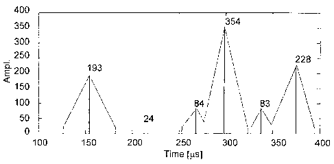
Fig. 3 the signal image of the ultrasound signal after selection
of the peak maxima in a vector representation;
Fig. 4 the envelope illustration of the ultrasound signal;
Fig. 5 the envelope illustration with exclusion of the vectors;
Fig. 6 the representation of the peaks and their averages;
Fig. 7 the representation of the peaks and the averages without a
defect.
Detailed Description of the Preferred Embodiments
The data reduction is intended particularly for use in an EMAT
pig which detects tears, corrosion and other
abnormalities/defects in the pipe wall during its run through a
pipe to be examined. During the measuring run, the analog
measured signals of the electromagnetic sensors are digitalized,
CA 02596609 2012-12-05
= 25213-94 =
- 4 -
compressed with the aid of computer modules and stored in a data
storage unit travelling along in the pig. The method for data
reduction/compression is subdivided into three basic
steps/methods for processing the resultant data set:
- the precompression,
- the extraction of features and
- the compression.
The EMAT sensor installed in the pig consists of at least one
transmitting and receiving unit. It produces an ultrasound wave
train, directed onto the pipe wall, of a preset wave type and
presettable frequency from the range of about 400 kHz to about
2 MHz, the echo of which is detected by the pipe wall with the
at least one EMAT receiver. The echo is reconverted into an
electric analog signal, digitalized with an (Analog/Digital) AD
converter and subsequently rectified (see Fig. 1).
The objective of improving the data reduction is achieved by
method steps which are divided into two procedural groups,
namely:
- determining the size of a defective point and
- determining the signal base in the adjoining area of a
defective point.
Determining the size:
The procedural steps for determining the size are based on an
algorithm for selecting peak values which delivers amplitude,
transit-time pairs, vectors, which indicate the maxima of the
ultrasound envelopes (see Fig. 3). The transit-time pairs are
here called vector. The vectors are selected below a parametric
threshold and the noise extracted in this way.
The envelopes are formed in such a way that, in addition, the
width of the ultrasound envelopes is determined for each selected
vector. For this purpose, the minima about each vector are
determined from the peak amplitudes immediately adjacent to the
CA 02596609 2007-08-01
- 5 -
vector which are below the preset threshhold. The interval of
the minima to the time value of the vector is stored. A
sufficiently accurate reconstruction of the envelopes is obtained
later (off-line) by interpolation between the individual time
values and vectors. If the peak amplitude does not fall below
the threshold between two vectors, the peak with the minimal
amplitude values is specified as minimum, which is the next
minimum of the first vector and preceding minimum of the
following vector (see Fig. 4).
The envelope vectors. whose width falls below a threshold value,
the so-called envelope width, are excluded. The envelope vectors
whose form cannot be allocated to a preset characteristic, the
so-called envelope form, are also excluded. Thus, the envelope
vector is determined by the amplitude and time value/time point
of the occurrence of the respective maximum as well as by the
envelope width and the envelope form. The characteristic of the
envelope form is determined by the ratio of the differences in
time between the time value of the maximum and the preceding and
subsequent peak minimum. If
the parametric ratio is, for
example, greater than the value 2 or less than 0.5, then the
envelope form does not correspond to the preset characteristic.
The envelope vector is then excluded.
Each envelope vector is recorded by three amplitude transit-time
pairs, i.e. by the maximum, the chronologically preceding peak
minimum and the chronologically subsequent peak minimum.
A characteristic extraction is performed by the envelope
formation, combined with the envelope width and the envelope
form, in the precompressed/vectorized ultrasound signal, in order
to decide whether the ultrasound signal contains information-
carrying characteristics and which should be stored with it.
Signal base:
Since, as already explained above, depending on the steel
CA 02596609 2012-12-05
25213-94
- 6 -
structure, it results in fluctuations of the signal base, the
determination thereof is of great significance for determining
the size of a tear in relation to the base, Therefore, in this
case, the time range of the ultrasound echo is divided into any
time intervals desired, however, at least with the duration of
4 linked wavelengths of the ultrasound frequency applied, with
a parametrisized start value and length value. In
these
intervals, the amplitudes of the peaks are summed up to form an
interval-specific summation value which is then divided by the
number of peaks. It is then used to provide an average. To
determine the size, each mean value is determined at the
allocated defective point and stored. To determine the size of
a defect, the base values adjacent to the defect are used as a
reference off-line in an azimuthal and radial direction.
The data of the selected features are then compressed without a
significant loss of information. The data are thereby coded
without information loss about the defect. The time values are
then stored with different resolution in dependence on the
significance, so that a higher compression factor is obtained as
a result. The time value of the maximum for
the local resolution of a detected defect is significant,
however, the time values of the envelope width are of secondary
importance. By coding with a different 'time resolution, a higher
compression factor is obtained.
The maximum is determined in a
defined interval of at least the size of half a wavelength
(scanning theory) (see Fig. 2).
In comparison= to conventional methods, the method substantially
increases the fidelity of reproduction, namely without increasing
the data set to be stored, or without increasing it
significantly. The
method enables a reconstruction of the
ultrasound envelopes from the data stored in a reduced manner
without information loss, at least without significant
CA 02596609 2012-12-05
. 25213-94
- 7 -
information loss, after the testing step of the EMAT pig. The
method evaluates the size of the envelope - and that is with a
decisive, efficient feature - by method groups in order to
determine the size. The size determination is determined in
the quality thereof by the method groups in order to determine
the signal base.
Figs. 1 to 4 were already connected above in the description
text. In Fig. 1, the significant echo of a tear is shown in
Fig. 1 at about 150 ps. At about 300 ps and at about 370 ps,
the transmission signals of the adjacent transmitters can be
seen. The sensor arrangement is described in GB 2 380 794 A
(in this connection, see especially Fig. 3 and the description
passages at page 6, line 26, to page 7, line 18).
In comparison to Fig. 3, the envelope vector illustration of
the ultrasound signal in Fig. 4 provides a better illustration
of the ultrasound envelopes than the pure vector illustration.
In Fig. 5, the envelope vector illustration is shown with
exclusion of the vectors, identified with 34, whose envelope
widths fall below a minimum width.
= CA 02596609 2007-08-01
- 8 -
The illustration of the peaks and averages of the peaks is shown
in three intervals in Fig. 6: 100 As - 150 As, 150 As - 200 As,
200 As - 250 As.
To elucidate, Fig. 7 shows, on a modified/enlarged scale,
vertically and horizontally, the illustration of the peaks and
the averages of the peaks in three intervals from Fig. 6 (100 As
- 150 As, 150 As - 200 its, 200 As - 250 As) of a US echo without
a defect.
