Note: Descriptions are shown in the official language in which they were submitted.
CA 02597177 2007-08-14
IMMUNOASSAY TEST DEVICE AND METHOD
OF USE
Background of the Invention
[0001] Disclosed is a novel device for detection of an analyte in a sample
using a lateral flow assay. The device and method of the present
invention may be easily and inexpensively assembled, and suitable for
use by personnel with little specialized training. The device further
provides for sanitary handling and disposal of biological samples. The
device and method disclosed herein may be used with labels
conventionally used with lateral flow assays such as colloidal metals,
or may also be used with colorimetric or fluorescent labels that require
instrumentation for detection.
[0002] Immunoassays
[0003] The present invention relates to assays utilizing test strips, in
particular, a lateral flow immunoassay. The immunoassay, in general,
is a sensitive technique used to measure levels of a substance using the
reaction of an antibody or antibodies to its antigen. Immunoassays
generally rely on binding of an antibody to an antigen. Monoclonal
antibodies, in particular, are often used because such antibodies
generally bind to only one site of a particular molecule. This specific
binding enhances the specificity and accuracy of binding to a particular
analyte. The antibodies used in immunoassays typically have a high
affinity for the antigen such that a high proportion of the antigen binds
to the antibody.
[0004] Immunoassays are powerful and versatile biomedical diagnostic tools
that can be used, for example, to monitor drug and hormone levels in
body fluids, diagnose infectious and autoimmune diseases, and
diagnose and monitor treatment of cancer.
CA 02597177 2007-08-14
-2-
[0005] One analyte in particular that is ideally suited for detection using
immunoassay techniques is influenza. Influenza is a highly contagious
epidemic to pandemic acute viral respiratory disease caused by several
genera of the Orthomyxoviridae family. Influenzavirus A and
lnfluenzavirus B are the two genera most commonly associated with
disease in humans. Influenza infection rates tend to be highest in
pediatric populations, while serious complications from influenza
disease are more common in the elderly. Clinical signs and symptoms
begin after a 1-4 day incubation period and include cough, fever,
myalgia and malaise. The clinical presentation of influenza can range
from asymptomatic infection to fatal pneumonia. Influenza co-
circulates with other respiratory pathogens; hence it is important to
differentiate influenza from other respiratory diseases. Rapid influenza
detection tests facilitate the more timely administration of antiviral
drugs, which, in general, are of clinical benefit when administered
within 48 hours of the appearance of symptoms. Not all antiviral drugs
are effective against both influenza A and influenza B; therefore it is
important to distinguish between the two.
[0006] Influenza A and B can be detected in human respiratory samples by a
variety of methods including tissue culture, immunofluorescent assay
and enzyme immunoassay. Tissue culture isolation remains the gold
standard for the detection of influenza, yet the procedure can take up to
7 days to complete. Immunofluoresccnt antibody-based tests are
moderately sensitive, yet highly dependent on specimen quality and
preparation. The rapid detection of influenza using enzyme and
microparticle-based immunoassays has become an important aspect of
patient management in patients of all ages with acute respiratory
disease due to influenza. Test results can be used to support data
available from the patient's clinical evaluation and assist the physician
in determining the course of action.
CA 02597177 2011-04-21
-3-
[0007] Immunoassay techniques typically employ a detectable label that
permits the user to determine whether the analyte is present in the
sample. The label can be conjugated to a particle such as an antibody
that binds to the analyte (referred to herein as a first "binding
reagent"). The type of label used may vary, and may include visually
detectable labels as well as labels that require instrumentation for
detection. Non-limiting examples of labels that can be used with
immunoassay techniques include enzymes, radioisotopes, fluorescent
tags, carbon particles, beads, or metal sol tags such as colloidal gold.
[0008] Lateral Flow Immunoassays
[0009] Lateral flow assays (or "flow-through" assays) are well known in the
art and are described in Ching et al., U.S. 6,534,320, May et al., U.S.
6,228,660, Charlton et al, U.S. Patent 5,989,921, Charlton U.S. Patent
6,485,982, Charlton US Patent 5,714,389, Rosenstein, U.S. RE38,430.
100101 Lateral flow assays are characterized in that a liquid solution
containing an analyte to be detected is transported by capillary action
laterally along a membrane strip. The membrane strip typically has
reagents impregnated in the membrane. Sample is applied to one end
of the strip (typically at a first absorbent pad) and sometimes with the
aid of a solvent such as water. The sample may be mixed with a
labeling reagent having a first binding reagent before contact with the
strip, or the strip may contain labeling reagent therein. As the liquid
passes through a "detection zone," second binding reagents
immobilized on the strip permit visualization of the assay results. The
lateral flow assay is typically rapid and provides sensitive and accurate
detection of analytes, depending in part on the selection of the binding
reagents used.
CA 02597177 2011-04-21
-4-
[00111 Lateral flow assays may employ "competitive" or "noncompetitive"
techniques, both of which are well-known in the art. In the
competitive-type immunoassay, analyte in a sample is mixed with
analyte that is conjugated to a detectable label. The mixture is then
contacted with a lateral flow test strip. The mixture then migrates
along a flow path defined by a membrane. The unlabeled analyte
(from the sample) and labeled analyte compete for a limited number of
binding sites on a binding agent immobilized on the test strip. The
amount of labeled analyte detected at the detection region in a
competitive assay is inversely proportional to the concentration of
analyte in the sample (i.e., a greater amount of accumulated label
indicates lower levels of analyte in the test sample).
[00121 In contrast, in "non-competitive" or "sandwich"-type immunoassays,
antigen in the sample binds to a first binding reagent (such as an
antibody) conjugated to a label (the "labeling reagent"). The sample
containing antigen bound to the labeling reagent is then contacted with
a lateral flow assay test strip. As the mixture migrates by capillary
action along the membrane, the analyte-labeling reagent complex
contacts and binds to a second binding reagent immobilized in the
membrane. The label-analyte complex accumulates on the membrane,
and a visible indicator line results. The amount of accumulated label is
directly proportional to the concentration of the antigen in the sample.
Both competitive-type and non-competitive-type assays are described
in Ching et at, U.S. 6,534,320.
[0013] The lateral flow immunoassays typically employ the same basic
components. These are described in, for example, Ching et al, US
6,534,320 and May et al. US 6,228,660. These components are: a first
absorbent material, a membrane (such as nitrocellulose), and a second
absorbent material, wherein the test strip has reagents impregnated
therein for the detection of analytes.
CA 02597177 2007-08-14
-5-
[0014] Lateral flow devices can also be categorized as using either a one-step
or two-step method. The two-step method (also referred to as the
"pour on" method) is described in European Patent Application 0 250
137 A2, entitled "Colloidal Gold Immunoassay," published December
12, 1987 ("Mochnal"). In this method, the sample and labeling reagent
are mixed prior to contacting the sample with the lateral flow test strip.
After mixing sample with labeling reagent, the mixture is contacted
with a first absorbent material to initiate the lateral flow assay. The
sample then flows along the membrane, contacting one or more
immobilized second binding reagents. Analyte in the sample binds to
the second binding reagent and accumulated label results in a visible
reaction. The two-step method is characterized by the initial step of
pre-mixing liquid sample with labeling reagent prior to contacting the
mixture to the test strip.
[0015] In contrast, in the "dried-on" or "one-step" method, sample is not
mixed with labeling reagent prior to contacting a test strip. In the one-
step method, the labeling reagent is pre-dried and embedded within the
test strip, typically within the first absorbent pad. Liquid sample
applied directly to the first absorbent pad solubilizes the dried labeling
reagent. As the liquid sample flows laterally along the test strip
towards the test site, analyte binds to and transports the labeling
reagent bound to analyte to an immobilized second binder. As in the
two-step method described above, the analyze reacts with a second
binding reagent immobilized on the matrix to effect a visual result.
The one-step method is distinct from the two-step method primarily in
that all of the reagents necessary for the assay are present in dry form
on the test strip, eliminating the need for a separate mixing step.
[0016] Additionally, cross-contamination and sanitation is often a concern in
the use of lateral flow assays. Test strips used for detection of analytes
in biological samples, such as urine, saliva or feces, pose a potential
CA 02597177 2007-08-14
-6-
contamination hazard when the test strips are contacted with sample
and then transported to a different location. Contamination can occur
when the test strips are in use, or upon disposal of the strips. As such,
it is desirable to have a device that provides sanitary handling and
disposal, minimizing cross contamination of test strips or personnel.
[00171 The invention described herein provides a support for a test strip, in
particular, a lateral flow immunoassay test strip, and a device for
conducting assays using test strips, that provide for improved ease of
use, assembly, sanitary handling and disposal. The invention further
relates to a device that may be used for detection of multiple labeling
reagents including those that emit light or that require the use of
instrumentation such as spectrophotometers.
Brief Summary of the Invention
[00181 Discl osed herein is a device for determining the presence or absence
of
an analyte in a sample, wherein the device comprises a receptacle, a
holder, and a test strip. In one embodiment, the holder comprises an
elongated region for affixing a test strip, a stop feature, a closure for
the receptacle, a grip member, an alignment feature and retention
features. The holder may further comprise secondary pins for securing
the test strip.
[0019] In another embodiment, the holder comprises a grip member, a stop
feature, a closure, an alignment feature, and a hinge wherein the grip
member and closure are fanned by folding the top portion of die grip
member upon the lower portion of the grip member at the hinge
[0020] Another embodiment of the present invention is further related to a
device for determining the presence or absence of an analyte in a
sample, the device comprising a receptacle containing a labeling
reagent that binds with the analyte and a holder. In one embodiment,
the holder contains a test strip comprising a first absorbent pad, a
CA 02597177 2007-08-14
-7-
membrane strip and a second absorbent pad defirting a flow path for
transporting a liquid sample, the test strip having at least one detection
region. The test strip is held in a recess within the holder which further
comprises an elongated support containing the recess and having an
alignment feature, a closure, a stop feature and a grip member. In one
embodiment, the holder is formed by folding the top of the
unassembled (unfolded) holder at a hinge such that the top portion of
the grip member is folded upon the lower portion of the grip member
thereby capturing the second absorbent pad of the test strip between
the two surfaces of the grip member of the holder. The holder also
comprises a closure that substantially seals the receptacle when the
holder is inserted into the receptacle.
[0021] The device may be provided in the form of a kit containing 1) a
receptacle containing the dried and dispensed gold conjugate, 2) a
holder and strip assembly, 3) a swab or transfer pipette and (in a four-
part embodiment) 4) a rack or other assembly for maintaining the
device in an upright position during testing.
(0022] Described herein are various embodiments of the invention, one or
more examples of which are set forth below. Each example is
provided by way of explanation, and not limitation, of the invention. [t
will be apparent to those skilled in the all that modifications may be
made in the present invention without departing from the scope or
spirit of the invention. Thus, it is intended that the present invention
covers such modifications and variations as come within the scope of
the claims and their equivalents.
CA 02597177 2007-08-14
-8-
Brief Description of the Drawing
[0023] The invention will now be illustrated with respect to the following
drawings illustrating embodiments of the invention in which:
[0024] FIG. 1 is a front view of an embodiment of the invention.
[0025] FIG. 2 is an exploded view of the unassembled holder and test strip.
[0026] FIG. 3 is an enlarged isometric view of the holder 2.
[0027] FIG. 4 is an enlarged view of the holder.
[0028] FIG. 5 is an enlarged view of the holder.
[0029] FIG. 6 is a bottom view of the holder in Fig. 2.
[0030] FIG. 7 is an enlarged view of the holder in Fig. 2.
[0031] FIG. 8 is a perspective view of the holder.
Detailed Description of the Invention
[0032] Definitions
[0033] The singular forms -a7', "an", and "the" include plural references
unless the context clearly dictates otherwise.
[0034] As used herein, the tern "analyte" generally refers to a substance to
be
detected. For instance, analytes may include antigenic substances,
haptens, antibodies, and combinations thereof. The analyte may be
any analyte described in the art.
[0035] An "analyte detection region' or "detection region' is any region of an
assay device in which the analyte or label may be detected and/or
measured to determine the presence or absence of analyte in a sample.
The analyte detection region may be qualitative or quantitative in
nature. Thus in a lateral flow device, for example, the analyte
CA 02597177 2007-08-14
-9-
detection region may be part of a porous matrix which contains
binding reagents for immobilizing a detectable label. One or more
detection regions may be present. Depending on the assay format, the
amount of immobilized label in the analyto detection region may
increase or decrease in the presence of analyte. For example, in a
sandwich assay format, the amount of immobilized label will increase,
while in a competition assay format, the amount of immobilized label
will decrease.
[0036] The term "emission signal" refers to electromagnetic radiation emitted
when an atom in an excited higher energy state decays to a lower
energy state.
[0037] The term "excitation signal" refers to the energy, for example, that
form electromagnetic radiation, which causes an electron of an atom to
move from a lower energy state into an "excited" higher energy state.
[0038] The term "label" as used herein refers to any substance that is capable
of producing a detectable signal, whether visibly or by using suitable
instrumentation. Various labels suitable for use in the present invention
include, but are not limited to, chromatogens, fluorescent or
chemiluminescent compounds, catalysts, enzymes, enzymatic
substrates, dyes, colloidal metallic and nonmetallic particles, and
organic polymer latex particles.
[0039] The term "luminescence" refers to any emission of light that does not
derive energy from the temperature of an energy source (for example, a
source of electromagnetic radiation, a chemical reaction, mechanical
energy). In general, the source causes an electron of an atom to move
from a lower energy state into an "excited" higher energy state; then
the electron releases that energy in the form of emitted light when it
falls back to a tower energy state. Such emission of light usually
occurs in the visible or near-visible range of the electromagnetic
CA 02597177 2007-08-14
- 10-
spectrum. The term "luminescence" includes, but is not limited to,
such light emission phenomena such as phosphorescence,
fluorescence, bioluminescence, radioluminesccnec. clcctro-
luminescence, and thereto-luminescence.
[0040] The term "luminescent later' refers to a label that generates a
luminescent signal, e.g. an emission of light that does not derive
energy from the temperature of the emitting source. The luminescent
label may be, for example, a fluorescent molecule, a phosphorescent
molecule, a radiluminescent molecule, a luminescent chelate, a
phosphor or phosphor-containing compound, or a quantum dot.
[0041] As used herein. the term "porous material" refers to any material
capable of providing capillary action. This would include material
such as, for example, nitrocellulose, nitnxxllulace blends with
polyester or cellulose, untreated paper, porous paper, rayon, glass fiber,
acrylonitrile copolymer, or nylon. One skilled in the art will have
knowledge of other porous materials that allow lateral flow.
[0042] As used herein, the term "test sample" generally refers to a biological
material suspected of containing an analyte. The test sample may, for
instance, include materials obtained directly from a source, as well as
materials pretreated using techniques, such as, but not Tunitcd to,
filtration, precipitation, dilution. distillation, mixing, concentration,
inactivation of interfering components, the addition of reagents, lysing,
and so forth. The test sample may be obtained or derived from any
biological source, such as a physiological fluid, including, blood,
interstitial fluid, saliva, ocular lens fluid, cerebral spinal fluid, sweat,
urine, milk, ascites fluid, mucous, synovial fluid, peritoneal fluid.
vaginal fluid, amniotic fluid, and so forth. Besides physiological fluids,
other liquid samples may be used such as water, food products, and so
forth, for the performance of environmental or food production assays.
In addition, a solid material suspected of containing the analyte may be
CA 02597177 2007-08-14
- 11 -
used as the test sample. The test sample may be used directly as
obtained from the biological source or following a pretreatment to
modify the character of the sample. For example, such pretreatment
may include preparing plasma from blood, diluting viscous fluids, and
so forth. Methods of pretreatment may also involve filtration,
precipitation, dilution, distillation, mixing, concentration, inactivation
of interfering components, the addition of reagents, etc. Moreover, it
may also be beneficial to modify a solid test sample to form a liquid
medium or to release the analyte.
[0043] As used herein, the term "detection zone' when used to refer to a test
strip, refers to the region on the mcntbranc containing binding
reagents, whether binding to molecules that indicate a positive or
negative control, or to molecules that indicate presence or absence of
analyte. The binding reagents may include those that bind to analyte,
labeling reagent, label. or any other molecules such that a visual signal
is obtained.
[0041] Test Device
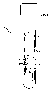
[0045] Fig. 1 depicts one embodiment of the present invention. In this
embodiment, the test device 1 comprises a receptacle 3, a holder 2, and
a test strip 4 wherein the test strip 4 allows fluids to flow laterally
along its length. The test may be any test strip known in the art. The
receptacle 3 may be used to receive sample, diluent and/or labeling
reagent. The holder 2 is used to hold any suitable test strip 4 known in
the art, such as a lateral flow immunoassay test strip ac shown in Figs.
1,2 and 8.
[o04b] The Receptacle
[0047] As shown in Fig. 1. the receptacle 3 of the device 1 generally has an
elongated shape that may be rounded on the bottom (having a test-tube
CA 02597177 2007-08-14
-12-
like shape, as shown) or flattened (not shown). Where the bottom of
the receptacle 3 is flattened, the receptacle 3 may be self-standing.
[0048) The receptacle 3 may also be cuvette-shaped or having the properties
of a cuvette such that the receptacle 3 is compatible for use with
spectrophotometric instruments or the like. However, the receptacle 3
may be of any shape suitable to receive both sample and the holder 2.
[0049] In embodiments in which the receptacle 3 has the properties of a
cuvette, the receptacle 3 may have a cuvette-like shape such as, for
example, cuvcttcs provided by Ocean Optics Inc. CVD-UV and CVD-
VIS Disposable Cuvettes, manufactured and sold by Ocean Optics Inc.,
are plastic cuvettes that work in the UV range--transmitting light
between 220-900 nm. The CVD-VIS Cuvettes transmit light from
350-900 nm and are suited for use in VIS applications. The cuvette
may be square or triangular in shape. Any cuvettes known in the art
can be used with the present invention.
[0050] In embodiments using a receptacle 3 having cuvette-like properties, the
receptacle 3 can be used with labeling reagents that use fluorescent or
other luminescent labels, and can be used in conjunction with
spectrofluorometers, for example, without the need to transfer sample
to a second container. In this embodiment, the sample need not be
removed from the receptacle 3 to determine, for example, light
absorption or refraction. The holder 2 may or may not be removed
prior to evaluation or detection. Where removed prior to evaluation or
detection, the receptacle 3 may be closed with any cap known to seal a
cuvette or test tube (not shown).
[0051] In embodiments using a test-tube shaped receptacle 3. the receptacle 3
may be shaped to allow use with a vortexing machine having a test-
tube shaped cup, and may be compatible with standard test-tube racks
that can maintain the device in an upright position during use.
CA 02597177 2007-08-14
-13-
[0052] Regardless of the shape, receptacle 3 of device 1 may be manufactured
from glass, plastic or any other material suitable for use with the
analyte, or any diluent, eta. The receptacle 3 may be comprised of a
material that provides chemical resistance, permitting use with organic
solvents, as well as acids and bases.
[0053] The receptacle 3 may be wholly or partly transparent to visible light,
or
may be wholly or partly opaque. La some embodiments, the receptacle
3 may be opaque, with the exception of viewing windows on the
receptacle 3. The viewing windows may take a variety of different
shapes, and may be present at varied locations on the receptacle 3,
depending on the desired use of the device, and the nature of the labels.
[0051] For example, where visually detectable labels are used with the device,
the viewing windows in the receptacle 3 may be positioned such that
the detection regions of the test strip are visible through the windows
when holder 2 is placed in receptacle 3. Alternatively, the windows
may be placed such that light reflectance or absorption of a sample
may be measured using appropriate instrumentation.
[0055] In a further embodiment, receptacle 3 may be shaped in a manner that
provides magnification of the contents or test strip 4 therein. For
example, a portion of receptacle 3 may be curved or suitably shaped to
magnify the regions of an enclosed test strip 4 such that viewing of the
accumulated label is enhanced,
[0056] The receptacle 3 may be shaped so that it can be used with test tube or
cuvette racks available, or may be used with specialty-designed racks
that maintain the device in an upright position during use. Such a
specially designed rack may be provided to the end-user as part of a
kit, described below.
[00571 The receptacle 3 used in the device 1 disclosed herein may be
disposable or may be able to be reused.
CA 02597177 2007-08-14
-14-
It should be understood that receptacle 3 is such that a user may use
both a lateral flow assay test strip to analyze the sample along with
other methods to analyze the sample. For example, the test strip 4 may
be used and removed, then the remaining sample may be assayed using
other methods known in the art.
[0059] Holder
[0460] As shown in Figs- 1, 2 and 8. the holder 2 of device 1 is used to hold
a
lateral flow test strip 4. Holder 2 is shaped to perform one or more of
the following functions: 1) substantially seal the receptacle 3; 2)
provide a support structure for the test strip 4; 3) provide a means for
positioning the test strip 4 relative to the receptacle 3 and sample; 4)
minimize contamination of sample and personnel; 5) minimize
contamination of the test strip 4 when manipulating or inserting the
holder 2 into the receptacle 3, and; 6) provide one or more detection
guides 13 ("reading guides") for detection of results on the test strip 4.
[0061] The holder 2 is preferably manufactured as a single piece using
standard techniques. For example, it can be injection molded,
compression molded or machined. The holder is shown in an open or
unassembled position in Fig. 2. The holder 2 may be formed from any
suitable material that will withstand the environment of the sample,
diluent, labeling reagents, and any additional reagents that may be
added to the receptacle 3. Preferably, it is made of plastic or the like,
more preferably, polypropylene, polystyrene or the like.
[0062] As depicted in Figs. 1-8, the holder 2 has an elongated portion 6. The
elongated portion 6 may further comprise an alignment feature 5, one
or more retention features 8, and shield regions 12. The holder 2 may
further comprise secondary pins 19 (shown in Fig, 6)_ Upon folding the
unassembled holder at the hinge 7 shown in Fig. 2, an assembled
holder 2 (ac shown in Figs. I and 8, shown with test strip 4) is formed.
CA 02597177 2007-08-14
-15-
In one embodiment. folding the top portion of the grip member upon
the lower portion of the grip member at the hinge region forms the grip
member 11 and the closure 10.
[0063] The grip member 11 of the holder comprises a top portion and lower
portion separated at the hinge 7. The hinge 7 may be a living hinge. A
living hinge is a hinge or flexure bearing with no moving parts,
generally a thin section of plastic or other material that connect two
segments of a part to keep them together and allows movement. The
grip member 11 comprises a top portion and a bottom portion. The top
portion is folded over or snapped into place on the lower portion at the
hinge to capture the upper edge of a test strip. When the top portion
and lower portion of the grip member 11 are aligned, the test strip is
secured in place. It will also be understood by one of ordinary skill in
the art that the hinge 7 is not essential, such that the holder 2 may be
manufactured by two separate pieces, one piece identical to the portion
below the hinge 7, the other piece identical to the portion above the
hinge 7.
[0064] Once folded, the holder 2 has a general shape of that depicted in Figs.
I and 8. To assemble the holder 2 as shown in Figs. I and 8, a lateral
flow test strip 4 or the like is first placed within the alignment feature
5. In a preferred embodiment, the alignment feature S may be a recess
with a solid wall on each side or a periodic wall (for example, guide
posts, or serial guides, formed in the elongated portion 6 of the holder
2. Upper and lower retention features 8 (shown in a close-up view in
Figs. 3, 4, and 7) may be present to secure the second and first
absorbent pads. respectively. The holder 2 is folded at a hinge 7.
thereby capturing the upper edge of the test strip 4, Secondary pins 19,
as shown in Fig. 6 (bottom view of holder without a test strip) may
also be provided. The secondary pins 19 may be present to further
secure the test strip 4 in place by locking into the second absorbent pad
CA 02597177 2007-08-14
1b
17. In one embodiment, the secondary pins 19 secure the test strip by
penetrating into the second absorbent pad approximately S mm.
10065 The assembled holder 2 as shown in Fig. 8 may further comprise a stop
feature 9. The stop feature 9 is formed upon assembling the top
portion and the lower portion of the holder 2 together at the hinge 7. In
one embodiment, best viewed in Fig. 8, the stop feature 9 is comprised
of a ledge-like region that extends outward from the closure 10. The
stop feature 9 prevents holder 2 from being inserted into the receptacle
3 beyond a fixed point. The stop feature 9 positions the distal end of
the holder 2 and the test strip 4 in the proper position within receptacle
3 relative to the sample, at a distance necessary for initiation of the
reaction, but such that the lower edge of test strip 4 contacts the sample
but the lower edge of the holder 2 does not contact the test sample.
The stop feature 9 and alignment feature S are in relation to one
another such that appropriate positioning of the test strip 4 relative to
the sample is achieved when the holder is placed in the receptacle 3.
[0066] The assembled holder further comprises a closure 10 (as shown in
Figs. I and 8) for the receptacle 3. The closure 10, in one embodiment,
is generally plug-like in shape, though any suitable shape that
substantially seals the receptacle 3 is contemplated within the present
invention. The closure 10 is shaped such that it substantially seals the
device 1 when inserted into receptacle 3. The closure 10 shown in Fig.
8 has hollowed-out portions that facilitate manufacturing of the device,
though it will be readily understood that the invention is not limited to
this embodiment, and the closure 10 may take a variety of different
forms.
[0067] The assembled holder 2 further comprises a grip member 11. In the
embodiment shown in Fig. I and 8, the grip member 11 has a generally
rectangular shape. However, the grip member 11 need not have this
particular shape, but may have any other shape that allows easy
CA 02597177 2007-08-14
-17-
handling of the holder 2. For example, the grip member 11 may have
rounded edges, or may also comprise raised ribs, ridges or other
texture to facilitate removal and insertion of the holder Z The grip
member 11 is sized such that a user may manipulate and use the holder
2,
[0068] As seen in Fig. 2, the holder 2 may also have shield regions 12 that
help prevent contamination of the test strip 4 when inserting holder 2
into receptacle 3. In this embodiment, the shield regions 12 may be
raised edges on the elongated support 6 that shield the sides of a test
strip 4. These raised edges can be a solid wall, as shown, or a broken
wall. In use, it is necessary for the user to insert the holder 2 inside the
receptacle 3 to initiate the reaction. Liquid or other contaminants may
exist on the rim of the receptacle 3. Shield regions 12 protect the test
strip 4 from rim contamination when inserted into the receptacle 3.
[0069] As shown in Figs. I and 8, the elongated portion 6 may also have one
or more "detection guides" 13 to show the user where labeling reagent
should accrue. For example, one or more visihic marks may be made
on a shield region 12 of the test strip or on the elongated portion 6 that
corresponds to a region on the test strip 4 that contains a binding
reagent for the analyte. The detection guides 13 may be a line or other
demarcation, and may be indicated by color or a raised portion of the
holder 2. In one embodiment, there are at least three reading lines,
corresponding to a detection region for influenza A, a detection region
for influenza B and a control line. However, it will be readily
understood to one of ordinary skill in the art that the detection guides
13 may take a variety of different forms and may correspond to one or
more different analyzes and control regions.
[0070] The Test Strip
CA 02597177 2007-08-14
-18-
[0071] The holder 2 of the present invention may be used to hold any test
strip
used in the art. In one embodiment, a lateral flow assay test strip as
known in the art (and depicted in Figs. 1, 2 and 8) is placed in the
holder 2. The test strip 4 may use the one-step or two-step method. In
the one-step method, the test strip 4 has a first labeling reagent
diffusively immobilized in the first absorbent pad 14. In the two-step
method, as described above, the first labeling reagent is separate from
the test strip 4. In a two-step embodiment, a labeling reagent such as a
dried conjugate is provided in the receptacle 3 separately from the test
strip 4. The labeling reagent may also be added by the end-user.
[0072] In embodiments using a lateral flow test strip, the test strip 4 can be
any lateral flow test strip known in the art. For example, the test strip
4 preferably is comprised of a first absorbent pad 14, a membrane 15,
and a second absorbent pad 17, as shown in Figs. 1, 2 and B. Fig. I
depicts a test strip 4 placed in the device, and Fig. 2 depicts the test
strip prior to placing in the holder 2 of the device 1. Referring to Fig.
2, suitable labeling reagents comprising a first binding reagent and a
label may be impregnated in the rust absorbent pad 14, and suitable
second binding reagents are impregnated in the membrane 15. The
binding reagents are selected based on the analyte to be detected, and
suitable selection of binding reagents will be readily understood by one
of ordinary skill in the art. The second binding reagents impregnated
in the membrane 15 form one or more "detection region(s)" 16 further
comprising regions containing second binding reagents for the analyte
and/or "control regions." Suitable reagents for control regions will be
readily understood by one of ordinary skill in the art. The test strip 4
may have one or more detection regions for analyte and one or more
control regions, as desired.
[0073] The test strip 4 used with the device I may also be any other strip
known in the an and compatible with the holder 2 as described above,
CA 02597177 2007-08-14
-19-
and is not limited to test strips for lateral flow immunoassays. For
example, the present invention may employ a membrane used for thin
layer chromatography. In such an embodiment, the thin-layer
chromatography membrane comprises an appropriate membrane or
other material affixed to the elongated portion 6 of the holder 2. The
sample, either liquid or a solid dissolved in a volatile solvent, is
deposited in the receptacle 3 of the device 1 or directly on the test strip
4. The constituents of a sample can be identified by simultaneously
running standards with the unknown. The solvent containing sample
moves up the elongated portion 6 of the holder 2 or the membrane/test
strip 4 contained thereon by capillary action. When the solvent front
reaches the upper edge of the holder 2, the separated spots may be
visualized using appropriate detection methods such as ultraviolet light
or placing the plate in iodine vapor. The different components in the
mixture move up the plate at different rates due to differences in their
partitioning behavior between the mobile liquid phase and the
stationary phase.
[00741 Where the device 1 is used in combination with a lateral flow assay
test strip 4, the test strip 4 is assembled as understood in the an. Fig. 2
depicts an example of a lateral flow test strip 4 that may be used. In
one embodiment, the immunoassay test strip comprises a bibulous
membrane strip 15 such as nitrocellulose, a fast absorbent pad 14, and
a second absorbent pad 17. "Bibulous" materials include untreated
forms of paper, nitrocellulose and the like which effect
chromatographic separation of components contained in liquids which
are passed therethrough. In contrast, in "nonbibulous" liquid flow all
of the dissolved or dispersed components of the liquid which are not
permanently entrapped or `filtered out' are carried at substantially
equal rates and with relatively unimpaired flow though the membrane
or support. Bibulous flow results in preferential retention of one or
more components. Test strips as disclosed in Chariton, et aL, U.S.
CA 02597177 2011-04-21
-20-
5,989,921 "Test Device and Method for Colored Particle
Immunoassay" issued Nov. 23, 1999, may be used.
100751 In embodiments using a lateral flow test strip, the membrane is
generally a porous carrier such as nitrocellulose. The test strip 4 may
further comprise a backing layer, such as Mylar, or may be directly
adhered to the elongated portion 6 of the holder 2. The test strip 4 may
have a backing of one continuous piece of laminate or separate pieces.
The backing may also be a laminate such as vinyl but one skilled in the
art will recognize that numerous materials can be used to provide
support to the test strip. In embodiments where the test device is used
with methods other than the lateral flow immunoassay, the strip may
comprise chromatographic paper or other materials suitable for the
type of assay desired.
[0076] First Absorbent pad
100771 In embodiments of the present invention using a lateral flow assay test
strip, a first absorbent pad is preferably used. Referring to Fig. 2, the
first absorbent pad 14 is placed at the end of the membrane 15 where
the sample is to be contacted with the strip, typically at the distal end
furthest from the holder. The first absorbent pad 14 contacts the
sample when the holder 2 is inserted into the receptacle 3. The first
absorbent pad 14 may extend beyond the lower edge of the holder 2
such that sample contacts the test strip 4 without contacting the lower
edge of the holder 2. This first absorbent pad 14 extends into the
sample volume when the holder 2 is secured in the receptacle 3.
Positioning of the first absorbent pad 14 relative to the sample and the
receptacle 3 is fixed via the alignment feature 5 and the stop feature 9
of the holder 2. Contact of the pad 14 to sample or sample-diluent
initiates the assay by permitting the pad 14 to absorb sample,
conducting flow along the membrane 15. Flow along the membrane
CA 02597177 2007-08-14
-21
15 permits analytc in the sample to contact second binding reagents
immobilized on the membrane. The first absorbent pad 14 can further
serve as a filter for separating liquid sample from particulate matter
that could interfere with capillary flow, further reducing the possibility
of false positives.
[0078] Absorbent pads used with lateral flow immunoassays are well known
in the art. Non-limiting examples of pads that may be used with the
present invention include Whatman D29, Whatman 1.5WF, Whatman
3MM CHR, available from Ahlstrom, 122 West Butler Street, Mount
Holly Springs, PA 17065, or Whatman, 200 Park Ave., Florham Park,
NJ 07932.
[00791 Matrix strip
[0080] Again referring to Fig. 2, the matrix strip is a porous membrane 15
that
may be any suitable membrane known in the art, in general, the
porous membrane 15 may be made from any of a variety of materials
through which the detection probes are capable of passing. For
example, the materials used to form the porous membrane 15 may
include, but are not limited to, natural, synthetic, or naturally occurring
materials that are synthetically modified, such as polysaccharides (e.g.,
cellulose materials such as paper and cellulose derivatives, such as
cellulose acetate and nitrocellulose); potyether sulfone; polyethylene;
nylon; polyvinylidene fluoride (PVDF); polyester; polypropylene;
silica; inorganic materials, such as deactivated alumina, diatomaceous
earth, MgSO4. or other inorganic finely divided material uniformly
dispersed in a porous polymer matrix, with polymers such as vinyl
chloride, vinyl chloride-propylene copolymer, and vinyl chloride-vinyl
acetate copolymer; cloth, both naturally occurring (e.g., cotton) and
synthetic (e.g., nylon or rayon): porous gels, such as silica gel, agarose,
dextran, and gelatin: polymeric films, such as polyacrylarnide; and the
like.
CA 02597177 2007-08-14
-22-
[0081] The pore size of the membrane 15 may preferably be about 0.05 to
about 20 microns.
[0082] In embodiments using a lateral flow immunoassay, the test strip 4
further comprises one or more detection regions 16, as. described
above, and as shown in Figs. 1, 2, and 8. The test strip 4 may further
comprise one or more control zones, as desired by the user. The one or
more detection regions 16 comprise binding reagents impregnated on
the matrix strip at predetermined points.
[0063] The detection regions 16 comprise unlabeled binding reagents
immobilized in the membrane 15 that bind to the analyte labeting
reagent complex. Accumulation of bound analyte results in a visible
signal. The control region is comprised of immobilized reagents that
typically bind to a region of the labeling reagent (such as the Fc region
of the first labeling reagent, where the first labeling reagent is an
antibody) and accumulated labeling reagent at the control region
indicates successful completion of the assay.
[0064] The one or more detection regions 16 may contain the same binding
reagents, or may contain different binding reagents for capturing
multiple analytes. For example, the detection region 16 may include
two or more distinct binding regions (e.g., lines, dots, etc.) for the
detection of one or more analytes and one or more control regions for
confirmation of assay completion and integrity. Preferably, the
binding and control regions may be disposed in the form of lines in a
direction that is substantially perpendicular to the flow of the test
sample through the device 1. However, in some embodiments, the
binding and control regions may be disposed in the form of lines in a
direction that is substantially parallel to the flow of the test sample
through the assay device.
CA 02597177 2007-08-14
-23-
[0085] The control region is generally located at a site on the membrane 15
downstream from the detection regions that contain binding reagents
specific to analyte. The reagent may bind to both complexed and
uncomplexed conjugate particles, and is therefore generally different
from the first binding reagent. In one embodiment, the reagent is a
biological binding reagent (e.g., antigens, haptens, pmtein A or G,
neutravidin, avidin. streptavidin, primary or secondary antibodies (e.gõ
polyclonal, monoclonal, etc.), and complexes thereof) that is different
than the first binding reagent. For example, the first binding reagent
may be a monoclonal antibody while the second binding reagent may
be avidin (a highly cationic 66,000-dalton glycoprotein), strcptavidin
(a non-glycasylated 52,800-daltnn protein), neutravidin (a deglysolated
avidin derivative), and/or capiavidin (a nitrated avidin derivative). In
this embodiment, the second binding reagent may bind to biotin. which
is biotinylated or contained on detection probes conjugated with a
monoclonal antibody different than the monoclonal antibody of the
first binding reagent.
[0086] In addition, various non biological materials may be used for the
second binding reagent of the control region as are known to one of
ordinary skill in the art.
[0087] Second absorbent pad 17
[0088] Where lateral flow immunoassay test strips are employed such as those
depicted in Figs. 1, 2 and S. the test strip 4 also comprises a second
absorbent pad 17. The first absorbent pad 14, membrane 15, and
second absorbent pad 17, as described above, comprise a flow path for
the liquid containing the analyte to be detected. The second absorbent
pad 17 serves as a reservoir for collection of sample liquid that has
passed through the membrane 15 via capillary action. Suitable
sorbcnts include commercial types available, for example, from
Alstrom or Whattnan.
CA 02597177 2007-08-14
-24-
[0089] In one embodiment, the test strip 4 employs a qualitative, rapid,
lateral-
flow immunoassay as described above, wherein the analytes to be
detected are influenza A and influenza B viral nucleoprotein antigens
in human nasal wash. nasopharyngeal aspirate, throat swah, or nasal
and nasopharyngeal swab samples. In this embodiment, the membrane
15 is comprised of nitrocellulose and further comprises two separate
detection regions further comprising dried monoclonal or polyclonal
antibodies (second binding reagents) for influenza A and influenza B.
A first detection region comprises antibodies to influenza A. and a
second detection region comprises antibodies to influenza B. The
antibodies are immobilized in the membrane, When analyte
conjugated to the first labeling reagent binds to antibody immobilized
in the test strip 4, a visibly detectable reaction occurs. Where
colloidal gold is used as the label, the detection region becomes a pink
to red color. In this embodiment, any suitable antibody may be used at
the control region. In one embodiment, the antibody used is goat anti-
mouse antibody, which is then immobilized at the control region of the
test strip 4.
[0090] Labeling Reagent (Conjugate)
[0091] Where a lateral flow immunoassay test strip is employed, a suitable
labeling reagent is selected. Depending on the method chosen, a
predetermined amount of at least one type of labeling reagent is
deposited in the receptacle 3, impregnated in the first absorbent pad 14,
or provided separately to the end-user.
[0092] The labeling reagent used may be any particle, protein or molecule that
recognizes or binds to the analyte in question, having attached,
conjugated or otherwise bound a detectable label. The exact nature of
the labeling reagent depends on whether the assay uses the competitive
or sandwich type assay.
CA 02597177 2007-08-14
-25-
[00931 In one embodimen% the particle, protein or molecule is a natural or
non-natural monoclonal or polyclonal antibody. Polyclonal and
monoclonal antibodies or fractions thereof having specific binding
properties and high affinity for virtually any antigenic substance are
known and commercially available or can be produced from stable cell
lines using well known cell fusion and screening techniques.
[00941 The labeling reagent of the present invention may be lyophilized,
freeze-dried or the like, and placed in the receptacle 3. In one
embodiment, the labeling reagent may be lyophilized onto a glass fiber
or other suitable pad. The labeling reagent may contain additional
cryoprotective agents or meta-soluble proteins as described in Ching et
al, US 6,534,320. Where the reagent is stable in a liquid form, the
reagent need not be lyophilized, The quantity of the labeled reagent is
calculated or experimentally optimized for achieving the desired assay
sensitivity.
[00951 In one embodiment, the labeling reagent comprises one or more
antibodies, for example, influenza A or B antibodies, conjugated to
gold. In another embodiment; the labeling reagent is manufactured as
a LyoSphereTM by Biolyph LLC 1317 Fifth Street South, Hopkins, MN
55343-7807 USA. In this embodiment, one or more antibodies (for
example, antibodies to influenza A antibody-1) arc conjugated to gold
and provided in a liquid state in Gold Conjugate Dry Buffer. The gold
conjugate dry buffer comprises Tris, Sodium Citrate, Sucrose, EDTA,
Sodium Azide, and Triton X-405. Microliter aliquots of liquid are then
lyophilized as a precise and durable unit in the form of a sphere. The
LyoSpheresTM are dispensed at the precise volume required in aliquots
ranging from 13 pL to 250 pL. If more volume per device is required,
multiple LyoSpheresTM can easily be packaged inside a single device.
In one embodiment, the LyoSpherem( spheres comprise approximately
about 15-50 microliters or about 25-30 microliters each.
CA 02597177 2011-04-21
-26-
[0096] The LyoSpheresTM are packaged inside the receptacle 3 immediately
after manufacture. The receptacle 3 may be vacuum sealed and
packaged with a desiccant to prevent degradation. Lyophilized
reagents are handled inside packaging suites operating at below 2%
relative humidity (RH).
[0097] Labels
[0098] Where a label is required for detection of results, any substance
generally capable of generating a signal that is detectable visually or
by an instrumental device may be used. Non-limiting examples of
suitable substances include chromogens, catalysts, luminescent
compounds (e.g., fluorescent, phosphorescent, etc.), radioactive
compounds, visual labels including colloidal metallic (e.g., gold) and
non-metallic particles, dyed particles, enzymes or substrates, or
organic polymer latex particles, liposomes or other vesicles containing
signal producing substances, and the like. See for example, U.S.
2005/0 1 1 2 703, Song et at. and U.S. 2006/0127886, Kaylor et al.
[0099] Metal sols and other types of colored particles useful as labels in
immunoassay procedures are known and commonly used in the art for
lateral flow immunoassays. See for example, Ching et at, U.S.
6,534,320 for a description of colloidal particles suitable as labels. See
also U.S. No. 4,313,734 and US 6,485,982.
[00100] In some embodiments, enzymes may be used as labels. Non-limiting
examples of enzymes suitable for use as detection probes are disclosed
in U.S. Pat. No. 4,275,149. One example of an enzyme/substrate
system is the enzyme alkaline phosphatase and the substrate nitro blue
tetrazolium-5-bromo-4-chloro-3-indolyl phosphate, or derivative or
analog thereof, or the substrate 4-methylumbelliferyl-phosphate. Other
suitable labels may be described in U.S. Pat. Nos. 5,670,381 and
CA 02597177 2007-08-14
-27-
5,252,459. In some embodiments, the label may contain a fluorescent
compound that produces a detectable signal. The fluorescent
compound may be a fluorescent molecule, polymer. dendrimer,
particle, and so forth. Some examples of suitable fluorescent
molecules, for instance, include, but are not limited to, fluorescein,
europium chelates, phycobiliprotein, rhodamine and their derivatives
and analogs.
[00101] The labels, such as described above, may be used alone or in
conjunction with a microparticle (sometimes referred to as "beads" or
"micrubeads'). For instance, naturally occurring microparticles, such
as bacteria, polysaccharides (e.g., agarose), and so forth, may be used.
Further, synthetic microparticles may also be utilized. For example,
latex microparticles that are labeled with a fluorescent or colored dye
may be used. Although any latex microparticlc may be used in the
present invention, the latex microparticles are typically formed from
polystyrene, butadiene styrenes, styrencacrylic-vinyl terpolymer,
polymethylmethacrylate, polyethylmethacrylate, styrene-malcic
anhydride copolymer, polyvinyl acetate, polyvinylpyridine,
polydivinylbenzene, polybutyleneterephthalate, acrylonitrile,
vinylchloride-acrylates, and so forth, or an aldehyde, carboxyl, amino,
hydroxyl, or hydrazide derivative thereof. Other suitable rnicroparticks
may be described in U.S. Pat. Nos. 5,670,381 and 5,252,459.
Commercially available examples of suitable fluorescent particles
include fluorescent carboxylated mierospheres said by Molecular
Probes, Inc., 29851 Willow Creek Road, Eugene, OR 97402 USA
under the trade names "FluoSphere" (Red 580/605) and
"TransfluoSphere" (543/620), as well as "Texas Red" and 5- and 6-
carboxytetramethyldiodanilne, which are also sold by Molecular
Probes. Inc. In addition, non-limiting commercially available examples
of suitable colored, latex microparticles include carboxylated latex
CA 02597177 2007-08-14
-28-
beads sold by Bang's Laboratory, Inc., 9025 Technology Drive,
Fishers, IN 46038-2886
[00102] When used, the shape of the particles may generally vary. In one
particular embodiment, for instance, the particles are spherical in
shape. However, it should be understood that other shapes are also
contemplated by the present invention, such as plates, rods, discs, bars,
tubes, irregular shapes, etc. In addition, the size of the particles may
also vary. For instance, the average size (e.g., diameter) of the
particles may range from about 0.1 nanometers to about 1,000 microns,
in some embodiments, from about 1 nanometer to about 100 microns,
and in some embodiments, from about 10 nanometers to about 10
microns. For instance,'"micron-scale" particles are often desired. When
utilized, such "micron-scale" particles may have an average size of
from about I micron to about 1,000 microns, in some embodiments
from about l micron to about 100 microns, and in some embodiments.
from about 1 micron to about 10 microns. Likewise, "nano-scale"
particles may also be utilized. Such "nano-scale" particles may have an
average size of from about 0.1 to about 80 nanometers, in some
embodiments from about 0.1 to about 5 nanometers, and in some,
embodiments, from about 1 to about 20 manometers.
[00103] In some instances, it is desired to modify the particles in some
manner
so that they are more readily able to bind to the analyte. In such
instances, the particles may be modified with certain specific binding
members that are adhered thereto to form conjugated particles. Specific
binding members generally refer to a member of a specific binding
pair, i.e., two different molecules where one of the molecules
chemically and/or physically hinds to the second molecule. For
instance, immunoreactive specific binding members may include
antigens, haptens, aptamers, antibodies (primary or secondary), and
complexes thereof, including those formed by recombinant DNA
CA 02597177 2007-08-14
-29-
methods or peptide synthesis. An antibody may be a monoclonal or
polyclonal antibody, a recombinant protein or a mixture(s) or
fragment(s) thereof, as well as a mixture of an antibody and other
specific binding members. The details of the preparation of such
antibodies and their suitability for use as specific binding members are
well known to those skilled in the art. Other common specific binding
pairs include but are not limited to, biotin and avidin (or derivatives
thereof), biotin and streptavidin, carbohydrates and lectins,
complementary nucleotide sequences (including probe and binding
nucleic acid sequences used in DNA hybridization assays to detect a
target nucleic acid sequence), complementary peptide sequences
including those formed by recombinant methods, effector and receptor
molecules, hormone and hormone binding protein, enzyme cofactors
and enzymes, enzyme inhibitors and enzymes, and so forth.
Furthermore. spec Inc binding pairs may include members that are
analogs of the original specific binding member. For example, a
derivative or fragment of the analyte. i.e.. an analytc-analog, may be
used so long as it has at least one epitope in common with the analyte.
[001041 The specific binding members may generally be attached to the
particles acing any of a variety of well-known techniques. For instance,
covalent attachment of the specific binding members to the detection
probes (e.g., particles) may be accomplished using carboxylic, amino,
aldehyde, bromoacetyl, iodoacety 1, thiol, epoxy and other reactive or
linking functional groups, as well as residual free radicals and radical
cations, through which a protein coupling reaction may be
accomplished. A surface functional group may also be incorporated as
a funclionalized co-monomer because the surface of the particle may
contain a relatively high surface concentration of polar groups. In
addition, although conjugate particles are often funclionalized after
synthesis, in certain cases, such as poly(thiophenol), the microparticles
CA 02597177 2007-08-14
-30-
are capable of dimct covalent linking with a protein without the need
for further modification.
[00105] In some embodiments, the first or second binding reagent may be a
biological binding reagent. Such biological binding reagents are well
known in the art and may include, but are not limited to, antigens,
haptens, protein A or G, neutravidin, avidin, streptavidin, captavidin,
primary or secondary antibodies (e.g., polyclonal, monoclonal, etc.),
and complexes thereof. In many cases, it is desired that these
biological binding reagents are capable of binding to a specific binding
member (e.g., antibody) present on the conjugate particles.
[00106] It may also be desired to use various non-biological materials for the
first or second binding reagent. For instance, in some embodiments,
the reagent may include a polyelectrolyte. The polyelectrolytes may
have a net positive charge or a negative charge, or a net charge that is
generally neutral. Some suitable examples of polyelectrolytes having a
net positive charge include, but are not limited to. polylysine
(commercially available from Sigma-Aldrich Chemical Co., Inc., St.
Louis, Mo.), polyethylenimine; epichlorohydrin-functionalized
polyamines and/or polyamidoamines, such as poly(dimethylamine-co-
epichlorohydrin); polydiallyldimethyl-ammonium chloride; cationic
cellulose derivatives, such as cellulose copolymers or cellulose
derivatives grafted with a quaternary ammonium water-soluble
monomer; and so forth. In one embodiment, CelQuat SC-230M or H-
100 (available from National Starch & Chemical, Inc. 742 Grayson
Street, Berkeley, CA 94710-2677). which are cellulosic derivatives
containing a quaternary ammonium water-soluble monomer, may be
utilized. Some suitable examples of polyelectrolytes having a net
negative charge include, but are not limited to, polyacrylic acids, such
as polyethylene-co-methacrylic acid, sodium salt), and so forth. It
should also be understood that other polyelectrolytes may also be used.
CA 02597177 2007-08-14
-31-
Some of these, such as amphiphilic polyelectrolytes (i.e., having polar
and non-polar portions) may have a net charge that is generally neutral.
For instance, some examples of suitable amphiphilic polyelectrolytes
include, but are not limited to, poly(styryl-b-N-methyl 2-vinyl
pyridinium iodide) and poly(styryl-b-acrylic acid), both of which are
available from Polymer Source, Inc. of Dorval, Canada.
[00107] Diluent
[00108] The diluent may be provided in a separate container such as a vial or
in
a closed pipette.
[001091 The diluent used with the present invention may be supplied by the
end-user or supplied as part of a kit, in a concentrated or ready-to-use
formulation. The diluent may be added before or after the addition of
sample, and may be added regardless of whether a one-step method or
two-step method is used, where the test strip 4 is a lateral flow
immunoassay. One purpose of the diluent is to re-suspend and carry
the conjugate particles. The diluent maybe any liquid that will
sufficiently solubilize and resuspend the labeling reagent such that
binding and subsequent labeling of the anatyte of interest will occur in
the solution. The diluent must also be capable of carrying the labeling
reagent-analyte complex via capillary action along the wicking
membrane 15 and across the detection regions 16. Diluent can also
serve the added benefit of decreasing the amount of body fluid
required.
[00110] Assay performance may be optimized by limiting the total volume of
sample and diluent in the receptacle 3 to a level such that liquid
contacts the first absorbent pad 14 without contacting the elongated
portion 6 of the holder 2. Contact of the diluent-sample solution with
the elongated portion 6 of the device I permits undesired wicking of
the solution between the test strip 4 and holder 2. Wicking behind the
CA 02597177 2007-08-14
-32-
test strip 4 interferes with the proper flow of the solution along the test
strip 4. As such, the level of solution is preferably restricted to a level
below the bottom edge of the holder 2, which can be achieved via
either or both the alignment feature 5 and stop feature 9 of the holder 2
of the device 1.
[00111] Examples of suitable diluents include phosphate buffered saline (PBS)
solution (pH of 7.2), Iris-buffered saline (TBS) solution (pH of 8.2) or
2-(N-morpholino) ethane sulfonic acid (MES) (pH of 5.3). These may
contain other additives to aid the performance of the assay, such as
polyethylene glycol, proteinaceous materials such as gelatin, casein,
and bovine serum albumin, detergents such as sodium dodecyl sulfate,
sodium deoxycholate, and TRITON X-100 (polyethylene glycol tert-
octylphenyt ether), water-soluble polymers, and preservatives. In one
embodiment, the diluent may comprise about 10 to about 13 g/L, or
about 12.1 g/L Tris-base; about 0.9 to about 2.0 g/L, or about 1.86 g/L
EDTA; about 5 to about IS F/L, or about 10.00 g/L BSA; about 1 to
about 3 mLIL, or about 2.0 mL/L Thesit; about 0.94 g/L Sodium azide;
about 8.5 to about 30 g/1., or about 29.22 g/L sodium chloride; about 8
to about 30 g/L, or about 25 g/L CHAPS; about 0.32 rnIiL Gentanticin
(50ug/mL) adjusted to a pH of about 7 to about 9. In one embodiment,
the pH is about 9Ø
[00112] Method of Use
[00113] Test Sample
[00114] As described above, the test sample used may be derived From various
sources. The sample used depends in part on the availability of the
sample and the analyte to be detected. The sample may be processed
prior to use with the device described herein. Contemplated samples
that may be used with the present invention incfudc, but are not limited
CA 02597177 2007-08-14
-33-
to, swabs of oral or nasal mucosa, urine samples, nasal wash,
nasopharyngeal aspirate, throat swab or the like.
[00115] Anal Ms
[00116] The device described herein is suitable for any analyte for which a
suitable binding partner is available and which is capable of migrating
along a strip with the liquid sample via lateral now, Exemplary
analytes are described above, and are understood to one of skill in the
art.
[00117] The device may be used with lateral flow immunoassay test strips that
employ either the one-step or two-step method as described above. For
example, in one cmhodiment of the present invention, the labeling
reagent used may be impregnated on the first absorbent pad 14 of the
test strip 4, thus employing the "one-step" method. The user may
directly apply, contact or deposit the test sample to the first absorbent
pad 14. Diluent may be added before or after sample is contacted with
the test strip 4. The diluent may be applied to the receptacle 3 by a
separate source such as by pipette or any other effective means known
to those skilled in the art. The diluent travels through the first
absorbent pad 14 that is in liquid communication with the porous
membrane 15, to one or more detection regions 16. In this
embodiment, the labeling reagent need not be pre-dispensed into the
receptacle 3. Further, in this embodiment, the holder 2 containing the
test strip 4 may be provided u) the consumer already fitted inside the
receptacle 3.
[00118] Alternatively, the device I may he used with lateral flow immunoassay
test strips that employ the two-step or "pour on" method- In this
embodiment, a sample is first mixed with a labeling reagent prior to
contacting the sample to a test strip 4. The sample and labeling reagent
may be mixed inside the receptacle 3, or in a separate container. The
CA 02597177 2007-08-14
-34-
holder 2 containing the test strip 4 is then contacted with the mixture
containing sample and labeling reagent.
[00119] In one embodiment employing the two-step method, the labeling
reagent is provided pre-dispensed in a receptacle 3. The receptacle 3
may be provided to a consumer containing the labeling reagent and
sealed with a cap, plug or similar closure. In this embodiment, the
labeling reagent may be provided in a variety of forms, including, for
example, dried onto receptacle 3, dried into pellet, dried into a powder,
vacuum dried, freeze dried, forced air-high temperature dried.
lyophilized using standard methods, or lyophilized into spheres as
described below. The labeling reagent may further be lyophilized onto
a glass fiber or other suitable pad, or may be dried onto the bottom of
the receptacle 3. The user may then open the receptacle 3 and add
diluent to solubilize the labeling reagent, or the labeling reagent may
be solubilized, where necessary, with the addition of sample. Diluent
may be added before or after sample is placed in the receptacle 3.
[00120] The user, regardless of the type of test strip 4 used, initiates
lateral
flow along the test strip 4 by inserting the holder 2 containing a
suitable test strip 4. The holder 2 and test strip 4 may be assembled
prior to providing the device 1 to the consumer, or the holder 2 and test
strip 4 may be provided separately for assembly prior to use.
[00121] Upon inserting the holder 2 containing a suitable test strip 4 into
the
receptacle 3 containing the sample, lateral flow is initiated. In
embodiments using a lateral flow immunoassay-type test strip, the
sample and/or diluent travels through the first absorbent pad 14 in
liquid communication with the porous membrane 15 having one or
more detection regions 16. Liquid sample and/or diluent then
accumulates in the second absorbent pad 17.
[00122] Detecting Test Results
CA 02597177 2007-08-14
-35-
(00123] A variety of labels may be used with the present invention as
discussed
above. The type of label used to determine the manner in which the
label is detected. Non-limiting examples of label detection that may be
used with the device are set forth below.
[001241 Colored Particles
[00125] Colored particles such as a metal sot (for example, colloidal gold)
may
be used, especially in embodiments utilizing lateral flow
immunoassays. In embodiments using these types of labels. color
development at the reaction zone may be visually observed without the
aid of additional instrumentation. Where a control region is present,
presence or absence of color at the control region indicates whether the
test was successfully completed. For example, where no line appears
at the control region, it may be concluded that the test is inconclusive,
whether as a result of reagent degradation or insufficient sample.
Where the reaction is quantitative in nature, color development may be
compared with the color of one or more standards of internal controls
to determine the approximate level of analyte concentration. Any
suitable colored particle known in the art may be employed with the
present invention, and such particles will be known to one of ordinary
skill in the art.
[00126] Luminescent Labels
[001271 An alternative to colored particles as labels are those labels using
luminescence. Visually read assay systems using colored labels such
as gold sot or blue latex particles may provide only limited sensitivity.
[00128] A technique known as "time-resolved fluorescence detection" may also
be used in the present invention. Time-resolved fluorescence detection
is designed to reduce background signals from the emission source or
from scattering processes (resulting from scattering of the excitation
radiation) by taking advantage of the fluorescence characteristics of
CA 02597177 2007-08-14
-36-
certain fluorescent materials. such as lanthanide chelates of europium
(Eu (111)) and terbium (Tb (ID)). Chelates may exhibit strongly red-
shi fttd., narrow-band, long-lived emission after excitation of the
chelate at substantially shorter wavelengths. Typically, the chelate
possesses a strong ultraviolet absorption hand due to a chromophore
located close to the lanthanide in the molecule. Subsequent to light
absorption by the chromophore, the excitation energy may be
transferred from the excited chromophore to the lanthanide. This is
followed by a fluorescence emission characteristic of the lanthanide.
The use of pulsed excitation and time-gated detection, combined with
narmw band emission filters, allows for specific detection of the
fluorescence from the lanthanide chelate only, rejecting emission from
other species present in the sample that are typically shorter-lived or
have shorter wavelength emission.
[00129] Fluorescence detection may be used to detect the presence of analyte
in
the detection and control zones and generally utilizes wavelength
filtering to isolate the emission photons from the excitation photons,
and a detector that registers emission photons and produces a
recordable output. usually as an electrical signal or a photographic
image. Examples of the types of detectors include spectrofluorometers
and microplate readers; fluorescence microscopes; fluorescence
scanners; and flow cytometers. One suitable fluorescence detector for
use with the present invention is a FluoroLog ITt Spectrofluorometer,
which is sold by SPEX Industries, Inc. of Edison, N.J. Label in the
binding zone may be confined to one or more discrete binding regions.
[00130] The luminescent label determinable by any of the subject assay readers
may be a fluorescent label, such as those described in US App.
2(X)410151632, Badley, ct al. In such embodiments, the emission
signal may he a fluorescent emission signal. in certain embodiments,
CA 02597177 2007-08-14
-37-
the light source may be an ultra-violet light source. The excitation
signal may be ultra-violet light in certain embodiments.
[00131] Radioactive Labels may also be used, and detection is achieved using
standard methods as known in the art. The holder Z may or may not be
removed for detection of radioactive labels.
Exam les
[00132] Example I
[00133] The following examples relate to an embodiment using the device I
wherein the test strip 4 is a rapid, qualitative, lateral-flow
immunoassay for detecting both influenza A and influenza B viral
nucleoprotein antigens in samples such as human nasal wash,
nasopharyngeal aspirate, throat swab, and nasal or nasopharyngeal
swab samples.
[00134] Test Kit and Components
[0013,5] A test kit for detection of Influenza A and B is prepared, comprising
a
test vibe shaped receptacfe 3, a holder 2. sample diluent, and
instructions. The receptacle 3 contains a lyophilized bead of colloidal
gold linked monoclonal antibodies to influema A and influenza B
("detector antibodies"). The holder 2 carries a nitrocellulose
membrane 15 with dried capture antibodies at separate lines for
influenza A and influenza B. The holder 2 is engaged with the
receptacle 3 during testing and subsequent disposal to reduce exposure
to potential pathogens. The holder 2 also provides one or more
detection guides 13 for the test strip 4.
[00136] The kit includes a test strip 4 with a holder 2 assembled as shown in
Fig. 8 enclosed in a foil pouch with a desiccant and a desiccant
indicator that is used to indicate moisture levels inside the foil pouch.
The test strip 4 carries monoclonal anti-influenza A and influenza B
CA 02597177 2007-08-14
-38-
capture antibodies for the test lines and a goat anti-mouse antibody for
a control. The influenza strains used to produce the monoclonal
antibodies incorporated into the test strip 4 and labeling reagents are
A/Texas, AIHIN3, BlSingapore and B/Beijing/184/93. The holder 2 is
used to substantially seal receptacle 3. The elongated portion 6 of the
holder 2 prevents the test strip 4 from bending while the receptacle 3 is
capped. The test strip 4 is ready to use as supplied. The pouch
containing the test strip 4 and holder 3 is stored at 2-25 C when not in
use.
[00137] The kit further includes a capped receptacle 3 containing a labeling
reagent in the form of a conjugate bead. The receptacle 3 is enclosed
in a foil pouch to prevent moisture contamination. The labeling
reagent comprises a gold-conjugated anti-influenza A and anti-
influenza B which serve as the detector antibodies. The influenza
strains used to produce the monoclonal antibodies incorporated into the
test strip 4 and labeling reagent are A/Texas, AIH1N1, B/Singapore
and B/BeijingIl84193. The foil pouch is stored at 2-25 C when not in
use. The cap closing the receptacle 3 is not removed prior to use.
[00138] Sample Diluent/Negative Control
[00139] The kit further includes diluent provided in a dropper vial that
serves
as a negative control. The solution is stored at about 2 C to about 25
C when not in use.
[00140] Plastic transfer pipettes with 50 uL and 100 uL volume marks are also
provided with the kit.
[00141] The labeling reagent provided in the receptacle 3 is in the form of a
lyophilized bead. The lyophilized bead is a LyoSphereTM bead
available from Biolyph. A LyoSphere comprises a blend of three
antibodies including influenza A antibody-1, influenza A antibody-2,
and influenza B antibody.I conjugated to gold. It is prepared from a
CA 02597177 2007-08-14
-39-
liquid "gold conjugate dry buffer" which is supplied to Biolyph, a
company based in Hopkins Minnesota and specializing in Life Science
& Diagnostic Reagents, http:/Iwww.biolyph.com. The gold conjugate
dry buffer is comprised of Tris, PEG-20,000, sodium citrate, PVP-40,
sucrose (0.5%), BSA, EDTA, non-fat dry milk, sodium azide, tween-
20, triton X-405, adjusted to a pH of about 9.0 to about 9.5
Approximately 25-30 microliters is dried into a single bead.
[00142] The nitrocellulose membrane 15 for the test strip 4 is prepared in the
following manner. Fust, appropriate binding reagents as described
herein are applied to the nitrocellulose in the presence of a "test/control
line buffer" comprising sodium phosphate, sodium chloride, and
sodium azide. The nitrocellulose is then air dried in a heat tower for
several minutes. A "block buffer," comprising sodium phosphate.
tween-20, sodium chloride, triton X-405, BSA, sodium azide, and non-
fat dry milk, is applied to the nitrocellulose. The nitrocellulose is air
dried a second time in a heat tower for several minutes, laminated and
cut into test strips. The test strip is assembled, including an adhesive
backing, blocked nitrocellulose, upper wicking pad, and lower sample
pad. The test strip 4 is assembled in a humidity controlled
environment.
[00143] The test strip 4 is placed in a pouch with the gold conjugate tubes
and
desiccant in a humidity controlled environment.
[00144] Method of using the Device
[00145] Specimen Collection
[00146] Specimens are collected and transported in standard containers and
stored at about 2-8 C until tested. Ideally, the specimen is tested as
soon as possible, but may be held up to 72 hours at 2-8 C prior to
testing. If testing cannot be performed within this time frame,
specimens may be frozen immediately on receipt and stored frozen (<_
CA 02597177 2007-08-14
-40-
about -20 C) for up to two weeks until tested. A single heezelthaw
cycle should not affect test results.
[00147] Transport media appropriate to the sample to be analyzed may be used.
For example, for the collection of oral fluids, the following transport
media are acceptable for collection of specimens: M4, M4-RT, M5,
Stuart's, Hank's Balanced Salt, Amies, Dulbecco's PBS, 0.856 saline,
available from Fisher Scientific, 4500 Turnberry Drive, Hanover Park
Illinois 60133.
[00148] The following types of swabs are used (Swab/Handle): cotton/plastic,
rayon/plastic, foam/plastic, polycstcr/metal, polyester/plastic,
rayon/metal, cotton/metal, flocked nylon, and the like. Calcium
alginate swabs are not preferred because the chemical decreases
positive reactions.
[00149] Specimen Preparation
[00150] Specimens and reagents are first brought to room temperature (20-250
C) before testing.
[00151] Where nasal wash, nasopharyngeal aspirate or swab specimenss in
transport media am used, the following steps are followed:
[00152] 1. A receptacle 3 containing labeling reagent is removed from its foil
pouch. The receptacle 3 is labeled appropriately.
[00153] 2. The cap is removed from the receptacle 3.
[00154] 3. Three drops (approx. 100 pl.) of Sample Diluent is added to the
receptacle 3 using a dropper vial.
[00155] 4. Sample is thoroughly mixed regardless of consistency. One of the
transfer pipettes supplied with the kit may be used to mix the sample
gently but thoroughly by squeezing the pipette hulh three limes in the
CA 02597177 2007-08-14
-41-
sample. Alternatively, the sample may be mixed for at least 10
seconds using a vortex mixer.
[00156] 5. Using the same pipette, approximately 100 lal of specimen is drawn
and added to the receptacle 3.
[00137] 6. Using the same pipette, the sample and labeling reagent is
thoroughly but gently mixed by squeezing the pipette bulb three times.
Alternatively, sample and labeling reagent may be mixed for at least 10
seconds using a vortex mixer. The pipette is then discarded.
[00158] Where nasal, throat and nasopharyngeal swab specimens are collected
immediately without transport media, the following steps are followed:
[00159] 1. One receptacle 3 containing labeling reagent is removed from its
foil
pouch. The receptacle 3 is appropriately Labeled.
[00160] 2. The cap is removed from the receptacle 3 and discarded.
[00161] 3. Using the dropper vial, 8 drops (approximately 300 pL) of Sample
Diluent are immediately added to the receptacle 3. For heavily viscous
samples, up to 12 drops (approximately 500p1) of sample diluent can
be added.
[00162] 4. The swab is then dipped into the receptacle 3 and rotated three
times in the liquid. The swab is pressed against the side of the tube as
it is removed to squeeze out as much fluid as possible.
(00163] Test Procedure
[00164] To use the device, the conjugate bead is re-hydrated in the receptacle
3
containing labeling reagent with diluent. Sample is then added as
described above. The contents are mixed by swirling the receptacle 3
gently before the holder 2 containing the test strip 4 is added. The test
is then incubated at about 20 to about 25 C (approximately room
temperature), permitting influenza A or influenza B antigens in the
CA 02597177 2007-08-14
-42-
diluted sample to bind to the corresponding monoclonal antibody-
colloidal gold conjugate as the sample moves up the test strip. The
second binding reagent, a monoclonal antibody for influenza A is
bound to the nitrocellulose membrane at a "test-FLU A' position.
When the antigen-influenza A antibody-colloidal gold complex binds
to the second binding reagent, a visible pink-red line is created.
Similarly, the monoclonal antibody for influenza B is bound to the
membrane at a "test-FLU B" position. Binding of analyte to this
position results in a pink to red line when it captures antigen-influenza
B antibody-colloidal gold complexes. When no antigen is present, no
complexes are formed and no pink-red line will appear at either the test
FLU A or the test FLU B position of the Test Strip. An internal
control line is placed upstream of the FLU A and FLU B positions to
determine whether adequate flow has occurred through the test strip
during a test run. The control line may be any suitable antibody, as
understood by one of ordinary skill in the art. For example, the control
line may comprise a goat-anti-mouse antibody, which is bound at the
control position of the test strip. A visible pink and line at the control
position of the test strip is present each time a specimen or control is
tested, provided the test has functioned properly. If no pink-red
control line is seen, the test is considered invalid.
[00165] To conduct a test using the device, the following steps are performed:
[00166] 1. The holder 2 containing a test strip 4, provided in a foil pouch,
is
removed from the pouch.
[00167] 2. The elongated portion 6 of the holder 2 containing the test strip 4
is
inserted into the receptacle 3 containing sample and rehydrated
colloidal gold conjugated to Influenza A and Influenza B antibodies
(the labeling reagent).
CA 02597177 2007-08-14
- 43
[00168] 3. The holder 2 is firmly pressed down to substantially seal the
receptacle 3.
[00169] 4. The device 1 is then incubated at 20-25 C for 15 minutes.
[00170] 5. The results may then be read within 1 minute. The holder 2 may be
removed from the receptacle 3 if the test results are difficult to read.
The receptacle 3 may be recapped with the holder 2 or other cap and
discarded when testing is completed.
[00171] Internal Controls
[00172] Internal controls are contained within the test strip 4 and therefore
can
be evaluated with each test. A pink or red band appearing at the
"control line" serves as an internal positive control and indicates that
the test has been performed correctly, that sampic was added, that it
flowed properly. and that the tcst reagents wcrc active at the time of
use. A colorless background around the Control or Test Lines serves
as a negative control. A background that obscures the reading of
results invalidates the test and is an indication of reagent deterioration,
inappropriate sample or improper test performance.
[00173] External Control Tests
[00174] An external control test may be performed comprising the following
steps:
[00175] 1. All test components, reagents and samples are brought to room
temperature (20-25 C) prior to testing.
[00176] 2. One receptacle 3 and test strip 4 is used for positive control
testing
and one receptacle 3 and test strip 4 is used for negative control testing.
[00177] 3. The receptacles 3 is removed from the foil pouch and the tubes are
labeled accordingly. The pouches are discarded.
[00178] 4. The caps are removed from the receptacles 3.
CA 02597177 2007-08-14
-44-
[00179] 5. Three to five drops (about 90 uL to about 210 uL)of the Positive
Control reagent is added to the receptacle 3 marked for the Positive
Control.
[00180] 6. Exactly Three to seven drops (about 120 uL to about 280 uL) of
Sample Diluent/Negative Control is added to the to the receptacle 3
marked for the Negative Control
[00181] 7. The contents of the receptacles 3 arc vortcxcd or mixed for 10
Seconds.
[00182] S. The holder 2 containing lateral flow test strips 4 as described
above
are removed from the foil pouches.
[00183] 9. The holder 2 containing the test strip 4 is added to each
receptacle
3. Each receptacle 3 is closed by pressing firmly on the top of the
holder 2.
[001841 10. Both receptacles 3 are incubated at 20-25 C for 15 minutes.
[001851 11. The results are read within 1 minute.
[00186] Reading Results
f00M A negative test result is determined if there is a pink to red band at
the
control line position only.
[00188] A positive test for Influenza A is determined if a pink to red band
develops at the control and Flu A positions, with no band present at the
Flu B position. The appearance of a Flu A test line, even if very weak,
indicates the presence of influenza A antigen. The intensity of the test
line may be less than that of the control line.
[04189] Positive test result for Influenza B: PINK-RED bands at the Control
and Flu B line positions. No bands at the Flu A test line. The
appearance of a Flu B test line, even if very weak, indicates the
CA 02597177 2007-08-14
-45-
presence of influenza B antigen. The intensity of the Test line can be
less than that of the Control Line.
[00190] Invalid test results are determine where no band is observed at the
designated position for the control line. The test is invalid since the
absence of a control band indicates the test procedure was performed
improperly or that deterioration of reagents has occurred. Test results
are also considered invalid where a pink to red band appears at either
the FLU A or FLU B test line positions of the device after 16 minutes
of incubation, or a band of any color other than pink to red develops.
False positive results may occur if tests are incubated too long. Bands
with colors other than pink to red may indicate reagent deterioration.
[00191] am 11
[00192] In this example, the kit and method are substantially the same as that
described in Example 1, with the exception of the labeling reagent
used. In this example, the labeling reagent is prepared on a pad that is
then placed in the receptacle 3, instead of the lyophilized sphere
described in Example I.
[00193] To prepare the pad containing the labeling reagent, each antibody is
conjugated separately by adding the antibody to a colloidal gold
solution at the optimal pH and protein concentration determined for
each antibody. The gold conjugate is then blocked with BSA and
PEG-20,000, then centrifuged. The supernatant is discarded and the
gold conjugate pellet is resuspended in gold conjugate dry buffer. The
three conjugates arc blended together at the proper ratios to ensure
appropriate and consistent rcactivities. The liquid gold conjugate is
then sprayed on a glass fiber pad and air dried using a beat tower. The
dried conjugate is cut into 8x10 mm sections and placed in a test-tube
shaped receptacle in a humidity controlled environment. The
CA 02597177 2007-08-14
-46-
receptacle containing the conjugate pad may then be used following
the same protocol as described above in Example I.