Note: Descriptions are shown in the official language in which they were submitted.
CA 02598613 2007-08-21
WO 2006/094916 PCT/EP2006/060304
Formulations with anti-angiogenesis-dependent tumour
action
The subject of the present invention is a method for
treating a tumor-affected mammalian by administering to
said mammalian an effective amount of defibrotide
and/or oligotide; in particular it relates to the use
of oligotide and/or defibrotide for the treatment of
angiogenesis-dependent tumors.
Background of the invention
Angiogenesis is a multi-step process leading to the
formation of new blood vessels from pre-existing
vasculature and it is necessary for primary tumor
growth, invasiveness and development of metastases
(20) . It is normally suppressed in the adult, where
angiogenesis occurs transiently only during
reproduction, development and wound healing. Beyond a
critical volume, a tumor cannot expand further in the
absence of neovascularization (12). To promote this, a
tumor must acquire the angiogenic phenotype which is
the result of the net balance between positive (pro-
angiogenic) and negative (anti-angiogenic) regulators
(16). However, tumors are highly heterogenous in
vascular architecture, differentiation, and functional
blood supply (24). These differences in size of
avascular preangiogenic tumors may be due in part to
the capacity of tumor cells to survive under differing
degrees of hypoxia (18).
Evidence for the angiogenesis-dependency of certain
tumors, such as multiple myeloma, even non-solid
leukemias and lymphomas (8) and (21), as well as breast
(25), colorectal (7), gastric (26), prostate (9),
cervix (19), hepatocellular (23), and non-small cell
lung cancer (13) came from the observation that the
CA 02598613 2007-08-21
WO 2006/094916 PCT/EP2006/060304
2
measure of the degree of angiogenesis, the microvessel
density, is an independent prognostic factor for
survival in the mentioned clinical entities (17) . In a
recent clinical study, again in breast carcinoma, it
became clear that angiogenesis-related genes are
important for clinical outcome, for example the
vascular endothelial cell growth factor VEGF, the VEGF
receptor FLT1, and metalloproteinase MMP9 (6).
Definitions
The term oligotide is herein used to identify any
oligodeoxyribonucleotide having a molecular weight of
4000-10000 Dalton. Preferably it identifies any
oligodeoxyribonucleotide having the following
analytical parameters:
molecular weight (mw): 4000-10000 Dalton,
hyperchromicity (h): <10,
A+T/C+G: 1.100-1.455,
A+G/C+T: 0.800-1.160,
specific rotation: +30 - +46.8 , preferably +30 -
+46.2 .
The oligotide may be produced by extraction from animal
and/or vegetable tissues, in particular, from mammalian
organs, or may be produced synthetically. Preferably,
when produced by extraction, it will be obtained in
accordance with the method described in (1), (2), and
(3) which are incorporated herein by reference. The
oligotide is known to be endowed with a significant
anti-ischemic activity.
The term defibrotide identifies a
polydeoxyribonucleotide that is obtained by extraction
from animal and/or vegetable tissues but which may also
CA 02598613 2007-08-21
WO 2006/094916 PCT/EP2006/060304
3
be be produced synthetically; the polydesoxyribo-
nucleotide is normally used in the form of an alkali-
metal salt, generally a sodium salt, and generally has
a molecular weight of about 45-50 kDa (CAS Registry
Number: 83712-60-1) . Preferably, defibrotide presents
the physical/chemical characteristics described in (4)
and (5), which are incorporated herein by reference.
DESCRIPTION OF THE INVENTION
We have recently developed a model for an alternative
pathway of tumor angiogenesis. In addition to the
endothelial cell sprouting from pre-existing vessels,
we suggest that blood borne endothelial cells might
also give rise to the tumor vasculature. These
endothelial-like cells (ELC) can transdifferentiate
from tumor-associated dendritic cells under specific
culture conditions (11). Briefly, monocytes are
elutriated from leukapheresis products of healthy human
blood donors and cultured in the presence of
granulocyte-macrophage-colony stimulating factor (GM-
CSF) and interleukin 4 (IL-4) to stimulate the
differentiation of dendritic cells (DC) . In addition,
cells are treated with a cocktail specifically released
by tumor cells (M-CSF, IL.6 and lactate, Gottfried et
al., manucript submitted) to promote the outgrowth of
tumor-associated dendritic cells (TuDC).
These TuDC-ELC acquire the phenotype of endothelial
cells (FactorVIII related Ag, vWF) while they lose
monocytic (CD14) and dendritic cell markers (CDla).
Importantly, they do not express CD34, nor CD133 or
CD146 which proves that they are real
transdifferentiation products and no contaminants of
either circulating endothelial progenitors (CD34,
CD133) or mature circulating endothelial cells (CD146).
CA 02598613 2007-08-21
WO 2006/094916 PCT/EP2006/060304
4
In addition, they are able to form tube-like structures
in MatrigelTM, an in vitro assay of angiogenesis.
The MatrigelTM assay is one of the most popular and
widely used in vitro angiogenesis assays (22).
MatrigelTM is a semisolid synthetic mixture of
extracellular matrix proteins which simulate the matrix
that physiologically exist beneath the endothelial cell
wall of a blood vessel. When the cells of question are
seeded onto this matrix in microscopic chamber slides,
they are activated to form tubular structures in 3-7
days, but only in the case that they have an
endothelial phenotype. Therefore, this assay is
suitable to show the potential capacity of cells to
give rise to a tumor vasculature.
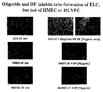
Our data data demonstrate that oligotide and/or
defibrotide in clinical and subclinical concentrations
can inhibit tube formation of transdifferentiating ELC
(TuDC-ELC) in MatrigelTM. TuDC-ELC and mature,
differentiated endothelial cells, [human umbilical vene
(HUVEC) or microvascular endothelial cells (HMEC) as
"stable" controls] were incubated in the presence or
absence of oligotide or Defibrotide (10pg/mL each) for
7 days. Importantly, after a single addition of
Defibrotide, HUVEC and HMEC are not affected in their
tube formation potential, suggesting that Defibrotide
and/or oligotide only target transdifferentiating
endothelial cells (Figure 1 A). However, when
Defibrotide was added repeatedly, it could also block
angiogenesis of mature, fully differentiated
endothelial cells (see below).
By the help of a complimentary software from the NIH
(Image J, http://rsb.info.nih.gov/ij/), we are able to
quantify these effects, the total length of tubes and
CA 02598613 2007-08-21
WO 2006/094916 PCT/EP2006/060304
the area of the photograph are assessed, the
microvascular density (MVD) is then given in total
length/area [pix-1] . DF significantly (p=0.02, TTEST)
downregulates MVD of TuDC-ELC (Figure 1 B).
To support these data with an alternative angiogenesis
assay the sprouting of rat aorta endothelial cells in
MatrigelTM was prevented by nearly 100%, when DF was
applied on a daily basis (Figure 2), suggesting that DF
not only acts on transdifferentiating, but also on
mature, fully differentiated endothelial cells.
The aortic ring assay investigates macrovascular
endothelial cells. But often, the tumor vasculature
consists of microvascular endothelial cells. Therefore,
a third in vitro angiogenesis assay was performed on
the basis of microvascular endothelial cells
vascularizing through a layer of dermal fibroblasts
after 9-11 days of culture. These vessel-like
structures can subsequently be visualized by staining
for CD31 and vWF.
As demonstrated in Figure 3 (A and B), DF can also
block angiogenesis of human microvascular endothelial
cells with a superiority for the daily application.
Interestingly, concentrations around 10 pg/mL appear to
be the most effective. A single application of DF could
not significantly block angiogenesis.
Taken together, our data strongly suggest that
defibrotide and/or oligotide can block angiogenesis of
tumor-associated transdifferentiating endothelial cells
and those that arise from already existing vascular
cells.
It is subject to ongoing studies whether oligotide and
defibrotide also inhibit angiogenesis in vivo. We are
currently performing a dorsal skin chamber assay (14)
CA 02598613 2007-08-21
WO 2006/094916 PCT/EP2006/060304
6
that investigates the effect of defibrotide in a highly
vascularized human gastric carcinoma mouse model
(Xenograft system) . First data clearly show that the
microvascular density (MVD) of DF-treated tumors is
lower than that of control tumors. This set of
experiments will be reproduced in due time.
The mechanism of action by which DF can block
angiogenesis remains to be elucidated, but preliminary
evidence from Western Blot analyses suggest a
downregulating effect of DF on activated p70S6 kinase
(p-p70S6), a mitogen-activated protein kinase.
Additional evidence for the impact of p70S6 kinase was
obtained from another tube formation assay with HMEC
incubated in the presence or absence of the p70S6
kinase inhibtor DRB.
There are also first clinical data available for
patients (pts.) having received allogeneic stem cell
transplantation (SCT) : In a cohort of 17 defibrotide-
treated pts a striking decline in serum VEGF levels has
been seen, also suggesting that defibrotide might act
through growth factor withdrawal for sprouting tumor
endothelial cells.
Defibrotide and oligotide are strong candidates for a
therapy of angiogenesis-dependent tumors and might be
used alone or in combination with other anti-
angiogeneic agents, such as rapamycin (14).
Interestingly, rapamycin has the negative side effect
of pro-thrombotic activity (15) that could be
attenuated by the simultaneous application of the anti-
thrombotic and fibrionolytic defibrotide.
References
1. US5646127
CA 02598613 2007-08-21
WO 2006/094916 PCT/EP2006/060304
7
2. US5646268
3. US6046172
4. US4985552
5. US5223609
6. 't Veer,L.J., et al.(2002) Gene expression
profiling predicts clinical outcome of breast
cancer. Nature, 415, 530-536.
7. Abdalla,S.A., et al. (1999) Prognostic relevance of
microvessel density in colorectal tumours.
Oncol.Rep., 6, 839-842.
8. Andersen,N.F., et al. (2005) Syndecan-1 and
angiogenic cytokines in multiple myeloma:
correlation with bone marrow angiogenesis and
survival. Br.J.Haematol., 128, 210-217.
9. Bostwick,D.G. & Iczkowski,K.A. (1998) Microvessel
density in prostate cancer: prognostic and
therapeutic utility. Semin.Urol.Oncol., 16, 118-
123.
10. Eissner,G., et al. (2002) Fludarabine induces
apoptosis, activation, and allogenicity in human
endothelial and epithelial cells: protective effect
of defibrotide. Blood, 100, 334-340.
11. Fernandez,P.B., et al. (2001) Dendritic cells
derived from peripheral monocytes express
endothelial markers and in the presence of
angiogenic growth factors differentiate into
endothelial-like cells. Eur.J.Cell Biol., 80, 99-
110.
12. Folkman,J., et al. (1971) Isolation of a tumor
factor responsible for angiogenesis. J.Exp.Med.,
133, 275-288.
CA 02598613 2007-08-21
WO 2006/094916 PCT/EP2006/060304
8
13. Fontanini,G., et al. (1995) Microvessel count
predicts metastatic disease and survival in non-
small cell lung cancer. J.Pathol., 177, 57-63.
14. Guba,M., et al. (2002) Rapamycin inhibits primary
and metastatic tumor growth by antiangiogenesis:
involvement of vascular endothelial growth factor.
Nat.Med., 8, 128-135.
15. Guba,M., et al. (2005) Rapamycin induces tumor-
specific thrombosis via tissue factor in the
presence of VEGF. Blood.
16. Hanahan,D. & Folkman,J. (1996) Patterns and
emerging mechanisms of the angiogenic switch during
tumorigenesis. Cell, 86, 353-364.
17. Hasan,J., et al. (2002) Intra-tumoural microvessel
density in human solid tumours. Br.J.Cancer, 86,
1566-1577.
18. Helmlinger,G., et al. (1997) Interstitial pH and
p02 gradients in solid tumors in vivo: high-
resolution measurements reveal a lack of
correlation. Nat.Med., 3, 177-182.
19. Kainz,C., et al. (1995) Prognostic value of tumour
microvessel density in cancer of the uterine cervix
stage IB to IIB. Anticancer Res., 15, 1549-1551.
20. Morabito,A., et al. (2004) Antiangiogenic
strategies, compounds, and early clinical results
in breast cancer. Crit Rev.Oncol.Hematol., 49, 91-
107.
21. Podar,K. & Anderson,K.C. (2005) The
pathophysiologic role of VEGF in hematologic
malignancies: therapeutic implications. Blood, 105,
1383-1395.
CA 02598613 2007-08-21
WO 2006/094916 PCT/EP2006/060304
9
22. Staton,C.A., et al. (2004) Current methods for
assaying angiogenesis in vitro and in vivo.
Int.J.Exp.Pathol., 85, 233-248.
23. Sun,H.C., et al. (1999) Microvessel density of
hepatocellular carcinoma: its relationship with
prognosis. J.Cancer Res.Clin.Oncol., 125, 419-426.
24. Verheul,H.M., et al. (2004) Are tumours
angiogenesis-dependent? J.Pathol., 202, 5-13.
25. Weidner,N., et al. (1992) Tumor angiogenesis: a new
significant and independent prognostic indicator in
early-stage breast carcinoma. J.Natl.Cancer Inst.,
84, 1875-1887.
26. Xiangming,C., et al. (1998) Angiogenesis as an
unfavorable factor related to lymph node metastasis
in early gastric cancer. Ann.Surg.Oncol., 5, 585-
589.
