Note: Descriptions are shown in the official language in which they were submitted.
DEMANDE OU BREVET VOLUMINEUX
LA PRESENTE PARTIE DE CETTE DEMANDE OU CE BREVET COMPREND
PLUS D'UN TOME.
CECI EST LE TOME 1 DE 2
CONTENANT LES PAGES 1 A 60
NOTE : Pour les tomes additionels, veuillez contacter le Bureau canadien des
brevets
JUMBO APPLICATIONS/PATENTS
THIS SECTION OF THE APPLICATION/PATENT CONTAINS MORE THAN ONE
VOLUME
THIS IS VOLUME 1 OF 2
CONTAINING PAGES 1 TO 60
NOTE: For additional volumes, please contact the Canadian Patent Office
NOM DU FICHIER / FILE NAME:
NOTE POUR LE TOME / VOLUME NOTE:
CA 02598713 2007-08-22
WO 2006/091965 PCT/US2006/007013
NOD1 AS AN ANTI-TUMOR AGENT
This application claims benefit of the filing dates of U.S. Provisional Ser.
No. 60/656,175, filed February 25, 2005, and U.S. Provisional Ser. No.
60/752,794, filed December 22, 2005, the contents of which are incorporated
herein by reference.
Government Funding
The invention described herein was made with United States Govermnent
support under Grant Number AI15136 awarded by the National Institutes of
Health. The United States Government has certain rights in this invention.
Field of the Invention
The invention relates to Nodl and its function in apoptosis of
transformed, malignant cells.
Background of the Invention
Cancer is a disease that afflicts many people and is a leading cause of
death in huinans and non-human animals. Cancers typically involve uncontrolled
division of a few cells that then create many new cells. Accordingly, many
anti-
cancer drugs are agents that inhibit or stop cell growth. While such
chemotherapeutic agents have improved the survival rate of patients having
neoplastic diseases, the serious side effects associated with many
chemotherapeutic agents limits their usage and undermines the health of
patients
already weakened by cancer. New agents are therefore needed that exhibit
enhanced selectivity for cancer cells or that are capable of controlling
proliferation of oncocytes.
One major problem with many anticancer agents is their specificity. An
anti-cancer drug needs to distinguish between cells that are cancerous and
cells
that are not cancerous. However, the vast bulk of anticancer drugs are
indiscriminate at this regard. Typically anticancer agents have negative
CA 02598713 2007-08-22
WO 2006/091965 PCT/US2006/007013
hematological effects (e.g., cessation of mitosis and disintegration of formed
elements in marrow and lymphoid tissues), and immunosuppressive action (e.g.,
depressed cell counts), and can also have a severe impact on epithelial
tissues
(e.g., intestinal mucosa), reproductive tissues (e.g., impairment of
spermatogenesis), and the nervous system. See, e.g., P. Calabresi and B. A.
Chabner, In: Goodman and Gilman, The Pharmacological Basis of Therapeutics
(Pergainon Press, 8th Edition) (pp. 1209-1216).
What is needed are anticancer agents that can beneficially treat selected
tumor types, or preferably a wide variety of tumor types, and that is
particularly
suitable for invasive tumors. Moreover, while such anticancer agents should be
effective, they should also exhibit have little or no toxicity.
Summary of the Invention
The invention provides compositions and methods for promoting
apoptosis in tumor cells that involve increasing Nodl expression or NOD 1
activity.
Tlius, one aspect of the invention is a method of promoting tumor
regression in a mammal that involves administering to the mammal an agent that
increases Nodl expression or NOD1 activity. Examples of tunzors that can be
treated with the methods of the invention include brain, bladder, cervix,
colon,
gall bladder, kidney, liver, lung, pancreas, ovary, prostate, skin, stomach,
or
thyroid tumors. In some embodiments, the tumor is an estrogen-sensitive tumor
or a breast tumor.
Examples of agents that increase NOD 1 activity include peptides having
the following sequences: D-Ala-L-Glu-Diaminopimelic acid (yTriDAP), y-D-
glutamy-meso-diaminopimelic acid (iE-DAP), y-D-Gln-DAP (iQ-DAP), D-Ala-
L-Glu-Diaminopimelic acid (yTriDAP), and coinbinations thereof. These
peptides can activate the NOD1 protein, and hence the Nodl-dependent pathway
leading to apoptosis. Another example of an agent that can increase NOD 1
activity is a NOD1 polypeptide. In some embodiments, the NODl polypeptide
can be a human NOD1 polypeptide, for example, a human NOD1 polypeptide
with SEQ ID NO:1 or SEQ ID NO:3.
2
CA 02598713 2007-08-22
WO 2006/091965 PCT/US2006/007013
One example of an agent that can increase Nodl expression is a nucleic
acid that comprises a segment encoding a NOD 1 polypeptide. Examples of
sequences for NOD1 polypeptides include SEQ ID NO:1 or SEQ ID NO:3. One
example of a nucleic acid segment encoding NOD 1 polypeptide comprises SEQ
ID NO:2. The nucleic acid can further include a regulatory element, for
example, a promoter, enhancer, transcriptional termination signal, or a
coinbination tllereof. The nucleic acid can be part of an expression cassette
or an
expression vector or a gene delivery vehicle.
Additional active ingredients can be administered in conjunction with the
agent that increases Nodl expression or NOD1 activity. For example, an
effective amount of tumor necrosis factor a can be administered with such
agents. In some embodiments, tumor necrosis factor a can enhance the Nod-
dependent apoptotic pathway. In addition, an effective amount of cycloheximide
can be administered with the agents at increase Nodl expression or NOD1
activity. Moreover, one or more chemotherapeutic compounds can be
administered in conjunction with the agent.
Examples of chemotherapeutic compounds that may be used in the
compositions and methods of the invention include Altretamine, Bleomycin,
Busulphan, Calcium Folinate, Capecitabine, Carboplatin, Carmustine,
Chlorambucil, Cisplatin, Cladribine, Crisantaspase, Cyclophosphamide,
Cytarabine, Dacarbazine, Dactinomycin, Daunorubicin, Docetaxel, Doxorubicin,
Epirubicin, Etoposide, Fludarabine, Fluorouracil, Gemcitabine, Hydroxyurea,
Idarubicin, Ifosfamide, Irinotecan, Liposomal doxorubicin, Lomustine,
Melphalan, Mercaptopurine, Methotrexate, Mitomycin, Mitoxantrone,
Oxaliplatin, Paclitaxel, Pentostatin, Procarbazine, Raltitrexed, Streptozocin,
Tegafur-uracil, Temozolomide, Thiotepa, Tioguanine/Thioguanine, Topotecan,
Treosulfan, Vinblastine, Vincristine, Vindesine, Vinorelbine, and a
combination
thereof.
The agent can be administered locally to the site of the tumor and/or be
formulated for sustained release.
Another aspect of the invention is a composition that includes a carrier, a
nucleic acid that comprises a segment encoding a NOD 1 polypeptide and an
effective amount of D-Ala-L-Glu-Diaminopimelic acid (yTriDAP), y-D-
3
CA 02598713 2007-08-22
WO 2006/091965 PCT/US2006/007013
glutamy-meso-diaminopimelic acid (iE-DAP), y-D-Gln-DAP (iQ-DAP), or D-
Ala-L-Glu-Diaminopimelic acid (,yTriDAP), wherein the composition is
formulated for local adininistration to a tumor. The NOD1 polypeptide can, for
example, include SEQ ID NO: l or SEQ ID NO:3. An exainple of a nucleic acid
segment that encodes a NOD1 polypeptide is SEQ ID NO:2. The nucleic acid
employed in the composition can include a regulatory element, for example, a
promoter, enliancer, transcriptional termination signal, or a combination
thereof.
The nucleic acid can be an expression cassette or an expression vector. The
nucleic acid comprises a gene delivery vehicle. The composition of the
invention can also include other active ingredients, for example, an effective
amount of tumor necrosis factor a or a chemotherapeutic compound. The
composition can be formulated for local administration to the site of the
tumor
and/or be formulated for sustained release.
Another aspect of the invention is a method of promoting apoptosis in
breast tuinor cells comprising contacting the breast tumor cells with an
effective
ainount of D-Ala-L-Glu-Diaminopimelic acid (yTriDAP).
Another aspect of the invention is a method of promoting apoptosis in
estrogen-sensitive tumor cells comprising contacting the breast tumor cells
with
an effective amount of D-Ala-L-Glu-Diaminopimelic acid (yTriDAP).
Description of the Figures
FIG. lA-C illustrates Nodl involvement in TNF-induced apoptosis. FIG.
lA provides a schematic diagram of the mutated gene that gave rise to a TNFa-
resistant phenotype, and was later identified as a Nodl mutant bearing a
blasticidine (blast) gene insertion. The cell line bearing this Nodl mutation
is
the MCF7-C20 cell line. The insertion from the pDisrup retroviral construct
was
mapped to the Nodl gene. The junction of blasticidine fused with the Nodl gene
occurred at the 3' end of the Nod1 gene between leucine-rich region 8 (LRR8)
and leucine-rich region 9 (LRR9). Hence, the LRR9 and LRR10 regions are 3'
to the blasticidine insertion. FIG. 1B shows that the NOD 1 protein is not
present
in detectable amounts in MCF-7 C20 cells. Cell extracts from MCF-7 parental
(called "wt") and MCF-7 C20 cells were prepared and either
iminunoprecipitated with a monoclonal anti-NOD 1 antibody (upper panel) or
4
CA 02598713 2007-08-22
WO 2006/091965 PCT/US2006/007013
directly loaded onto an SDS-PAGE gel (lower panel), then transferred to PVDF
membranes. Blots were analyzed by immnunoblotting using the same
monoclonal anti-NOD1 antibody. FIG. 1C shows that MCF-7 cells are more
resistant to TNF-induced apoptosis than MCF-7 C20 cells. MCF-7 and MCF-7
C20 cells were treated with increasing concentrations of TNF (0-40 ng/ml) for
20 h. Cell viability was determined by propidium iodide (PI) exclusion assay
and flow cytometry. The graph shows that MCF-7 C20 cells are significantly
more likely to undergo apoptosis.
FIG. 2A-D shows that MCF-7 cells undergo apoptosis upon yTriDAP
treatment. FIG. 2A illustrates that NOD1 is needed for yTriDAP-induced cell
death. MCF-7 Blasto cells that express normal levels of NOD 1, MCF-7 C20
cells that express little or no NOD 1, or MCF-7 Nodl cells that over-express
NOD 1 were treated with yTriDAP or aTriDAP (50 ug/ml each) in the presence
(shaded bars) or absence (open bars) of cycloheximide (CHX)(3 ug/ml) for 48 h.
Control assays received medium (Med) instead of yTriDAP or aTriDAP. After
the 48 h incubation, cells were harvested and incubated with propidium iodide
(PI) (4 ug/ml). Cell viability was measured by flow cytometry analysis. Data
shown are representative experiments of at least four independent experiments.
FIG. 2B shows the levels of NOD 1 expression in MCF-7 Blasto cells that were
transfected with vector alone, MCF-7 C20 cells that have a disruption in the
endogenous Nodl gene or MCF-7 Nod1 cells that were engineered to over-
express NOD 1. Expression of NOD 1 in MCF-7 Blasto, MCF-7 C20 and MCF-7
Nodl cells was analyzed by western blotting using monoclonal anti-NODl
antibody. FIG. 2C illustrates the morphological changes in yTriDAP-treated
MCF-7 Nod1 cells. Cells were seeded in 4-well chamber slides and treated with
yTriDAP/cycloheximide (CHX) (panels b, d, f) or CHX alone (panels a, c, e).
Cells were stained with DAPI (panels c, d) or TUNEL (panels e, f), fixed and
observed under a phase contrast (panels a, b) or fluorescence (panels c-f)
microscopes. FIG. 2D shows that yTriDAP-induced apoptosis in MCF-7 Nodl
cells was diininished or abolished by two broad spectrum caspase inhibitors, z-
VAD-FMK and Boc-D-FMK. MCF-7 Nod1 cells were pretreated with z-VAD
or Boc-D-FMK caspase inhibitors (50 uM each) for 30 min before addition of
5
CA 02598713 2007-08-22
WO 2006/091965 PCT/US2006/007013
7TriDAP/CHX for 48 h. Cells were incubated with propidium iodide (PI) and
apoptotic cell death was ineasured by flow cytometry.
FIG. 3 illustrates by western analysis that addition of 7TriDAP, but not
the inactive control tri-peptide aTriDAP, to MCF-7 cells resulted in
proteolytic
cleavage of poly(ADP-ribose)polymerase (PARP) and of capases 6, 7, 8 and 9.
Cleavage of PARP and various caspases was detected by Western blot analysis
of cell MCF-7 Blasto, MCF-7 C20 and MCF-7 Nodl after stimulation with
yTriDAP, aTriDAP or medium (control) in the presence or absence of CHX (0.5
ug/ml) for 24 h. Cells were harvested, subjected to western blotting and PARP,
caspases, p20, p41/43 and p35 were detected with antibodies reactive thereof.
FIG. 4A-C show that NOD 1 mutant V41 Q is responsive to yTriDAP and
remains functional in the apoptosis pathway, whereas NOD1 mutant K208R is
not responsive to yTriDAP and is not active in the apoptosis pathway. FIG. 4A
graphically illustrates the percentage of apoptotic cells in different cell
lines after
treatment with medium (Med., a control) or yTriDAP. The NOD 1 V41 Q and
K208R mutants were constructed by site-directed mutagenesis. The V41 Q
mutation is in the CARD domain of NOD1, wllereas the K208R mutation is
thought to block conformational changes required for oligomerization mediated
by the Nod/NBD domain. Constructs encoding these NOD 1 V41 Q and K208R
mutant polypeptides were transfected into MCF-7 C20 cells and apoptotic assays
were performed. As shown, the V41 Q mutant retains NOD 1 activity but the
K208R inutant does not. FIG. 4B shows that the expression levels of the NODl
mutant and wild type polypeptides were substantially identical. FIG. 4C
illustrates by western analysis that NOD 1 expression in MCF-7 cells is needed
for proteolytic cleavage of PARP and of capases 6, 7, 8 and 9. Cleavage of
PARP and various caspases was detected by Western blot analysis of MCF-7
C20 cells that do not express NOD1 and of MCF-7 C20 Nodl cells in which a
Nodl construct has been recoinbinantly introduced into MCF-7 C20 cells. Cells
were stimulated with yTriDAP or medium (control) in the presence or absence of
cycloheximide (CHX) (0.5 ug/ml) for 24 h. Cells were then harvested, subjected
to western blotting. PARP, caspases, p20, p41/43 and p35 were detected with
antibodies reactive therewith.
6
CA 02598713 2007-08-22
WO 2006/091965 PCT/US2006/007013
FIG. 5A-C illustrate that Nod2 does not induce apoptosis in MCF-7 cells.
In FIG. 5A, MCF-7 Blasto, MCF-7 Nodl and MCF-7 Nod2 were treated with
the NOD1 ligand yTriDAP or the Nod2 ligand muramyl dipeptide (MDP)(20
ug/ml each) in the presence or absence of CHX for 48 h. Cells were then
incubated with PI and apoptotic cell death was measured by flow cytometry. As
shown, yTriDAP stimulates apoptosis, but the Nod2 ligand does not. In FIG.
5B, expression of NOD1 and NOD2 was confirmed by Western blot analysis
using anti-Myc antibodies for detection of the recombinant proteins. FIG. 5C
shows that MCF-7 Nod2 cells respond to MDP as detected by interleukin-8 (IL-
8) secretion. MCF-7 Blasto, MCF-7 Nodl and MCF-7 Nod2 cells were
stimulated with yTriDAP or MDP in the presence or absence of CHX (0.5
ug/ml) for 24 h. Cell supernatants were then harvested and assayed for IL-8
secretion.
FIG. 6A-D illustrate that caspase 8 and caspase 9 are required for
7TriDAP-induced apoptosis. FIG. 6A shows the effects of different caspase
inliibitors on yTriDAP-induced apoptosis. MCF-7 Nodl cells were pretreated
with inhibitors of the caspases listed along the x-axis of FIG. 6A for 30 min
prior
to stimulation with yTriDAP/CHX for 48 h. Cells were then incubated with
propidium iodide and cell viability was measured by flow cytometry. All
inhibitors were used at a concentration of 100 uM. FIG. 6B shows that a high
molecular weight form of NOD1 is detected when Nodl is co-expressed with
caspase 9, indicating that NOD1 interacts with caspase 9. In this experiment,
293 cells were co-transfected with vectors encoding for FLAG-caspase 9 in the
presence of empty vector, Myc-NOD1, or Myc-NOD2. Cell extracts were
immunoprecipitated (IP) with anti-FLAG antibody and co-precipitated proteins
were revealed by immunoblotting (WB) using polyclonal anti-Myc antibody.
FIG. 6C shows that CLARP completely prevented yTriDAP-induced apoptosis,
whereas Bcl2 inhibited yTriDAP-induced apoptosis only partially. MCF-7
CLARP, MCF-7 CLARP/Nod1, MCF-7 Bc12 and MCF-7 Bc12/Nod1 cells were
untreated or treated with yTriDAP in the presence or absence of CHX (3 ug/ml)
for 48 h. Cells were then incubated with propidium iodide and apoptotic cell
death was measured by flow cytometry. FIG. 6D shows western blots
illustrating that CLARP, Bc12 and NOD1 are expressed in MCF-7 cells,
7
CA 02598713 2007-08-22
WO 2006/091965 PCT/US2006/007013
indicating that the results observed in FIG. 6C are due to functional
differences
between CLARP and Bcl2, rather than differences in the expression levels of
this
two proteins.
FIG. 7A-C illustrate that both wild-type RIP2 and a kinase-deficient
(KD) mutant of RIP2 are functional in the NOD1 apoptosis pathway. However,
RIP2 lacking the CARD domain acts as a dominant negative inhibitor of NOD 1
signaling. FIG. 7A graphically illustrates the percentage of cells that were
apoptotic in populations of wild type RIP2, RIP2 KD and RIP2 ACARD cells.
MCF-7 cells were stably transfected with wild type Myc-RIP2, Myc-RIP2 KD,
and Myc-RIP2 ACARD and were left untreated or were treated with yTriDAP in
the presence or absence of CHX (3 ug/ml) for 48 h. Cells were incubated with
PI
and apoptotic cell death was measured by flow cytometry. FIG. 7B shows that
RIP2 polypeptides were expressed as confirmed by immunoprecipitation of cell
extracts with polyclonal anti-Myc antibody and immunoblotting using
monoclonal anti-Myc 9E10 antibody. FIG. 7C illustrates yTriDAP-induction of
phosphoiylation of JNK in RIP2 wild types, MCF-7 RIP2 KD, and RIP2
ACARD cells. As shown, exposure of MCF-7 cells expressing wild type RIP2
or RIP2 KD to yTriDAP in the presence of cycloheximide for 2 h induced
phosphorylation of JNK. However, no such phosphorylation of JNK was
observed in RIP2ACARD cells that were treated in the saine manner.
FIG. 8A-D illustrate that a synergistic relationship exists between NOD 1
and TNFa. FIG. 8A graphically illustrates that the percentage of apoptotic
cells
increases in dose-specific manner as the concentration of NOD 1 increases from
0.0 to 100 g/ml and the concentration of TNFa increases from 0.5 ng/inl to 1
ng/ml. FIG. 8B graphically illustrates that the percentage of apoptotic cells
increases in dose-specific manner as the concentration of cyclohexiinide
increases from 0.0 to 3 g/inl and the concentration of TNFa increases from
0.5
ng/ml to 1 ng/ml. FIG. 8C graphically illustrates that while yTriDAP increases
apoptosis in the presence of TNFa, the inactive control tri-peptide aTriDAP
may
actually inhibit apoptosis, even at higher doses of TNFa. FIG. 8D illustrates
NOD1 expression at various time points after exposure of MCF-7 cells to TNFa.
FIG. 9A-C illustrates NOD1 expression in another human breast cancer
cell line, the SKBR3 cancer cell line. FIG. 9A (top) graphically illustrates
the
8
CA 02598713 2007-08-22
WO 2006/091965 PCT/US2006/007013
percentage of apoptotic SKBR3 wild type and SKBR3 Nodl cells as a function
of TNFa concentration. FIG. 9A(bottom) graphically illustrates the percentage
of apoptotic SKBR3 wild type and SKBR3 Nodl cells as a function of yTriDAP
concentration. FIG. 9B (top) graphically illustrates the percentage of
apoptotic
SKBR3 cells that were observed under different conditions by propidium iodide
(PI) or Dioc6. The conditions employed are indicated below the western blots.
The abbreviations used are as follows: CHX (cycloheximide), TNF (TNFa),
yTri (yTriDAP), aTri (aTriDAP). FIG. 9B2 provides a western analysis
illustrating proteolytic cleavage of poly(ADP-ribose)polymerase (PARP) and of
capases 3, 7, and 8. Cleavage of PARP and various caspases was detected by
Western blot analysis of cell MCF-7 cells after stinzulation under the
conditions
specified below the western blot. Cells were harvested, subjected to western
blotting and PARP, caspases, p20, p41/43 and p35 were detected with antibodies
reactive thereof. FIG. 9C graphically illustrates the percentage of apoptotic
wild
type, NOD 1-expressing and CLARP-expressing SKBR3 cells that were
observed under the conditions indicated below the bar graph. Abbreviations
used: CHX (cycloheximide), TNF (TNFa), gTri (yTriDAP), gTC (yTriDAP +
CHX), aTC (aTriDAP + CHX).
FIG. l0A , B and C provide images of mice inoculated with wild type
MCF-7 breast cancer cells, NOD 1 knockout MCF-7 cells and NOD 1-transfected
MCF-7 cells, respectively. Mice were inoculated subcutaneously with 3x10⁶
human breast cancer cells. The arrowheads indicated the subcutaneous tumors,
which are shown in the close-up images to the right of FIG. l OB.
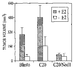
FIG. 1 lA-D illustrate that the presence of Nodl prevents tumor growth
in SCID mice. FIG. 11A graphically illustrates the tumor volume in mice
injected with MCF-7 C20 (left) and MCF-7 C20/Nodl (right) cells as a function
of time. The cells (3 x 10⁶ cells/mouse) were injected into-the flanks of
female
SCID mice. Tumor size was measured once a week and voluine was determined
according to the formula (W2 x L)/2. Each line represents tumor growth
observed in one mouse (n = 4). As illustrated, the tumor volume diminished in
mice that received Nodl expressing tumor cells (C20/Nodl). In contrast, the
tumor volume increased in mice that received tumor cells that did not express
Nodl (C20 cells). FIG. 11B graphically illustrates that implanting estrogen
9
CA 02598713 2007-08-22
WO 2006/091965 PCT/US2006/007013
pellets into mice prior to injection of MCF-7 Blasto, MCF-7 C20 and MCF-7
C20/Nodl cells (3 x 10⁶ cells/mouse) increases tumor growth except when the
tumor cells express Nod1 (the C20/Nodl cells). The shaded bar represents
results obtained for mice that received estrogen pellets while the open bar
represents results obtained for mice that did not receive estrogen pellets.
The
tumor volume was determined as described above. FIG. 11 C graphically
illustrates the tumor volume over time in mice injected with MCF-7 Blasto,
MCF-7 C20 and MCF-7 RIP2ACARD cells. As shown, tumor volume in mice
receiving MCF-7 RIP2ACARD cells (*) was greater than in mice that express
some Nodl (Blasto, filled diamonds), but less than in mice that do not express
Nodl (C20, filled squares). FIG. 11D graphically illustrates tumor cell growth
as a function of estrogen concentration. As shown, tumor cells that do not
express Nodl (the MCF-7 C20 cells) and tumor cells that express inutant RIP2
(the MCF-7 RIP2ACARD cells) are more sensitive to estrogen and exhibit
increased cell growth. MCF-7 Blasto, MCF-7 C20, MCF-7 C20/Nodl and
MCF-7 RIP2ACARD cells were exposed to increasing concentrations of 17(3-
estradiol, pulsed with 3H-thimidine and cell growth was determined by liquid
scintillation. Data are representative of at least 3 independent experiments.
FIG. 12A-C illustrates that estrogen increases yTriDAP-induced
apoptosis and tamoxifen inhibits yTriDAP-induced apoptosis in various MCF-7
tumor cell lines. FIG. 12A graphically illustrates the percent apoptosis in
MCF-
7 Blasto, MCF-7 C20 and MCF-7 C20/Nodl cells cultured in increasing
amounts or estrogen and either cycloheximide (CHX) or CHX plus yTriDAP
(yTri). FIG. 12B graphically illustrates the percent apoptosis in MCF-7
Blasto,
MCF-7 C20 and MCF-7 C20/Nodl cells cultured in increasing amounts of
tamoxifen and either cycloheximide (CHX) or CHX plus yTriDAP (yTri). For
the estrogen studies, MCF-7 cells were cultured in charcoal-treated medium and
stimulated with yTriDAP/CHX in the presence of increasing concentrations of
estrogen (E2) and cell viability was measured by propidium iodide (PI)
exclusion. For the tamoxifen studies, cells were treated with tamoxifen prior
to
addition of yTriDAP/CHX and cell viability measured by PI exclusion. FIG.
12C shows immunoblots of cell lysates from in vitro cultured cells (left) and
in
vivo tumor cells (right) illustrating that expression of estrogen receptor is
CA 02598713 2007-08-22
WO 2006/091965 PCT/US2006/007013
reduced in cells that express Nod1. Total protein extracts from MCF-7 Blasto,
MCF-7 C20 and MCF-7 C20/Nodl cells and from cells isolated from tumors
were analyzed by immunoblotting using antibody against estrogen receptor
(ERa), and ERK2 as loading control.
FIG. 13A-D illustrate that RIP2 is an important component of the Nodl
apoptotic pathway. FIG. 13A shows that stable expression of kinase-deficient
RIP2 (RIP2 KD). in tumor cell lines sensitizes those cells to apoptotic cell
death
induced by Nodl or Nod2 activators. MCF-7 cells were stably transfected with
Myc-Nod1, Myc-RIP2 (wild type), Myc-RIP2 K D, and Myc-RIP2 OCARD.
Test cells were then treated with yTriDAP, aTriDAP and MDP (20 g/ml each)
in the presence or absence of CHX (3 g/ml) for 48 hr, while control cells
were
not treated with these agents. Cells were then incubated with propidium iodide
(PI) and apoptotic cell death was measured by flow cytometry. FIG. 13B
illustrates the effect of TNF upon apoptosis in the same MCF-7 cell lines
described in FIG. 13A. These cells lines were treated with TNF (10 ng/ml) for
18 hr and apoptotic cell death was assessed as described for FIG. 13A. TNF
increased apoptosis in all the cell lines tested. FIG. 13C shows that MCF-7
RIP2
KD cells exhibited increased yTriDAP-induced apoptosis than MCF-7 Nodl
cells. Cells were incubated in the presence of increasing concentrations of
ti
yTriDAP or MDP in the presence of CHX for 481ir and apoptotic cell death was
assessed as described for FIG. 13A. FIG. 13D illustrates IL-8 secretion by
MCF-7 Blasto, MCF-7 Nodl and MCF-7 RIP2 wild type cells after stimulation
with yTriDAP in the presence of CHX (0.5 ing/ml) or TNF. After incubation,
cell supernatants were harvested and assayed for IL-8 secretion.
Detailed Description of the Invention
According to the present invention, increased Nodl expression or NOD1
activity leads to tumor regression. The invention therefore involves
administering NOD1 polypeptides, Nod1 nucleic acids and agents that increase
Nod1 expression or NOD1 activity to subjects as an anti-tumor treatment. In
some embodiments, the agents that increase Nodl expression or activity include
peptide activators of NOD1 such as y-D-glutamy-meso-diaminopimelic acid (iE-
DAP),,y-D-Gin-DAP (iQ-DAP), D-AIa-L-Glu-Diaminopimelic acid (,yTriDAP),
11
CA 02598713 2007-08-22
WO 2006/091965 PCT/US2006/007013
and combinations thereof. While the invention conteinplates treating any type
of
tuinor, the coinpositions and methods of the invention may have particular
utility
for treating estrogen-sensitive tumors and/or breast cancer tumors.
NOD1
The innate immune system is comprised of families of receptors that
recognize components of micro-organisms, viruses, abnormal/damaged host
cells and the like, and initiate host responses to eliminate and kill invading
organisms or to remove atypical host cells. Recently a family of
intracellular,
cytosolic proteins known as the Nod/Caterpillar family has been linked to
innate
immune responses. All members of this fainily have two conserved domains;
the nucleotide-binding oligomerization domain (NBD/NOD) and the carboxyl-
terminus leucine-rich repeat (LRR) region. The ainino-terminal regions of
family members, termed the effector domains, contain variable structures that
include caspase recruitment domain (CARD), pyrin, BIR and other domains.
Two members of the Nod/Caterpillar family have been particularly
linked to innate iminune responses to infection. These members are known as
NOD 1(CARD4) and NOD2 (CARD 15). The effector domains of NOD l and
NOD2 are made up of one and two CARD domains, respectively. NOD1 and
NOD2 were first suggested to be intracellular proteins acting as receptors for
bacterial lipopolysaccharide (LPS). Subsequently, it was discovered that the
NOD ligands are derived from bacterial peptidoglycan (PGN) and not from LPS.
Thus NOD1 and NOD2 are likely to function as intracellular sensors of bacteria
or bacterial products during infection. However, studies performed to date
indicate that mice deficient in NODl or NOD2 do not manifest obvious
phenotypes associated with immunodeficiency or increased sensitivity to
infection.
Nucleic acid and amino acid sequences for NOD1 and other members of
the NOD/Caterpillar family can be found in the art, for example, in the NCBI
database. See website at ncbi.nlm.nih.gov. For example, one amino acid
sequence for human NOD1 is provided below for easy reference as SEQ ID
NO:1 (NCBI accession nuinber Q9Y239; gi: 20137579).
12
CA 02598713 2007-08-22
WO 2006/091965 PCT/US2006/007013
1 MEEQGHSEME IIPSESHPHI QLLKSNRELL VTHIRNTQCL
41 VDNLLKNDYF SAEDAEIVCA CPTQPDKVRK ILDLVQSKGE
81 EVSEFFLYLL QQLADAYVDL RPWLLEIGFS PSLLTQSKVV
121 VNTDPVSRYT QQLRHHLGRD SKFVLCYAQK EELLLEEIYM
161 DTIMELVGFS NESLGSLNSL ACLLDHTTGI LNEQGETIFI
201 LGDAGVGKSM LLQRLQSLWA TGRLDAGVKF FFHFRCRMFS
241 CFKESDRLCL QDLLFKHYCY PERDPEEVFA FLLRFPHVAL
281 FTFDGLDELH SDLDLSRVPD SSCPWEPAHP LVLLANLLSG
321 KLLKGASKLL TARTGIEVPR QFLRKKVLLR GFSPSHLRAY
361 ARRMFPERAL QDRLLSQLEA NPNLCSLCSV PLFCWIIFRC
401 FQHFRAAFEG SPQLPDCTMT LTDVFLLVTE VHLNRMQPSS
441 LVQRNTRSPV ETLHAGRDTL CSLGQVAHRG MEKSLFVFTQ
481 EEVQASGLQE RDMQLGFLRA LPELGPGGDQ QSYEFFHLTL
521 QAFFTAFFLV LDDRVGTQEL LRFFQEWMPP AGAATTSCYP
561 PFLPFQCLQG SGPAREDLFK NKDHFQFTNL FLCGLLSKAK
601 QKLLRHLVPA AALRRKRKAL WAHLFSSLRG YLKSLPRVQV
641 ESFNQVQAMP TFIWMLRCIY ETQSQKVGQL AARGICANYL
681 KLTYCNACSA DCSALSFVLH HFPKRLALDL DNNNLNDYGV
721 RELQPCFSRL TVLRLSVNQI TDGGVKVLSE ELTKYKIVTY
761 LGLYNNQITD VGARYVTKIL DECKGLTHLK LGKNKITSEG
801 GKYLALAVKN SKSISEVGMW GNQVGDEGAK AFAEALRNHP
841 SLTTLSLASN GISTEGGKSL ARALQQNTSL EILWLTQNEL
881 NDEVAESLAE MLKVNQTLKH LWZIQNQITA KGTAQLADAL
921 QSNTGITEIC LNGNLIKPEE AKVYEDEKRI ICF
A nucleotide sequence for human Nodl is also available as NCBI
accession number BC040339 (gi: 25955660), and reproduced below as SEQ ID
NO:2 for easy reference.
1 CCCGGCCCCG GCGTCCCCGG ACCATGGCGC TCTCCGGGCT
41 CTTCTCTAGC TCTCAGCGGC TGCGAAGTCT GTAAACCTGG
81 TGGCCAAGTG ATTGTAAGTC AGGAGACTTT CCTTCGGTTT
121 CTGCCTTTGA TGGCAAGAGG TGGAGATTGT GGCGGCGATT
161 ACAGAAAACG TCTGGGAAGA CAAGTTGCTG TTTTTATGGG
201 AATCGCAGGC TTGGAAGAGA CAGAAGCAAT TCCAGAAATA
13
CA 02598713 2007-08-22
WO 2006/091965 PCT/US2006/007013
241 AATTGGAAAT TGAAGATTTA AACAATGTTG TTTTAAAACA
281 TTCTAACTTC AAAGAATGAT GCCAGAAACT TAAAA.AGGGG
321 CTGCGCAGAG TAGCAGGGGC CCTGGAGGGC GCGGCCTGAA
361 TCCTGATTGC CCTTCTGCTG AGAGGACACA CGCAGCTGAA
401 GATGAATTTG GGAAAAGTAG CCGCTTGCTA CTTTAACTAT
441 GGAAGAGCAG GGCCACAGTG AGATGGAAAT AATCCCATCA
481 GAGTCTCACC CCCACATTCA ATTACTGAAA AGCAATCGGG
521 AACTTCTGGT CACTCACATC CGCAATACTC AGTGTCTGGT
561 GGACAACTTG CTGAAGAATG ACTACTTCTC GGCCGAAGAT
601 GCGGAGATTG TGTGTGCCTG CCCCACCCAG CCTGACAAGG
641 TCCGCAAAAT TCTGGACCTG GTACAGAGCA AGGGCGAGGA
681 GGTGTCCGAG TTCTTCCTCT ACTTGCTCCA GCAACTCGCA
721 GATGCCTACG TGGACCTCAG GCCTTGGCTG CTGGAGATCG
761 GCTTCTCCCC TTCCCTGCTC ACTCAGAGCA AAGTCGTGGT
801 CAACACTGAC CCAGTGAGCA GGTATACCCA GCAGCTGCGA
841 CACCATCTGG GCCGTGACTC CAAGTTCGTG CTGTGCTATG
881 CCCAGAAGGA GGAGCTGCTG CTGGAGGAGA TCTACATGGA
921 CACCATCATG GAGCTGGTTG GCTTCAGCAA TGAGAGCCTG
961 GGCAGCCTGA ACAGCCTGGC CTGCCTCCTG GACCACACCA
1001 CCGGCATCCT CAATGAGCAG GGTGAGACCA TCTTCATCCT
1041 GGGTGATGCT GGGGTGGGCA AGTCCATGCT GCTACAGCGG
1081 CTGCAGAGCC TCTGGGCCAC GGGCCGGCTA GACGCAGGGG
1121 TCAAATTCTT CTTCCACTTT CGCTGCCGCA TGTTCAGCTG
1161 CTTCAAGGAA AGTGACAGGC TGTGTCTGCA GGACCTGCTC
1201 TTCAAGCACT ACTGCTACCC AGAGCGGGAC CCCGAGGAGG
1241 TGTTTGCCTT CCTGCTGCGC TTCCCCCACG TGGCCCTCTT
1281 CACCTTCGAT GGCCTGGACG AGCTGCACTC GGACTTGGAC
1321 CTGAGCCGTG TGCCTGACAG CTCCTGCCCC TGGGAGCCTG
1361 CCCACCCCCT GGTCTTGCTG GCCAACCTGC TCAGTGGGAA
1401 GCTGCTCAAG GGGGCTAGCA AGCTGCTCAC AGCCCGCACA
1441 GGCATCGAGG TCCCGCGCCA GTTCCTGCGG AAGAAGGTGC
1481 TTCTCCGGGG CTTCTCCCCC AGCCACCTGC GCGCCTATGC
1521 CAGGAGGATG TTCCCCGAGC GGGCCCTGCA GGACCGCCTG
1561 CTGAGCCAGC TGGAGGCCAA CCCCAACCTC TGCAGCCTGT
14
CA 02598713 2007-08-22
WO 2006/091965 PCT/US2006/007013
1601 GCTCTGTGCC CCTCTTCTGC TGGATCATCT TCCGGTGCTT
1641 CCAGCACTTC CGTGCTGCCT TTGAAGGCTC ACCACAGCTG
1681 CCCGACTGCA CGATGACCCT GACAGATGTC TTCCTCCTGG
1721 TCACTGAGGT CCATCTGAAC AGGATGCAGC CCAGCAGCCT
1761 GGTGCAGCGG AACACACACA GCCCAGTGGA GACCCTCCAC
1801 GCCGGCCGGG ACACTCTGTG CTCGCTGGGG CAGGTGGCCC
1841 ACCGGGGCAT GGAGAAGAGC CTCTTTGTCT TCACCCAGGA
1881 GGAGGTGCAG GCCTCCGGGC TGCAGGAGAG AGACATGCAG
1921 CTGGGCTTCC TGCGGGCTTT GCCGGAGCTG GGCCCCGGGG
1961 GTGACCAGCA GTCCTATGAG TTTTTCCACC TCACCCTCCA
2001 GGCCTTCTTT ACAGCCTTCT TCCTCGTGCT GGACGACAGG
2041 GTGGGCACTC AGGAGCTGCT CAGGTTCTTC CAGGAGTGGA
2081 TGCCCCCTGC GGGGGCAGCG ACCACGTCCT GCTATCCTCC
2121 CTTCCTCCCG TTCCAGTGCC TGCAGGGCAG TGGTCCGGCG
2161 CGGGAAGACC TCTTCAAGAA CAAGGATCAC TTCCAGTTCA
2201 CCAACCTCTT CCTGTGCGGG CTGTTGTCCA AAGCCAAACA
2241 GAAACTCCTG CGGCATCTGG TGCCCGCGGC AGCCCTGAGG
2281 AGAAAGCGCA AGGCCCTGTG GGCACACCTG TTTTCCAGCC
2321 TGCGGGGCTA CCTGAAGAGC CTGCCCCGCG TTCAGGTCGA
2361 AAGCTTCAAC CAGGTGCAGG CCATGCCCAC GTTCATCTGG
2401 ATGCTGCGCT GCATCTACGA GACACAGAGC CAGAAGGTGG
2441 GGCAGCTGGC GGCCAGGGGC ATCTGCGCCA ACTACCTCAA
2481 GCTGACCTAC TGCAACGCCT GCTCGGCCGA CTGCAGCGCC
2521 CTCTCCTTCG TCCTGCATCA CTTCCCCAAG CGGCTGGCCC
2561 TAGACCTAGA CAACAACAAT CTCAACGACT ACGGCGTGCG
2601 GGAGCTGCAG CCCTGCTTCA GCCGCCTCAC TGTTCTCAGA
2641 CTCAGCGTAA ACCAGATCAC TGACGGTGGG GTAAAGGTGC
2681 TAAGCGAAGA GCTGACCAAA TACAAAATTG TGACCTATTT
2721 GGGTTTATAC AACAACCAGA TCACCGATGT CGGAGCCAGG
2761 TACGTCACCA AAATCCTGGA TGAATGCAAA GGCCTCACGC
2801 ATCTTAAACT GGGAAAAAAC AAAATAACAA GTGAAGGAGG
2841 GAAGTATCTC GCCCTGGCTG TGAAGAACAG CAAATCAATC
2881 TCTGAGGTTG GGATGTGGGG CAATCAAGTT GGGGATGAAG
2921 GAGCAAAAGC CTTCGCAGAG GCTCTGCGGA ACCACCCCAG
CA 02598713 2007-08-22
WO 2006/091965 PCT/US2006/007013
2961 CTTGACCACC CTGAGTCTTG CGTCCAACGG CATCTCCACA
3001 GAAGGAGGAA AGAGCCTTGC GAGGGCCCTG CAGCAGAACA
3041 CGTCTCTAGA AATACTGTGG CTGACCCAAA ATGAACTCAA
3081 CGATGAAGTG GCAGAGAGTT TGGCAGAAAT GTTGAAAGTC
3121 AACCAGACGT TAAAGCATTT ATGGCTTATC CAGAATCAGA
3161 TCACAGCTAA GGGGACTGCC CAGCTGGCAG ATGCGTTACA
3201 GAGCAACACT GGCATAACAG AGATTTGCCT AAATGGAAAC
3241 CTGATAAAAC CAGAGGAGGC CAAAGTCTAT GAAGATGAGA
3281 AGCGGATTAT CTGTTTCTGA GAGGATGCTT TCCTGTTCAT
3321 GGGGTTTTTG CCCTGGAGCC TCAGCAGCAA ATGCCACTCT
3361 GGGCAGTCTT TTGTGTCAGT GTCTTAAAGG GGCCTGCGCA
3401 GGCGGGACTA TCAGGAGTCC ACTGCCTCCA TGATGCAAGC
3441 CAGCTTCCTG TGCAGAAGGT CTGGTCGGCA AACTCCCTAA
3481 GTACCCGCTA CAATTCTGCA GAA.AAAGAAT GTGTCTTGCG
3521 AGCTGTTGTA GTTACAGTAA ATACACTGTG AAGAGACTTT
3561 ATTGCCTATT ATAATTATTT TTATCTGAAG CTAGAGGAAT
3601 AAAGCTGTGA GCAAACAGAG GAGGCCAGCC TCACCTCATT
3641 CCAACACCTG CCATAGGGAC CAACGGGAGC GAGTTGGTCA
3681 CCGCTCTTTT CATTGAAGAG TTGAGGATGT GGCACAAAGT
3721 TGGTGCCAAG CTTCTTGAAT AAAACGTGTT TGATGGATTA
3761 GTATTATACC TGAAATATTT TCTTCCTTCT CAGCACTTTC
3801 CCATGTATTG ATACTGGTCC CACTTCACAG CTGGAGACAC
3841 CGGAGTATGT GCAGTGTGGG ATTTGACTCC TCCAAGGTTT
3881 TGTGGAAAGT TAATGTCAAG GAAAGGATGC ACCACGGGCT
3921 TTTAATTTTA ATCCTGGAGT CTCACTGTCT GCTGGCAAAG
3961 ATAGAGAATG CCCTCAGCTC TTAGCTGGTC TAAGAATGAC
4001 GATGCCTTCA AAATGCTGCT TCCACTCAGG GCTTCTCCTC
4041 TGCTAGGCTA CCCTCCTCTA GAAGGCTGAG TACCATGGGC
4081 TACAGTGTCT GGCCTTGGGA AGAAGTGATT CTGTCCCTCC
4121 AZAAGAAATAG GGCATGGCTT GCCCCTGTGG CCCTGGCATC
4161 CAAATGGCTG CTTTTGTCTC CCTTACCTCG TGAAGAGGGG
4201 AAGTCTCTTC CTGCCTCCCA AGCAGCTGAA GGGTGACTAA
4241 ACGGGCGCCA AGACTCAGGG GATCGGCTGG GAACTGGGCC
4281 AGCAGAGCAT GTTGGACACC CCCCACCATG GTGGGCTTGT
16
CA 02598713 2007-08-22
WO 2006/091965 PCT/US2006/007013
4321 GGTGGCTGCT CCATGAGGGT GGGGGTGATA CTACTAGATC
4361 ACTTGTCCTC TTGCCAGCTC ATTTGTTAAT AA.AATACTGA
4401 AAACACTAAA AAAAAAAAAA AA
The NODl protein appears to have an important role in bacterial
recognition and may function as a specific host pattern recognition receptor
in
intracellular compartments. Recent studies have shown that NOD 1 as essential
in
host recognition of bacterial peptidoglycan containing diaminopimelic acid
(Chamaillard et al., Nature Immunology, DOI:10.1038/ni945, June 8, 2003).
The core structure recognized by NOD 1 is a dipeptide, y-D-glutamyl-meso-
diaminopimelic acid (also referred to as iE-DAP). This dipeptide known to
exist
only in limited number of bacteria (Escherichia coli and several gram-positive
bacteria, such as Bacillus subtilis and Listeria monocytogefzes).
The inventors have discovered that NOD1 sensitizes cells to TNFa-
induced apoptosis and that a NOD 1-specific ligand induces apoptosis in tumor
cells in the absence of any other known apoptotic triggers. In vivo studies
using
an animal model illustrate and highlight the role that NOD 1 has in tumor
regression. In particular, as shown herein, xenografts of MCF-7 breast tumor
cells placed in SCID mice typically form tumors. However, after a short while
these tumors typically regress, even without anti-tumor treatment. But, when
Nodl-/- MCF-7 tumor cells are grafted into mice, the tumors do not regress
and,
instead, continue to grow (see FIG. 10). When NOD1 function is added back to
Nodl-- MCF-7 tumor cells (by transfection of the appropriate genetic
construct),
tumors generated by grafting these NOD 1-expressing cells will now regress.
Hence, NOD 1 expression can help control tumor cell growth and can lead to
apoptosis of tumor cells.
The invention also provides a NOD 1 mutant (V41 Q) polypeptide that
retains apoptosis activity. The sequence of this V41Q mutant NOD1
polypeptide is provided below as SEQ ID NO:3, with the V41Q mutation in bold
and underlined.
1 MEEQGHSEME IIPSESHPHI QLLKSNRELL VTHIRNTQCL
41 QDNLLKNDYF SAEDAEIVCA CPTQPDKVRK ILDLVQSKGE
81 EVSEFFLYLL QQLADAYVDL RPWLLEIGFS PSLLTQSKVV
17
CA 02598713 2007-08-22
WO 2006/091965 PCT/US2006/007013
121 VNTDPVSRYT QQLRHHLGRD SKFVLCYAQK EELLLEEIYM
161 DTIMELVGFS NESLGSLNSL ACLLDHTTGI LNEQGETIFI
201 LGDAGVGKSM LLQRLQSLWA TGRLDAGVKF FFHFRCRMFS
241 CFKESDRLCL QDLLFKHYCY PERDPEEVFA FLLRFPHVAL
281 FTFDGLDELH SDLDLSRVPD SSCPWEPAHP LVLLANLLSG
321 KLLKGASKLL TARTGIEVPR QFLRKKVLLR GFSPSHLRAY
361 ARRMFPERAL QDRLLSQLEA NPNLCSLCSV PLFCWIIFRC
401 FQHFRAAFEG SPQLPDCTMT LTDVFLLVTE VHLNRMQPSS
441 LVQRNTRSPV ETLHAGRDTL CSLGQVAHRG MEKSLFVFTQ
481 EEVQASGLQE RDMQLGFLRA LPELGPGGDQ QSYEFFHLTL
521 QAFFTAFFLV LDDRVGTQEL LRFFQEWMPP AGAATTSCYP
561 PFLPFQCLQG SGPAREDLFK NKDHFQFTNL FLCGLLSKAK
601 QKLLRHLVPA AALRRKRKAL WAHLFSSLRG YLKSLPRVQV
641 ESFNQVQAMP TFIWMLRCIY ETQSQKVGQL AARGICANYL
681 KLTYCNACSA DCSALSFVLH HFPKRLALDL DNNNLNDYGV
721 RELQPCFSRL TVLRLSVNQI TDGGVKVLSE ELTKYKIVTY
761 LGLYNNQITD VGARYVTKIL DECKGLTHLK LGKNKITSEG
801 GKYLALAVKN SKSISEVGMW GNQVGDEGAK AFAEALRNHP
841 SLTTLSLASN GISTEGGKSL ARALQQNTSL EILWLTQNEL
881 NDEVAESLAE MLKVNQTLKH LWLIQNQITA KGTAQLADAL
921 QSNTGITEIC LNGNLIKPEE AKVYEDEKRI ICF
The mutation V41 Q occurs in the CARD domain of Nodl and has previously
been reported to disrupt binding of Caspase 9 to Nodl. However, contrary to
previous results indicating that the V41 Q mutation inhibits Nodl-dependent
apoptosis, experiinents conducted by the inventors show that the V41Q mutant
polypeptide is active in the apoptosis pathway (FIG. 4).
Tumors
The compositions and methods of this invention are useful in the
treatment of a variety of cancers and tumors including, but not limited to
estrogen-sensitive tumors as well as tumors of the breast, bladder, cervix,
colon,
gall bladder, kidney, liver, lung, pancreas, ovary, prostate, skin, stomach,
thyroid, and the like. In some embodiments the compositions of the invention
can be used to treat or prevent carcinomas such as bladder, breast, colon,
kidney,
liver, lung, including small cell lung cancer, esophagus, gall bladder, ovary,
18
CA 02598713 2007-08-22
WO 2006/091965 PCT/US2006/007013
pancreas, stomach, cervix, thyroid, prostate, and skin, including squamous
cell
carcinoma; hematopoietic tumors of lymphoid lineage, including leukemia, acute
lymphocytic leukemia, acute lyinphoblastic leukemia, B-cell lymphoma, T-cell-
lymphoma, Hodgkin's lymphoma, non-Hodgkin's lymphoma, hairy cell
lymphoma and Burkett's lymphoma; hematopoietic tumors of myeloid lineage,
including acute and chronic myclogenous leukemias, myelodysplastic syndrome
and promyelocytic leukemia; tumors of inesencliymal origin, including
fibrosarcoma and rhabdomyosarcoma; tumors of the central and peripheral
nervous system, including astrocytoma, neuroblastoma, glioma and
schwannomas; other tumors, including melanoma, seminoma, teratocarcinoma,
osteosarcoma, xeroderma pigmentosum, keratoxanthoma, thyroid follicular
cancer and Kaposi's sarcoma. In some embodiments, the invention can be used
to treat or prevent breast cancers and tumors.
For example, a NOD1 polypeptide, Nodl nucleic acid, agent that can
increase NOD1 expression or activity, or a combination thereof, can be
injected
into or adjacent to a tumor alone, or in combination with other factors suclz
as
TNF, to cause the tumor cells to undergo apoptosis. Accordingly, the
compositions and methods of the invention may be used to treat cancer.
Modulating NOD1 Expression and Activity
According to the invention, increased NOD1 expression and/or NOD1
activity promotes regression of tumors. Hence, the invention provides methods
for treating and preventing tumor growth in a mammal by administering to the
mammal NOD1 polypeptides, Nodl nucleic acids, agents that increase NODl
expression and/or activity, or a combination thereof. Agents that increase NOD
1
expression or activity include any agent that can increase the transcription,
translation or activity of NOD 1.
Thus, the incidence of tumor regression can be increased or promoted by
administering NOD1 polypeptides (e.g. SEQ ID NO: 1), nucleic acids that
encode NOD1 polypeptides (e.g. a nucleic acid comprising SEQ ID NO:2).
Nucleic acids that encode NOD 1 can be placed in an expression cassette and/or
maintained in a vector for easy manipulation, expression and replication.
Methods for generating a nucleic acid that encodes NOD 1 and can express
NODl polypeptides are described in more detail below.
19
CA 02598713 2007-08-22
WO 2006/091965 PCT/US2006/007013
Agents that increase NOD1 expression or activity include small peptidyl
ligands that enhance NOD 1 activity. For example, y-D-glutamy-meso-
diaminopimelic acid (iE-DAP), y-D-GIn-DAP (iQ-DAP), D-Ala-L-Glu-
Diaminopimelic acid (,yTriDAP), and combinations thereof, can increase NOD1
expression or activity. In some embodiments, only y-D-glutamy-meso-
diaininopimelic acid (iE-DAP), or only y-D-Gln-DAP (iQ-DAP), or only D-Ala-
L-Glu-Diaminopimelic acid (yTriDAP) is used or administered. In other
einbodiments, combinations of y-D-glutamy-meso-diaminopimelic acid (iE-
DAP), y-D-Gln-DAP (iQ-DAP), and/or D-Ala-L-Glu-Diaminopimelic acid
(yTriDAP) are used or administered.
The present invention further provides a metllod of modulating NOD1
activity in a subject, comprising providing an agent that is capable of
altering a
subject's NOD1 activity; and adininistering the agent to a subject under
conditions such that the subject's NOD1 activity is altered. In some
embodiments, administering the agent to the subject results in regression of a
tumor within the subject. The present invention is not limited to a particular
compound. Indeed, a variety of compounds are contemplated including, but not
limited to, a peptide comprising D-Ala-L-Glu-Diaininopimelic acid (yTriDAP),
glutamine-diaminopimelic acid dipeptide and a peptide comprising a glutamic
acid-diaminopimelic acid dipeptides (e.g., iE-DAP, iQ-DAP, an analog of iE-
DAP or iQ-DAP, or a small molecule mimetic of iE-DAP or iQ-DAP).
Combination Therapies
The invention contemplates compositions and methods that employ
combinations of NOD 1 -promoting agents with other available anti-tumor
therapeutics. Dosages of conventional anti-tumor agents are often kept as low
as possible because side effects may be observed at higher dosages. According
to the invention, a combination of NOD 1 and/or agents that increase NOD 1
expression or activity, with available anti-tumor agents may improve the
spectrum of cancers against which those anti-tumor agents are effective and
reduce the required dosage of those anti-tumor agents. Thus, the invention
contemplates combinations of the present NOD1-related agents with one or more
anti-tumor or carcinostatic agents. Any anti-tumor and carcinostatic agent
available to one of skill in the art can be used with the present NOD1-related
CA 02598713 2007-08-22
WO 2006/091965 PCT/US2006/007013
agents. However, in some embodiments, the selected anti-tumor or carcinostatic
agents have different mechanisms of actions, or operate against somewhat
different types of cancers or tumors. For example, the NOD1-related agents of
the invention can be combined with a carcinostatic agent or an immune
activator
to combine the pro-apoptotic effects of NOD 1 with the anti-neoplastic effect
of
the carcinostatis agent and/or the pro-iinmune responses induced by the immune
activator. Further, in some cases, radiotherapy or surgical treatment is
performed
in addition to these methods to improve the effect of the treatment.
Exainples of other chemotherapeutic agents that may be used in
conjunction with the NOD 1-related agents of the invention include
Altretamine,
Bleomycin, Busulphan, Calcium Folinate, Capecitabine, Carboplatin,
Carmustine, Chlorambucil, Cisplatin, Cladribine, Crisantaspase,
Cyclophosphamide, Cytarabine, Dacarbazine, Dactinomycin, Daunorubicin,
Docetaxel, Doxorubicin, Epirubicin, Etoposide, Fludarabine, Fluorouracil,
Gemcitabine, Hydroxyurea, Idarubicin, Ifosfamide, Irinotecan, Liposomal
doxorubicin, Lomustine, Melphalan, Mercaptopurine, Methotrexate, Mitomycin,
Mitoxantrone, Oxaliplatin, Paclitaxel, Pentostatin, Procarbazine, Raltitrexed,
Streptozocin, Tegafur-uracil, Temozolomide, Thiotepa,
Tioguanine/Thioguanine, Topotecan, Treosulfan, Vinblastine, Vincristine,
Vindesine, Vinorelbine and a combination thereof.
In some embodiments, the NOD1-related agents are administered with
one or more hormones. For example, the NOD1-related agents can be
administered with one or more androgens, progesterones, estrogens or anti-
estrogens. Anti-estrogens act by exerting antagonistic effects on cells or
tissues
that are responsive to estrogen, or by competing with estrogens for access to
receptor sites located on the cell surface. For example, the drugs tamoxifen
(brand name: Nolvadex) or Arimidex (Anastrozole), are anti-estrogens that can
be used. Tamoxifen has been used in the treatment of breast cancer and to
reduce the breast cancer incidence in high-risk women. As shown herein,
addition of tamoxifen partially blocked yTriDAP-Nodl induced cell death.
Hence, in some instances tamoxifen may not be used in the NOD1 compositions
of the invention. However, in other instances, tamoxifen may be useful when
included in a therapeutic regimen that includes administration of NOD1 agents.
21
CA 02598713 2007-08-22
WO 2006/091965 PCT/US2006/007013
In another embodiment, the NOD 1-related agents of the invention are
administered in conjunction with tumor necrosis factor a(TNFa). TNFa is
available commercially, for example, from Pro-Spec Tany TechnoGene Ltd.
(Israel). Sequences for tumor necrosis factors can be found in the NCBI
database at ncbi.nlm.nih.gov. One example of a sequence for huinan TNFa is
provided below as SEQ ID NO:4.
1 MSTESMIRDV ELAEEALPKK TGGPQGSRRC LFLSLFSFLI
41 VAGATTLFCL LHFGVIGPQR EESPRDLSLI SPLAQAVRSS
81 SRTPSDKPVA HVVANPQAEG QLQWLNRRAN ALLANGVELR
121 DNQLVVPSEG LYLIYSQVLF KGQGCPSTHV LLTHTISRIA
161 VSYQTKVNLL SAIKSPCQRE TPEGAEAKPW YEPIYLGGVF
201 QLEKGDRLSA EINRPDYLDF AESGQVYFGI IAL
Apoptosis
As described herein, NODl can promote apoptosis of tumor cells. To
treat or prevent tumor growth, one of skill in the art may choose to employ a
combination of anti-tumor agents with the NOD 1-related agents described
herein. Compositions containing a variety of anti-tumor agents, along with the
NOD1-related agents of the invention, can be tested in a variety of ways
available to the skilled artisan to ascertain whether those compositions
optimally
promote tumor regression and/or apoptosis of tumor cells.
For example, apoptosis can be assayed by detecting TUNEL (TdT-
mediated dUTP nick-end labeling) labeling of the 3'-OH free end of DNA
fragments produced during apoptosis (Gavrieli et al. (1992) J. Cell Biol.
119:493). TUNEL assays generally consist of catalytically adding a nucleotide,
which has been conjugated to a chromogen system or to a fluorescent tag, to
the
3'-OH end of the 180-bp (base pair) oligomer DNA fragments in order to detect
the fragments. The presence of a DNA ladder of 180-bp oligomers is indicative
of apoptosis. Procedures to detect cell death based on the TUNEL method are
available cominercially, e.g., from Boehringer Mannheim (Cell Death Kit) and
Oncor (Apoptag Plus).
Another apoptosis marker that is currently available is annexin, sold
under the trademark APOPTESTTM. The annexin marker is used in the
"Apoptosis Detection Kit," which is also commercially available, for example,
22
CA 02598713 2007-08-22
WO 2006/091965 PCT/US2006/007013
from R&D Systems. During apoptosis, a cell membrane's phospholipid
asymmetry changes such that the phospholipids are exposed on the outer
membrane. Annexins are a homologous group of proteins that bind
phospholipids in the presence of calcium. A second reagent can be used in
conjunction with the reagent that detects annexin, propidium iodide (PI),
which
is a DNA binding fluorochrome. When a cell population is exposed to both
reagents, apoptotic cells stain positive for annexin and negative for PI,
necrotic
cells stain positive for both, while live cells stain negative for both. Other
methods of testing for apoptosis are known in the art and can be used in the
methods of the invention.
Tumor regression can be assessed by using animal models, for example,
any animal model available to one of skill in the art or the xenograft model
described and illustrated herein.
Xenograft Model
The invention also provides a xenograft model that includes cell lines
capable of forming tumors in mice. When mice are inoculated with these
xenograft cells, tumors appear. The xenograft cell lines of the invention lack
Nodl function and are sometimes referred to herein as Nod1-/- cells.
Surprisingly, cells with an identical genetic background except for the
presence
of a wild type as opposed to a null Nodl allele, form tumors that quickly
regress.
Only the Nodl-/- cells that lack Nodl function form tumors that continue to
grow. According to the invention, these isolated Nodl-/- cells are useful for
studying tuinors and tumor regression. The Nodl-/- cells of the invention can
therefore be used to develop chemotherapeutic agents and to investigate the
mechanisms of tumor development.
One example of an isolated Nodl-/- cell line of the invention is the MCF-
7 C20 cell line. The inventors have observed more robust tumor growth when
the C20 clone was transplanted into male mice. Polyrrierase chain reaction
amplification studies determined that the progesterone receptor was missing in
MCF-7 C20 cells and in MCF-7 C20 cells in which a functional Nodl allele had
been introduced recombinantly (i.e., MCF-7 C20Nodl cells). Further PCR
studies revealed that the estrogen receptor alpha was present in each of all
the
23
CA 02598713 2007-08-22
WO 2006/091965 PCT/US2006/007013
tliree MCF7 cell types (wild type MCF-7 cells, MCF-7 C20 cells and MCF-7
C20Nod1 cells).
Nod1 Expression Cassettes and Vectors
According to the invention, NOD1 polypeptides can be produced
recombinantly and then purified for administration as anti-tuinor agents to
subjects. In anotlier embodiment, nucleic acids that encode NOD1 can be placed
in expression cassettes and/or expression vectors. These Nod1 expression
cassettes and expression vectors can also be administered as anti-tumor agents
to
subjects. Hence, the invention provides Nodl expression cassettes and Nodl
expression vectors.
Mammalian expression of NOD 1 polypeptides can be accomplished as
described in Dijkema et al., EMBO J. (1985) 4: 761, Gorman et al., Proc. Natl.
Acad. Sci. USA (1982b) 79: 6777, Boshart et al., Cell (1985) 41: 521 and U.S.
Pat. No. 4,399,216. Other features of mammalian expression can be facilitated
as described in Ham and Wallace, Meth. Enz. (1979) 58: 44, Barnes and Sato,
Anal. Biochem. (1980) 102: 255, U.S. Pat. Nos. 4,767,704, 4,657,866,
4,927,762, 4,560,655, WO 90/103430, WO 87/00195, and U.S. Pat. No. RE
30,985. Use of such Nod1 nucleic acids can augment or replace the expression
of endogenous Nodl genes.
Nodl nucleic acids can be placed within linear or circular molecules.
They can be placed within autonomously replicating molecules or within
molecules without replication sequences. They can be regulated by their own or
by other regulatory sequences, as is known in the art. Nucleic acid constructs
encoding NOD 1 may include transcriptional regulatory eleinents, such as a
promoter element, an enhancer or UAS element, and a transcriptional terminator
signal, for controlling transcription of the Nodl sequences in the cells.
Nodl nucleic acids can be used in expression cassettes or gene delivery
vehicles, for the purpose of delivering a Nod1 mRNA, a full-length NOD1
protein, a NOD1 fusion protein, a NOD1 polypeptide, or a fragment of a NOD1
polypeptide, into a cell, preferably a eukaryotic cell. According to the
present
invention, a gene delivery vehicle can be, for example, naked plasmid DNA, a
viral expression vector, or a Nodl nucleic acid of the invention in
conjunction
witli a liposome or a condensing agent.
24
CA 02598713 2007-08-22
WO 2006/091965 PCT/US2006/007013
Nodl nucleic acids can be introduced into suitable host cells using a
variety of tecluliques that are available in the art, such as transferrin-
polycation-
mediated DNA transfer, transfection with naked or encapsulated nucleic acids,
liposome-mediated DNA transfer, intracellular transportation of DNA-coated
latex beads, protoplast fusion, viral infection, electroporation, use of
nucleic acid
microprojectile procedures and calcium phosphate-mediated transfection.
In one embodiment of the invention, the gene delivery vehicle comprises
a promoter and a NOD 1-encoding nucleic acid. Examples of promoters that can
be used include tissue-specific promoters and promoters that are activated by
cellular proliferation, such as the thymidine kinase and thymidylate synthase
promoters. Other preferred promoters include promoters that are activated by
infection with a virus, such as the a- and (3-interferon promoters, and
promoters
that can be activated by a hormone, such as estrogen. Other promoters that can
be used include the Moloney virus LTR, the CMV promoter, and the mouse
albumin promoter.
A gene delivery vehicle can comprise viral sequences such as a viral
origin of replication or packaging signal. These viral sequences can be
selected
from viruses such as astrovirus, coronavirus, orthomyxovirus, papovavirus,
paramyxovirus, parvovirus, picornavirus, poxvirus, retrovirus, togavirus or
adenovirus. In some embodiments, the gene delivery vehicle is a recombinant
retroviral vector. Recombinant retroviruses and various uses thereof have been
described in numerous references including, for example, Maim et al., Cell
33:153, 1983, Cane and Mulligan, Proc. Nat'l. Acad. Sci. USA 81:6349, 1984,
Miller et al., Human Gene Therapy 1:5-14, 1990, U.S. Pat. Nos. 4,405,712,
4,861,719, and 4,980,289, and PCT Application Nos. WO 89/02,468, WO
89/05,349, and WO 90/02,806. Numerous retroviral gene delivery vehicles can
be utilized in the present invention, including for example those described in
EP
0,415,731; WO 90/07936; WO 94/03622; WO 93/25698; WO 93/25234; U.S.
Pat. No. 5,219,740; WO 9311230; WO 9310218; Vile and Hart, Cancer Res.
53:3860-3864, 1993; Vile and Hart, Cancer Res. 53:962-967, 1993; Ram et al.,
Cancer Res. 53:83-88, 1993; Takainiya et al., J. Neurosci. Res. 33:493-503,
1992; Baba et al., J. Neurosurg. 79:729-735, 1993 (U.S. Pat. No. 4,777,127, GB
2,200,651, EP 0,345,242 and W091102805).
CA 02598713 2007-08-22
WO 2006/091965 PCT/US2006/007013
Examples of retroviruses that can be utilized include avian leukosis virus
(ATCC Nos. VR-535 and VR-247), bovine leukemia virus (VR-1315), murine
leukemia virus (MLV), mink-cell focus-inducing virus (Koch el al., J. Vir.
49:828, 1984; and Oliff et al., J. Vir. 48:542, 1983), inurine sarcoma virus
(ATCC Nos. VR-844, 45010 and 45016), reticuloendotheliosis virus (ATCC
Nos. VR-994, VR-770 and 45011), Rous sarcoma virus, Mason-Pfizer monkey
virus, baboon endogenous virus, endogenous feline retrovirus (e.g., RD114),
and
mouse or rat gL30 sequences used as a retroviral vector. Strains of MLV from
which recombinant retroviruses can be generated include 4070A and 1504A
(Hartley and Rowe, J. Vir. 19:19, 1976), Abelson (ATCC No. VR-999), Friend
(ATCC No. VR-245), Graffi (Ru et al., J. Vir. 67:4722, 1993; and Yantchev
Neopksma 26:397, 1979), Gross (ATCC No. VR-590), Kirsten (Albino et al., J.
Exp. Med. 164:1710, 1986), Harvey sarcoma virus (Manly el al., J. Vir.
62:3540,
1988; and Albino et al., J. Exp. Med. 164:1710, 1986) and Rauscher (ATCC No.
VR-998), and Moloney MLV (ATCC No. VR-190). A non-inouse retrovirus
that can be used is Rous sarcoma virus, for example, Bratislava (Manly et al.,
J.
Vir. 62:3540, 1988; and Albino et al., J. Exp. Med. 164:1710, 1986), Bryan
high
titer (e.g., ATCC Nos. VR-334, VR-657, VR-726, VR-659, and VR-728), Bryan
standard (ATCC No. VR-140), Carr-Zilber (Adgighitov et al., Neoplasma
27:159, 1980), Engelbreth-Holm (Laurent et al., Biochem Biophys Acta
908:241, 1987), Harris, Prague (e.g., ATCC Nos. VR-772, and 45033), or
Schmidt-Ruppin (e.g. ATCC Nos. VR-724, VR-725, VR-354) viruses.
Any of the above retroviruses can be readily utilized in order to assemble
or construct retroviral gene delivery vehicles given the disclosure provided
herein and standard recombinant techniques (e.g., Sambrook et al., Molecular
Cloning: A Laboratory Manual, 2nd Edition (1989), Sambrook et al., Molecular
Cloning: A Laboratory Manual, 3rd Edition (2001), and Kunkle, Proc. Natl.
Acad. Sci. U.S.A. 82:488, 1985). Portions of retroviral expression vectors can
be derived from different retroviruses. For example, retrovirus LTRs can be
derived from a murine sarcoma virus, a tRNA binding site from a Rous sarcoma
virus, a packaging signal from a murine leukemia virus, and an origin of
second
strand synthesis from an avian leukosis virus. These recombinant retroviral
vectors can be used to generate transduction competent retroviral vector
particles
26
CA 02598713 2007-08-22
WO 2006/091965 PCT/US2006/007013
by introducing them into appropriate packaging cell lines (see Ser. No.
071800,921, filed Nov. 29, 1991).
Recombinant retroviruses can be produced that direct the site-specific
integration of the recombinant retroviral genome into specific regions of the
host
cell DNA. Such site-specific integration is useful for mutating or replacing
the
endogenous NOD1 gene. Site-specific integration can be mediated by a
chimeric integrase incorporated into the retroviral particle (see Ser. No.
08/445,466 filed May 22, 1995). It is preferable that the recombinant viral
gene
delivery vehicle is a replication-defective recombinant virus.
Packaging cell lines suitable for use with the above-described retroviral
gene delivery vehicles can be readily prepared (see WO 92/05266) and used to
create producer cell lines (also termed vector cell lines or "VCLs") for
production of recombinant viral particles. In some embodiments of the present
invention, packaging cell lines are made from lhuman (e.g., HT1080 cells) or
mink parent cell lines, thereby allowing production of recombinant retroviral
gene delivery vehicles that are capable of surviving inactivation by human
serum. The construction of such recombinant retroviral gene delivery vehicles
is
described in detail in WO 91/02805. These recombinant retroviral gene delivery
vehicles can be used to generate transduction competent retroviral particles
by
introducing them into appropriate packaging cell lines. Similarly, adenovirus
gene delivery vehicles can also be readily prepared and utilized given the
disclosure provided herein (see also Berkner, Biotechniques 6:616-627, 1988,
and Rosenfeld et al., Science 252:431-434, 1991, WO 93/07283, WO 93/06223,
and WO 93/07282).
A gene delivery vehicle can also be a recombinant adenoviral gene
delivery vehicle. Such vehicles can be readily prepared and utilized given the
disclosure provided herein and information available in the art (see, e.g.,
Berkner, Biotechniques 6:616, 1988, and Rosenfeld et al., Science 252:43 1,
1991, WO 93/07283, WO 93/06223, and WO 93/07282). Adeno-associated
viral gene delivery vehicles can also be constructed and used to deliver
proteins
or nucleic acids of the invention to cells in vitro or in vivo. The use of
adeno-
associated viral gene delivery vehicles in vitro is described in Chatteijee et
al.,
Science 258: 1485-1488 (1992), Walsh et al., Proc. Nat'l. Acad. Sci. 89: 7257-
27
CA 02598713 2007-08-22
WO 2006/091965 PCT/US2006/007013
7261 (1992), Walsh et al., J. Clin. Invest. 94: 1440-1448 (1994), Flotte et
al., J.
Biol. Chem. 268: 3781-3790 (1993), Ponnazhagan et al., J. Exp. Med. 179: 733-
738 (1994), Miller et al., Proc. Nat'l Acad. Sci. 91: 10183-10187 (1994),
Einerhand et al., Gene Ther. 2: 336-343 (1995), Luo et al., Exp. Hematol. 23:
1261-1267 (1995), and Zhou et al., Gene Therapy 3: 223-229 (1996). In vivo
use of these vehicles is described in Flotte et al., Proc. Nat'l Acad. Sci.
90:
10613-10617(1993), and Kaplitt et al., Nature Genet. 8:148-153 (1994).
In another embodiment of the invention, a gene delivery vehicle is
derived from a togavirus. Such togaviruses include alphaviruses such as those
described in U.S. Ser. No. 08/405,627, filed Mar. 15, 1995, WO 95/07994.
Alpha viruses, including Sindbis and ELVS viruses can be gene delivery
vehicles for nucleic acids of the invention. Alpha viruses are described in WO
94/21792, WO 92/10578 and WO 95/07994. Several different alphavirus gene
delivery vehicle systems can be constructed and used to deliver nucleic acids
to a
cell according to the present invention. Representative examples of such
systems include those described in U.S. Patent Nos. 5,091,309 and 5,217,879.
In
some embodiments, alphavirus gene delivery vehicles for use in the present
invention include those that are described in WO 95/07994.
The recombinant viral vehicle can also be a recombinant alphavirus viral
vehicle based on a Sindbis virus. Sindbis constructs, as well as numerous
similar constructs, can be readily prepared. Sindbis viral gene delivery
vehicles
typically comprise a 5' sequence capable of initiating Sindbis virus
transcription,
a nucleotide sequence encoding Sindbis non-structural proteins, a viral
junction
region inactivated so as to prevent fragment transcription, and a Sindbis RNA
polymerase recognition sequence. Optionally, the viral junction region can be
modified so that nucleic acid transcription is reduced, increased, or
maintained.
As will be appreciated by those of ordinary skill in the art, corresponding
regions
from other alphaviruses can be used in place of those described above.
The viral junction region of an alphavirus-derived gene delivery vehicle
can comprise a first viral junction region that has been inactivated in order
to
prevent transcription of the nucleic acid and a second viral junction region
that
has been modified such that nucleic acid transcription is reduced. An
alphavirus-derived vehicle can also include a 5' promoter capable of
initiating
28
CA 02598713 2007-08-22
WO 2006/091965 PCT/US2006/007013
synthesis of viral RNA from cDNA and a 3' sequence that controls transcription
termination.
Other recombinant togaviral gene delivery vehicles that can be utilized in
the present invention include those derived from Semliki Forest virus (ATCC
VR-67; ATCC VR-1247), Middleberg virus (ATCC VR-370), Ross River virus
(ATCC VR-373; ATCC VR-1246), Venezuelan equine encephalitis virus
(ATCC VR923; ATCC VR-1250; ATCC VR-1249; ATCC VR-532), and those
described in U.S. Patent Nos. 5,091,309 and 5,217,879, as well as in WO
92/10578.
Other viral gene delivery vehicles suitable for use in the present
invention include, for example, those derived from poliovirus (Evans et al.,
Nature 339:385, 1989, and Sabin et al., J. Biol. Standardization 1:115, 1973)
(ATCC VR-58); rhinovirus (Arnold et al., J. Cell. Biochem. L401, 1990) (ATCC
VR-1110); pox viruses, such as canary pox virus or vaccinia virus (Fisher-Hoch
et al., PROC. NATL. ACAD. SCI. U.S.A. 86:317, 1989; Flexner et al., Ann.
N.Y. Acad. Sci. 569:86, 1989; Flexner et al., Vaccine 8:17, 1990; U.S. Patent
Nos. 4,603,112 and 4,769,330; WO 89/01973) (ATCC VR-111; ATCC VR-
2010); SV40 (Mulligan et al., Nature 277:108, 1979) (ATCC VR-305), (Madzak
et al., J. Gen. Vir. 73:1533, 1992); influenza virus (Luytjes et al., Cell
59:1107,
1989; McMicheal et al., The New England Journal of Medicine 309:13, 1983;
and Yap et al., Nature 273:238, 1978) (ATCC VR-797); parvovirus such as
adeno-associated virus (Samulski et al., J. Vir. 63:3822, 1989, and Mendelson
et
al., Virology 166:154, 1988) (ATCC VR-645); herpes simplex virus (Kit et al.,
Adv. Exp. Med. Biol. 215:219, 1989) (ATCC VR-977; ATCC VR-260); Nature
277: 108, 1979); human immunodeficiency virus (EPO 386,882, Buchschacher
et al., J. Vir. 66:2731, 1992); measles virus (EPO 440,219) (ATCC VR-24); A
(ATCC VR-67; ATCC VR-1247), Aura (ATCC VR-368), Bebaru virus (ATCC
VR-600; ATCC VR-1240), Cabassou (ATCC VR-922), Chikungunya virus
(ATCC VR-64; ATCC VR-1241), Fort Morgan (ATCC VR-924), Getah virus
(ATCC VR-369; ATCC VR-1243), Kyzylagach (ATCC VR-927), Mayaro
(ATCC VR-66), Mucambo virus (ATCC VR-580; ATCC VR-1244), Ndumu
(ATCC VR-371), Pixuna virus (ATCC VR-372; ATCC VR-1245), Tonate
(ATCC VR-925), Triniti (ATCC VR-469), Una (ATCC VR-374), Whataroa
29
CA 02598713 2007-08-22
WO 2006/091965 PCT/US2006/007013
(ATCC VR-926), Y-62-33 (ATCC VR-375), O'Nyong virus, Eastern
encephalitis virus (ATCC VR-65; ATCC VR-1242), Western encephalitis virus
(ATCC VR-70; ATCC VR-1251; ATCC VR-622; ATCC VR-1252), and
coronavirus (Hamre et al., Proc. Soc. Exp. Biol. Med. 121:190, 1966) (ATCC
VR-740).
A nucleic acid of the invention can also be combined with a condensing
agent to forin a gene delivery vehicle. In some embodiments, the condensing
agent is a polycation, such as polylysine, polyarginine, polyornithine,
protamine,
spermine, spermidine, and putrescine. Many suitable methods for making
linkages between condensing agents and nucleic acids are known in the art
(see,
for example, Ser. No. 08/366,787, filed Dec. 30, 1994).
In an alternative embodiment, a Nodl nucleic acid or a Nod1 polypeptide
is associated with a liposome to form a gene delivery vehicle. Liposomes are
small, lipid vesicles comprised of an aqueous compartment enclosed by a lipid
bilayer, typically spherical or slightly elongated structures several hundred
Angstroms in diameter. Under appropriate conditions, a liposome can fuse with
the plasma membrane of a cell or with the membrane of an endocytic vesicle
within a cell that has internalized the liposome, thereby releasing its
contents
into the cytoplasm. Prior to interaction with the surface of a cell, however,
the
liposome membrane acts as a relatively impermeable barrier that sequesters and
protects its contents, for example, from degradative enzymes. Additionally,
because a liposome is a synthetic structure, specially designed liposomes can
be
produced that incorporate desirable features. See, Stryer, Biochemistry, pp.
236-
240, 1975 (W. H. Freeman, San Francisco, Calif.); Szoka et al., Biochim.
Biophys. Acta 600:1, 1980; Bayer et al., Biochim. Biophys. Acta. 550:464,
1979; Rivnay et al., Meth. Enzymol. 149:119, 1987; Wang et al., PROC. NATL.
ACAD. SCI. U.S.A. 84: 7851, 1987, Plant et al., Anal. Biochem. 176:420, 1989,
and U.S. Pat. No. 4,762,915. Liposomes can encapsulate a variety of nucleic
acid
and polypeptide molecules including DNA, RNA, plasmids, expression
constructs comprising nucleic acids such those disclosed in the present
invention, and Nodl polypeptides.
Liposomal preparations for use in the present invention include cationic
(positively charged), anionic (negatively charged) and neutral preparations.
CA 02598713 2007-08-22
WO 2006/091965 PCT/US2006/007013
Cationic liposomes have been shown to mediate intracellular delivery of
plasmid
DNA (Felgner et al., Proc. Natl. Acad. Sci. USA 84:7413-7416, 1987), mRNA
(Malone et al., Proc. Natl. Acad. Sci. USA 86:6077-6081, 1989), and purified
transcription factors (Debs et al, J. Biol. Chem. 265:10189-10192, 1990), in
functional form. Cationic liposomes are readily available. For example, N[1-
2,3-
dioleyloxy)propyl]-N,N,N-triethylammonium (DOTMA) liposomes are
available under the trademark LipofectinTM, from GIBCO BRL, Grand Island,
N.Y. See also Feigner et al., Proc. Natl. Acad. Sci. US491: 5148-5152.87,
1994.
Other commercially available liposomes include Transfectace (DDAB/DOPE)
and DOTAP/DOPE (Boerhinger). Other cationic liposomes can be prepared
from readily available materials using techniques available in the art. See,
e.g.,
Szoka et al., Proc. Natl. Acad. Sci. USA 75:4194-4198, 1978; and WO 90/11092
for descriptions of the synthesis of DOTAP (1,2-bis(oleoyloxy)-3-
(trimethylammonio)propane) liposomes.
Similarly, anionic and neutral liposomes are readily available, such as
from Avanti Polar Lipids (Birmingham, Ala.), or can be easily prepared using
readily available materials. Such materials include phosphatidyl choline,
cholesterol, phosphatidyl ethanolamine, dioleoylphosphatidyl choline (DOPC),
dioleoylphosphatidyl glycerol (DOPG), dioleoylphoshatidyl ethanolamine
(DOPE) and the like. These materials can also be mixed with the DOTMA and
DOTAP starting materials in appropriate ratios. Methods for making liposomes
using these materials are well known in the art.
The liposomes can comprise multilamellar vesicles (MLVs), small
unilamellar vesicles (SUVs), or large unilamellar vesicles (LUVs). The various
liposome-nucleic acid complexes are prepared using methods known in the art.
See, e.g., Straubinger et al., METHODS OF IMMUNOLOGY (1983), Vol. 101, pp.
512-527; Szoka et al., Proc. Natl. Acad. Sci. USA 87:3410-3414, 1990;
Papahadjopoulos et al., Biochim. Biophys. Acta 394:483, 1975; Wilson et al.,
Cell 17:77, 1979; Deamer and Bangham, Biochim. Biophys. Acta 443:629,
1976; Ostro et al., Biochem. Biophys. Res. Cominun. 76:836, 1977; Fraley et
al.,
Proc. Natl. Acad Sci. USA 76:3348, 1979; Enoch and Strittmatter, Proc. Natl.
Acad Sci. USA 76:145, 1979; Fraley et al., J. Biol. Chem. 255:10431, 1980;
31
CA 02598713 2007-08-22
WO 2006/091965 PCT/US2006/007013
Szoka and Papahadjopoulos, Proc. Natl. Acad. Sci. USA 75:145, 1979; and
Schaefer-Ridder et al., Science 215:166, 1982.
In addition, lipoproteins can be included with a nucleic acid of the
invention for delivery to a cell. Examples of such lipoproteins include
chylomicrons, HDL, IDL, LDL, and VLDL. Mutants, fragments, or fusions of
these proteins can also be used. Modifications of naturally occurring
lipoproteins
can also be used, such as acetylated LDL. These lipoproteins can target the
delivery of nucleic acids to cells expressing lipoprotein receptors. In some
embodiments, if lipoproteins are included with a nucleic acid, no otlier
targeting
ligand is included in the composition.
Receptor-mediated targeted delivery of Nodl nucleic acids to specific
tissues can also be used. Receptor-mediated DNA delivery techniques are
described in, for example, Findeis et al. (1993), Trends in Biotechnol. 11,
202-
05; Chiou et al. (1994), GENE THERAPEUTICS: METHODS AND APPLICATIONS OF
DIRECT GENE TRANSFER (J. A. Wolff, ed.); Wu & Wu (1988), J. Biol. Chem.
263, 621-24; Wu et al. (1994), J. Biol. Chem. 269, 542-46; Zenke et al.
(1990),
Proc. Natl. Acad. Sci. U.S.A. 87, 3655-59; Wu et al. (1991), J. Biol. Chem.
266,
338-42.
In another embodiment, naked nucleic acid molecules are used as gene
delivery vehicles, for example, as described in WO 90/11092 and U.S. Patent
No. 5,580,859. Such gene delivery vehicles can be either DNA or RNA and, in
certain embodiments, are linked to killed adenovirus. Curiel et al., Hum.
Gene.
Ther. 3:147-154, 1992. Other suitable vehicles include DNA-ligand (Wu et al.,
J.
Biol. Chem. 264:16985-16987, 1989), lipid-DNA combinations (Feigner et al.,
Proc. Natl. Acad. Sci. USA 84:7413 7417, 1989), liposomes (Wang et al., Proc.
Natl. Acad Sci. 84:7851-7855, 1987) and microprojectiles (Williams et al.,
Proc.
Natl. Acad. Sci. 88:2726-2730, 1991).
One can increase the efficiency of naked nucleic acid uptake into cells by
coating the nucleic acids onto biodegradable latex beads. This approach takes
advantage of the observation that latex beads, when incubated with cells in
culture, are efficiently transported and concentrated in the perinuclear
region of
the cells. The beads will then be transported into cells wlien injected into
muscle.
Nucleic acid-coated latex beads will be efficiently transported into cells
after
32
CA 02598713 2007-08-22
WO 2006/091965 PCT/US2006/007013
endocytosis is initiated by the latex beads and thus increas& gene transfer
and
expression efficiency. This method can be improved further by treating the
beads to increase their hydrophobicity, thereby facilitating the disruption of
the
endosome and release of nucleic acids into the cytoplasm.
NOD 1-encoding nucleic acids can be introduced into cells in a similar
manner. Mechanical methods, such as microinjection, liposome-mediated
transfection, electroporation, or calcium phosphate precipitation, can be used
to
introduce the NOD 1 nucleic acid construct into cells promote apoptosis and or
tumor cell death. Alternatively, if it is desired that the cells stably retain
the
DNA construct, the DNA construct can be supplied on a plasmid and maintained
as a separate element or integrated into the genome of the cells, as is known
in
the art.
Expression of an endogenous NODl gene in a cell can also be altered by
introducing in frame with the endogenous NOD1 gene a DNA construct
comprising a NOD 1 targeting sequence, a regulatory sequence, an exon, and an
unpaired splice donor site by homologous recombination, such that a
homologous recombinant cell comprising the DNA construct is formed. The new
transcription unit can be used to turn the NODl gene on or off as desired.
This
method of affecting endogenous gene expression is taught in U.S. Patent No.
5,641,670.
hitegration of a delivered NOD 1 nucleic acid into the genome of a cell
line or tissue can be monitored by any means known in the art. For example,
Southern blotting of the delivered NOD1 nucleic acid can be performed. A
change in the size of the fraginents of a delivered nucleic acid indicates
integration. Replication of a delivered nucleic acid can be monitored inter
alia
by detecting incorporation of labeled nucleotides combined with hybridization
to
a NOD 1 probe. Expression of a NOD 1 nucleic acid can be monitored by
detecting production of NOD1 mRNA that hybridizes to the delivered nucleic
acid or by detecting NOD1 protein. NOD1 protein can be detected
immunologically.
RIP2 Modulators
According to the invention, cells are sensitized to apoptosis when
"kinase-deficient" RIP2 is expressed in the cells. As used herein, "kinase-
33
CA 02598713 2007-08-22
WO 2006/091965 PCT/US2006/007013
deficient" RIP2 means a RIP2 polypeptide that has substantially no kinase
function. Hence, the invention provides RIP2 polypeptides without kinase
function, RIP2 kinase inhibitors as well as expression cassettes and
expression
vectors for expression kinase-deficient RIP2.
RIP2 is a serine/threonine kinase that contains a CARD domain at its
carboxyl terminus and has been shown to induce NF-xB activation in over-
expression systems. RIP2 has also been shown to play a role in regulating both
the innate and adaptive immune responses. Mice deficient in Rip2 mounted only
an attenuated immune response against Toll-like receptor agonists such as
lipopolysaccaride (LPS).
Sequences for RIP2 are available, for example, in the NCBI database.
See website at ncbi.nlm.nih.gov. For example, one amino acid sequence for
human RIP2 is provided below for easy reference as SEQ ID NO:5 (NCBI
accession number AAC27722; gi: 3342910).
1 MNGEAICSAL PTIPYHKLAD LRYLSRGASG TVSSARHADW
41 RVQVAVKHLH IHTPLLDSER KDVLREAEIL HKARFSYILP
81 ILGICNEPEF LGIVTEYMPN GSLNELLHRK TEYPDVAWPL
121 RFRILHEIAL GVNYLHNMTP PLLHHDLKTQ NILLDNEFHV
161 KIADFGLSKW RMMSLSQSRS SKSAPEGGTI IYMPPENYEP
201 GQKSRASIKH DIYSYAVITW EVLSRKQPFE DVTNPLQIMY
241 SVSQGHRPVI NEESLPYDIP HRARMISLIE SGWAQNPDER
281 PSFLKCLIEL EPVLRTFEEI TFLEAVIQLK KTKLQSVSSA
321 IHLCDKKKME LSLNIPVNHG PQEESCGSSQ LHENSGSPET
361 SRSLPAPQDN DFLSRKAQDC YFMKLHHCPG NHSWDSTISG
401 SQRAAFCDHK TTPCSSAIIN PLSTAGNSER LQPGIAQQWI
441 QSKREDIVNQ MTEACLNQSL DALLSRDLIM KEDYELVSTK
481 PTRTSKVRQL LDTTDIQGEE FAKVIVQKLK DNKQMGLQPY
521 PEILVVSRSP SLNLLQNKSM
As indicated above, a.nd described in more detail in the Example, cells
expressing kinase-deficient RIP2 are sensitized to apoptosis, when the other
domains and functions of RIP2 (e.g. the CARD domain) are largely functional.
Therefore, the invention relates to a method of sensitizing cells to apoptosis
by
contacting the cells with an agent that can inhibit RIP2 kinase. In anotlier
34
CA 02598713 2007-08-22
WO 2006/091965 PCT/US2006/007013
embodiment, the invention relates to methods of treating cancer in an animal
by
administering to the mammal a therapeutically effective amount of a RIP2
kinase
inhibitor. The RIP2 kinase inhibitor can also be administered with NOD1
polypeptides or Nodl nucleic acids, or agents that modulate NOD1 activity.
Any available RIP2 inhibitor may be used in the compositions and
methods of the invention. In some embodiments, RIP2 kinase fiulction can be
inhibited using p38 inhibitors. For example, p38 inhibitors that can be used
to
inhibit RIP2 include 2-(4-chlorophenyl)-4-(4-fluorophenyl)-5-pyridin-4-yl-1,2-
dihydropyrazol-3-one, SC68376, SB203580(Iodo), SB202190, SB203580,
SB203580 (Sulfone), PD169316, SB220025, SKF-86002, SB239063, or ML
3163. Structures for SB220025, SB203580 and PD169316 are provided below.
H
~__N~
N N r 0
H,N N h l hJ GH;
3t
~;Ã sw ,.~J
F F
SB220025
i: " -k~
Ãtt : ', ~ h102
F'
These inhibitors can be used at concentrations comparable to those used to
inhibit p38.
In anotller embodiment, the invention provides a method of identifying a
RIP2 inhibitor by observing whether a teat agent can inhibit RIP2 kinase
activity. This method can be performed in vitro or in vivo. A control can be
included where the RIP2 assay is performed in the absence of the test agent.
Decreased activity of RIP2 in the presence of the test agent indicates that
the test
agent is a RIP2 inhibitor.
CA 02598713 2007-08-22
WO 2006/091965 PCT/US2006/007013
Compositions
The NOD 1 polypeptides and NOD 1 ligands, including their salts, as well
as the NOD1 nucleic acids and/or RIP2 kinase inhibitors are administered to
promote apoptosis and tumor regression, modulate NOD1 activity or to achieve
a reduction in at least one symptom associated with a cancerous condition or
other disease associated with inappropriate cellular growtll. Other agents can
be
included as described herein such as anti-tumor agents, chemotherapeutic
agents,
TNF, RIP2 kinase inhibitors, RIP2 kinase-deficient polypeptides, nucleic acids
encoding RIP2 kinase-deficient polypeptides, and the like.
To achieve the desired effect(s), the NOD1 polypeptides, ligands, nucleic
acids, and combinations with other agents, may be administered as single or
divided dosages. For example, NODI polypeptides, ligands and nucleic acids
can be administered in dosages of at least about 0.01 mg/kg to about 500 to
750
mg/kg, of at least about 0.01 mg/kg to about 300 to 500 mg/kg, at least about
0.1
mg/kg to about 100 to 300 mg/kg or at least about 1 mg/kg to about 50 to 100
mg/kg of body weight, although other dosages may provide beneficial results.
The amount administered will vary depending on various factors including, but
not limited to, the polypeptide, ligand or nucleic acid chosen, the disease,
the
weight, the physical condition, the health, the age of the mammal, whether
prevention or treatment is to be achieved, and if the polypeptide, ligand or
nucleic acid is chemically modified. Such factors can be readily determined by
the clinician employing aniinal models or other test systems that are
available in
the art.
Administration of the therapeutic agents in accordance with the present
invention may be in a single dose, in multiple doses, in a continuous or
intermittent manner, depending, for example, upon the recipient's
physiological
condition, whether the purpose of the adininistration is therapeutic or
prophylactic, and other factors known to skilled practitioners. The
administration of the polypeptides, ligands, nucleic acids and other agents of
the
invention may be essentially continuous over a preselected period of time or
may
be in a series of spaced doses. Both local and systemic administration is
contemplated.
36
CA 02598713 2007-08-22
WO 2006/091965 PCT/US2006/007013
To prepare the composition, polypeptides, ligands, nucleic acids and
agents are synthesized or otherwise obtained, purified as necessary or desired
and then lyophilized and stabilized. The polypeptide, ligand, or nucleic acid
can
then be adjusted to the appropriate concentration, and optionally combined
with
other agents. The absolute weight of a given polypeptide, ligand, or nucleic
acid
included in a unit dose can vary widely. For example, about 0.01 to about 2 g,
or
about 0.1 to about 500 mg, of at least one polypeptide, nucleic acid or
antibody
of the invention, or a plurality of polypeptides, ligands, and nucleic acids
can be
administered. Alternatively, the unit dosage can vary from about 0.01 g to
about
50 g, from about 0.01 g to about 35 g, from about 0.1 g to about 25 g, from
about
0.5 g to about 12 g, from about 0.5 g to about 8 g, from about 0.5 g to about
4 g,
or from about 0.5 g to about 2 g.
Daily doses of the polypeptides, ligands, or nucleic acids of the invention
can vary as well. Such daily doses can range, for example, from about 0.1
g/day
to about 50 g/day, from about 0.1 g/day to about 25 g/day, from about 0.1
g/day
to about 12 g/day, from about 0.5 g/day to about 8 g/day, from about 0.5 g/day
to
about 4 g/day, and from about 0.5 g/day to about 2 g/day.
Thus, one or more suitable unit dosage forms comprising the therapeutic
polypeptides, ligands, or nucleic acids of the invention can be administered
by a
variety of routes including oral, parenteral (including subcutaneous,
intravenous,
intrainuscular and intraperitoneal), rectal, dermal, transdermal,
intrathoracic,
intrapulmonary and intranasal (respiratory) routes. In some embodiments, the
NOD1 polypeptides, ligands or nucleic acids are administered locally to tuinor
or cancer sites.
The therapeutic agents may also be formulated for sustained release (for
example, using microencapsulation, see WO 94/ 07529, and U.S. Patent
No.4,962,091). The formulations may, where appropriate, be conveniently
presented in discrete unit dosage forms and may be prepared by any of the
methods well known to the pharmaceutical arts. Such methods may include the
step of mixing the therapeutic agents with liquid carriers, solid matrices,
semi-
solid carriers, finely divided solid carriers or combinations thereof, and
then, if
necessary, introducing or shaping the product into the desired delivery
system.
37
CA 02598713 2007-08-22
WO 2006/091965 PCT/US2006/007013
When the therapeutic agents of the invention are prepared for oral
administration, they are generally combined with a pharmaceutically acceptable
carrier, diluent or excipient to form a pharmaceutical formulation, or unit
dosage
form. For oral administration, the therapeutic agents may be present as a
powder, a granular formulation, a solution, a suspension, an emulsion or in a
natural or synthetic polymer or resin for ingestion of the active ingredients
from
a chewing gum. The therapeutic agents may also be presented as a bolus,
electuary or paste. Orally administered therapeutic agents of the invention
can
also be formulated for sustained release, e.g., the therapeutic agents can be
coated, micro-encapsulated, or otherwise placed within a sustained delivery
device. The total active ingredients in such formulations comprise from 0.001
to
99.9% by weight of the formulation.
By "pharmaceutically acceptable" is meant a carrier, diluent, excipient,
and/or salt that is compatible with the other ingredients of the formulation,
and
not deleterious to the recipient thereof.
Pharmaceutical formulations containing the therapeutic agents of the
invention can be prepared by procedures known in the art using well-known and
readily available ingredients. For example, the therapeutic agents can be
formulated with common excipients, diluents, or carriers, and formed into
tablets, capsules, solutions, suspensions, powders, aerosols and the like.
Examples of excipients, diluents, and carriers that are suitable for such
formulations include buffers, as well as fillers and extenders such as starch,
cellulose, sugars, mannitol, and silicic derivatives. Binding agents can also
be
included such as carboxymethyl cellulose, hydroxymethylcellulose,
hydroxypropyl methylcellulose and other cellulose derivatives, alginates,
gelatin,
and polyvinyl-pyrrolidone. Moisturizing agents can be included such as
glycerol, disintegrating agents such as calcium carbonate and sodium
bicarbonate. Agents for retarding dissolution can also be included such as
paraffin. Resorption accelerators such as quatemary ammonium compounds can
also be included. Surface active agents such as cetyl alcohol and glycerol
monostearate can be included. Adsorptive carriers such as kaolin and bentonite
can be added. Lubricants such as talc, calcium and magnesium stearate, and
solid polyethyl glycols can also be included. Preservatives may also be added.
38
CA 02598713 2007-08-22
WO 2006/091965 PCT/US2006/007013
The compositions of the invention can also contain thickening agents such as
cellulose and/or cellulose derivatives. They may also contain gums such as
xanthan, guar or carbo gum or gum arabic, or alternatively polyethylene
glycols,
bentones and montmorillonites, and the like.
For example, tablets or caplets containing the therapeutic agents of the
invention can include buffering agents such as calcium carbonate, magnesium
oxide and magnesiuin carbonate. Caplets and tablets can also include inactive
ingredients such as cellulose, pregelatinized starch, silicon dioxide, hydroxy
propyl methyl cellulose, magnesium stearate, microcrystalline cellulose,
starch,
talc, titanium dioxide, benzoic acid, citric acid, corn starch, mineral oil,
polypropylene glycol, sodium phosphate, zinc stearate, and the like. Hard or
soft
gelatin capsules containing at least one therapeutic agent of the invention
can
contain inactive ingredients such as gelatin, microcrystalline cellulose,
sodium
lauryl sulfate, starch, talc, and titanium dioxide, and the like, as well as
liquid
vehicles such as polyethylene glycols (PEGs) and vegetable oil. Moreover,
enteric-coated caplets or tablets containing one or more therapeutic agents of
the
invention are designed to resist disintegration in the stomach and dissolve in
the
more neutral to alkaline environment of the duodenum.
The therapeutic agents of the invention can also be formulated as elixirs
or solutions for convenient oral administration or as solutions appropriate
for
parenteral administration, for instance by intramuscular, subcutaneous,
intraperitoneal or intravenous routes. The pharmaceutical formulations of the
therapeutic agents of the invention can also take the form of an aqueous or
anhydrous solution or dispersion, or alternatively the form of an emulsion or
suspension or salve.
Thus, the therapeutic agents may be formulated for parenteral
adininistration (e.g., by injection, for example, bolus injection or
continuous
infusion) and may be presented in unit dose form in ampoules, pre-filled
syringes, small volume infusion containers or in multi-dose containers. As
noted
above, preservatives can be added to help maintain the shelve life of the
dosage
form. The active polypeptides, nucleic acids or antibodies and other
ingredients
may form suspensions, solutions, or emulsions in oily or aqueous vehicles, and
may contain formulatory agents such as suspending, stabilizing and/or
dispersing
39
CA 02598713 2007-08-22
WO 2006/091965 PCT/US2006/007013
agents. Alternatively, the active polypeptides, nucleic acids or antibodies
and
other ingredients may be in powder form, obtained by aseptic isolation of
sterile
solid or by lyophilization from solution, for constitution with a suitable
vehicle,
e.g., sterile, pyrogen-free water, before use.
These formulations can contain pharmaceutically acceptable carriers,
vehicles and adjuvants that are well known in the art. It is possible, for
example,
to prepare solutions using one or more organic solvent(s) that is/are
acceptable
from the physiological standpoint, chosen, in addition to water, from solvents
such as acetone, ethanol, isopropyl alcohol, glycol ethers such as the
products
sold under the name "Dowanol," polyglycols and polyethylene glycols, C1-C4
alkyl esters of short-chain acids, ethyl or isopropyl lactate, fatty acid
triglycerides such as the products marketed under the name "Miglyol,"
isopropyl
myristate, animal, mineral and vegetable oils and polysiloxanes.
It is possible to add, if desired, additional ingredients chosen from
antioxidants, surfactants, other preservatives, film-forming, keratolytic or
comedolytic agents, perfumes, flavorings and colorings. Antioxidants such as t-
butylhydroquinone, butylated hydroxyanisole, butylated hydroxytoluene and a-
tocopherol and its derivatives can be added.
Additionally, the polypeptides, ligands and nucleic acids are well suited
to formulation as sustained release dosage forms and the like. The
formulations
can be so constituted that they release the therapeutic agents, for example,
in a
particular part of the intestinal or respiratory tract, possibly over a period
of time.
Coatings, envelopes, and protective matrices may be made, for example, from
polymeric substances, such as polylactide-glycolates, liposomes,
microemulsions, microparticles, nanoparticles, or waxes. These coatings,
envelopes, and protective matrices are useful to coat indwelling devices,
e.g.,
stents, catheters, peritoneal dialysis tubing, draining devices and the like.
For topical administration, the therapeutic agents may be formulated as is
known in the art for direct application to a target area. Forms chiefly
conditioned for topical application take the forin, for example, of creams,
inilks,
gels, dispersion or microemulsions, lotions thickened to a greater or lesser
extent, impregnated pads, ointments or sticks, aerosol formulations (e.g.,
sprays
or foams), soaps, detergents, lotions or cakes of soap. Other conventional
forms
CA 02598713 2007-08-22
WO 2006/091965 PCT/US2006/007013
for this purpose include wound dressings, coated bandages or other polymer
coverings, ointments, creams, lotions, pastes, jellies, sprays, and aerosols.
Thus,
the therapeutic agents of the invention can be delivered via patches or
bandages
for dermal administration. Alternatively, the polypeptides, ligands and/or
nucleic acids can be formulated to be part of an adhesive polymer, such as
polyacrylate or acrylate/vinyl acetate copolymer. For long-term applications
it
inight be desirable to use microporous and/or breathable backing laminates, so
hydration or maceration of the skin can be minimized. The backing layer can be
any appropriate thickness that will provide the desired protective and support
functions. A suitable thickness will generally be from about 10 to about 200
inicrons.
Ointments and creams may, for exainple, be formulated with an aqueous
or oily base with the addition of suitable thickening and/or gelling agents.
Lotions may be formulated with an aqueous or oily base and will in general
also
contain one or more emulsifying agents, stabilizing agents, dispersing agents,
suspending agents, thickening agents, or coloring agents. The therapeutic
agents
can also be delivered via iontophoresis, e.g., as disclosed in U.S. Patent
Nos.
4,140,122; 4,383,529; or 4,051,842. The percent by weight of a therapeutic
agent of the invention present in a topical formulation will depend on various
factors, but generally will be from 0.001 % to 95% of the total weight of the
formulation, and typically 0.01-85% by weight.
Drops, such as eye drops or nose drops, may be formulated with one or
more of the therapeutic agents in an aqueous or non-aqueous base also
comprising one or more dispersing agents, solubilizing agents or suspending
agents. Liquid sprays are conveniently delivered from pressurized packs. Drops
can be delivered via a simple eye dropper-capped bottle, or via a plastic
bottle
adapted to deliver liquid contents dropwise, via a specially shaped closure.
The therapeutic agents may further be formulated for topical
administration in the mouth or throat. For example, the active ingredients may
be formulated as a lozenge further comprising a flavored base, usually sucrose
and acacia or tragacanth; pastilles comprising the composition in an inert
base
such as gelatin and glycerin or sucrose and acacia; and mouthwashes comprising
the coinposition of the present invention in a suitable liquid carrier.
41
CA 02598713 2007-08-22
WO 2006/091965 PCT/US2006/007013
The pharmaceutical formulations of the present invention may include, as
optional ingredients, pharmaceutically acceptable carriers, diluents,
solubilizing
or emulsifying agents, and salts of the type that are available in the art.
Examples of such substances include norinal saline solutions such as
physiologically buffered saline solutions and water. Specific non-limiting
examples of the carriers and/or diluents that are useful in the pharmaceutical
formulations of the present invention include water and physiologically
acceptable buffered saline solutions such as phosphate buffered saline
solutions
pH 7.0-8Ø
The therapeutic agents of the invention can also be administered to the
respiratory tract. Thus, the present invention also provides aerosol
pharmaceutical formulations and dosage forms for use in the methods of the
invention. In general, such dosage forms comprise an amount of at least one of
the agents of the invention effective to treat or prevent the clinical
symptoms of
a,specific cancer, tumor, indication or related disease. Any statistically
significant attenuation of one or niore symptoms of a cancer that has been
treated
pursuant to the method of the present invention is considered to be a
treatment of
such cancer within the scope of the invention.
Alternatively, for administration by inhalation or insufflation, the
composition may take the form of a dry powder, for example, a powder mix of
the therapeutic agents and a suitable powder base such as lactose or starch.
The
powder composition may be presented in unit dosage form in, for example,
capsules or cartridges, or, e.g., gelatin or blister packs from which the
powder
may be administered with the aid of an inhalator, insufflator, or a metered-
dose
inhaler (see, for exa.inple, the pressurized metered dose inhaler (MDI) and
the
dry powder inhaler disclosed in Newman, S. P. in Aerosols and the Lung,
Clarke, S. W. and Davia, D. eds., pp. 197-224, Butterworths, London, England,
1984).
Therapeutic agents of the present invention can also be administered in
an aqueous solution when adininistered in an aerosol or inhaled form. Thus,
other aerosol pharmaceutical formulations may comprise, for example, a
physiologically acceptable buffered saline solution containing between about
0.1
mg/ml and about 100 mg/ml of one or more of the therapeutic agents of the
42
CA 02598713 2007-08-22
WO 2006/091965 PCT/US2006/007013
present invention specific for the indication or disease to be treated. Dry
aerosol
in the form of finely divided solid polypeptide, ligand, or nucleic acid
particles
that are not dissolved or suspended in a liquid are also useful in the
practice of
the present invention. Polypeptides, ligands or nucleic acids of the present
invention may be formulated as dusting powders and comprise finely divided
particles having an average particle size of between about 1 and 5 m,
alternatively between 2 and 3 m. Finely divided particles may be prepared by
pulverization and screen filtration using techniques well known in the art.
The
particles may be administered by inhaling a predetermined quantity of the
finely
divided material, which can be in the form of a powder. It will be appreciated
that the unit content of active ingredient or ingredients contained in an
individual
aerosol dose of each dosage form need not in itself constitute an effective
amount for treating the particular infection, indication or disease since the
necessary effective amount can be reached by administration of a plurality of
dosage units. Moreover, the effective amount may be achieved using less than
the dose in the dosage form, either individually, or in a series of
administrations.
For administration to the upper (nasal) or lower respiratory tract by
inhalation, the therapeutic agents of the invention are conveniently delivered
from a nebulizer or a pressurized pack or other convenient means of delivering
an aerosol spray. Pressurized packs may comprise a suitable propellant such as
dichlorodifluoromethane, trichlorofluoromethane, dichlorotetrafluoroethane,
carbon dioxide or other suitable gas. In the case of a pressurized aerosol,
the
dosage unit may be determined by providing a valve to deliver a metered
amount. Nebulizers include, but are not limited to, those described in U.S.
Patent Nos. 4,624,251; 3,703,173; 3,561,444; and 4,635,627. Aerosol delivery
systems of the type disclosed herein are available from numerous commercial
sources including Fisons Corporation (Bedford, Mass.), Schering Corp.
(Kenilworth, NJ) and American Pharmoseal Co., (Valencia, CA). For intra-nasal
administration, the therapeutic agent may also be administered via nose drops,
a
liquid spray, such as via a plastic bottle atoinizer or metered-dose inhaler.
Typical of atomizers are the Mistometer (Wintrop) and the Medihaler (Riker).
Furtherinore, the active ingredients may also be used in combination with
other therapeutic agents, for example, pain relievers, anti-inflammatory
agents,
43
CA 02598713 2007-08-22
WO 2006/091965 PCT/US2006/007013
antihistamines, antimicrobial agents, bronchodilators and the like, whether
for
the conditions described or some other condition.
The present invention further pertains to a packaged pharmaceutical
composition for modulating Nodl expression such as a kit or other container.
The kit or container holds a therapeutically effective ainount of a
pharmaceutical
composition for modulating Nodl gene expression, and instructions for using
the
pharmaceutical composition for modulating Nod1 gene expression. The
pharmaceutical composition includes at least one Nodl nucleic acid of the
present invention, in a therapeutically effective amount such that Nodl gene
expression is modulated. The composition can also contain an anti-tuinor agent
or a chemotherapeutic agent.
In another embodiment, the invention provides a packaged
pharmaceutical composition for modulating NOD1 activity. The kit or container
holds a therapeutically effective ainount of a pharmaceutical composition for
modulating NOD 1 activity and instructions for using the pharmaceutical
composition for modulating NOD1 activity. The pharmaceutical composition
includes at least one NOD1 polypeptide or NOD1 ligand of the present
invention, in a therapeutically effective amount such that NOD 1 activity is
modulated.
The invention will be further described by reference to the following
detailed examples, which are given for illustration of the invention, and are
not
intended to be limiting thereof.
EXAMPLE 1: NOD1 Induction Leads to Tumor Regression
The Example illustrates that Nodl sensitizes cells to TNFa-induced
apoptosis and that a NOD 1 -specific ligand induces apoptosis in MCF-7 breast
cancer cells in the absence of any other known apoptotic triggers. An in vivo
animal model was employed to demonstrate the role of NOD 1 in tumor
regression that involved a xenograft performed with SCID mice. These data
indicate Nodl plays a key role in controlling tumor cell growth.
44
CA 02598713 2007-08-22
WO 2006/091965 PCT/US2006/007013
Materials and Methods
Cell culture. Human breast cancer cell lines MCF-7 and SKBR3 were
maintained in Dulbecco's modified Eagle's medium supplemented with 10 %
fetal bovine serum, 2 mM glutamine, 100 U/ml penicillin and 10 g/ml
streptomycin
Mammalian expression constructs and site-directed mutagenesis.
Human FLAG-Nodl, FLAG-Nod2 cDNAs were obtained from Dr Gabriel Nuez
and are described in Y. Ogura et al., J. Biol. Chem. 276, 4812 (2001). Huinan
Myc-RIP2 wt and Myc-RIP2 (K47A) were a gift from Dr. C. Vincenz
(University of Michigan Medical School). Nodl mutants (V41Q and K208R)
were constructed by site-directed mutagenesis. Myc-RIP2 ACARD was
generated by deletion of the carboxy-terminal CARD domain and cloning into
the pcDNA4/Myc/His plasinid (Invitrogen, Carlsbad, CA). The nucleotide
sequences were all confirmed by DNA sequencing.
Retroviral transfections. The various genes used in this study were
cloned into pMSCV-Blasto, pBabe-Puro or pBabe-Neo retroviral vectors. MCF-
7 cells were stably transfected using available procedures. Briefly,
amphotropic
293 cells were transfected with retroviral vectors encoding selected Nodl,
Nod2
and other proteins. Twenty-four hours post-transfection, cells were incubated
at
32 C overnight to produce viral particles. Target cells were infected the
next
day with virus-containing 293 cell supernatants containing recombinant
retroviral particles. Cells were selected with 10 g/ml blasticidin S
(Calbiochem
EMD Biosciences Inc., San Diego, CA), 500 g/ml gentamycin (Invitrogen,
Carlsbad, CA) or 5 g/ml puromycin (Calbiochem). The expression of all
constructs was confirmed by Western blot analysis. To determine the effects of
170-estradiol (Calbiochem), cells were cultured in phenol red-free DMEM
supplemented with 5 % charcoal-stripped FCS. Cells were seeded in 96 well
plates with various concentration of 170-estradiol (E2) for 24 h and pulsed
with
3H-thymidine (1 Ci/well, MP Biomedicals, Irvine, CA). Cells were harvested
on glass fiber filters and radioactivity measured by liquid scintillation.
Westef=n blot analysis and immunoprecipitation. Cells were washed
extensively and lysed using lysis buffer containing 50 mM Hepes, 100 mM
NaCI, 2 mM EDTA, 10 % glycerol, 1 % Nonidet P-40, 14 M pepstatin A, 100
CA 02598713 2007-08-22
WO 2006/091965 PCT/US2006/007013
uM leupeptin, 3 mM benzamidine, 1 mM PMSF, 1 mM sodium pyrophosphate,
mM sodiuin orthovanadate, 100 U/ml aprotinin, and 100 mM sodium
fluoride. After incubation for 30 min on ice, cell lysates were centrifuged
(14000 rpm, 10 min, 4 C) and the supernatants were recovered. For
5 immunoprecipitation, cell lysates were mixed with 5 g of antibody for 3
hour at
4 C under constant agitation. Immune complexes were allowed to bind to 20 l
protein A-Sepharose beads overnight, beads were washed three times with lysis
buffer. Immunoprecipitates were separated on SDS-PAGE and transferred to
PVDF meinbranes.
10 Cell viability assays. Pf=opidium iodide exclusion assay: Cells were
stimulated as indicated for 2 days. Subsequently, cells were harvested, washed
twice in FACS buffer (PBS containing 1 % FCS and 0.1 % NaN3) and
resuspended in propidium iodide (PI)-containing FACS buffer (4 ug/ml). The
extent of cell death was analyzed with a FACSCalibur flow cytometer (Becton
Dickinson, Mountain View, CA). DAPI staining: MCF-7 cells were plated in
chamber slides, and stimulated for 2 days. Cells were washed with PBS and
apoptotic nuclei were stained witll l g/ml DAPI (Sigma Cheinical Co., St.
Louis, MO). Cells were fixed in 4 % paraformaldehyde and exainined by
fluorescence microscopy.
TUNEL staining: Cells were treated with yTriDAP and apoptotic nuclei
were monitored by TUNEL (TdT-mediated UTP nick-end labeling) assay
according to manufacturer's instruction (ROCHE, Indianapolis, IN).
IL-8 ELISA. Concentrations of IL-8 in the culture supernatants of
transfected HeLa cells were measured by ELISA using 96-well Immunlon plates
(Dynatech Laboratories, Chantilly, VA). ELISA was performed using the mAb
MAB208 for capture and a biotinylated polyclonal rabbit anti-human IL-8 Ab
(R&D Systems, Minneapolis, MN) followed by streptavidin HRP for detection.
Hunzan xenografts in nude mice. MCF-7 Blasto, MCF-7 C20 and
MCF-7 C20/Nodl cells were trypsinized, washed once with PBS and
resuspended to a concentration of 1.5 x 10⁷/ml. Two hundred inicroliters of
each
suspension were inoculated subcutaneously into the flank of athymic
SCID/SCID or SCID/Nod(non-obese diabetic)female mice. Tumor size was
assessed once a week. Tumor volume was calculated.
46
CA 02598713 2007-08-22
WO 2006/091965 PCT/US2006/007013
Results
Nodl has a role in TNFa- and Nodl ligand-induced apoptosis. The
breast cancer epithelial cell line MCF-7 has been widely used to study
apoptosis
induced by biological signals such as TNFa or by cytotoxic drugs. The MCF-7
cell line has also been used as a model to study estrogen positive breast
cancer
(Simstein et al., Exp. Biol. Med. 228:995-1003 (2003)).
MCF-7 cells were initially used here in a genetic screen employing
retrovirus-mediated mutagenesis in order to identify genes required for TNFa-
induced cell death. After exposure of MCF-7 cells to the retroviral construct,
clones were selected for TNFa resistance and inutated genes were then
identified in the TNFa-resistant clones. One of the resistant clones contained
a
disrupted Nodl gene; a cell line of this TNFa resistant clone was tenned MCF7-
C20. The pDisrup insertion from the retroviral construct was mapped to the 3'
portion of the Nodl gene, in the region of leucine-rich regions 9 and 10 (LRR9-
10; see FIG. 1A). This insertion placed a blasticidine open reading frame in
the
Nodl coding region. A schematic diagram of the blasticidine-Nodl fusion
mRNA is shown schematically in FIG. 1A. Western blot analysis using an anti-
human Nodl monoclonal antibody was performed to test whether NOD1 protein
was expressed in MCF7-C20 cells. Endogenous NOD 1 protein was detected in
parental MCF-7 cell lysates (labeled "wt") but western blots of MCF-7 C20 cell
lysates failed to reveal detectable expression of Nodl, indicating that the
functional Nodl allele in MCF-7C20 was disrupted (FIG. 1B).
The MCF-7 C20 cell line was deposited under the terms of the Budapest
Treaty on or about February 23, 2006 with the American Type Culture
Collection (10801 University Blvd., Manassas, Va., 20110-2209 USA (ATCC))
as ATCC Accession No. ATCC Number
Loss of Nodl function had a significant effect upon apoptosis. In
particular, MCF-7 C20 cells (without Nodl expression) were significantly more
resistant to TNFa-induced apoptosis than the parent MCF-7 cells that did
express Nodl (FIG. 1 C). These data suggest that Nodl is as a sensitizer in
the
TNFa pathway within MCF-7 cells and Nodl promotes apoptosis in MCF-7
breast cancer cells.
47
CA 02598713 2007-08-22
WO 2006/091965 PCT/US2006/007013
To further investigate the role of Nodl in apoptosis, a variety of cell
types were incubated with the Nodl ligand, D-Ala-L-Glu-Diaminopimelic acid
(yTriDAP), which specifically activates Nodl. The cell types tested included
the
parental MCF-7 cell line, the Nod1-deficient MCF-7C20 clone, and a cell line
engineered to contain an additional huinan Nodl allele (MCF-7Nodl) so that
Nodl was over-expressed. To determine whether the resistance of C20 cells to
yTriDAP-induced apoptosis resulted from the absence of Nod1, human Nodl
was stably expressed in MCF-7 C20 cells to produce Nod 1 -sufficient C20 cells
(C20/Nodl). As another control, the parental MCF-7 cell line was transfected
witll an empty retroviral vector to generate MCF-7 Blasto cells. Cells were
treated with yTriDAP in the presence or absence of cyclohexiinide (CHX) and
cell death was then determined by propidium iodide staining and flow
cytometry.
Exposure of wild-type MCF-7 cells to yTriDAP in the presence of
cycloheximide induced about 25% cell death (FIG. 2A, upper left panel, shaded
bars). No cell death was observed in wild type cells treated with
cycloheximide
alone (open bars) or yTriDAP alone. In contrast, MCF-7C20 (without Nodl
expression) cells were totally resistant to the effects of yTriDAP plus
cycloheximide and substantially no increase in apoptosis was observed in these
cells (FIG. 2A, upper right panel). Reintroduction of Nodl into MCF-7C20 cells
restored full sensitivity of these cells to yTriDAP-induced apoptosis (FIG.
2A,
lower right panel). The combination of cycloheximide and yTriDAP induced
extensive cell death in MCF-7 cells that stably over-expressed Nodl - cell
death
in these MCF-7Nodl cells reached almost 60% after 48 h treatment (FIG. 2A,
lower right panel).
The Nodl ligand,,yTriDAP was needed for optimal induction of
apoptosis. An inactive control tri-peptide named aTriDAP, where mesoDAP
was bound to Glu in the a position rather than in the y position as in
yTriDAP,
did not induce cell death when incubated with MCF-7 cells in the presence of
cycloheximide (aTri in FIG. 2A).
As a control, medium ("Med") was used instead of yTriDAP or
aTriDAP. Addition of medium had substantially no effect on apoptosis.
Expression of endogenous Nod1, and over-expression of Nod1, in the MCF-7
48
CA 02598713 2007-08-22
WO 2006/091965 PCT/US2006/007013
Blasto and MCF-7Nodl cells was confirmed by immunoprecipitation and
Western blot analysis (FIG. 2B). No such expression was seen in MCF-7 C20
cells (FIG. 2B). These data show that Nodl in the presence of yTriDAP
sensitizes MCF-7 breast cancer cells to apoptosis.
However, treatment of cells with 7TriDAP alone did not induce apoptosis
in wild type cells. Instead, addition of both yTriDAP and cycloheximide was
needed before apoptosis was observed. Pro-apoptotic agents such as
cycloheximide are frequently needed before apoptosis is observed. However,
the changes in sensitivity to yTriDAP or TNFa that were noted with MCF-7
cells were not seen with other apoptotic stimuli, including doxirubicin and
camptothecin, where parental and C20 cells were equally sensitive to cell
death
(data not shown).
Light microscopic observation of MCF-7 cells treated with yTriDAP
revealed morphologic changes characteristic of apoptosis and not necrosis
(FIG.
2C, panels a-b). However, to confirm that the cell death induced by yTriDAP
was indeed apoptosis, DAPI and TUNEL staining was performed. As shown in
FIG. 2C, MCF-7 cell nuclei contained condensed chromatin after treatment with
,jTriDAP, as shown by DAPI staining (panels c-d), with nuclear fragmentation
as
observed by TUNEL staining (panels e-f). No nuclear staining was detectable in
untreated cells.
Further confirmation that yTriDAP induces apoptosis was obtained using
two broad spectrum caspase inhibitors, z-VAD-FMK and Boc-D-FMK. Both z-
VAD-FMK and Boc-D-FMK abrogated yTriDAP-induced cell death (FIG. 2D).
Finally, addition of yTriDAP, but not the inactive control tri-peptide
aTriDAP,
to MCF-7 cells resulted in proteolytic cleavage of poly(ADP-ribose)polymerase
(PARP) and capases 6, 7, 8 and 9 (FIG. 3, 9B). MCF-7 cells are known to lack
caspase 3, and expression of caspase 3 in parental MCF-7 cells or MCF-7 C20
cells did not change the response patterns to yTriDAP (data not shown).
Thus, multiple lines of evidence indicate that there is a specific apoptotic
pathway induced in MCF-7 cells by yTriDAP, the cognate ligand for Nodl, and
that this pathway requires the presence of the Nodl protein.
To further establish that the resistance of MCF-7C20 cells to yTriDAP-
induced apoptosis resulted from the absence of Nodl, human Nodl was stably
49
CA 02598713 2007-08-22
WO 2006/091965 PCT/US2006/007013
expressed in MCF-7C20 cells to produce Nodl-expressing MCF-7C20 cells
(MCF-7C20/Nodl). Reintroduction of Nod1 into these Nodl-deficient cells
restored full sensitivity of these cells to yTriDAP (FIG. 2A), and led to
caspase
and PARP cleavage (FIG. 3).
To deterinine if Nodl -dependent apoptosis occurs in other huinan cell
lines, a series of epithelial cell lines were examined where Nodl was stably
expressed to enhance sensitivity to activation of the Nodl pathway. Cells were
treated with TNFa (T) and yTriDAP (y) in the presence or absence of CHX (C)
and monitored for viability by PI staining as a measure of apoptosis (Table
1).
Table 1: Cell Viability after TNFa and yTriDAP Treatment
Cell Apoptosis IL-8 Production
Lines Ctrl T y y/C T/C y/T y/T/C Ctrl T y/C y/T
SK-BR3 - - - - + - +++ - ++ ++ ++
A431 - + - - + - ++ - + +++ +++
293 - - - - - - - - + +++ +++
HT29 - ++ - - +++ ++ +++ ++ ++ ++ +++
CaCo2 - + - - ++ + ++ - + - ++
Of the cell lines tested, both SK-BR3 and A431 cells revealed a Nodl-
dependent apoptotic pathway. However, in contrast to the MCF-71ine, these
cells only undergo apoptosis when yTriDAP was added together with TNFa and
CHX. CaCo2 and HT29 cells undergo apoptosis in response to TNFa and CHX
but no synergistic effects were observed when the Nodl pathway was activated
by yTriDAP at the same time. Other lines such as 293 cells were resistant to
yTriDAP-induced cell death even when TNFa and CHX were added. The lack
of Nodl-dependent apoptosis is not a result of an inability of yTriDAP to
activate NOD 1 because 293 cells that express Nodl do release IL-8 in response
to Nodl ligand. The HT29 and CaCo2 cells were poorly responsive to yTriDAP
in both the apoptosis and IL-8 release assays despite the presence of NOD 1
protein, suggesting that one or more positive regulators are absent, or a
strong
negatively regulatory pathway is present. Despite the complex interactions
CA 02598713 2007-08-22
WO 2006/091965 PCT/US2006/007013
involved, Nodl-dependent apoptosis is demonstrable in several epithelial cell
lines, including the SK-BR3 and A431 cell lines.
NOD 1 is expressed in the SKBR3 human breast cancer cell line and, as
indicated above, also modulates apoptosis in those cancer cells. SKBR3 wild
type cells exhibit less apoptosis as a function of TNFa or yTriDAP
concentration, than SKBR3 cells that over-express Nod1 (FIG. 9A). Moreover,
the percentage of apoptotic SKBR3 cells varies depending upon the culture
conditions (FIG. 9B). As shown in FIG. 9B, apoptosis of SKBR3 cells is highest
when the cells are exposed yTriDAP (yTri) plus TNFa (TNF) and cycloheximide
(CHX). Substitution of the inactive yTriDAP analog, aTriDAP (aTri), led to
reduced apoptosis. Western analysis of lysates of SKBR3 cells that had been
exposed to these agents showed that poly(ADP-ribose)polylnerase (PARP) and
capases 3, 7, and 8 underwent proteolytic cleavage. FIG. 9C graphically
illustrates the percentage of apoptotic wild type, NOD1-expressing and CLARP-
expressing SKBR3 cells that were observed after culturing SKBR3 cells in the
presence of different agents. As indicated, apoptosis of SKBR3 cells is
highest
when cycloheximide (CHX or C), TNFa (TNF or T) and yTriDAP (yTri) are
present (i.e., the "gTC" combination). However, CLARP expression did not
increase apoptosis under a variety of culture conditions.
Previous structure-function studies of human Nod1 indicate that a P-loop
residue (K208) is required for transiently over-expressed Nodl protein to
activate NF-kB in 293 cells. Other studies indicate that mutation of K208
blocks
conformational changes required for oligomerization mediated by the
nucleotide-binding/oligomerization (NBD/NOD) domain. The mutation V41 Q
in the CARD domain of Nodl has also been shown to disrupt binding of Caspase
9 to Nodl, resulting in inhibition ofNodl-dependent apoptosis during transient
transfection studies in 293 cells.
Further experiments were performed to determine which NOD 1
structural domains were needed for apoptosis. As shown in FIG. 4A, the K208R
mutation abrogated the effects of yTriDAP plus cycloheximide, while the V41 Q
inutation had little or no significant effect. These mutant Nodl polypeptides
were efficiently expressed, as shown by the western blot depicted in FIG. 4B.
Thus, the different effects of these polypeptides were not due to differences
in
51
CA 02598713 2007-08-22
WO 2006/091965 PCT/US2006/007013
expression levels. Instead, these data indicate that the nucleotide-
binding/oligomerization (NBD/NOD) domain, which mediates the
conformational changes required for oligomerization, are needed for Nod1
apoptosis.
Further experimentation showed that while yTriDAP-induced IL-8
production was inhibited in MCF-7C20 cells (data not shown), such IL-8
production was restored when wild-type Nodl was expressed in MCF7-
C20Nod1 cells (FIG. 5C), or when the V41 Q Nodl inutant was expressed in
MCF-7C20 (i.e., MCF-7C20V41 Q) cells (data not sliown). However,
expression of the K208R Nodl muta.nt polypeptide in MCF-7C20 (i.e., MCF-
7C20K208R) cells did not restore yTriDAP-induced apoptosis.
Nod2 activation does szot ifzduce apoptosis in MCF-7 cells
Experiments were also performed to ascertain whether activation of
Nod2 by its specific activator, muramyl dipeptide (MDP), would initiate
apoptosis in parental MCF-7 cells or in MCF-7 cells over-expressing Nod2
(MCF-7 Nod2). Thus, yTriDAP or MDP were added to each of these lines and
apoptosis (FIG. 5A) and IL-8 production (FIG. 5C) were measured. MCF-7
Nod2 cells treated with MDP plus CHX did not undergo increased apoptosis. In
contrast, yTriDAP addition produced cell death, as expected. MDP treatment did
result in IL-8 secretion in MCF-7 Nod2 cells (FIG. 5C). Expression of Nodl
and Nod2 in MCF-7 stable transfectants was confirmed by immunoblotting
(FIG. 5B). Compleinentation of MCF-7 C20 cells with Nod2 did not result in
apoptosis after addition of either MDP or yTriDAP (data not shown).
Caspase Iszvolvemeut in Nodl-depeizdefztApoptosis
It is now understood that apoptosis occurs following activation of distinct
intracellular pathways initiated by specific caspases. To obtain inforlnation
about the initiating caspases in Nodl-dependent pathways, pharmacologic and
biologic inhibitors were used that had varying specificities towards caspases.
The broad-spectrum inhibitor z-VAD blocked yTriDAP-induced apoptosis
nearly completely (FIG. 6A). In contrast, specific inhibitors of caspases 1,
2, 6,
and 7 had only minimal effects on apoptosis (FIG. 6A). However, the caspase 9
inhibitor LEHD and caspase 8 inhibitor IETD had marked inhibitory effects with
levels of inhibition similar to those seen with z-VAD (FIG. 6A). These data
52
CA 02598713 2007-08-22
WO 2006/091965 PCT/US2006/007013
suggest a possible role for caspase 8 and 9 in the initiation of yTriDAP-
induced
apoptosis.
Two major apoptotic pathways have been described, the intrinsic
(mitochondria, stress induced) and the extrinsic (receptor mediated) pathways.
Caspases that participate in apoptosis appear to be organized into
hierarchical
cascades with caspase 9 and caspase 8 being the upstream initiators in the
intrinsic and extrinsic pathways, respectively. To distinguish between these
pathways several protein inhibitors were used that were introduced into cells
by
transfection.
First, CLARP (Flip) transfectants were tested. CLARP is believed to be
a specific inhibitor of caspase 8, has two death effector domains (DEDs) and
has
an inactive caspase domain. CLARP is known to interact with caspase 8 and
FADD and thereby specifically inhibit apoptosis induced by various ligand-
receptor pairs including Fas, TNFa and TRAIL. To detennine the effects of
CLARP on yTriDAP-induced cell death, an MCF-7 cell line was established that
stably expressed CLARP (MCF-7 CLARP) alone or in the presence of Nod1.
When MCF-7CLARP cells were incubated with yTriDAP/CHX, cell death was
totally inhibited, suggesting that the Nodl-induced apoptotic pathway overlaps
with a pathway initiated by Caspase 8 (FIG. 6C, top panel).
Another anti-apoptotic protein, Bcl-2, has been shown to prevent the
release of cytochrome c from the mitochondria thereby blocking activation of
the
Apafl/caspase 9 complex. A stable MCF-7 cell line was created that over-
expressed Bcl-2 and cell death was assessed in this Bcl-2-expressing MCF-7
cell
upon Nodl activation. As shown in FIG. 6C (bottom panel), overexpression of
Bcl-2 only partially prevented yTriDAP-induced cell death.
MCF-7 cells are known to lack Caspase III. Further studies by the
inventors determined that expression of Caspase III in the parental MCF-7 or
MCF-7C20 cells did not change the response patterns of these cells to the Nod1
ligand yTriDAP (data not shown). These experiments indicate that Caspase 8
plays a predominant role in initiating yTriDAP-induced apoptosis in MCF-7
cells. Additional support, albeit indirect, derived from the finding that the
V41 Q
mutant of Nodl, which fails to interact with Caspase 9, was equivalent to wild-
53
CA 02598713 2007-08-22
WO 2006/091965 PCT/US2006/007013
type Nodl in supporting yTriDAP induced apoptosis (FIG. 4A) and IL8
production (data not shown).
The CARD donzain but not kinase activity is f=equired for Nod1-induced
apoptosis.
RIP2 is a protein kinase containing a CARD domain. RIP2 has been
shown to be important in Nodl signaling that leads to NF-kB activation
(Kobayashi et al. Nature 416: 194-99 (2002); Chin et al. Nature 416: 190-94
(2002)). Binding of RIP2 to Nodl via CARD-CARD interactions is believed to
be essential for NF-kB activation because RIP2 lacking the CARD domain acts
as a dominant negative inhibitor of Nodl signaling.
Several RIP2 inutants were stably expressed in MCF-7 cells to evaluate
the role of the RIP2 kinase activity in yTriDAP-induced apoptosis. Expression
of RIP2 lacking its CARD domain completely abrogated Nodl-induced cell
death (FIG. 7A). In contrast, expression of wild type RIP2 or of a
catalytically
inactive RIP2 (RIP2 KD) increased the extent of apoptosis relative to the
apoptosis levels seen in parental MCF-7Blasto cells, which expressed normal.
levels of Nodl (FIG 7A, 13A). Cells expressing wild type RIP2 and RIP2 KD
constructs were sensitive to lower concentrations of yTriDAP and died faster
than cells expressing only Nodl (FIG. 13C) or than parental MCF-7Blasto cells
(data not shown). Moreover, MDP was effective at inducing high levels of
apoptosis in cells expressing RIP2 KD, but not in cells expressing either wild
type RIP2 or RIP2ACARD (FIG. 13A). MDP also induced higher levels of
apoptosis in RIP2 KD cells than in Nodl-espressing cells (FIG. 13C).
However, TNFa induced similar levels of apoptosis in cells that expressed wild
type RIP2, RIP2 KD and RIP2ACARD (FIG. 13B). yTriDAP-induced IL-8
secretion was highest in cells expressing wild type RIP2 (FIG. 13D). Each of
the transfected cell lines studied expressed approxiinately the same amount of
wild type or mutant RIP2 (FIG. 7B), indicating that expression levels were not
the cause of the differences in cell death. Thus the Nodl -dependent apoptotic
pathway requires the RIP2 CARD domain but, surprisingly, does not require
RIP2 kinase activity.
The effect of RIP2 expression on yTriDAP-induced JNK phosphorylation
was also examined (FIG. 7C). Exposure of MCF-7 cells expressing wild type
54
CA 02598713 2007-08-22
WO 2006/091965 PCT/US2006/007013
RIP2 or RIP2 KD to yTriDAP in the presence of cycloheximide for 2 h induced
phosphorylation of JNK (FIG. 7C). However, 7TriDAP and cyclohexiinide had
no such effect RIP2ACARD cells.
Thus Nodl needs RIP2 (a scaffold protein kinase) for regulating
estrogen-sensitive tumor growth. However, while the Nodl-RIP2 regulation of
estrogen-sensitive tumor growth requires the RIP2 CARD domain, it does not
need RIP2 kinase activity to provide appropriate downstream signals for both
Nodl-dependent apoptosis and suppression of tuinor growth. Similar results
were obtained with p38 (data not shown). The absence of MAPK activation in
RIP2ACARD cells was not due to altered MAPK kinase signaling because IL-1
strongly induced JNK phosphorylation in all transfectants. Thus, the activity
of
yTriDAP in MCF-7 cells requires RIP2 but not its kinase activity.
These data indicate that RIP2/RICK, a protein kinase downstreain of
Nodl, may be an essential component of Nodl pro-apoptotic pathway because
expression of a dominant negative form of RIP2 abolished yTriDAP-induced cell
death.
Nodl controls tunaos= formation. Nodl-dependent apoptotic pathways
might be important in a number of biological processes, including tuinor cell
growth regulation and a failure of malignant cells to undergo cell death,
leading
to tumorigenesis. A xenograft model of tumor growth in SCID mice was used to
examine the role that Nodl plays in tumor growth and tumor rejection.
Populations of MCF-7Blasto, MCF-7C20 and MCF-7C20Nod1 cells (each
totaling about 3 x 10⁶ cells), were separately injected subcutaneously into
the
flanks of female mice to induce tumor growth in SCID/SCID or SCID/Nodl
mice. Animals were scored for tumor formation once a week post-injection for
up to 8 weeks.
All three cell populations grew tumors equally during the first several
weeks after injection so that by 15 days post-injection there was an
approximate
calculated tumor volume of 10 mm3 in all mice. Thus, MCF-7Blasto, MCF-
7C20 and MCF-7C20Nodl cells could all give rise to tumors by about 15 days
after introduction into mice.
However, the tumors formed from MCF-7Blasto and MCF-7C20Nod1
cells almost disappeared over the remainder of the 8 week experiment. The
CA 02598713 2007-08-22
WO 2006/091965 PCT/US2006/007013
initial tumors produced by MCF-7Blasto and MCF-7C20Nod1 cells quickly
diminished in size and regressed (FIG. 10). Only the injection of MCF-7C20
cells produced large round tumors at the site of injection that did not
regress and
continued to grow. These results indicate that the absence of Nodl allows for
tumor growth, while the presence of Nod1 leads to tumor regression.
Because these studies were performed in SCID mice, the role of an
immune system in tumor growth of the xenograft was negligible. Moreover, the
absence of tumors in animals receiving MCF-7Blasto and MCF-7C2ONodl cells
was not due to a decreased potential to proliferate, because each of the MCF-7
lines studied here, including cell lines expressing MCF-7Blasto, MCF-7Nodl,
MCF-7C20 and MCF-7C2ONodl, had identical growth characteristics in tissue
culture conditions and in soft-agar colony forming assays (data not shown).
A summary of results of three separate experiments are shown in Table 2,
where SCID/SCID mice were used in Experiinents 1 and 2, and SCID/Nod mice
were used for experiment 3.
Table 2: Incidence of Tumors Induced by MCF-7 Cell Lines
Cell Line Experiment Experiment Experiment Total
1 2 3
Wild type 2/8 0/8 0/6 2/22
MCF-7
MCF-7 C20 7/8 4/8 5/6 16/22
MCF-7 C20 1/8 1/8 0/6 2/22
Nodl
These results indicate that loss of Nodl function (as when MCF-7 C20 cells
were used) increases the probability that tumors will not regress. Replacement
of Nodl function (as when MCF-7 C20 Nod1 cells were used), or induction of
Nodl expression, increases the probability that tumors will regress.
One experiment was also performed where tumors were harvested from
tumor-bearing SCID/SCID mice injected 3.5 weeks earlier with MCF-7C20 and
MCF-7C2ONodl cells. The tumor tissue was then minced and placed into tissue
culture flasks. After about one week, any remaining solid tumor tissue was
reinoved and 10 g/ml blasticidin was added. The seeded cells were then
56
CA 02598713 2007-08-22
WO 2006/091965 PCT/US2006/007013
maintained under standard conditions for 6 passages in the presence of
blasticidin. These cells were then used to inject naive SCID mice and the time
course of changes in tumor volume was measured during a 60 day period.
Mice containing either of the tumor-derived MCF-7C20 or MCF-
7C2ONodl cells began to grow tumors. By 15 days after injection both cell
types gave rise to an approximate calculated tumor volume of 50 rmn3. During
the remaining 40 days of this experiment the tumor formed from MCF7-
C20Nodl cells regressed to a minimally detectable size (<10 mm3 ) while the
tumors produced by MCF-7C20 cells grow to a maximum volume of 200-270
mm3 (FIG. 11A-C).
These results further confirm that Nodl expression in tumor cells can
lead to tumor cell apoptosis and tuinor regression.
Because the MCF-7 cells undergo apoptosis in response to TNFa, a
hamster monoclonal antibody that neutralizes murine TNFa (Bancroft et al. J.
Immunol. 143: 127-30 (1989)) was used to further examine the effects of TNFa
upon Nodl-induced apoptosis. The presence of this antibody did not inhibit
apoptosis because no tumors formed wllen MCF-7 Blasto or MCF-7 Nodl cells
were inoculated. Moreover, as observed in the previous experiments, tumors did
grow when MCF-7 C20 cells were injected into mice.
Again, the absence of tumors after injecting Nodl-expressing tumor cells
was not due to a decreased proliferation rate of MCF-7 Blasto and MCF-7
C20/Nodl cells compared to MCF-7 C20 cells, because each of the MCF-7 lines
studied had identical growth characteristics in culture and in soft-agar
colony
forming assays. Furthermore, simple blockade of apoptosis by other factors was
likely not the mechanism that gave rise to tumor growth because CLARP (c-
FLIP) is a specific inhibitor of caspase 8, and MCF-7 c-FLIP/CLARP cells were
unable to form tumors in nude mice (data not shown).
Role of Estrogesz
In preliminary experiments, more robust tumor growth was observed
when the C20 clone was transplanted into male mice, suggesting a role for
female-related hormones in tumor regression. Further investigation by
polymerase chain reaction amplification of a variety of genetic targets, led
to the
observation that the progesterone receptor was absent in the C20 cell line
while
57
CA 02598713 2007-08-22
WO 2006/091965 PCT/US2006/007013
it is present in wild-type cells, and more importantly in C20Nod1 cell.
Moreover, PCR analysis also revealed that the estrogen receptor alpha was
present in each of the three MCF7 cell types.
In most studies, tumor formation by MCF-7 cells requires
supplementation of estrogens for tumorigenesis in nude mice, even when cells
are inoculated at high concentration. To further examine the role of estrogen
in
tumorigenesis, all three cell lines were injected into mice along with
estrogen
pellets (FIG. 11B). As expected tumors grew in mice injected with MCF-7
Blasto cells when estrogen was present. Mice injected with MCF-7 C20 cells
produced tumors that grew even larger when estrogen pellets were present.
Interestingly, mice that received MCF-7 C20/Nodl did not grow tumors in the
presence of estrogen pellets. These data indicate that Nodl suppresses
estrogen-
dependent tumor growth.
To fur ther support the role of Nodl pathway in tumor growth, expression
of a dominant negative allele of RIP2 (RIP2ACARD) was investigated to
determine whether this allele could also interfere with the ability of MCF-7
cells
to grow tumors. FIG. 11 C shows that these RIP2ACARD cells grew tumors,
although the tumors were smaller than the tumors observed in the MCF-7 C20
cells. Taken together, these data indicate that Nodl has a critical role in
tumor
growth and that the presence of Nodl acts as a brake on estrogen-dependent
tumor growth.
To obtain additional support for this hypothesis, the sensitivity of the
MCF-7 cell lines to estrogen-induced proliferation was observed under
conditions where the cells were grown in the absence of estrogen in the
culture
medium. Under these conditions, the MCF-C20 cells as well as MCF-7
RIP2ACARD cells undergo a strong proliferative response to added estrogen
while neither the parental nor MCF-7 C20/Nodl cell lines were stimulated to
proliferate (FIG. 1 1D). However, the estrogen-induced proliferation observed
for MCF-7 C20 cells was blocked by addition of tamoxifen in the culture
medium (data not shown).
To determine whether the presence of estrogens modulates Nodl-induced
apoptotic pathway, cells were cultured in medium containing charcoal-treated
serum. Estrogen had little or no effect upon apoptosis in C20 cells, which
58
CA 02598713 2007-08-22
WO 2006/091965 PCT/US2006/007013
express substantially no Nodl and which exhibited substantially no apoptosis
(FIG. 12A, middle panel). C20/Nodl and Blasto cells were more resistant to
yTriDAP-induced apoptosis when cultured in the absence of steroids (C20/Nodl
cells exhibited 10% apoptosis without steroids vs 80 % with steroids, FIG.
12A,
lower panel). Thus, the resistance to apoptosis was reversed by addition of
estrogens such that apoptosis increased in a dose-dependent manner with
estrogen concentration (FIG. 12A). Conversely, addition of tamoxifen partially
blocked 7TriDAP-induced cell death (FIG. 12B). Finally, overexpression of
Nodl markedly reduced expression of endogenous estrogen receptor-a (ERa)
without affecting that of ERK2, which was used as loading control (FIG. 12C).
Similarly, a decrease in ERa expression was observed in re-cultured cells
isolated from tumors (FIG. 12C, right panel). These data indicate that Nodl
pathway influences ERa expression levels and therefore the sensitivity of MCF-
7 breast cancer cells to develop tumors.
All patents and publications referenced or mentioned herein are
indicative of the levels of skill of those skilled in the art to which the
invention
pertains, and each such referenced patent or publication is hereby
incorporated
by reference to the same extent as if it had been incorporated by reference in
its
entirety individually or set forth herein in its entirety. Applicants reserve
the
right to physically incorporate into this specification any and all materials
and
information from any such cited patents or publications.
The specific methods and compositions described herein are
representative of preferred embodiments and are exemplary and not intended as
limitations on the scope of the invention. Other objects, aspects, and
embodiments will occur to those skilled in the art upon consideration of this
specification, and are encompassed within the spirit of the invention as
defined
by the scope of the claims. It will be readily apparent to one skilled in the
art
that varying substitutions and modifications may be made to the invention
disclosed herein without departing from the scope and spirit of the invention.
The invention illustratively described herein suitably may be practiced in the
absence of any element or elements, or limitation or limitations, which is not
specifically disclosed herein as essential. The methods and processes
illustratively described herein suitably may be practiced in differing orders
of
59
CA 02598713 2007-08-22
WO 2006/091965 PCT/US2006/007013
steps, and that they are not necessarily restricted to the orders of steps
indicated
herein or in the claims. As used herein and in the appended claims, the
singular
forms "a," "an," and "the" include plural reference unless the context clearly
dictates otherwise. Tlius, for example, a reference to "an antibody" includes
a
plurality (for example, a solution of antibodies or a series of antibody
preparations) of such antibodies, and so forth. Under no circumstances may the
patent be interpreted to be limited to the specific examples or einbodiments
or
methods specifically disclosed herein. Under no circumstances may the patent
be interpreted to be limited by any statement made by any Examiner or any
other
official or employee of the Patent and Trademark Office unless such statement
is
specifically and without qualification or reservation expressly adopted in a
responsive writing by Applicants.
The terms and expressions that have been employed are used as terms of
description and not of limitation, and there is no intent in the use of such
terms
and expressions to exclude any equivalent of the features shown and described
or portions thereof, but it is recognized that various modifications are
possible
within the scope of the invention as claimed. Thus, it will be understood that
althougli the present invention has been specifically disclosed by preferred
embodiments and optional features, modification and variation of the concepts
herein disclosed may be resorted to by those skilled in the art, and that such
modifications and variations are considered to be within the scope of this
invention as defined by the appended claims.
The invention has been described broadly and generically herein. Each
of the narrower species and subgeneric groupings falling within the generic
disclosure also form part of the invention. This includes the generic
description
of the invention with a proviso or negative limitation removing any subject
matter from the genus, regardless of whether or not the excised material is
specifically recited herein.
Other embodiments are within the following claims. In addition, where
features or aspects of the invention are described in terins of Markush
groups,
those skilled in the art will recognize that the invention is also thereby
described
in terms of any individual member or subgroup of members of the Markush
group.
DEMANDE OU BREVET VOLUMINEUX
LA PRESENTE PARTIE DE CETTE DEMANDE OU CE BREVET COMPREND
PLUS D'UN TOME.
CECI EST LE TOME 1 DE 2
CONTENANT LES PAGES 1 A 60
NOTE : Pour les tomes additionels, veuillez contacter le Bureau canadien des
brevets
JUMBO APPLICATIONS/PATENTS
THIS SECTION OF THE APPLICATION/PATENT CONTAINS MORE THAN ONE
VOLUME
THIS IS VOLUME 1 OF 2
CONTAINING PAGES 1 TO 60
NOTE: For additional volumes, please contact the Canadian Patent Office
NOM DU FICHIER / FILE NAME:
NOTE POUR LE TOME / VOLUME NOTE: