Note: Descriptions are shown in the official language in which they were submitted.
CA 02598863 2007-08-21
WO 2006/090232 PCT/IB2006/000345
Radiolabeled Gallium Complexes, Methods for Synthesis and Use for
PET Imagiu of EGFR Expression in Malilinant Tumors
Field of the Invention
The present invention relates to radiolabeled gallium complexes and methods
of synthesis thereof. The radiolabeled gallium complexes according to the
present
invention are useful as radiopharmaceuticals, specifically for use in Positron
Emission
Tomography (PET). They are particularly useful for the detection of epidermal
growth factor receptor (EGFR) expression in malignant tumors.
Back2round of the Invention
The epidennal growth factor receptor (EGFR), also known as HERl and
ErbB-1, is a transmembrane protein belonging to the tyrosine kinase receptor
family.
Activation of EGFR causes signaling leading to cell division, increasing
motility and
suppression of apoptosis (Yarden Y, Sliwkowski MX. Untangling the ErbB
signaling
network. Nat Rev Mol Cell Biol. 2001; 2(2): 127-137). In a number of
carcinomas,
amplification or translocation of EGFR genes causes an increased transcription
and a
subsequent high level of EGFR expression (Collins VP. Amplified genes in human
gliomas. Sernin Cancer Biol. 1993; 4(1): 27-32; Bigner SH, Burger PC, Wong AJ,
et
al. Gene amplification in malignant human gliomas: clinical and
histopathologic
aspects. JNeuropathol Exp Neurol. 1988; 47(3): 191-205). Overexpression of
EGFR
1
CONFIRMATION COPY
CA 02598863 2007-08-21
WO 2006/090232 PCT/IB2006/000345
is documented in e.g. carcinomas of breast (Walker RA, Dearing SJ. Expression
of
epidermal growth factor receptor mRNA and protein in primary breast
carcinomas.
Breast Cancer Res Treat. 1999; 53(2): 167-176; Witton CJ, Reeves JR, Going JJ,
Cooke TG, Bartlett JM. Expression of the HERI-4 family of receptor tyrosine
kinases
in breast cancer. JPathol. 2003; 200(3): 290-297), lung (Hirsch FR, Varella-
Garcia
M, Bunn PA, Jr., et al. Epidermal growth factor receptor in non-small-cell
lung
carcinomas: correlation between gene copy number and protein expression and
impact
on prognosis. J Clin Oncol. 2003; 21(20): 3798-3807) and urinary bladder (Neal
DE,
Mellon K. Epidermal growth factor receptor and bladder cancer: a review. Urol
Int.
lo 1992; 48(4): 365-371). A high level of EGFR expression could provide
malignant
cells with an advantage in survival by increasing cell proliferation and
inetastatic
spread, and a decreasing apoptosis. For the moment, a number of approaches to
suppress tumor growth by inactivation of EGFR signaling are in clinical use or
under
active evaluation. These approaches are based either on blocking ligand
binding to
the EGFR extracellular domain using anti-EGFR antibodies or preventing
intracellular signaling with selective tyrosine kinase inhibitors (Castillo L,
Etienne-
Grimaldi MC, Fischel JL, Formento P, Magne N, Milano G. Pharmacological
background of EGFR targeting. Ann Oncol. 2004; 15(7): 1007-1012).
Detection of EGFR expression in tumors has documented prognostic and
predictive values. It was shown that overexpression of EGFR is associated with
poor
survival and recurrences in colon (Resnick MB, Routhier J, Konlcin T, Sabo E,
Pricolo VE. Epidermal growth factor receptor, c-MET, beta-catenin, and p53
expression as prognostic indicators in stage II colon cancer: a tissue
microarray study.
2
CA 02598863 2007-08-21
WO 2006/090232 PCT/IB2006/000345
Clin Cancer Res. 2004; 10(9): 3069-3075), rectal (Kopp R, Rothbauer E, Mueller
E,
Schildberg FW, Jauch KW, Pfeiffer A. Reduced survival of rectal cancer
patients with
increased tumor epidermal growth factor receptor levels. Dis Colon Rectum.
2003;
46(10): 1391-1399), non-small-cell lung (Selvaggi G, Novello S, Toni V, et al.
Epidermal growth factor receptor overexpression correlates with a poor
prognosis in
completely resected non-small-cell lung cancer. Ann Oncol. 2004; 15(1): 28-32)
and
breast cancer (Witton CJ, Reeves JR, Going JJ, Cooke TG, Bartlett JM.
Expression of
the HER1-4 family of receptor tyrosine kinases in breast cancer. JPathol.
2003;
200(3): 290-297; Tsutsui S, Kataoka A, Ohno S, Murakami S, Kinoshita J,
Hachitanda Y. Prognostic and predictive value of epidermal growth factor
receptor in
recurrent breast cancer. Clin Cancer Res. 2002; 8(11): 3454-3460). It was
suggested
that EGFR expression status could identify a subgroup of patients within
advanced
nasopharyngeal carcinoma that will have a poor outcome after induction
chemotherapy and radiotherapy (Chua DT, Nicholls JM, Sham JS, Au GK.
Prognostic
value of epidermal growth factor receptor expression in patients with advanced
stage
nasopharyngeal carcinoma treated with induction chemotherapy and radiotherapy.
Int
JRadiat Oncol Biol Plays. 2004; 59(1): 11-20). There are evidences that
expression
of EGFR correlates with disease relapse aiid progression to androgen-
independence in
prostate cancer (Di Lorenzo G, Tortora G, D'Armiento FP, et al. Expression of
epidermal growth factor receptor correlates with disease relapse and
progression to
androgen-independence in human prostate cancer. Clin Cancer Res. 2002; 8(11):
3438-3444). Apparently, detection of EGFR in clinical practice might influence
patient management including questions of relevance of the use of EGFR-
targeted
drugs.
3
CA 02598863 2007-08-21
WO 2006/090232 PCT/IB2006/000345
Detection of EGFR is possible in surgical samples or samples of fine-needle
biopsies using immunohistochemistry or FISH technique. However, nuclear
medicine
visualization may provide advantages due to evaluation of the whole volume of
both
the primary tumor and metastases, and enabling to avoid false-negative results
associated with sampling errors and heterogeneity of EGFR expression.
Indium-111 labelled anti-EGFR antibody 425 was successfully used for
detection of malignant gliomas (Dadparvar S, Krishna L, Miyamoto C, et al.
Indium-
lo 111-labeled anti-EGFr-425 scintigraphy in the detection of malignant
gliomas.
Caracer. 1994; 73(3 Suppl): 884-889). Tc-99m-labellled anti-EGFR humanized
antibodies hR3 and C225 are under clinical evaluation (Vallis KA, Reilly RM,
Chen
P, et al. A phase I study of 99mTc-hR3 (DiaCIM), a humanized immunoconjugate
directed towards the epidermal growth factor receptor. Nucl Med Commun. 2002;
23(12): 1155-1164; Schechter NR, Wendt RE, 3rd, Yang DJ, et al. Radiation
dosimetry of 99mTc-labeled C225 in patients with squamous cell carcinoma of
the
head and neck. JNucl Med. 2004; 45(10): 1683-1687). It should be noted,
however,
that the use of bulky antibody proteins might complicate radioconjugate
diffusion
through healthy tissues and into tumor. An alternative to anti-EGFR antibodies
might
be the use of a natural ligand, epidermal growth factor (EGF) as a targeting
vector for
delivery of radionuclides to tumor cells (Schechter NR, Wendt RE, 3rd, Yang
DJ, et
al. Radiation dosimetry of 99mTc-labeled C225 in patients with squamous cell
carcinoma of the head and neck. JNucl Med. 2004; 45(10): 1683-1687). The small
molecular weight of EGF, 6.2 kDa, might enable fast tumor penetration and fast
blood
4
CA 02598863 2007-08-21
WO 2006/090232 PCT/IB2006/000345
clearance, providing good contrast of the image. Earlier, 131I-labellled EGF
has been
successfully used for visualization of lung cancer (Cuartero-Plaza A, Martinez-
Miralles E, Rosell R, Vadell-Nadal C, Farre M, Real FX. Radiolocalization of
squamous lung carcinoma with 131I-labeled epidermal growth factor. Clin Cancer
Res. 1996; 2(1): 13-20). However, poor cellular retention of radiohalogens
might lead
to decreased tumor accumulation and suboptimal imaging contrast, and the use
of
radiometals might be a better choice for labeling of EGF (Orlova A, Bruskin A,
Sjostrom A, Lundqvist H, Gedda L, Tolmachev V. Cellular processing of (125)1-
and
(111)in-labeled epidermal growth factor (EGF) bound to cultured A431 tumor
cells.
l0 Nucl Med Biol. 2000; 27(8): 827-835). Different single-photon radiometal
labels for
EGF have been proposed. 111In (T tiz = 2,8 d) has been attached to EGF using
the
monoamide DTPA (Orlova A, Bruskin A, Sjostrom A, Lundqvist H, Gedda L,
Tolmachev V. Cellular processing of (125)1- and (111)in-labeled epidermal
growth
factor (EGF) bound to cultured A431 tumor cells. Nucl Med Biol. 2000; 27(8):
827-
835; Reilly RM, Gariepy J. Factors influencing the sensitivity of tumor
imaging with
a receptor-binding radiopharmaceutical. JNucl Med. 1998; 39(6): 1036-1043) or
isothiocyanate-benzyl-DTPA (Sundberg AL, Orlova A, Bruskin A, et al.
[(111)In]Bz-
DTPA-hEGF: Preparation and in vitro characterization of a potential anti-
glioblastoma targeting agent. Cancer Biother Radiopharna. 2003; 18(4): 643-
654).
MAG3 (Hnatowich DJ, Qu T, Chang F, Ley AC, Ladner RC, Rusclcowski M.
Labeling peptides with technetium-99m using a bifunctional chelator of a N-
hydroxysuccinimide ester of mercaptoacetyltriglycine. JNucl Med. 1998; 39(1):
56-
64), introduced SH-group (Capala J, Barth RF, Bailey MQ, Fenstermaker RA,
Marek
MJ, Rhodes BA. Radiolabeling of epidermal growth factor with 99mTc and in vivo
5
CA 02598863 2007-08-21
WO 2006/090232 PCT/IB2006/000345
localization following intracerebral injection into normal and glioma-bearing
rats.
Bioconjug Chem. 1997; 8(3): 289-295) or HYNIC (Tolmachev, unpublished data)
have been applied for labeling of EGF with generator-produced 99mTc (T y2 = 6
h). It
may be of advantage, however, to use a positron-emitting label for EGF, since
positron emission tomography (PET), compared to SPECT, is a superior detection
technique in sensitivity, resolution, and quantification (Lundqvist H,
Lubberink M,
Tolmachev V. Positron Emission Tomography. Euf opean Journal of Physics. 1999;
19: 537-552; Lundqvist H, Tolmachev V. Targeting peptides and positron
emission
tomography. Biopolvmers. 2002; 66(6): 381-392).
PET imaging is a tomographic nuclear imaging technique that uses radioactive
tracer molecules that emit positrons. When a positron meets an electron, the
both are
annihilated and the result is a release of energy in fonn of gamma rays, which
are
detected by the PET scanner. By employing natural substances that are used by
the
body as tracer molecules, PET does not only provide information about
structures in
the body but also information about the physiological function of the body or
certain
areas therein. A common tracer molecule is for instance 2-fluoro-2-deoxy-D-
glucose
(FDG), which is similar to naturally occurring glucose, with the addition of
an 18F-
atom. Gamma radiation produced from said positron-emitting fluorine is
detected by
-the PET scanner and shows the metabolism of FDG in certain areas or tissues
of the
body, e.g. in the brain or the heart. The choice of tracer molecule depends on
what is
being scanned. Generally, a tracer is chosen that will accumulate in the area
of
interest, or be selectively talcen up by a certain type of tissue, e.g. cancer
cells.
Scanning consists of either a dynamic series or a static image obtained after
an
6
CA 02598863 2007-08-21
WO 2006/090232 PCT/IB2006/000345
interval during which the radioactive tracer molecule enters the biochemical
process
of interest. The scanner detects the spatial and temporal distribution of the
tracer
molecule. PET also is a quantitative imaging method allowing the measurement
of
regional concentrations of the radioactive tracer molecule.
Commonly used radionuclides in PET tracers are 11C, 1sF, 150 13N or 76Br.
Recently, new PET tracers were produced that are based on radiolabelled metal
complexes comprising a bifunctional chelating agent and a radiometal.
Bifunctional
chelating agents are chelating agents that coordinate to a metal ion and are
linked to a
targeting vector that will bind to a target site in the patient's body. Such a
targeting
vector may be a peptide that binds to a certain receptor, probably associated
with a
certain area in the body or with a certain disease. A targeting vector may
also be an
oligonucleotide specific for e.g, an activated oncogene and thus aimed for
tumour
localisation. The advantage of such complexes is that the bifunctional
chelating agents
may be labelled with a variety of radiometals like, for instance, 68Ga, 213B1
or 86Y. In
this way, radiolabelled complexes with special properties may be "tailored"
for
certain applications.
68Ga is of special interest for the production of Ga-radiolabelled metal
complexes used as tracer molecules in PET imaging. 68Ga is obtained from a
68Ge/68Ga generator, which means that no cyclotron is required. 68Ga decays to
89%
by positron emission of 2.92 MeV and its 68 min half life is sufficient to
follow many
biochemical processes in vivo without unnecessary radiation. With its
oxidation state
7
CA 02598863 2007-08-21
WO 2006/090232 PCT/IB2006/000345
of +111, 68Ga forms stable complexes with various types of chelating agents
and 68Ga
tracers have been used for brain, renal, bone, blood pool, lung and tumour
imaging.
The short half-life of this nuclide is compatible with quick blood clearance
of
EGF. The use of derivatives of macrocyclic chelators such as DOTA or NOTA
provided stable gallium labeling of somatostatin analogues and
oligonucleotides
(Hofinann M, Maecke H, Bomer R, et al. Biokinetics and imaging with the
somatostatin receptor PET radioligand (68)Ga-DOTATOC: preliminary data. Eur J
Nucl Med. 2001; 28(12): 1751-1757; Ugur 0, Kothari PJ, Finn RD, et al. Ga-66
labeled somatostatin analogue DOTA-DPhel-Tyr3-octreotide as a potential agent
for
positron emission tomography imaging and receptor mediated internal
radiotherapy of
somatostatin receptor positive tumors. Nucl Med Biol. 2002; 29(2): 147-157;
Eisenwiener KP, Prata MI, Buschmann I, et al. NODAGATOC, a new chelator-
coupled somatostatin analogue labeled with [67/68Ga] and [111In] for SPECT,
PET,
and targeted therapeutic applications of somatostatin receptor (hsst2)
expressing
tumors. Bioconjug Clzem. 2002; 13(3): 530-541 ; Froidevaux S, Eberle AN,
Christe
M, et al. Neuroendocrine tumor targeting: study of novel gallium-labeled
somatostatin
radiopeptides in a rat pancreatic tumor model. Int J Cancer. 2002; 98(6): 930-
937;
Velikyan I, Beyer GJ, Langstrom B. Microwave-supported preparation of (68)Ga
bioconjugates with high specific radioactivity. Bioconjug Chem. 2004; 15(3):
554-
560; Velikyan I, Lendvai G, Valila M, et al. Microwave accelerated Ga-68-
labelling
of oligonucleotides. Journal ofLabelled Conzpounds & Radiopharmaceuticals.
2004;
47(1): 79-89). Earlier experiments with acyclic chelators demonstrated that
their
attachment to EGF did not reduce affinity of EGF binding to its receptor. For
these
8
CA 02598863 2007-08-21
WO 2006/090232 PCT/IB2006/000345
reasons, coupling of DOTA to EGF might provide an appropriate way for its
labeling
with gallium. However, there has been no suggestion or teaching in the prior
art of
how to employ these scientific observations in the imaging of EGFR
overexpression
in tumors.
Therefore, there is a long-standing need within the medical community for a
non-invasive PET tracer for detecting EGFR overexpression in tumors. Such a
tracer
would be extremely useful in the development of an in vivo non-invasive PET
procedure with high sensitivity. Detection of EGFR overexpression in many
carcinomas provides important diagnostic infoimation, which can influence
patient
management. Thus, it is desirable to provide a method for the production of a
positron-emitting tracer on the basis of the natural ligand to EGFR, the human
recombinant epidermal growth factor (hEGF) and use such a tracer in the
imaging of
EGFR overexpression in tumors.
Discussion or citation of a reference herein shall not be construed as an
admission that such reference is prior art to the present invention.
Summary of the Invention
The present invention provides a method for labeling synthesis of radiolabeled
gallium complex, comprising:
(a) providing a 68Ga3+ radioisotope,
9
CA 02598863 2007-08-21
WO 2006/090232 PCT/IB2006/000345
(b) reacting said 68Ga3+ radioisotope with a chelating agent using
microwave activation, and
(c) collecting the resultant radiolabeled gallium complex.
The present invention further provides such radiolabeled gallium complexes as
PET tracers. A preferred tracer according to the instant invention is 68Ga-
DOTA-
hEGF.
In yet another embodiment, the invention also provides a method for imaging
of EGFR overexpression in tumors comprising administering a radiolabeled
gallium
complex to a human, wherein the radiolabelled gallium complex is capable of
being
imaged by positron emission tomography, detecting of EGFR overexpression'in
tumors by performing positron emission tomography process. In still another
embodiment, the invention provides a kit which could be used to obtain 68Ga
and a
kit, which could be used for the production of 68Ga-radiolabelled complexes.
Brief Description of the Figures
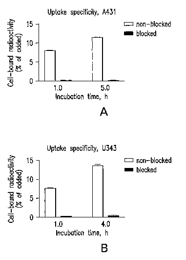
Fig. 1 shows specificity of 68Ga-DOTA-EGF binding to A431 carcinoma (a)
and U343 glioma (b) cell lines. At all time points, EGF receptors on control
cells were
blocked with a 100-fold excess amount of non-labelled EGF. The binding was
specific, since the binding could be suppressed. The presented data are mean
values of
three measurements and standard deviations.
CA 02598863 2007-08-21
WO 2006/090232 PCT/IB2006/000345
Fig. 2 shows saturation of 68Ga -DOTA-hEGF binding to cultured carcinoma
A431 and glioma U343 cells, incubated with different concentrations of 68Ga -
DOTA-
hEGF (0.26-16.9 nM, for A431 cells and 0.14-36 nM for U343 cells) for 2 h on
ice in
presence or absence of unlabellled hEGF to get non-specific and total binding,
respectively. The data was analyzed by GraphPad Prism 3Ø All data points are
mean
values of at least three data points, and maximal variations are shown.
Fig. 3 shows Internalisation of 68Ga -DOTA-EGF after binding to carcinoma
A431 and glioma U343 cells. Internalization was determined by acid wash at two
different time points. Radioactivity, which was removed from cells by an
acidic buffer
was considered as membrane-bound, and the rest as internalized. The presented
data
are mean values of three measurements and standard deviations.
Fig. 4 shows cell-associated 68Ga radioactivity as a function of time after
interrupted incubation of A431(solid line) and U343(dotted line) with 68Ga-
DOTA-
EGF . The cell associated radioactivity at time zero after the interrupted
incubation
was considered as 100%. All data points are mean values of three measurements
and
standard deviations. Both A431 and U343 cell cultures were incubated with 68Ga-
DOTA-hEGF for 4 h.
Fig. 5(A) is biodistribution of 68Ga -DOTA-EGF expressed as % injected dose
per gram tissue in tumour bearing nude mice at 30 min time point. Fig. 5(B)
shows
tumour-to-Organ ratios of 68Ga -DOTA-EGF in tumour bearing nude mice at 30 min
11
CA 02598863 2007-08-21
WO 2006/090232 PCT/IB2006/000345
time point. Mice were intravenously injected with either 0.016 or 0.16 nmol of
radiotracer and killed at 30 min time point. Data are presented as mean SD
(n = 4).
Fig. 6 Left) is an image showing a summation of frames 20-24 (x-30 min after
injection). The tumours can clearly be seen at either side of the head. Right)
is a
photograph of the positioning of the mouse.
Fig. 7 are pharmacokinetic curves showing the rapid distribution of 68Ga -
DOTA-EGF (0.16 nmol injected) to liver, kidney and tuinours. The excretion
into
urine is continuous throughout the observation time.
Detailed Description of the Invention
One object of the invention is to provide a method for synthesizing
radiolabeled gallium complexes which are useful as radiopharmaceuticals,
specifically for use in PET. They are particularly useful for the detection of
epidermal growth factor receptor (EGFR) expression in malignant tumors. This
is
achieved by the method described in the invention.
68Ga is obtainable from a 68Ge/68Ga generator. Such generators are known in
the art, see for instance C. Loc'h et al, J. Nucl. Med. 21, 1980, 171-173 or
J.
Schuhmacher et al. Int. J. appl. Radiat. Isotopes 32, 1981, 31-36. 68Ge may be
obtained by cyclotron production by irradiation of, for instance Ga2(S04)3
with 20
MeV protons. It is also commercially available, e.g. as 68Ge in 0.5 M HC1.
Generally,
68Ge is loaded onto a column consisting of organic resin or an inorganic metal
oxide
12
CA 02598863 2007-08-21
WO 2006/090232 PCT/IB2006/000345
like tin dioxide, aluminium dioxide or titanium dioxide. 68Ga is eluted from
the
column with aqueous HCl yielding 68GaC13. 6sGa3+ is particularly preferred in
the
method according to the invention as its production does not require a
cyclotron and
its 68 min half-life is sufficient to follow many biochemical processes in
vivo by PET
imaging without long radiation.
Suitable columns for 68Ge/68Ga generators consist of inorganic oxides like
aluminium dioxide, titanium dioxide or tin dioxide or organic resins like
resins
comprising phenolic hydroxyl groups (US-A-4264468) or pyrogallol (J.
Schulunacher
et al., Int. J. appl. Radiat. Isotopes 32, 1981, 31-36). In a preferred
embodiment, a
68Ge/68Ga generator comprising a column comprising titanium dioxide is used in
the
method according to the invention.
The concentration of the aqueous HCl used to elute 68Ga from the 68Ge/68Ga
generator column depends on the column material. Suitably 0.05 to 5 M HCl is
used
for the elution of 68Ga. In a preferred embodiment, the eluate is obtained
from a
68Ge/68Ga generator comprising a column comprising titanium dioxide and 68Ga
is
eluted using 0.05 to 0.1 M HCI, preferably about 0.1 M HCI.
In a preferred embodiment of the method according to the invention, a strong
anion exchanger comprising HCO3" as counterions, preferably a strong anion
exchanger comprising HCO3- as counterions, is used. In a further preferred
embodiment, this anion exchanger comprises quaternary amine functional groups.
In
another further preferred embodiment, this anion exchanger is a strong anion
exchange resin based on polystyrene-divinylbenzene. In a particularly
preferred
embodiment, the anion exchanger used in the method according to the invention
is a
13
CA 02598863 2007-08-21
WO 2006/090232 PCT/IB2006/000345
strong anion exchange resin comprising HC03- as counterions, quatemary amine
functional groups and the resin is based on polystyrene-divinylbenzene.
Suitably, water is used to elute the 68Ga from the anion exchanger in the
method according to the invention.
The 68Ga obtained according to the method of the invention is preferably used
for the production of 68Ga-radiolabelled complexes, preferably for the
production of
68Ga-radiolabelled PET tracers that comprise a bifunctional chelating agent,
i.e. a
chelating agent linked to a targeting vector.
Thus, another aspect of the invention is a method for producing a 68Ga-
radiolabelled complex by
a) obtaining 68Ga by contacting the eluate from a 68Ge/68Ga generator with an
anion
exchanger comprising HC03" as counterions and eluting 68Ga3+ from said anion
exchanger, and
b) reacting the 68Ga with a chelating agent.
Preferred chelating agents for use in the metliod of the invention are those
which present 68Ga in a physiologically tolerable form. Further preferred
chelating
agents are those that form complexes with 68Ga that are stable for the time
needed for
diagnostic investigations using the radiolabelled complexes.
14
CA 02598863 2007-08-21
WO 2006/090232 PCT/IB2006/000345
Suitable chelating agents are, for instance, polyaminopolyacid chelating
agents
like DTPA, EDTA, DTPA-BMA, DOA3, DOTA, NOTA, HP-DOA3, TMT or DPDP.
Those chelating agents are well known for radiopharmaceuticals and
radiodiagnosticals. Their use and synthesis are described in, for example, US-
A-
4647447, US-A-5362 475, US-A-5534241, US-A-5358704, US-A-5198208, US-A-
4963344, EP-A-230893, EP-A-130934, EP-A-606683, EP-A-438206, EP-A-434345,
WO-A- 97/00087, WO-A-96/40274, WO-A-96/30377, WO-A-96/28420, WO-A-
96/16678, WO-A-96/11023, WO-A-95/32741, WO-A-95/27705, WO-A-95/26754,
WO-A-95/28967, WO-A-95/28392, WO-A-95/24225, WO-A-95/17920, WO-A-
1o 95/15319, WO-A-95/09848, WO-A-94/27644, WO-A-94/22368, WO-A-94/08624,
WO-A-93/16375, WO-A-93/06868, WO-A-92/11232, WO-A-92/09884, WO-A-
92/08707, WO-A-91/15467, WO-A-91/10669, WO-A-91/10645, WO-A-91/07191,
WO-A-91/05762, WO-A-90/12050, WO-A-90/03804, WO-A-89/00052, WO-A-
89/00557, WO-A-88/01178, WO-A-86/02841 and WO-A-86/02005.
Suitable chelating agents include macrocyclic chelating agents, e.g. porphyrin-
like molecules and pentaaza-macrocycles as described by Zhang et al., Inorg.
Chem.
37(5), 1998, 956-963, phthalocyanines, crown ethers, e.g. nitrogen crown
ethers such
as the sepulchrates, cryptates etc., hemin (protoporphyrin - IX chloride),
heme and
chelating agents having a square-planar symmetry.
Macrocyclic chelating agents are preferably used in the method of the
invention. In a preferred embodiment, these macrocyclic chelating agents
comprise at
least one hard donor atom such as oxygen and/or nitrogen like in polyaza- and
CA 02598863 2007-08-21
WO 2006/090232 PCT/IB2006/000345
polyoxomacrocycles. Preferred examples of polyazainacrocyclic chelating agents
include DOTA, NOTA, TRITA, TETA and HETA with DOTA being particularly
preferred.
Particularly preferred macrocyclic chelating agents coinprise functional
groups such as carboxyl groups or amine groups which are not essential for
coordinating to Ga3+ and thus may be used to couple other molecules, e.g.
targeting
vectors, to the chelating agent. Examples of such macrocyclic chelating agents
comprising functional groups are DOTA, NOTA, TRITA or HETA.
In a further preferred embodiment, bifunctional chelating agents are used in
the method according to the invention. "Bifunctional chelating agent" in the
context
of the invention means chelating agents that are linked to a targeting vector.
Suitable
targeting vectors for bifunctional chelating agents useful in the method
according to
the invention are chemical or biological moieties, which bind to target sites
in a
patient's body, when the 68Ga-radiolabelled complexes comprising said
targeting
vectors have been administered to the patient's body. A preferred targeting
vector for
bifunctional chelating agents useful in the method according to the invention
is the
natural ligand to EGFR, epidermal growth factor (EGF) or a part, a fragment, a
derivative or a complex thereof. The small molecular weight of EGF, 6.2 kDa,
enables fast tumour penetration and fast blood clearance, providing good
contrast of
the image. The use of a positron-emitting label for EGF is particularly
advantageous,
since PET, compared with SPECT, is a superior detection technique in
sensitivity,
resolution and quantification. Particularly preferred targeting vector is the
human
16
CA 02598863 2007-08-21
WO 2006/090232 PCT/IB2006/000345
recombinant epidermal growth factor (hEGF) or a part, a fragment, a derivative
or a
complex thereof.
In a particularly preferred embodiment, macrocyclic bifunctional chelating
agents are used in the method according to the invention. Preferred
macrocyclic
bifunctional chelating agents comprise DOTA, NOTA, TRITA or HETA linked to a
targeting vector, preferably to an EGF or a part, a fragment, a derivative or
a complex
thereof; particularly preferably to an hEGF or a part, a fragment, a
derivative or a
complex thereof.
The targeting vector can be linked to the chelating agent via a linker group
or
via a spacer molecule. Examples of linker groups are disulfides, ester or
ainides,
examples of spacer molecules are chain-like molecules, e.g. lysin or
hexylamine or
short peptide-based spacers. In a preferred embodiment, the linkage between
the
targeting vector and the chelating agent part of radiolabelled gallium complex
is as
such that the targeting vector can interact with its target in the body
without being
blocked or hindered by the presence of the radiolabelled gallium complex.
A preferred aspect of the invention is a method for producing a 68 Ga-
radiolabelled complex by
c) obtaining 68Ga by contacting the eluate from a 68Ge/68Ga generator with an
anion
exchanger comprising HC03" as counterions and eluting 68Ga from said anion
exchanger, and
17
CA 02598863 2007-08-21
WO 2006/090232 PCT/IB2006/000345
d) reacting the 68Ga with a chelating agent, wherein the reaction is carried
out using
microwave activation.
It has been found that the use of microwave activation substantially improves
the efficiency and reproducibility of the 68Ga-chelating agent complex
formation. Due
to microwave activation, chemical reaction times could be shortened
substantially; i.e.
the reaction is completed within 2 min and less. This is a clear improvement
as a 10
minutes shortage of the reaction time saves about 10% of the 68Ga activity.
Furthermore, microwave activation also leads to fewer side reactions and to an
increased radiochemical yield, which is due to increased selectivity.
Suitably, a microwave oven, preferably a monomodal microwave oven is used
to carry out microwave activation. Suitably microwave activation is carried
out at 80
to 120 W, preferably at 90 to 110 W, particularly preferably at about 100 W.
Suitable
microwave activation times range from 20 s to 2 min, preferably from 30 s to
90 s,
particularly preferably from 45 s to 60 s.
A temperature control of the reaction is advisable when temperature sensitive
chelating agents, like for instance bifunctional chelating agents comprising
peptides
or proteins as targeting vectors, are employed in the method according to the
invention. Duration of the microwave activation should be adjusted in such a
way,
that the temperature of the reaction mixture does not lead to the
decomposition of the
chelating agent and/or the targeting vector. If chelating agents used in the
method
according to the invention comprise peptides or proteins, higher temperatures
applied
18
CA 02598863 2007-08-21
WO 2006/090232 PCT/IB2006/000345
for a shorter time are generally more favourable than lower temperatures
applied for a
longer time period.
Microwave activation can be carried out continuously or in several microwave
activation cycles during the course of the reaction.
Another aspect of the invention is a kit for obtaining 68Ga from a 68Ge/68Ga
generator, which comprises a generator colurmi and a second column that
comprises
an anion exchanger comprising HC03- as counterions.
In a preferred embodiment, the kit further comprises means to couple the
columns in series and/or aqueous HCl to elute the 68Ga from the generator
column
and/or water to elute the 68Ga from the anion exchanger colunm. The HCl and
the
water are preferably aseptically and in a hermetically sealed container.
In another preferred embodiment, the kit according to the invention further
comprises a chelating agent, preferably a bifunctional chelating agent, i.e. a
chelating
agent linked to a targeting vector.
The present invention further provides such radiolabeled gallium complexes as
PET tracers. A preferred tracer according to the instant invention is 68Ga-
DOTA-
hEGF.
19
CA 02598863 2007-08-21
WO 2006/090232 PCT/IB2006/000345
In yet another embodiment, the invention also provides a method for imaging
of EGFR overexpression in tumors comprising administering a radiolabeled
gallium
complex to a human, wherein the radiolabelled gallium complex is capable of
being
imaged by positron emission tomography, detecting of EGFR overexpression in
tumors by performing positron emission tomography process.
Examples
The invention is further described in the following exainples which are in no
way intended to limit the scope of the invention.
Example 1 - Cheinistry and Radiochemistry of 68Ga-DOTA-hEGF Preparation
1. Materials
Recombinant human epidermal growth factor (hEGF) was purchased from
Chemicon (Temecul, CA, USA). Sodium acetate (99.995%), HEPES (4-(2-
Hydroxyethyl) piperazine-l-ethanesulfonic acid), doubly distilled hydrochloric
acid
(Riedel de Haen) were obtained from Sigma-Aldrich Sweden (Stockholm, Sweden).
Sodium dihydrogen phosphate, di-sodium hydrogen phosphate and trifluoroacetic
acid (TFA) were obtained from Merck (Darmstadt, Germany). Sulfo-NHS ester of
DOTA (1,4,7,1 0-tetraazacyclododecane- 1,4,7,1 0-tetraacetic acid) was
purchased
from Macrocyclics (Dallas, TX, USA). The purchased chemicals were used without
further purification. Deionised water (18.2 MSZ), produced with a Purelab
Maxima
CA 02598863 2007-08-21
WO 2006/090232 PCT/IB2006/000345
Elga system (Bucks, the UK), was used in all reactions. 68Ga was obtained from
a
68Ge/68Ga generator (Cyclotron C., Obninsk, Russia).
II. HPLC AnaLysis
Analytical liquid chromatography (LC) was performed using a HPLC system
from Beckman (Fullerton, CA, USA) consisting of a 126 pump, a 166 UV detector
and a radiation detector coupled in series. Data acquisition and handling was
performed using the Beckman System Gold Nouveau Chromatography Software
Package. The column used was a Vydac RP 300 A HPLC column (Vydac, USA) with
the dimensions 150 mm x 4.6 mm, 5 m particle size. The applied gradient
elution
had the following parameters: A= 10 mM TFA; B = 70% acetonitrile (MeCN), 30%
H20, 10mM TFA with UV-detection at 220 nm; flow was 1.2 mL/min; 0-2 min
isocratic 20% B, 20-90% B linear gradient 8 min, 90-20% B linear gradient 2
min.
The quantity of 68Ga -DOTA-hEGF and radio-impurities retained on the column
could be obtained by measuring the activity of the sample injected on the
column and
the fractions collected from the outlet with a crystal scintillation counter.
The overall
loss on the system was 10%. The measured activity of the fractions of 68Ga -
DOTA-
hEGF and hydrophilic radio-impurities were in agreement with the respective
values
obtained from the HPLC chromatograms. The corresponding relative standard
deviation values were 7% and 0.5%, respectively for hydrophilic radio-
impurities and
68Ga -DOTA-hEGF.
III. Preparation of 68Ga -DOTA-hEGF
21
CA 02598863 2007-08-21
WO 2006/090232 PCT/IB2006/000345
hEGF (32-70 nanomols, 80-180 L) in 0.08 M borate buffer, pH 9.4, was
added to dry N-hydroxy-sulfosuccinimide ester of DOTA (10-20 fold excess)
under
stirring and the pH was further adjusted to 9.0 by adding borate buffer (240-
340 L).
The mixture was left at room temperature for 3-4 hours or overnight. The
conjugate
was purified on Bio-select RP C18 C-18 SPE column (Vydac). The reaction
mixtures
was passed slowly though extraction disc, which was then washed with 2 mL of
0.1 %
TFA. The product was eluted in 1 mL of 70% acetonitrile with 0.1 % TFA. The
solvent was evaporated using a vacuum centrifuge (Labconco CentriVap Console,
Kansas City, Missouri, USA), operated at 50 C, and the dry purified product
was
stored at a temperature below zero.
The labeling of the conjugate was performed using either non-concentrated
68Ga-eluate or eluate pre-concentrated, as described previously (Velikyan I,
Beyer GJ,
Langstrom B. Microwave-supported preparation of (68)Ga bioconjugates with high
specific radioactivity. Biocoyajug Claem. 2004; 15(3): 554-560). In some
cases, the
eluates from two generators were pre-concentrated in order to increase the
amount of
68Ga utilized in the labeling reaction. The amount of DOTA-hEGF used in the
labeling reaction was 6-10 and 2-5 nanomols, respectively, when using non-
concentrated and pre-concentrated 68Ga-eluate. Sodium acetate buffer, pH 5.0-
5.5,
was used for labeling with non-concentrated 68Ga, and HEPES buffer, pH 4.6-
4.8,
was used for pre-concentrated eluate. The labeling was performed by 1 min long
microwave heating. The product was purified on Bio-select RP C18 C-18 SPE
column as described above. The solvent was then exchanged to PBS (phosphate
22
CA 02598863 2007-08-21
WO 2006/090232 PCT/IB2006/000345
buffered saline) buffer on NAP-5 columns (Sephadex G-25; Amersham Pharmacia
Biotech AB, Uppsala, Sweden). Purity of the conjugate was assessed by HPLC,
and
concentration of the conjugate and the tracer was determined from UV-HPLC
calibration plots.
In order to verify, that binding of 68Ga to hEGF was DOTA mediated, a blank
experiment was performed. The manipulations were the same as described above,
but
non-conjugated hEGF was used.
69'71Ga of natural isotope composition was complexed to DOTA-hEGF using
the same protocol. 69'71Ga-DOTA-hEGF characterized with LC-ESI-MS was used for
the identification of the radio-HPLC chromatogram signals.
IV. Microwave Heating and LC-ESI-MS Analysis
The microwave heating was performed in a SinithCreatorTM monomodal
microwave cavity producing continuous irradiation at 2450 MHz (Personal
Chemistry
AB, Uppsala, Sweden). The temperature, pressure and irradiation power were
monitored during the course of the reaction. The reaction vial was cooled down
with
pressurized air after completed irradiation.
Liquid chromatography electrospray ionization mass spectrometry (LC-ESI-
MS) was performed using the Waters Micromass Quattro Premier Mass Spectrometer
23
CA 02598863 2007-08-21
WO 2006/090232 PCT/IB2006/000345
(Micromass, UK) and an HPLC system from Alliance (Waters 269, UK) with
Photodiode Array UV detector. The column used was an Antlantis, dC 18, RP HPLC
column with the dimensions 100 mm x 2.1 mm, 3,um particle size. Isocratic
elution
was applied with the following parameters: A= 10 mM Formic acid; B= 100%
acetonitrile (MeCN), with UV-detection at 210-400 nm; flow was 0.3 mL/min. LC-
ESI-MS was performed with positive mode scanning and selected ion recording
(SIR)
detecting [M+6H]6+, [M+7H]7+ and [M+8H]$+ species. hEGF was detected at m/z
=781.5 for [M+8H]$+, m/z = 893 for [M+7H]7+ and m/z = 1042 for [M+6H]6+
Reconstitution of the data gave M= 6244.6711.15. (DOTA)1-hEGF was detected at
1o m/z =829.75 for [M+8H]g+, m/z = 948.13 for [M+7H]7+ and m/z = 1105 for
[M+6H]6+. Reconstitution of the data gave M = 6629.95 0.05. (DOTA)2-hEGF was
detected at m/z =878 for [M+8H]8+, mlz = 1003.3 for [M+7H]7+ and m/z =1170.36
for [M+6H]6+. Reconstitution of the data gave M= 7016+0.08. (DOTA)3-hEGF was
detected at m/z =926.29 for [M+BH]8+, m/z = 1058.47 for [M+7H]7+ and m/z =
1234.72 for [M+6H]6+. Reconstitution of the data gave M = 7402 0.1. (Ga-DOTA)1-
hEGF was detected at m/z =838.5 for [M+8H]8+, m/z = 958.13 for [M+7H]7+ and
m/z
= 1117.66 for [M+6H]6+. Reconstitution of the data gave M = 6699.95 0.05. (Ga-
DOTA)2-hEGF was detected at m/z =896.05 for [M+8H]8+, mlz = 1023.3 for
[M+7H]'+ and m/z = 1193.69 for [M+6H]6+. Reconstitution of the data gave M
2o 7157.55 2.47. (Ga-DOTA)3-hEGF was detected at m/z =952.54 for [M+8H]8+, m/z
=
1088.47 for [M+7H]7+ and m/z = 1269.72 for [M+6H]6+. Reconstitution of the
data
gave M = 7612.31 0.05.
V. Results
24
CA 02598863 2007-08-21
WO 2006/090232 PCT/IB2006/000345
68Ga -DOTA-hEGF was synthesized by a two-step procedure where hEGF
was initially conjugated to a bifunctional chelator, DOTA, and thereafter
labeled with
68Ga via a complexation reaction of 68Ga with the chelator. In the conjugation
step,
the one of carboxylic groups of the DOTA chelate was coupled to an amine
functionality of the peptide forming an amide bond (Scheme 1). The basic pH
required for the conjugation reaction was provided by borate buffer. hEGF
contains
one terminal and two lysine amino groups. Consequently, the conjugation
reaction of
hEGF resulted in the formation of a mixture of molecules with one, two and
three
DOTA fragments, as determined by LC-ESI-MS analysis.
The microwave-accelerated labeling of the conjugates (Scheine 1) was
performed using a non-concentrated or a pre-concentrated generator 68Ga-
eluate. The
labeling yield was 60 10 % (N=3) in the case of non-concentrated conjugate.
The
use of pre-concentration enabled to increase 77 4 % (N =3). Pre-
concentration of
eluate allowed to obtain specific radioactivity of 28 MBq/nmol. Attachment of
68Ga to
hEGF was DOTA-mediated, since the same treatment of non-conjugated hEGF din
not provide any labeled peptide. The radiochemical purity of the tracers in
the study
exceeded 99%. The tracer proved to be stable in the PBS, with no additional
radio-
HPLC signals during the stability assay of four hours.
oo- o~o- oyo
00 ~o ( o
N~~N ~ NN O-NH-hEGF 88Ga3+ in HCI N Ga N C'NH-hEGF.
( 1
N H 9hEGF, RT hEGF-NH2 ( 1 r ee a+
o- ' J ~ p= ' ) Buffer, pH - 4.6 p- ' J
.~N,~N 0 /~~ p NN Microwave heating NN
O p~ o O p-~o O 0-~0
Scheme 1
CA 02598863 2007-08-21
WO 2006/090232 PCT/IB2006/000345
Example 2- Cell Binding and Retention Experiments
1. Cell Culture
The human squamous carcinoma cell line A431 (ATCC, CLR 1555,
Rocksville, MD, USA) and the malignant glioma cell line U343MGaC12:6
(Westermark B, Magnusson A, Heldin CH. Effect of epidermal growth factor on
membrane motility and cell locomotion in cultures of human clonal glioma
cells. J
Neurosci Res. 1982; 8(2-3): 491-507) (from now on denoted U343) were used in
all
cell experiments. This A431 cell line is reported to express approximately 2 x
106
EGFR per cell, and the U343 cell line express approximately 5.5 x 10⁵ EGFR per
cell.
The cells were cultured in Hain's F10 medium (Biochrom Kg), supplemented with
10% fetal calf serum (Sigma), L-glutamine (2 mM) and PEST (penicillin 100IU/ml
and streptomycin 100 g/ml) both from Biochrom Kg. During cell culture and
cell
experiments (unless otherwise stated) cells were grown at 37 C in incubators
with
humidified air, equilibrated with 5% COZ. The cells were trypsinized with
trypsin-
EDTA (0.25% trypsin, 0.02% EDTA in PBS without Ca and Mg) from Biochrom Kg.
II. Binding of 68Ga-DOTA-EGF to the Cells
A431 and U343 cells were cultured in 3 cm Petri dishes (approximately 3.5 x
105 and 1.9 x 10⁵ cells per dish, respectively). After washing the cells once,
68Ga -
DOTA-EGF in cell culture medium (35 ng/dish, 50 kBq/dish for A431 cells and 5
26
CA 02598863 2007-08-21
WO 2006/090232 PCT/IB2006/000345
ng/dish, 20 kBq/dish for U343 cells) was added. The concentration of the added
tracer
was 0.26-16.9 nM, for A431 cells and 0.14-36 nM for U343 cells. To some
dishes, a
molar excess of EGF (5 or 3 g/dish) was added together with the labelled
conjugate,
in order to estimate the binding specificity of the 68Ga -DOTA-EGF conjugate.
After
0.5-6 h incubation at 37 C, the cells were washed six times with cold serum
free
medium, and they were then harvested using 0.5 ml trypsin-EDTA (15 min, 37 C).
The trypsination was terminated with addition of 1 ml cell culture medium, and
part
of the cell suspension (0.5 ml) was used for cell counting while the rest was
measured
in a gamma counter.
In order to estimate the cellular internalization of the 68Ga -DOTA-EGF
conjugate, a number of additional cell dishes were used during the binding
study to
separate the membrane bound fraction of the conjugate from internalized
radioactivity. Instead of trypsinising the cells, treatment with 0.5 ml ice-
cold 0.1 M
glycin-HCl buffer, pH 2.5 for 6 min at 0 C was used to extract the membrane
bound
fraction of the conjugate. An additional 0.5 ml of the glycin-HCl buffer was
used to
wash the cells once. The remaining radioactivity, considered to be
internalized
radioactivity, was collected by treatment with 0.5 ml 1 M NaOH solution at 37
C for
about 60 min. Another 0.5 ml NaOH solution was used for washing. The collected
fractions were measured in an automated gamma counter.
The binding of 68Ga -DOTA-EGF to A431 cells and U343 cells on ice was
also studied, in order to determine the time required for binding in the
saturation
study. Cell dishes placed on ice were incubated with ice cold 68Ga -DOTA-EGF
27
CA 02598863 2007-08-21
WO 2006/090232 PCT/IB2006/000345
solution for 0.5-4 h. The cells were then washed, trypsinized and counted as
described
above.
III. Cellular Retention of Radioactivity, Saturation Assay and Animal Tumor
Model
The cellular retention of radioactivity was studied after 1 h of incubation
with
68Ga -DOTA-EGF. After the incubation, the cells were washed thoroughly to
eliminate unbound conjugate, and the incubation was then continued in fresh
cell
culture medium. After 0.5- 4 h, the cells were trypsinized, counted and
measured for
radioactivity, as described above.
The equilibrium dissociation constant, Kd, was determined from a saturation
study with 68Ga -DOTA-EGF on A431 cells and U343 cells. Cells cultured in 24-
well
dishes (approximately 3.1 X 10⁴ A431 cells per well and 7.8 x 10⁴ U343 cells
per well)
were placed on ice, and ice cold 68Ga -DOTA-EGF solutions of different
concentrations (0.26-16.9 nM for A431 and 0.14-36 nM for U343) were added. For
each concentration, the unspecific background binding was studied by adding a
100
times excess of unlabelled EGF to some wells. After 2 h of incubation (the
time was
determined from the results of the uptake study on ice), the cells were washed
six
times with cold serum fiee medium. The cells were then trypsinized with 0.5 ml
of
trypsin-EDTA (15 min at 37 C), and the cells were counted and measured for
radioactivity in a gamma counter.
28
CA 02598863 2007-08-21
WO 2006/090232 PCT/IB2006/000345
The in vivo studies were carried out in adult female Balb/c nu/nu mice (21-25
g) (Mollegard, Denmark) with tumor xenografts. All animals were handled
according
to the guidelines by the Swedish Animal Welfare Agency, and the experiments
were
approved by the local Ethics Committee for Animal Research. The mice were
injected
subcutaneously with A431 tumor cells (approximately 7 million cells per tumor
in
100 l cell culture medium) in both front legs. The tumors were allowed to
grow for
12-13 days before the experiments were performed, and had then reached a
weight of
0.1-0.8 g.
IV. Results
The binding specificity of 68Ga-DOTA-EGF to EGFR-expressing cell lines in
vitro is shown in Figure 1. Cervical carcinoma A431 and glioma U343 cell
lines,
which have a documented expression of EGFR, were used in the cell tests. In
order to
demonstrate that binding is receptor-specific, a large amount of non-labelled
EGF was
added to cells in the control experiments, in order to saturate EGFR. Results
of the
binding specificity experiments demonstrated that the binding of 68Ga -DOTA-
EGF to
both cell lines might be prevented by receptor saturation at all tested data
points. This
indicates that binding of the labelled conjugate is receptor specific.
The results of the saturation experiments with 68Ga -DOTA-EGF on cervical
carcinoma A431 and glioma U343 cell lines are shown in Figures 2A and 2B,
respectively. The specific binding in amol/cell is plotted against the total
molar
concentration of added radiolabelled conjugate, and the result is analyzed by
nonlinear regression using the GraphPad Prism Software. Both curves seem to
have
reached a maximum value, indicating saturation. The obtained Kd values were in
an
29
CA 02598863 2007-08-21
WO 2006/090232 PCT/IB2006/000345
excellent agreement, 2.0 nM for A431 and 2.3 nM for U343 cells. The maximum
amount of binding sites per cell, 7.8x10⁵ for U343 cells corresponds
reasonably well
with 5.4x10⁵ as previously determined for a [ 111 In] -Bz-DTPA-EGF conjugate
(22).
The number of binding sites for A431, 1.9 millions per cell is also in good
agreement
with literature data.
In this study, the degree of internalization was estimated by acid wash.
Radioactivity, which was removed from cells by an acidic buffer was considered
as
membrane-bound, and the rest as internalized. Results of such experiments are
shown
in Figure 3 which shows that internalization of 68Ga -DOTA-EGF is a rapid
process in
both cell lines. However, results of these experiments indicate that the
internalization
rate was faster in glioma U343 cells as compared to A43 1. This may possibly
be due
to the documented capacity of A431 cells to recycle internalized receptors to
the cell
surface. More than 50% of the radioactivity was internalized at 30 min after
the start
of incubation in the case of glioma U343 cells.
The retention pattern of radioactivity after interrupted incubation with 68Ga -
DOTA-EGF for A431 and U343 cells was similar for both cell lines (Figure 4).
An
initial drop of radioactivity, which was most likely due to dissociation of
membrane-
bound conjugates, was followed by a relatively constant amount of cell-bound
68Ga.
Botli cell-lines demonstrated good retention, when more than 70 % of the
radioactivity was still cell-associated 4 hours, more than 3 half-lives of the
labell,
after interrupted incubation.
Example 3 - Biodistribution Studies
CA 02598863 2007-08-21
WO 2006/090232 PCT/IB2006/000345
I. Biodistribution in Mice with A431 Tumor Xeno r~afts
In order to estimate an influence of amount injected conjugate on uptake in
tumors and normal tissues, a biodistribution study was performed. Mice with
A431
tumor xenografts were injected intravenously with 50 168Ga-DOTA-EGF solution
(0.16 nmol or 0.016 nmol in PBS per animal), and 30 min post injection the
animals
were sacrificed and dissected. The mice were anesthetised by an
intraperitoneal
injection of a mixture of Rompun (1 mg/ml) and Ketalar (10 mg/ml), 0.2 ml per
10 g
of animal weight, and killed by heart puncture. In addition to the tumors,
blood, heart,
pancreas, spleen, stomach, liver, kidneys, lungs, small and large intestine,
muscle,
bone and salivary gland were collected, weighed and measured in an automated
gamma counter. The tails were also measured for radioactive content, in order
to
determine the accuracy of the injections. Organ values were calculated as
percent of
inj ected activity per g of organ (%IA/g).
II. Results
A summary of the biodistribution data for 68Ga -DOTA-EGF in A431 tumour-
bearing mice is shown in Figure 5. The measurement of the organ radioactivity
30
min after i.v. administration of 68Ga -DOTA-EGF showed the highest values in
the
kidneys and liver for both conjugates. The lower level of radioactivity
accumulation
was observed in pancreas, salivary gland, small and large intestine, stomach
and
spleen. The uptake of 68Ga -DOTA-EGF in the A431 tumour xenografte was 1.51 ~
31
CA 02598863 2007-08-21
WO 2006/090232 PCT/IB2006/000345
0.16 %IA/g and 2.69 0.29 %IA/g, for 0.016 and 0.16 nmol of injected
conjugate
respectively ( p = 0.036). The radiotracer had a rapid blood clearance, with
less than 1
%IA/g remaining in the circulation at 30 min time point for both conjugates
(no
significant difference). There were statistically significant decrease of the
radioactivity uptake in pancreas, spleen and stomach, when larger ainount of
conjugate was injected. Influence of increased amount of conjugate was even
more
pronounced, when tumor-to-normal organs were considered. Though, there were no
difference in tumor-to-blood ratio, 4.42 1.81 %IA/g and 4.50 2.53 %IA/g,
for
0.016 and 0.16 nmol of injected conjugate respectively ( p= 0.036), there were
statistically significant increase of tumor-to-organ ratios for heart,
pancreas, stomach,
spleen, lungs, intestines, muscles and salivary glands in he case when 0.16
nmol of
conjugate was injected.
Example 4 - MicroPET Imagin
Imaging was performed on a microPET R4 scanner (Concorde Microsystems,
Inc.), with a computer-controlled bed and 10 cm transaxial and 8 cm axial
field of
view (FOV). It operates exclusively in 3-dimensional list mode and has no
septa. All
raw data were first sorted into 3-dimensional sinograms, followed by Fourier
rebinning and 2-dimensional filtered back projection image reconstruction
resulting in
images with 2 nun resolution. The mice were taken to the laboratory just
before the
experiment. After a short period of heating under a red-light bulb the animal
was
placed in a cylinder connected to an isoflurane vaporizer adjusted to deliver
2%
isoflurane in a 45/55% mixture of oxygen and air. When the animal was
unconscious
32
CA 02598863 2007-08-21
WO 2006/090232 PCT/IB2006/000345
a heparinised venous catheter was placed in a tail vein and connected to a 1
ml
syringe with 0.9% NaC1 and 10 IU heparin. The animal was subsequently placed
on
the camera bed with its abdomen down and forelegs with tumours stretched out
forward as much as possible from the body and covered with saran wrap to
minimize
heat and water loss. Heated air (40 C) was blown on the animal to reduce the
loss of
body temperature during the experiment. The tracer was injected as a bolus
dose
shortly after the camera start in a volume of 100 1 followed by 100 l
saline. After
completion of the study the animals were decapitated under anesthesia and
blood,
liver, and kidney samples were collected for radioactivity measurements.
Scatter correction, random counts and dead time correction were all
incorporated into the reconstruction algorithm. Radiation attenuation in each
animal
was measured with two rotating rod sources containing 68Ge/68Ga before tracer
injection and the images were corrected for radiation attenuation. All PET
studies
started with a 20 min transmission scan. The amount of the injected activity
was 2.0 +
0.5 MBq. Two different imaging protocols were employed in this study. The
acquisition times were as follows: Protocol 1(duration 120 min) 10 x 30 s, 5 x
120 s,
and 5 x 300 s, 8 x 600 s; Protocol 2 (duration 30 min) 10 x 30 s, 5 x 60 s, 10
x 120 s.
Regions of interest (ROIs) were drawn on liver, kidney, bladder, salivary
gland and
tumours. Pharmacokinetic curves, representing the radioactivity concentrations
(percentage of injected dose per gram of tissue), versus time after injection
were
determined accordingly. The uptake index was calculated as activity in organ
[kBq/mL]/injected dose [1c]Bq] x 100%.
33
CA 02598863 2007-08-21
WO 2006/090232 PCT/IB2006/000345
The localization of 68Ga -DOTA-EGF in tumor-bearing mice as determined by
microPET imaging (Figure 6) was followed by activity measurements of blood,
liver,
and kidney samples collected after decapitation of the animal. The image of a
tumor
bearing mouse 30 min after administration of 2.0 MBq (with specific
radioactivity of
12-20 MBq/nmol) 68Ga -DOTA-EGF is shown to the left of Figure 6. The
evaluation
results of the microPET image are correlated with the activity measurements of
blood,
liver, and kidney samples. Both right and left leg tumors were visible with
clear
contrast frdm the adjacent background. Prominent uptake was observed in the
liver
and kidneys, and clearance of the activity through the urinary bladder was
evident
(Figure 7). The distribution to tumors and salivary gland were slower. Uptake
data
derived from microPET and biodistribution studies were found to be in
agreement and
compared with data obtained from the post imaging tissue sampling.
Specific Embodiments, Citation of References
The present invention is not to be limited in scope by specific embodiments
described herein. Indeed, various modifications of the inventions in addition
to those
described herein will become apparent to these skilled in the art from the
foregoing
description and accompanying figures. Such modifications are intended to fall
within
the scope of the appended claims.
Various publications and patent applications are cited herein, the disclosures
of which are incorporated by reference in their entireties.
34