Note: Descriptions are shown in the official language in which they were submitted.
CA 02599455 2007-08-28
WO 2006/081556 PCT/US2006/003222
-1-
BIOPSY NEEDLE
Field of the Invention
The invention relates generally to biopsy needles, and more particularly to
methods
and apparatus for collecting and retaining tissue samples in endoscopic biopsy
needles.
Background of the Invention
Endoscopic biopsy is a minimally invasive medical procedure for detecting
various
types of cancer. During a biopsy, tissue samples are removed from the body and
analyzed.
Io Doctors use the biopsy samples to analyze the cellular composition of
tissue, and in core-
sampling biopsies, the histology (structure) of the tissue.
According to a national health study performed in 1996, over 1.2 million
endoscopic
biopsies are performed in the United States each year. Unfortunately, the
success rate of
obtaining a tissue sample with a coring biopsy needle is less than desired. In
many cases, the
obtained samples have poor cellular architecture quality and are not
adequately intact to
provide histological data.
Typical endoscopes include a hollow tube and a control handle. The hollow tube
provides a conduit for safe insertion of the needle into the body and the
control handle allows
the doctor to bend the endoscope head. Typically, the endoscope head contains
an ultrasound
device, a camera, and liquid or air flushing capabilities. For a typical oral
procedure, the
endoscope is inserted into a patient's mouth and navigated to the biopsy site.
After the
endoscope is in place, a needle is passed through the hollow tube of the
endoscope.
A typical endoscope and needle assembly, such as the one shown in FIG. 1,
includes
an endoscope control handle 10, a hollow tube 12, a needle control handle 14,
a needle 16
having a lumen 18, a sheath 20, a removable stylet 22. The sheath protects the
interior of the
endoscope from needle damage. The stylet travels through the lumen of the
needle and
prevents a needle with an open end tip 24 from collecting tissue before the
end tip reaches the
target sample site. Once the needle reaches the target sample site, the stylet
is removed
through the proximal end of the needle. A control handle is used to control
the extension and
location of the needle, and to remove the stylet.
Biopsy needles can be separated into two general types: side-cutting needles
and end-
cutting needles. A side-cutting needle typically includes a sliding sheath
which is moved past
CA 02599455 2007-08-28
WO 2006/081556 PCT/US2006/003222
-2-
an opening along the side of the needle. The sheath or needle is configured to
cut the tissue
and force the tissue into the interior lumen of the needle. An end-cutting
needle typically has
a slanted tip and a cutting leading edge for puncturing and cutting the
tissue. Once the needle
has been advanced over a column of tissue, the intent is for the tissue column
to remain in the
needle lumen when the needle is retracted. Some needle assemblies include an
aspiration
needle which uses suction to help retain the tissue sample.
A need exists for biopsy needles that increase the success rate of obtaining a
tissue
sample and/or improve the quality of the samples obtained.
Summary of the Invention
According to certain aspects of the invention, a biopsy needle includes tissue
capture
elements within the needle lumen to help hold a tissue sample within the
needle and maintain
its integrity. According to one aspect, several flexible members form a sort
of "tissue check-
valve" that allows tissue to enter the lumen during advancement of the needle
into tissue, and
Is prevents the tissue sample from exiting the needle during retraction of the
needle. The
flexible members may, in some embodiments, include cutting edges configured to
cut the
tissue sample from the target tissue mass at the start of needle extraction. -
According to one embodiment, a biopsy apparatus includes a needle having an
opening to permit tissue entry and having a sidewall defining a lumen, and a
tissue capture
element protruding into the lumen, the tissue capture element being configured
to permit
tissue to move past the tissue capture element within the lumen, and further
configured to
prevent tissue from exiting the needle via the opening.
According to another embodiment, a needle includes a distal tip having an
opening
with a first cross-sectional area, the distal tip further having a cutting
leading edge. The
needle further includes a first longitudinal portion of the needle, proximal
to the distal tip,
having a first passage with a second cross-sectional area that is smaller than
the first cross-
sectional area, and a second longitudinal portion of the needle, proximal to
the first
longitudinal portion of the needle, having a second passage with a third cross-
sectional area
that is larger than the second cross-sectional area.
According to a further embodiment, a method of obtaining a tissue sample
includes
inserting a distal section of a needle having a lumen into a tissue mass to
force a column of
tissue into the lumen, a portion of the column of tissue passing through a
tissue capture
CA 02599455 2007-08-28
WO 2006/081556 PCT/US2006/003222
-3-
element disposed within the lumen. The method further includes retracting the
needle to
remove the column of tissue from the tissue mass, the tissue capture element
providing
resistance to movement of the tissue in the distal direction that is greater
than any resistance
provided to movement of the tissue in the proximal direction through the
tissue capture
element.
Brief Description of the Drawings
Other advantages, features, and uses of the invention will become apparent
from the
following detailed description of non-limiting embodiments of the invention
when considered
to in conjunction with the accoinpanying drawings, which are schematic and
which are not
intended to be drawn to scale. For purposes of clarity, not every component is
labeled in
every figure, nor is every component of each embodiment of the invention shown
where
illustration is not necessary to allow those of ordinary skill in the art to
understand the
invention. In cases where the present specification and a document
incorporated by reference
include conflicting disclosure, the present specification shall control.
FIG. 1 shows a typical prior art endoscope and needle assembly;
FIG. 2a is a perspective view of a needle including flexible members angled in
the
proximal direction according to one embodiment of the invention;
FIG. 2b is a side view of the embodiment shown in FIG. 2a;
FIG. 2c is a front view looking into the needle of the embodiment shown in
FIGS. 2a
and 2b;
FIG. 3 is a front view looking into a needle which includes one flexible
member,
according to an alternative embodiment of the invention;
FIG. 4a is a cross-sectional side view of an alternative embodiment of the
invention
comprising a ring with a passage;
FIG. 4b is a front view of the embodiment shown in FIG. 4a;
FIG. 5 is a cross-sectional side view of another alternative embodiment of the
invention comprising an opening in the sidewall;
FIG. 6 is a cross-sectional side view of another alternative embodiment of the
invention comprising a needle having internal barbs; and
FIG. 7 shows a side view of a removable needle tip according to one aspect of
the
invention.
CA 02599455 2007-08-28
WO 2006/081556 PCT/US2006/003222
-4-
Detailed Description of the Invention
According to one aspect of the invention, methods and apparatus are provided
to
collect tissue samples by introducing tissue into a biopsy needle lumen during
needle
advancement and retaining the tissue sample in the needle lumen during needle
retraction.
According to another aspect of the invention, the amount of tearing that
occurs during tissue
sampling may be reduced by providing a cutting element within a biopsy needle.
According
to another aspect, the integrity of obtained samples may be improved, which
can be
particularly helpful wlien studying the histology of tissue samples. Not every
embodiment of
the invention includes all of the aspects of the invention described herein.
Some
embodiments do, however, include combinations of various aspects.
In some embodiments, the cutting and/or retaining of a tissue sample can be
achieved
in a passive manner, that is to say, active triggering of a cutting or
retaining device by a user
may not be required. Such an approach may reduce the complexity of use and/or
the cost to
manufacture.
Tissue capture elements, in some embodiments, are retaining members that
protrude
into the needle lumen and allow tissue to pass through a passage defined by
the members
and/or the interior wall of the needle. When the tissue contained within the
needle is pulled to
move in the opposite direction, i.e., during retraction of the needle from a
tissue mass, the
retaining members prevent the tissue from moving through the lumen by
substantially
constricting or closing off the previously available passage. In this manner,
the retaining
member or members act as a sort of one-way valve for the tissue sample.
In some embodiments, the retaining members do not open or constrict based on
the
movement of tissue through the lumen, but rather maintain a defined opening
space or
passage which acts as a one-way valve by virtue of the shape and structure of
the members.
Instead of a plurality of members, in some embodiments a ring with a passage
is used
within a needle to provide a tissue capture element. The entrance to the
passage, i.e., the side
of the ring into which tissue is introduced, may be slanted so as to gradually
compress the
tissue as it moves through the ring. The passage may include a section of
constant diameter,
and then open relatively abruptly into the full lumen diameter of the needle
on the exit side.
The abrupt change in diameter (or cross-sectional area in the case of a non-
cylindrical needle)
prevents tissue from re-entering the passage on the exit side when the needle
is retracted from
the tissue mass and the sample of tissue tends to be pulled in such a
direction. For purposes
CA 02599455 2007-08-28
WO 2006/081556 PCT/US2006/003222
-5-
herein, the term "passage" means any opening, aperture, hole, pathway or
channel through
which tissues or cells may pass.
In embodiments where the retaining members move to expand and constrict a
passage
for the tissue, the members may be coupled to the needle sidewall with a
flexural hinge. The
members may extend from the needle wall at an angle and toward a proximal end
of the
needle. In other embodiments, other suitable hinges and/or attachments may be
used to
couple the ineinbers to the needle sidewall. For purposes herein, the proximal
end of the
needle apparatus is the end toward the control handle, while the distal end of
the needle is the
end which is introduced into a tissue mass.
to The proximal side of the free ends of the retaining members may include a
cutting
surface such that upon needle extraction, when the tissue starts pushing
against the back side
of the needle members, the tissue is cut, thereby separating it from the main
tissue mass. In
this manner, the separation of the tissue sample from the tissue mass is
passive. In other
embodiments, active cutting devices may be used to separate the tissue
sanlple. In still
further embodiments, cutting of the tissue may be accomplished with a twisting
action.
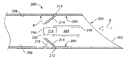
Referring now to the figures, one embodiment of a needle including a tissue
capture
element comprising four retaining members is shown in FIGs. 2a-2c. Needle 200
has a
slanted distal tip 201 and includes a cutting leading edge 202 which
circumferentially cuts
tissue as needle 200 is advanced into a tissue mass (not shown). An opening
205 permits the
introduction of tissue into a needle lumen 206. Four retaining members 204
extend from a
needle sidewa11208 and are spaced symmetrically about a center axis of needle
lumen 206.
Each retaining member 204 is coupled to needle 200 by a flexural hinge 210. To
form
this flexural hinge, a portion (or all) of retaining member 204 may be cut
(such as by laser
cutting), attached (such as by tacking or using adhesive), molded, or
otherwise formed from
needle wa11208. As shown in FIGs. 2a-2c, a bend 212 in the retaining member
may be
positioned at a distance from flexural hinge 210. Bend 212 provides retaining
member 204
with a distally-facing surface 214. When tissue moves into needle 200 (i.e.,
in the direction
of arrow A), the tissue pushes on distally-facing surface 214 and this force
pushes retaining
member 204 outwardly toward needle sidewall 208, i.e., in the direction of
arrow B. This
radial component of movement expands, or in some cases creates, a passage
between the
retaining members. The passage permits the fiuther advancement of needle 200
into the
tissue mass to push a column of tissue into lumen 206 and past tissue
retaining members 204.
CA 02599455 2007-08-28
WO 2006/081556 PCT/US2006/003222
-6-
When a suitable amount of tissue has been introduced into needle 200 and
pushed past
retaining members 204, the needle is retracted. With some prior art needles,
retraction of the
needle results in the tissue colunm moving in a direction opposite to arrow A,
i.e., out of
needle 200, because the tissue colunm remains attached at its end to the
tissue mass. In some
embodiments of the present invention, as the tissue starts to move in the
direction opposite to
arrow A, sharp edges 216 of retaining members 204 catch the outer edge of the
tissue column
and further movement of the tissue column urges retaining members 204 radially
inwardly
which cuts the tissue. Additionally, the radially inward movement of retaining
members 204
constricts, or in some cases closes, the passage that was previously present
between the
to retaining members. In this manner, the tissue present in needle 200 is
prevented from exiting
via end opening 205. For purposes herein, urging a tissue capture element to
constrict does
not necessarily require the retaining member to completely close the lumen or
any passage
through which the tissue passed. Urging a tissue capture member to constrict
encompasses
(but is not limited to) urging the tissue capture element to constrict by an
amount that does
not represent the fullest possible constriction. Similarly, for purposes
herein, urging a tissue
capture member to open does not necessarily require the tissue capture member
to completely
open.
For purposes herein, lumen 206 is defined as extending to the distal end of
the needle.
As such, a retaining element that extends from the sidewall into an area
partially radially
bordered by cutting leading edge 202 is considered to be protruding into the
lumen.
Bend 212 of retaining member 204 preferably forms an angle 218 with needle
sidewall 208 of approximately 45 . In some embodiments, angle 218 may be
between 30
and 60 inclusive, or another suitable angle. In some embodiments, retaining
member 204
may not include a bend at all. For example, retaining member 204 could be
coupled to the
interior sidewall at an angle and not include a further bend. Or, in other
embodiments,
retaining member 204 may be parallel with sidewall 208 (or disposed at a very
slight angle)
and include a free end edge that is configured to grab tissue moving in a
distal direction such
that the retaining member 204 is pulled inwardly as the tissue movement
continues.
While bend 212 is shown forming an acute angle with sidewall 208, retaining
member
204 may form a 90 or an obtuse angle with the sidewall.
Retaining members 204 need not be longitudinally linear, but may include
curves -
convex or concave or both. In some embodiments, retaining member 204 may have
a radius
of curvature (relative to a longitudinal axis) identical to or similar to
needle sidewall 208. In
CA 02599455 2007-08-28
WO 2006/081556 PCT/US2006/003222
-7-
other embodiments, retaining members 204 may be flat in a side-to-side and/or
longitudinal
direction. In some embodiments, lateral sides 220 of retaining member 204 near
sharp edge
216 may have a cutting edge. In this manner, twisting the needle may provide
further cutting
characteristics.
Four retaining members 204 are shown in the embodiment of FIGs. 2a-2c, but a
fewer
or a greater number of retaining members 204 may be used such as 1, 2, 3, 5,
or 6 or more.
Using an even number of retaining members 204 may provide advantages in
manufacturability because if laser cutting is einployed, opposing retaining
members may be
cut with a single cut. Stainless steel, or other suitable material, may be
used to construct the
!o needle and/or the retaining members. In some embodiments, the shapes and/or
sizes of the
retaining members may vary within the same needle 200. The longitudinal
positioning of the
retaining members may vary relative to one another and/or relative to cutting
leading edge
202. Additionally, retaining members 204 need not be positioned radially
symmetrically
about the longitudinal axis of lumen 206.
The ends and/or sides of retaining members 204 may be constructed and arranged
such that when they bend or move radially inwardly the ends and/or sides
contact one another
along complementary edges or surfaces. By doing so, a portion of a passage may
be entirely
blocked. For example, instead of having a flat distally-facing surface 214,
retaining members
204 may be shaped such that when the four retaining members 204 are pushed
together, there
is no passage along the center axis of lumen 206 (although small passages may
still exist
radially outwardly from the center axis). In still other embodiments,
retaining members 204
may be configured such that lutnen 206 is entirely blocked when retaining
members 204 are
pushed together.
FIG. 3 shows an embodiment that includes a single retaining member 204 as a
tissue
capture element. This embodiment is similar in many respects to the embodiment
shown in
FIGs. 2a-2c, but instead of four retaining members, one large retaining member
204 is used to
retain a tissue sample in needle 200. Retaining member 204 is shown with bend
212,
although, as with the embodiment of FIGs. 2a-2c, a bend is not required.
Retaining member
204 is coupled to sidewall 208 with a flexural hinge (not shown). For purposes
herein, when
describing one element as being "coupled to" another element, the term
"coupled to" means
any form of attachment (direct or indirect) and/or the elements being integral
to one another.
For example, in the embodiment of FIG. 3, retaining member 204 is coupled to
sidewall 208
because retaining meinber 204 includes a portion that is cut from sidewall
208. Retaining
CA 02599455 2007-08-28
WO 2006/081556 PCT/US2006/003222
-8-
member 204 would also be considered to be coupled to sidewall 208 if retaining
member 204
is an independently manufactured element that is attached to sidewall 208 via
adhesion or
welding or other process.
As shown in this embodiment, a tissue capture element need not completely
obstruct
lumen 206 to prevent a tissue sample from exiting needle 200.
A stationary tissue capture element is shown in FIGs. 4a and 4b. In this
embodiment,
a ring 402 has a passage 404 which includes a longitudinal section that has a
smaller cross-
sectional area than lumen 206 of needle 200. Passage 404 is configured such
that tissue can
more easily travel proximally through passage 404 than the tissue can travel
through passage
404 in the distal direction to exit the needle.
In the illustrated embodiment, ring 402 forms a passage that gradually narrows
along a
constricting length 406 in the proximal direction. A length 408 of constant
diameter is
present proximal to the constriction length 406. Proceeding proximally (to the
right in FIG.
4a) passage 404 then opens into the full lumen diameter (or into a lumen
having a diameter
greater than the diameter of constant diameter section 408). With this
configuration, tissue is
cut into a coluinn by leading edge 202, travels through distal end opening
205, is gradually
compressed by constricting length 406, passes through constant diameter length
408, and
passes into lumen 206 proximal to ring 402. When needle 200 is retracted from
the tissue
mass, a proximally-facing wall 410 of ring 402 prevents the collected tissue
from exiting
lumen 206 via passage 404. Constant diameter length 408 is not required, and
passage 404
may proceed immediately from a constriction length 406 to opening to a larger
lumen
diameter.
Ring 402 may include sharp edges or cutting elements (not shown) on proximally-
facing wall 410 to cut the connection of the tissue sample to the tissue mass.
In some
embodiments, proximally-facing wall 410 may have a slope instead of an abrupt
diameter
change, with the slope being steep enough to resist movement of tissue into
passage 404 in
the distal direction.
A stationary tissue capture element need not extend around the entire
circumference of
lumen 206. In some embodiments, non-flexible members similar in shape to the
flexible
retaining members illustrated in FIGs. 2a-2c may be employed. Instead of being
thin
members, however, the stationary members may be more volumetric, that is,
similar to the
ring in that they would have a larger contact area with sidewall 208 and have
a proximally-
facing wall similar to wall 410.
CA 02599455 2007-08-28
WO 2006/081556 PCT/US2006/003222
-9-
An alternative embodiment of the invention is shown in FIG. 5 in which a
sidewall
opening is used to introduce tissue into a needle rather than a distal end
opening. In this
embodiment, needle 200 is advanced into a tissue mass with a cutting leading
edge 502.
Tissue enters a lumen 506 through a sidewall opening 505. As needle 200 is
retracted from
the tissue mass, a cutting edge 508 cuts a length of tissue. Further
retraction moves the tissue
through retaining members 204. Force on a proximally-facing surface 514
enlarges a passage
through lumen 506. As with the embodiment illustrated in FIGs. 2a-2c,
retaining members
504 may include cutting elements 516, in this case facing in the distal
direction. After tissue
has moved past retaining members 504, movement of needle 200 in the distal
direction may
ro cause cutting elements 516 to shear the tissue from the tissue mass. In
some embodiments,
instead of, or in addition to, cutting edge 508, a sheath (not shown) that has
a cutting edge
may be used to partially or fully separate a tissue sample from the tissue
mass. In some
embodiments, the tissue capture element may be positioned proximal to sidewall
opening 505
such that tissue passes the tissue capture element while needle 200 is being
advanced.
While much of the description contained herein for various embodiments of the
invention uses terininology associated with cylindrical devices (e.g.,
circumference, column,
diameter), it is important to note that many of the embodiments may be
employed using non-
cylindrical components. For example, a needle having a square lumen may be
used, or, in
some embodiments, a passage in a ring or a passage through flexible retaining
members may
have a shape other than circular, cylindrical or substantially circular or
cylindrical.
FIG. 6 shows an embodiment of a needle 200 in which a tissue capture element
includes a plurality of angled barbs 602 disposed along the-interior of needle
sidewall 208.
The cross-sectional view of FIG. 6 only shows sets of barbs protruding into
the top and
bottom of lumen 206, but similar sets of barbs 602 protrude into lumen 206
along the left and
right sides of lumen 206 (as viewed from the distal end of the needle) as
well. In this
embodiment, twenty barbs are used per linear set of barbs, but other amounts
may be used.
Barbs 602 are angled in the proximal direction and may be any suitable
thiclcness, such as
approximately 0.1 mm. In some embodiments, fewer or greater numbers of sets of
barbs or
barbs per set may be used. The barbs illustrated in FIG. 6 are symmetrically
disposed about a
central axis of luinen 206, but in some embodiments, the barbs may be
positioned
asymmetrically. Additionally, barbs 602 need not be positioned linearly along
needle 200.
The angles that the barbs form with sidewall 208 may vary among the barbs. For
example, in
CA 02599455 2007-08-28
WO 2006/081556 PCT/US2006/003222
-10-
some embodiments, the barbs closer to the opening in the needle may form
larger angles than
the barbs that are farther from the opening.
In an alternative einbodiment, the interior surface of needle 200 may be
etched such
that the coefficient of friction encountered by tissue differs depending on
the direction that the
tissue is moving or attempting to move. For example, the tissue capture
element may include
an etched surface in which tissue can more easily move into the needle as
compared to the
tissue moving toward the opening. In some embodiments, the etching feature may
be
combined with other tissue capture elements disclosed herein.
FIG. 7 illustrates an embodiment of a needle 700 in which a removable needle
tip 702
!o is provided. With removable needle tip 702, a tissue sample may be removed
from the needle
assembly or endoscope assembly without removing the sainple from the section
of the needle
in which the sample was originally collected. In some embodiments, this
section of the
needle may be made of a clear material (transparent or translucent) such that
a doctor can
visually confirm the presence of a sample without removing the sample from the
needle. In
some embodiments, the removable needle tip 702 may include a tissue capture
element.
The removable needle tip may be made of polycarbonate, another plastic, or
other
suitable material, and removable using a shearing device or a scoring device
at a selected
longitudinal location 704. In some embodiments, the removable needle tip may
be attached
to a main needle body with threads and/or adhesive.
Removable needle tip 702 may be identified with an identifier such as a UPC
symbol
or an RFID tag before or after tissue collection to improve tracking of the
tissue sainple.
While several embodiments of the invention have been described and illustrated
herein, those of ordinary slcill in the art will readily envision a variety of
other means and
structures for perfonning the functions and/or obtaining the results or
advantages described
herein, and each of such variations or modifications is deemed to be within
the scope of the
present invention. More generally, those skilled in the art would readily
appreciate that all
parameters, dimensions, materials, and configurations described herein are
meant to be
exemplary and that actual parameters, dimensions, materials, and
configurations will depend
upon specific applications for which the teachings of the present invention
are used. Those
skilled in the art will recognize, or be able to ascertain using no more than
routine
experimentation, many equivalents to the specific embodiments of the invention
described
herein. It is, therefore, to be understood that the foregoing embodiments are
presented by
way of example only and that, within the scope of the appended claims and
equivalents
CA 02599455 2007-08-28
WO 2006/081556 PCT/US2006/003222
-11-
thereto, the invention may be practiced otherwise than as specifically
described. The present
invention is directed to each individual feature, system, material and/or
method described
herein. In addition, any combination of two or more such features, systems,
materials and/or
methods, if such features, systems, materials and/or methods are not mutually
inconsistent, is
s included within the scope of the present invention.
We claim: