Note: Descriptions are shown in the official language in which they were submitted.
CA 02599988 2007-09-04
WO 2006/104911 PCT/US2006/010850
METHODS AND COMPOSITIONS FOR MODULATING HYPERSTABILIZED
C-MET
RELATED APPLICATIONS
This application is a non-provisional application filed under 37 CFR
1.53(b)(1),
claiming priority under 35 USC 119(e) to provisional application number
60/665,482 filed
March 25, 2005, the contents of which are incorporated herein by reference.
TECHNICAL FIELD
The present invention relates generally to the fields of molecular biology and
growth factor
regulation. More specifically, the invention concerns modulators of the HGF/c-
met signaling
pathway, and uses of said modulators.
BACKGROUND
HGF is a mesenchyme-derived pleiotrophic factor with mitogenic, motogenic and
morphogenic activities on a number of different cell types. HGF effects are
mediated through
a specific tyrosine kinase, c-met, and aberrant HGF and c-met expression are
frequently
observed in a variety of tumors. See, e.g., Maulik et al., Cytokine & Growth
Factor Reviews
(2002), 13:41-59; Danilkovitch-Miagkova & Zbar, J. Clin. Invest. (2002),
109(7):863-867.
Regulation of the HGF/c-Met signaling pathway is implicated in tuinor
progression and
metastasis. See, e.g., Trusolino & Comoglio, Nature Rev. (2002), 2:289-300).
HGF binds the extracellular domain of the Met receptor tyrosine kinase (RTK)
and
regulates diverse biological processes such as cell scattering, proliferation,
and survival.
HGF-Met signaling is essential for normal embryonic development especially in
migration of
muscle progenitor cells and development of the liver and nervous system (Bladt
et al., Nature
(1995), 376, 768-771.; Hamanoue et al., Faseb J (2000), 14, 399-406; Maina et
al., Cell
(1996), 87, 531-542; Schmidt et al., Nature (1995), 373, 699-702; Uehara et
al., Nature
(1995), 373, 702-705). Developmental phenotypes of Met and HGF knockout mice
are very
similar suggesting that HGF is the cognate ligand for the Met receptor
(Schmidt et al., 1995,
supra; Uehara et al., 1995, supra). HGF-Met also plays a role in liver
regeneration,
angiogenesis, and wound healing (Bussolino et al., J Cell Biol (1992), 119,
629-641;
Matsumoto and Nakamura, Exs (1993), 65, 225-249; Nusrat et al., J Clin Invest
(1994) 93,
2056-2065). The precursor Met receptor undergoes proteolytic cleavage into an
extracellular a subunit and membrane spanning (3 subunit linked by disulfide
bonds (Tempest
et al., Br J Cancer (1988), 58, 3-7). The 0 subunit contains the cytoplasmic
kinase domain
1
CA 02599988 2007-09-04
WO 2006/104911 PCT/US2006/010850
and harbors a multi-substrate docking site at the C-terminus where adapter
proteins bind and
initiate signaling (Bardelli et al., Oncogene (1997), 15, 3103-3111; Nguyen et
al., J Biol
Chem (1997), 272, 20811-20819; Pelicci et al., Oncogene (1995), 10, 1631-1638;
Ponzetto et
al., Cell (1994), 77, 261-271; Weidner et al., Nature (1996), 384, 173-176).
Upon HGF
binding, activation of Met leads to tyrosine phosphorylation and downstream
signaling
through Gabl and Grb2/Sos mediated P13-kinase and Ras/MAPK activation
respectively,
which drives cell motility and proliferation (Furge et al., Oncogene (2000),
19, 5582-5589;
Hartmann et al., J Biol Chem (1994), 269, 21936-21939; Ponzetto et al., J Biol
Chem (1996),
271, 14119-14123; Royal and Park, J Biol Chem (1995), 270, 27780-27787).
Met was shown to be transforming in a carcinogen-treated osteosarcoma cell
line
(Cooper et al., Nature (1984), 311, 29-33; Park et al., Cell (1986), 45, 895-
904). Met
overexpression or gene-amplification has been observed in a variety of human
cancers. For
example, Met protein is overexpressed at least 5-fold in colorectal cancers
and reported to be
gene-amplified in liver metastasis (Di Renzo et al., Clin Cancer Res (1995),
1, 147-154; Liu
et al., Oncogene (1992), 7, 181-185). Met protein is also reported to be
overexpressed in oral
squamous cell carcinoma, hepatocellular carcinoma, renal cell carcinoma,
breast carcinoma,
and lung carcinoma (Jin et al., Cancer (1997), 79, 749-760; Morello et al., J
Cell Physiol
(2001), 189, 285-290; Natali et al., Int J Cancer (1996), 69, 212-217; Olivero
et al., Br J
Cancer (1996), 74, 1862-1868; Suzuki et al., Br J Cancer (1996), 74, 1862-
1868). In addition,
overexpression of mRNA has been observed in hepatocellular carcinoma, gastric
carcinoma,
and colorectal carcinoma (Boix et al., Hepatology (1994), 19, 88-91; Kuniyasu
et al., Int J
Cancer (1993), 55, 72-75; Liu et al., Oncogene (1992), 7, 181-185).
A number of mutations in the kinase domain of Met have been found in renal
papillary carcinoma which leads to constitutive receptor activation (Olivero
et al., Int J
Cancer (1999), 82, 640-643; Schmidt et al., Nat Genet (1997), 16, 68-73;
Schmidt et al.,
Oncogene (1999), 18, 2343-2350). These activating mutations confer
constitutive Met
tyrosine phosphorylation and result in MAPK activation, focus formation, and
tumorigenesis
(Jeffers et al., Proc Natl Acad Sci U S A (1997), 94, 11445-11450). In
addition, these
mutations enhance cell motility and invasion (Giordano et al., Faseb J (2000),
14, 399-406;
Lorenzato et al., Cancer Res (2002), 62, 7025-7030). HGF-dependent Met
activation in
transformed cells mediates increased motility, scattering, and migration which
eventually
leads to invasive tumor growth and metastasis (Jeffers et al., Mol Cell Biol
(1996), 16, 1115-
1125; Meiners et al., Oncogene (1998), 16, 9-20).
Met has been shown to interact with other proteins that drive receptor
activation,
transformation, and invasion. In neoplastic cells, Met is reported to interact
with a6(34
2
CA 02599988 2007-09-04
WO 2006/104911 PCT/US2006/010850
integrin, a receptor for extracellular matrix (ECM) components such as
laminins, to promote
HGF-dependent invasive growth (Trusolino et al., Cell (2001), 107, 643-654).
In addition,
the extracellular domain of Met has been shown to interact with a member of
the semaphorin
family, plexin B 1, and to enhance invasive growth (Giordano et al., Nat Cell
Biol (2002), 4,
720-724). Furthermore, CD44v6, which has been implicated in tumorigenesis and
metastasis,
is also reported to form a complex with Met and HGF and result in Met receptor
activation
(Orian-Rousseau et al., Genes Dev (2002), 16, 3074-3086).
Met is a member of the subfamily of receptor tyrosine kinases (RTKs) which
include
Ron and Sea (Maulik et al., Cytokine Growth Factor Rev (2002), 13, 41-59).
Prediction of
the extracellular domain structure of Met suggests shared homology with the
semaphorins and
plexins. The N-terminus of Met contains a Sema domain of approximately 500
amino acids
that is conserved in all semaphorins and plexins. The semaphorins and plexins
belong to a
large family of secreted and membrane-bound proteins first described for their
role in neural
development (Van Vactor and Lorenz, Curr Bio (1999),19, R201-204). However,
more
recently semaphorin overexpression has been correlated with tumor invasion and
metastasis.
A cysteine-rich PSI domain (also referred to as a Met Related Sequence domain)
found in
plexins, semaphorins, and integrins lies adjacent to the Sema domain followed
by four IPT
repeats that are immunoglobulin-like regions found in plexins and
transcription factors. A
recent study suggests that the Met Sema domain is sufficient for HGF and
heparin binding
(Gherardi et al., Proc Natl Acad Sci U S A (2003), 100(21):12039-44).
As noted above, the Met receptor tyrosine kinase is activated by its cognate
ligand
HGF and receptor phosphorylation activates downstream pathways of MAPK, PI-3
kinase
and PLC-y (1, 2). Phosphorylation of Y1234/Y1235 within the kinase domain is
critical for
Met kinase activation while Y1349 and Y1356 in the multisubstrate docking site
are
important for binding of src homology-2 (SH2), phosphotyrosine binding (PTB),
and Met
binding domain (MBD) proteins (3-5), to mediate activation of downstream
signaling
pathways. An additional juxtamembrane phosphorylation site, Y1003, has been
well
characterized for its binding to the tyrosine kinase binding (TKB) domain of
the Cbl E3-ligase
(6, 7). Cbl binding is reported to drive endophilin-mediated receptor
endocytosis,
ubiquitination, and subsequent receptor degradation (8). This mechanism of
receptor
downregulation has been described previously in the EGFR family that also
harbor a similar
Cbl binding site (9-11).
Dysregulation of Met and HGF have been reported in a variety of tumors. Ligand-
driven Met activation has been observed in several cancers. Elevated serum and
intra-tumoral
HGF is observed in lung, breast cancer, and multiple myeloma (12-15).
Overexpression of
3
CA 02599988 2007-09-04
WO 2006/104911 PCT/US2006/010850
Met and/or HGF, Met amplification or mutation has been reported in various
cancers such as
colorectal, lung, gastric, and kidney cancer and is thought to drive ligand-
independent
receptor activation (2, 16). Additionally, inducible overexpression of Met in
a liver mouse
model gives rise to hepatocellular carcinoma demonstrating that receptor
overexpression
drives ligand independent tumorigenesis (17). The most compelling evidence
implicating Met
in cancer is reported in familial and sporadic renal papillary carcinoma (RPC)
patients.
Mutations in the kinase domain of Met that lead to constitutive activation of
the receptor were
identified as germline and somatic mutations in RPC (18). Introduction of
these mutations in
transgenic mouse models leads to tumorigenesis and metastasis. (19).
Although the role of the Met lcinase domain has been investigated in detail,
and it has
been theorized that increased expression levels of HGF/c-met probably underlie
development
of some cancers, direct evidence for a biological role for non-kinase domains
of c-met has
been lacking. Indeed, despite being implicated in the etiology of a variety of
oncological
conditions, the HGF/-c-met pathway has been a difficult pathway to target
therapeutically.
Efforts in this regard have been impeded in large part by a lack of
understanding regarding
mechanisms of action by which dysregulation of HGF/c-met causes tumorigenesis.
Therefore, it is clear that the need for greater understanding of c-met-
related oncogenic
mechanisms of action is great. The invention provided herein meets this need
and provides
other benefits.
All references cited herein, including patent applications and publications,
are
incorporated by reference in their entirety.
DISCLOSURE OF THE INVENTION
The invention is based at least in part on the novel finding that certain
human tuinors
express a mutated c-met protein that exhibits decreased rates of down-
regulation
intracellularly, yet are capable of cell signaling. These "hyperstabilized" c-
met proteins were
found to have increased oncogenic activity compared to wild-type c-met. As
shown herein,
these tumors can be inhibited by anti-c-met inhibitors. Inhibition of
hyperstabilized c-met
activity provides numerous therapeutic advantages. For example, since these c-
met mutants
are particularly oncogenic, their targeted inhibition would be expected to
diminish
tumorigenesis driven by these mutants. Moreover, since c-met is found in many
cell types,
including normal cells, the ability to specifically target tumor-specific c-
met mutants would
be particularly beneficial, for example in reducing side-effects of c-met
inhibition therapy.
The invention provides methods and compositions based on the findings
described herein, and
are useful for targeting and/or treating tumors having hyperstabilized c-met.
4
CA 02599988 2007-09-04
WO 2006/104911 PCT/US2006/010850
In one aspect, the invention provides a substance capable of specifically
binding to
hyperstabilized c-met. In one embodiment, the substance comprises an
inhibitory activity
against biological activity associated with the hyperstabilized c-met. In
another embodiment,
the substance is capable of specific binding to the hyperstabilized c-met. In
one embodiment,
the substance binds to hyperstabilized c-met and inhibits c-met activity. In
one embodiment,
the substance binds to hyperstabilized c-met without substantially inhibiting
c-met activity.
These substances find a variety of uses, for example as molecules for
targeting therapeutic
agents to a cell expressing hyperstabilized c-met. Therapeutic agents include
any of the
agents described herein, e.g. toxins. Substances can be in any suitable form,
including in the
form of antibody-drug conjugations and fusion polypeptides.
In one aspect, the invention provides c-met antagonists that disrupt HGF/c-
inet
signaling associated with a hyperstabilized c-met protein. In one embodiment,
the invention
provides an antagonist that inhibits c-met signaling activity of a human
hyperstabilized c-met
polypeptide, wherein the hyperstabilized c-met polypeptide comprises a
deletion of at least a
portion of exon 14 such that its rate of degradation in a cell is diminished
compared to wild
type c-met, and wherein the hyperstabilized c-met polypeptide has c-met
signaling activity.
An antagonist of the invention can be of any form capable of specifically
inhibiting
activity of a hyperstabilized c-met molecule as described herein. In one
embodiment, an
antagonist of the invention comprises an antibody. In one embodiment, an
antibody of the
invention specifically binds to an epitope formed by in-frame splicing of exon
13 and exon 15
of c-met. In one embodiment, at least a portion of exon 14 is deleted as a
result of said in-
frame splicing. In another aspect, an antagonist of the invention comprises an
aptamer. In
one embodiment, an aptamer of the invention specifically binds to an epitope
formed by in-
frame splicing of exon 13 and exon 15 of c-met. In one embodiment, at least a
portion of
exon 14 is deleted as a result of said in-frame splicing. In one aspect, an
antagonist of the
invention coinprises an inhibitory RNA that preferentially/selectively
inhibits expression from
a nucleic acid molecule encoding a splice variant of c-met wherein exon 13 is
spliced to exon
15. In one embodiment, the nucleic acid encodes a hyperstabilized c-met in
which at least a
portion of exon 14 is deleted as a result of variant splicing. In one aspect,
the invention
provides an antagonist comprising an antisense oligonucleotide that
preferentially/selectively
inhibits a nucleic acid molecule encoding a splice variant of c-met wherein
exon 13 is spliced
to exon 15. In one embodiment, the nucleic acid molecule encodes a
hyperstabilized c-met in
which at least a portion of exon 14 is deleted as a result of variant
splicing.
Inhibition of c-met activity can be effected in any of a number of ways known
in the
art, so long as biological activity of hyperstabilized c-met is diminished in
a cell. For
5
CA 02599988 2007-09-04
WO 2006/104911 PCT/US2006/010850
example, in one embodiment, inhibition of c-met activity by an antagonist of
the invention
comprises enhancement of cellular degradation of the hyperstabilized c-met
protein. In
another embodiment, inhibition of c-met activity by an antagonist of the
invention comprises
inhibition of phosphorylation of the hyperstabilized c-met protein. In yet
another
embodiment, inhibition of c-met activity by an antagonist of the invention
comprises
inhibition of phosphorylation of a member of the HGF/c-met signaling pathway
by the
hyperstabilized c-met. Inhibition of c-met activity by ati antagonist of the
invention can also
be effected by reduction of levels of hyperstabilized c-met polypeptide in a
cell. Thus, for
example, in one enlbodiment, inhibition of c-met activity by an antagonist of
the invention
comprises inhibition of expression of hyperstabilized c-met protein, for
example transcription
and/or translation from a polynucleotide encoding a hyperstabilized c-met
polypeptide. In
another embodiment, inhibition of c-met activity by an antagonist of the
invention comprises
cell death associatd with a cytotoxin linked to a molecule (e.g., an antibody-
drug conjugate)
that specifically binds to hyperstabilized c-met in a cell.
In one embodiment, an antagonist of the invention is a monoclonal antibody,
antibody
fragment, chimeric antibody, humanized antibody, human antibody, multi-
specific antibody
or single-chain antibody. Antagonists employed in the methods of the invention
may
optionally be conjugated to a growth inhibitory agent or cytotoxic agent such
as a toxin,
including, for example, a maytansinoid or calicheamicin, an antibiotic, a
radioactive isotope, a
nucleolytic enzyme, or the like. In some embodiments of methods of the
invention, a
chemotherapeutic agent is also administered to the subject.
In general, effective c-met antagonists include c-met inhibitors that
interfere with
binding of a ligand such as HGF to hyperstabilized c-met. For example, a c-met
inhibitor
may bind to hyperstabilized c-met such that binding of HGF to c-met is
inhibited. In one
embodiment, an antagonist antibody is a chimeric antibody, for example, an
antibody
comprising antigen binding sequences from a non-human donor grafted to a
heterologous
non-human, human or humanized sequence (e.g., framework and/or constant domain
sequences). In one embodiment, the non-human donor is a mouse. In one
embodiment, an
antigen binding sequence is synthetic, e.g. obtained by mutagenesis (e.g.,
phage display
screening, etc.). In one embodiment, a chimeric antibody of the invention has
murine V
regions and human C region. In one embodiment, the murine light chain V region
is fused to
a human kappa light chain. In one embodiment, the murine heavy chain V region
is fused to a
human IgGl C region. In one embodiment, the antigen binding sequences comprise
at least
one, at least two or all three CDRs of a light and/or heavy chain. In one
embodiment, the
antigen binding sequences comprise a heavy chain CDR3. In one embodiment, the
antigen
6
CA 02599988 2007-09-04
WO 2006/104911 PCT/US2006/010850
binding sequences comprise part or all of the CDR and/or variable domain
sequences of the
monoclonal antibody produced by the hybridoma cell line deposited under
American Type
Culture Collection Accession Number ATCC HB-1 1894 (hybridoma 1A3.3.13) or HB-
1 1895
(hybridoma 5D5.11.6). In one embodiment, the antigen binding sequences
comprise at least
CDR3 of the heavy chain of the monoclonal antibody produced by the hybridoma
cell line
1A3.3.13 or 5D5.11.6. Humanized antibodies of the invention include those that
have amino
acid substitutions in the FR and affinity maturation variants with changes in
the grafted
CDRs. The substituted amino acids in the CDR or FR are not limited to those
present in the
donor or recipient antibody. In other embodiments, the antibodies of the
invention further
comprise changes in amino acid residues in the Fc region that lead to improved
effector
function including enhanced CDC and/or ADCC function and B-cell killing. Other
antibodies
of the invention include those having specific changes that improve stability.
Antibodies of
the invention also include fucose deficient variants having improved ADCC
function in vivo.
In one embodiment, an antibody fragment of the invention comprises an antigen
binding arm comprising a heavy chain comprising at least one, at least two or
all three of
CDR sequences selected from the group consisting of SYWLH (SEQ ID NO: 1),
MIDPSNSDTRFNPNFKD (SEQ ID NO:2) and YGSYVSPLDY (SEQ ID NO:3). In one
embodiment, the antigen binding arm comprises heavy chain CDR-H1 having amino
acid
sequence SYWLH. In one embodiment, the antigen binding arm comprises heavy
chain
CDR-H2 having amino acid sequence MIDPSNSDTRFNPNFKD. In one embodiment, the
antigen binding arm comprises heavy chain CDR-H3 having amino acid sequence
YGSYVSPLDY. In one embodiment, an antibody fragment of the invention comprises
an
antigen binding arm comprising a light chain comprising at least one, at least
two or all three
of CDR sequences selected from the group consisting of KSSQSLLYTSSQKNYLA (SEQ
ID
NO:4), WASTRES (SEQ ID NO:5) and QQYYAYPWT (SEQ ID NO:6). In one
embodiment, the antigen binding arm comprises heavy chain CDR-L1 having amino
acid
sequence KSSQSLLYTSSQKNYLA. In one embodiment, the antigen binding arm
comprises heavy chain CDR-L2 having amino acid sequence WASTRES. In one
embodiment, the antigen binding arm comprises heavy chain CDR-L3 having amino
acid
sequence QQYYAYPWT. In one embodiment, an antibody fragment of the invention
comprises an antigen binding arm comprising a heavy chain comprising at least
one, at least
two or all three of CDR sequences selected from the group consisting of SYWLH
(SEQ ID
NO:1), MIDPSNSDTRFNPNFKD (SEQ ID NO:2) and YGSYVSPLDY (SEQ ID NO:3) and
a light chain coinprising at least one, at least two or all three of CDR
sequences selected from
7
CA 02599988 2007-09-04
WO 2006/104911 PCT/US2006/010850
the group consisting of KSSQSLLYTSSQKNYLA (SEQ ID NO:4), WASTRES (SEQ ID
NO:5) and QQYYAYPWT (SEQ ID NO:6).
The invention provides a humanized antagonist antibody that binds human
hyperstabilized c-met, or an antigen-binding fragment thereof, wherein the
antibody is
effective to inhibit human hyperstabilized HGF/c-met activity in vivo, the
antibody
comprising in the H chain Variable region (VH) at least a CDR3 sequence of the
monoclonal
antibody produced by the hybridoma cell line deposited under American Type
Culture
Collection Accession Number ATCC HB-11894 (hybridoma 1A3.3.13) or HB-11895
(hybridoma 5D5.11.6) and substantially a liuman consensus sequence (e.g.,
substantially the
human consensus framework (FR) residues of human heavy chain subgroup III
(VHIII)). In
one embodiment, the antibody further comprises the H chain CDR1 sequence
and/or CDR2
sequence of the monoclonal antibody produced by the hybridoma cell line
deposited under
American Type Culture Collection Accession Number ATCC HB-1 1894 (hybridoma
1A3.3.13) or HB-11895 (hybridoma 5D5.11.6). In another embodiment, the
preceding
antibody comprises the L chain CDR1 sequence, CDR2 sequence and/or CDR3
sequence of
the monoclonal antibody produced by the hybridoma cell line deposited under
American Type
Culture Collection Accession Number ATCC HB-1 1894 (hybridoma 1A3.3.13) or HB-
11895
(hybridoma 5D5.11.6) with substantially the human consensus framework (FR)
residues of
human light chain x subgroup I(Vicl).
In one embodiment, an antibody fragment of the invention comprises an antigen
binding arm comprising a heavy chain variable domain having the sequence:
QVQLQQSGPELVRPGAS VKMSCRASGYTFTSYWLHW VKQRPGQGL
EWIGMIDPSNSDTRFNPNFKDKATLNVDRSSNTAYMLLSSLTSADSA
VYYCATYGSYVSPLDYWGQGTSVTVSS (SEQ ID NO:7)
In one embodiment, an antibody fragment of the invention comprises an antigen
binding arm comprising a light chain variable domain having the sequence:
DIMMSQSPSSLTV S VGEKVTVSCKSSQSLLYTSSQKNYLAWYQQKPGQSPKL
LIYWASTRESGVPDRFTGSGSGTDFTLTITSVKADDLAVYYCQQYYAYPWTFGGGTK
LEIK (SEQ ID NO:8)
Yet in other instances, it may be advantageous to have a c-met antagonist that
does
not interfere with binding of a ligand (such as HGF) to c-met. Accordingly, in
some
embodiments, an antagonist of the invention does not bind a ligand (such as
HGF) binding
site on c-met. In another embodiment, an antagonist of the invention does not
substantially
inhibit ligand (e.g., HGF) binding to c-met. In one embodiment, an antagonist
of the
invention does not substantially compete with a ligand (e.g., HGF) for binding
to c-met. In
one example, an antagonist of the invention can be used in conjunction with
one or more
8
CA 02599988 2007-09-04
WO 2006/104911 PCT/US2006/010850
other antagonists, wherein the antagonists are targeted at different processes
aiid/or functions
within the HGF/c-met axis. Thus, in one embodiment, a c-met antagonist of the
invention
binds to an epitope on c-met distinct from an epitope to which another c-met
antagonist, such
as the Fab fragment of the monoclonal antibody produced by the hybridoma cell
line
deposited under American Type Culture Collection Accession Number ATCC HB-
11894
(hybridoma 1A3.3.13) or HB-11895 (hybridoma 5D5.11.6), binds. In another
embodiment, a
c-met antagonist of the invention is distinct from (i.e., it is not) a Fab
fragment of the
monoclonal antibody produced by the hybridoma cell line deposited under
American Type
Culture Collection Accession Number ATCC HB-11894 (hybridoma 1A3.3.13) or HB-
11895
(hybridoma 5D5.11.6). In one embodiment, a c-met antagonist of the invention
does not
comprise a c-met binding sequence of an antibody produced by the hybridoma
cell line
deposited under American Type Culture Collection Accession Number ATCC HB-1
1894
(hybridoma 1A3.3.13) or HB-11895 (hybridoma 5D5.11.6). In one embodiment, an
antagonist of the invention inhibits c-met activity but does not bind to a
wild-type
juxtamembrane doinain of c-met.
In one embodiment of a c-met antagonist of the invention, binding of the
antagonist
to c-met inhibits c-met activation by HGF. In one embodiment of a c-met
antagonist of the
invention, binding of the antagonist to c-met in a cell inhibits
proliferation, scattering,
morphogenesis and/or motility of the cell. In one embodiment, a c-met
antagonist of the
invention binds to hyperstabilized c-met in a cell, resulting in cell death.
For example, in one
embodiment, the antagonist is linked to a toxin as described herein.
In some embodiments, a c-met antagonist of the invention is or comprises a
peptide
(e.g., an oligopeptide), antibody, antibody fraginent, aptamer,
oligonucleotide (e.g., antisense
oligonucleotide), inhibitory RNA or a combination thereof.
In some embodiments, a c-met antagonist of the invention is obtained by a
screening
or identification method of the invention as described herein.
In another aspect, the invention provides methods for screening for or
identifying a c-
met antagonist. In one example, said methods comprise contacting a candidate
substance
with a target molecule comprising at least a portion of hyperstabilized c-met,
whereby a
substance that specifically binds said target molecule is selected (as a c-met
antagonist). In
one embodiment, the methods further comprises determining that a selected
candidate
substance specifically binds to a mutated region of hyperstabilized c-met. For
example, if the
target molecule comprises a polypeptide, a selected candidate substance should
specifically
bind to an epitope comprising a mutated position (or region) of
hyperstabilized c-met. In
another example, if the target molecule comprises a nucleic acid encoding at
least a portion of
9
CA 02599988 2007-09-04
WO 2006/104911 PCT/US2006/010850
hyperstabilized c-met, a selected candidate substance should specifically
inhibit expression of
hyperstabilized c-met protein from a nucleic acid encoding hyperstabilized c-
met. In some
embodiments, screening methods of the invention further comprise contacting a
selected
substance with a cell expressing hyperstabilized c-met, wherein inhibition of
c-met activity in
the cell is assessed (e.g., wherein extent of downstream c-met signaling
(e.g., MAPK
phosphorylation) is detected or quantitated). Inliibition of c-met signaling
activity can be
assayed in a variety of ways known in the art, and based on any of a variety
of criteria known
in the art, some of which are described in greater detail herein. For example,
inhibition of c-
met signaling activity may be indicated by a decrease in amount of c-met
activation, which
may in turn be indicated by, for instance, ainount of c-met-associated cell
signaling within a
cell. Cell signaling can be assessed by a variety of methods and based on a
variety of criteria,
which are known in the art, some of which are described herein. For example,
occurrence of
cell signaling in the HGF/c-met pathway can manifest biologically in the form
of change in
phosphorylation of target molecules in the signaling pathway. Thus, e.g.,
amount of protein
phosphorylation associated with one or more known phosphorylation targets in
the HGF/c-
met pathway could be measured. Examples of such phosphorylation targets
include c-met
itself and mitogen activated protein kinase (MAPK).
In one aspect, the invention provides compositions comprising one or more
antagonists of the invention and a carrier. In one embodiment, the carrier is
pharmaceutically
acceptable.
In one aspect, the invention provides nucleic acids encoding a c-met
antagonist of the
invention. In one embodiment, a nucleic acid of the invention encodes a c-met
antagonist
which is or comprises a polypeptide (e.g., an oligopeptide). In one
embodiment, a nucleic
acid of the invention encodes a c-met antagonist which is or comprises an
antibody or
fragment thereof. In one embodiment, a nucleic acid of the invention is an
aptamer. In one
embodiment, a nucleic acid of the invention is an antisense oligonucleotide.
In one
embodiment, a nucleic acid of the invention is an inhibitory RNA (e.g., small
interfering
RNA).
In one aspect, the invention provides vectors comprising a nucleic acid of the
invention.
In one aspect, the invention provides host cells comprising a nucleic acid or
a vector
of the invention. A vector can be of any type, for example a recombinant
vector such as an
expression vector. Any of a variety of host cells can be used. In one
embodiment, a host cell
is a prokaryotic cell, for example, E. coli. In one embodiment, a host cell is
a eukaryotic cell,
for example a mammalian cell such as Chinese Hamster Ovary (CHO) cell.
CA 02599988 2007-09-04
WO 2006/104911 PCT/US2006/010850
In one aspect, the invention provides methods for making an antagonist of the
invention. For example, the invention provides a method of making a c-met
antagonist which
is or comprises an antibody (or fragment thereof), said method comprising
expressing in a
suitable host cell a recombinant vector of the invention encoding said
antibody (or fragment
thereof), and recovering said antibody. In another example, the invention
provides a method
of making a c-met antagonist which is or comprises a polypeptide (such as an
oligopeptide),
said method comprising expressing in a suitable host cell a recombinant vector
of the
invention encoding said polypeptide (such as an oligopeptide), and recovering
said
polypeptide (such as an oligopeptide).
In one aspect, the invention provides an article of manufacture comprising a
container; and a composition contained within the container, wherein the
composition
comprises one or more c-inet antagonists of the invention. In one embodiment,
the
composition comprises a nucleic acid of the invention. In one embodiment, a
composition
comprising antagonist further comprises a carrier, which in some embodiments
is
pharmaceutically acceptable. In one embodiment, an article of manufacture of
the invention
further comprises instructions for administering the composition (e.g., the
antagonist) to a
subject.
In one aspect, the invention provides a kit comprising a first container
comprising a
composition comprising one or more c-met antagonists of the invention; and a
second
container comprising a buffer. In one embodiment, the buffer is
pharmaceutically acceptable.
In one embodiment, a composition comprising antagonist further comprises a
carrier, which
in some embodiments is pharmaceutically acceptable. In one embodiment, a kit
further
comprises instructions for administering the composition (e.g., the
antagonist) to a subject.
In one aspect, the invention provides use of a c-met antagonist of the
invention in the
preparation of a medicament for the therapeutic and/or prophylactic treatment
of a disease,
such as a cancer, a tumor, a cell proliferative disorder, an immune (such as
autoimmune)
disorder and/or an angiogenesis-related disorder. The c-met antagonist can be
of any form
described herein, including antibody, antibody fragment, polypeptide (e.g., an
oligopeptide),
nucleic acid (e.g., an oligonucleotide, such as an antisense oligonucleotide,
inhibitory RNA),
an aptamer, or combination thereof.
In one aspect, the invention provides use of a nucleic acid of the invention
in the
preparation of a medicament for the therapeutic and/or prophylactic treatment
of a disease,
such as a cancer, a tumor, a cell proliferative disorder, an immune (such as
autoimmune)
disorder and/or an angiogenesis-related disorder.
11
CA 02599988 2007-09-04
WO 2006/104911 PCT/US2006/010850
In one aspect, the invention provides use of an expression vector of the
invention in
the preparation of a medicament for the therapeutic and/or prophylactic
treatment of a disease,
such as a cancer, a tumor, a cell proliferative disorder, an immune (such as
autoimmune)
disorder and/or an angiogenesis-related disorder.
In one aspect, the invention provides use of a host cell of the invention in
the
preparation of a medicament for the therapeutic and/or prophylactic treatment
of a disease,
such as a cancer, a tumor, a cell proliferative disorder, an immune (such as
autoimmune)
disorder and/or an angiogenesis-related disorder.
In one aspect, the invention provides use of an article of manufacture of the
invention
in the preparation of a medicament for the therapeutic and/or prophylactic
treatment of a
disease, such as a cancer, a tumor, a cell proliferative disorder, an immune
(such as
autoimmune) disorder and/or an angiogenesis-related disorder.
In one aspect, the invention provides use of a kit of the invention in the
preparation of
a medicament for the therapeutic and/or prophylactic treatment of a disease,
such as a cancer,
a tumor, a cell proliferative disorder, an immune (such as autoimmune)
disorder and/or an
angiogenesis-related disorder.
The invention provides methods and compositions useful for modulating disease
states associated with dysregulation of the HGF/c-met signaling axis
associated with delayed
down-regulation of c-met. The HGF/c-met signaling pathway is involved in
multiple
biological and physiological functions, including, e.g., cell proliferation
and angiogenesis.
Thus, in one aspect, the invention provides a method comprising administering
to a subject an
antagonist that targets hyperstabilized c-met, whereby HGF/c-met signaling is
modulated.
In one aspect, the invention provides a method of treating a tumor in a
subject, said
method comprising administering an antagonist of the invention to a subject,
whereby the
tumor is treated. In one embodiment, the tumor is determined to comprise
hyperstabilized c-
met. In one embodiment, the tumor is determined to comprise mutant c-met
comprising
deletion of at least a portion of exon 14.
In one embodiment of methods of the invention, a c-met inhibitor of the
invention is
administered in conjunction with an agent that induces and/or enhances
receptor protein
degradation.
In one aspect, the invention provides a method of inhibiting c-met activated
cell
proliferation, said method comprising contacting a cell or tissue with an
effective amount of a
c-met antagonist of the invention, whereby cell proliferation associated with
c-met activation
is inhibited.
12
CA 02599988 2007-09-04
WO 2006/104911 PCT/US2006/010850
In one aspect, the invention provides a method of treating a pathological
condition
associated with dysregulation of c-met activation in a subject, said method
comprising
administering to the subject an effective amount of a c-met antagonist of the
invention,
whereby said condition is treated.
In one aspect, the invention provides a method of inhibiting the growth of a
cell that
expresses c-met or hepatocyte growtli factor, or both, said method comprising
contacting said
cell with a c-met antagonist of the invention thereby causing an inhibition of
growth of said
cell. In one embodiment, the cell is contacted by HGF expressed by a different
cell (e.g.,
through a paracrine effect).
In one aspect, the invention provides a method of therapeutically treating a
mammal
having a cancerous tumor comprising a cell that expresses c-met or hepatocyte
growth factor,
or both, said method comprising administering to said manunal an effective
amount of a c-
met antagonist of the invention, thereby effectively treating said mammal. In
one
embodiment, the cell is contacted by HGF expressed by a different cell (e.g.,
through a
paracrine effect).
In one aspect, the invention provides a method for treating or preventing a
cell
proliferative disorder associated with increased expression or activity of c-
met or hepatocyte
growth, or both, said method comprising administering to a subject an
effective amount of a
c-met antagonist of the invention, thereby effectively treating or preventing
said cell
proliferative disorder. In one embodiment, said proliferative disorder is
cancer.
In one aspect, the invention provides a method for inhibiting the growth of a
cell,
wherein growth of said cell is at least in part dependent upon a growth
potentiating effect of
c-met or hepatocyte growth factor, or both, said method comprising contacting
said cell with
an effective amount of a c-met antagonist of the invention, thereby inhibiting
the growth of
said cell. In one embodiment, the cell is contacted by HGF expressed by a
different cell (e.g.,
through a paracrine effect).
In one aspect, the invention provides a method of therapeutically treating a
tumor in a
mammal, wherein the growth of said tumor is at least in part dependent upon a
growth
potentiating effect of c-met or hepatocyte growth factor, or both, said method
comprising
contacting said cell with an effective amount of a c-met antagonist of the
invention, thereby
effectively treating said tumor. In one embodiment, the cell is contacted by
HGF expressed
by a different cell (e.g., through a paracrine effect).
Methods of the invention can be used to affect any suitable pathological
state, for
example, cells and/or tissues associated with dysregulation of the HGF/c-met
signaling
pathway. In one embodiment, a cell that is targeted in a method of the
invention is a cancer
13
CA 02599988 2007-09-04
WO 2006/104911 PCT/US2006/010850
cell. For example, a cancer cell can be one selected from the group consisting
of a breast
cancer cell, a colorectal cancer cell, a lung cancer cell, a papillary
carcinoma cell (e.g., of the
thyroid gland), a colon cancer cell, a pancreatic cancer cell, an ovarian
cancer cell, a cervical
cancer cell, a central nervous system cancer cell, an osteogenic sarcoma cell,
a renal
carcinoma cell, a hepatocellular carcinoma cell, a bladder cancer cell,
aprostate cancer cell, a
gastric carcinoma cell, a head and neck squamous carcinoma cell, a lymphoma
cell, a
melanoma cell and a leukemia cell. In one embodiment, a cell that is targeted
in a method of
the invention is a hyperproliferative and/or hyperplastic cell. In one
embodiment, a cell that
is targeted in a method of the invention is a dysplastic cell. In yet another
embodiment, a cell
that is targeted in a method of the invention is a metastatic cell.
Methods of the invention can further comprise additional treatment steps. For
example, in one embodiment, a method further comprises a step wherein a
targeted cell and/or
tissue (e.g., a cancer cell) is exposed to radiation treatment and/or a
chemotherapeutic agent.
As described herein, c-met activation is an important biological process the
dysregulation of which leads to numerous pathological conditions. Accordingly,
in one
embodiment of methods of the invention, a cell that is targeted (e.g., a
cancer cell) is one in
which activation of c-met is enhanced as compared to a normal cell of the same
tissue origin.
In one embodiment, a method of the invention causes the death of a targeted
cell. For
example, contact with an antagonist of the invention may result in a cell's
inability to signal
through the c-met pathway, which results in cell death or inhibition of cell
growth. In another
example, an antagonist of the invention targets a linked toxin to a cell
expressing
hyperstabilized c-met.
Dysregulation of c-met activation (and thus signaling) can result from a
number of
cellular changes, including, for example, overexpression of HGF (c-met's
cognate ligand)
and/or c-met itself (due to delayed down-regulation/degradation, increased
expression levels,
etc.). Accordingly, in some embodiments, a method of the invention comprises
targeting a
cell wherein c-met or hepatoctye growth factor, or both, is more abundantly
expressed by said
cell (e.g., a cancer cell) as compared to a normal cell of the same tissue
origin. A c-met-
expressing cell can be regulated by HGF from a variety of sources, i.e. in an
autocrine or
paracrine manner. For example, in one embodiment of methods of the invention,
a targeted
cell is contacted/bound by hepatocyte growth factor expressed in/by a
different cell (e.g., via a
paracrine effect). Said different cell can be of the same or of a different
tissue origin relative
to a targeted cell. In one embodiment, a targeted cell is contacted/bound by
HGF expressed
by the targeted cell itself (e.g., via an autocrine effect/loop). C-met
activation and/or
14
CA 02599988 2007-09-04
WO 2006/104911 PCT/US2006/010850
signaling can also occur independent of ligand. Hence, in one embodiment of
methods of the
invention, c-met activation in a targeted cell occurs independent of ligand.
In one embodiment of methods of the invention, the methods further comprise a
step
of deterniining whetller a tumor cell comprises hyperstabilized c-met (e.g.,
by detecting a
polynucleotide or polypeptide mutation, as described herein).
BRIEF DESCRIPTION OF THE DRAWINGS
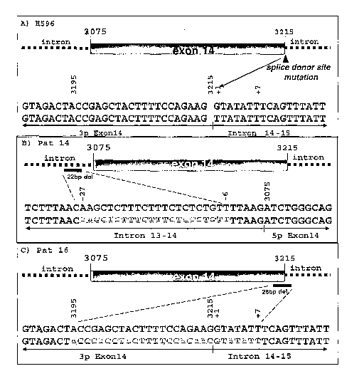
FIG. 1 depicts illustrative intronic mutations flanking exon 14 of Met. A
schematic
representation of Met exon 14 showing the corresponding nucleic acid
(NM_000245)
deletions and/or point mutations (light grey text) with respect to the
intron/exon structure.
(A) H596, lung cancer cell line. (B) pat. 14, patient 14 lung tumor specimen.
(C) pat. 16,
patient 16 lung tumor specimen. For reference, in tumor H596, there is a point
mutation from
G to T at position marked +1 in (A). In tumor Pat 14, there is a deletion of
the sequence from
position marked -27 to -6 in (B). In tumor Pat 16, there is a deletion of the
sequence from
position marked 3195 to +7 in (C).
FIG. 2. Delayed down regulation of hyperstabilized c-met is associated with
activation of
Met and MAPK. (A) 293 cells co-transfected with Met constructs and Cbl-flag
were
immunoprecipitated (IP) with V5 or Cbl antibodies followed by immunoblotting
(IB) with
V5, flag, or P-Tyr antibodies. Lysates were probed with flag or Cbl
antibodies. (B) 293 cells
were transfected with Met constructs followed by IP of endogenous Cbl.
Immunoblotting
with V5 antibody shows that Met WT, but not MetAEx14 co-IPs with endogenous
Cbl. The
membrane was stripped and reprobed with Y1003, YY1234/1235, Y1349, or Y1365
phospho-
specific antibodies. (C) Lysates from transient transfection of 293 cells were
immunoprecipitated with V5 antibody and immunoblotted with ubiquitin antibody
to detect
ubiquitinated Met. The membrane was stripped and reprobed with V5 antibody to
detect the
presence of Met. Lysates were probed with flag or actin antibodies to detect
Cbl-flag or actin
for equivalent expression. (D) 293 cells were transfected with the indicated
constructs and
treated with 10 [Lg/ml cycloheximide. Lysates were probed with V5 antibody or
actin. (E)
Serum-starved lung cancer cell lines were stimulated for 10 minutes with 50
ng/ml rhuHGF,
then rinsed and returned to serum-free media. Lysates were collected at the
indicated times
and immunoblotted for P-Met (Y1230/Y1234/Y1235), Met, P-MAPK, MAPK, P-Akt, or
Akt.
(F) Rat 1A stable clones were serum-starved and treated for 10 minutes with an
agonistic Met
monoclonal antibody 3D6 (5 g/ml), rinsed with PBS, and returned to serum-free
media. At
CA 02599988 2007-09-04
WO 2006/104911 PCT/US2006/010850
the indicated times, lysates were obtained and immunoblotted for P-MAPK, MAPK,
P-Akt,
or Akt.
FIG.3. Enhanced ligand-dependent proliferative potential in cell lines
harboring the Met
juxtamembrane deletion (A) HGF-stimulated growth in a panel of NSCLC cell
lines, was
determined after a 72 hour culture in the presence or absence of 50 ng/ml
rhuHGF. Results
are depicted as a stimulation index (SI), as determined from a minimum of
three separate
experiments. (B) Growth curves of subcutaneously inoculated Rat 1A stable cell
lines
expressing vector, Met WT, Met AEx14, in each case in the presence or absence
of an HGF
agonist antibody (3D6) in nude mice.
FIG. 4. Inhibition of ligand-dependent Met signaling and growth in H596 cells
with an anti-
Met mAb, OA-5D5. (A) Serum-starved H226 or H596 cells were incubated with OA-
5D5 for
30 minutes at the indicated concentrations and then stimulated witli 100 ng/ml
rhuHGF for 15
minutes. Lysates were obtained and immunoblotted for P-Met (Y1234/Y1235), Met,
P-Akt,
Akt, P-MAPK, or MAPK. (B) Cells were treated with OA-5D5 or a control Ig at
the indicated
concentrations in the presence or absence of 50 ng/ml rhuHGF and cell
viability was
determined after 72 hours.
FIG. 5. Quantification of phospho-kinase to kinase ratios in RatlA stable Met
cell lines. The
ratio of P-MAPK:MAPK (left) and P-Akt:Akt (right) was quantified using Odyssey
infrared
scanner that detects AlexaFluor680 and IR Dye800 conjugated secondary
antibodies.
FIG. 6. Quantification of phospho-kinase to kinase ratios in H596 and H226
cells treated with
OA-5D5. The ratio of P-Met:Met, P-Akt:Akt, or P-MAPK:MAPK for each cell line
was
quantified using Odyssey infrared scanner that detects AlexaFluor680 and
IRDye800
conjugated secondary antibodies.
FIG. 7 depicts illustrative cis-acting splicing elements expected to regulate
splicing of human
c-met exon 14. It is expected that a mutation at one or more positions within
these elements
would have a negative impact on wild type splicing of exon 14.
FIG. 8 depicts wild-type human c-met protein sequence based on RefSeq.
NM_000245.
16
CA 02599988 2007-09-04
WO 2006/104911 PCT/US2006/010850
FIG. 9 depicts light and heavy chain variable domain sequences for the OA-5D5
antibody
referred to in the Examples.
MODES FOR CARRYING OUT THE INVENTION
The invention provides methods, coinpositions, kits and articles of
manufacture for
identifying inhibitors of the HGF/c-rnet signaling pathway (in particular,
inhibitors of
hyperstabilized c-met), and methods of using such inhibitors.
Details of these methods, compositions, kits and articles of manufacture are
provided
herein.
General Techniques
The practice of the present invention will employ, unless otherwise indicated,
conventional
techniques of molecular biology (including recombinant techniques),
microbiology, cell biology,
biochemistry, and immunology, which are within the skill of the art. Such
techniques are
explained fully in the literature, such as, "Molecular Cloning: A Laboratory
Manual", second
edition (Sambrook et al., 1989); "Oligonucleotide Synthesis" (M. J. Gait, ed.,
1984); "Animal Cell
Culture" (R. I. Freshney, ed., 1987); "Methods in Enzymology" (Academic Press,
Inc.); "Current
Protocols in Molecular Biology" (F. M. Ausubel et al., eds., 1987, and
periodic updates); "PCR:
The Polymerase Chain Reaction", (Mullis et al., ed., 1994); "A Practical Guide
to Molecular
Cloning" (Perbal Bernard V., 1988).
Definitions
The term "hyperstabilized c-met", and variations thereof, as used herein,
refers to a
naturally-occuring mutant human c-met that is degraded/down-regulated at a
rate that is
detectably slower than that of a wild-type c-met. Methods of comparing
degration/down-
regulation rates between wild-type c-met and a hyperstabilized c-met would be
evident to one
skilled in the art, including, for example, as described in the Examples
below. In one
instance, delayed degradation/down-regulation is assessed based on
quantitating receptor
protein levels in a cell. In another instance, delayed degradation/down-
regulation is
determined based detection of a mutation in a c-met site that is associated
with Cbl binding to
c-met. In one instance, the mutation is in a c-met site that is associated
with c-met
ubiquitination (e.g., in c-met exon 14) and receptor protein degradation/down-
regulation.
These mutations can arise in any form that results in expression of a mutated
c-met protein
that is degraded/down-regulated at a slower rate than wild type c-met, wherein
the mutated c-
met protein is capable of wild-type c-met-associated activity (e.g.,
phosphorylating
downstream molecules such as MAPK, stimulating cell proliferation and/or
induction of
17
CA 02599988 2007-09-04
WO 2006/104911 PCT/US2006/010850
tumorigenic events). For example, these mutations include those that are
associated with
expression of a functional, in-frame c-met splice variant lacking at least a
portion of exon 14
that is associated with receptor protein degradation/down-regulation.
Illustrative examples of
mutations include those found in a splicing element as depicted in Figures 1
and 7. In one
embodiment, presence of a hyperstabilized c-met protein of the invention in a
cell is
associated with prolonged and/or increased phosphorylation of downstream
molecules in the
HGF/c-met pathway as compared with a similar amount of wild-type c-met protein
in a cell.
The term "vector," as used herein, is intended to refer to a nucleic acid
molecule capable of
transporting another nucleic acid to which it has been linked. One type of
vector is a"plasmid",
which refers to a circular double stranded DNA loop into which additional DNA
segments may be
ligated. Another type of vector is a phage vector. Another type of vector is a
viral vector, wherein
additional DNA segments may be ligated into the viral genome. Certain vectors
are capable of
autonomous replication in a host cell into which they are introduced (e.g.,
bacterial vectors having
a bacterial origin of replication and episomal mammalian vectors). Other
vectors (e.g., non-
episomal mammalian vectors) can be integrated into the genome of a host cell
upon introduction
into the host cell, and thereby are replicated along with the host genome.
Moreover, certain
vectors are capable of directing the expression of genes to which they are
operatively linked. Such
vectors are referred to herein as "recombinant expression vectors" (or simply,
"recombinant
vectors"). In general, expression vectors of utility in recombinant DNA
techniques are often in the
form of plasmids. In the present specification, "plasmid" and "vector" may be
used
interchangeably as the plasmid is the most commonly used form of vector.
"Polynucleotide," or "nucleic acid," as used interchangeably herein, refer to
polymers of
nucleotides of any length, and include DNA and RNA. The nucleotides can be
deoxyribonucleotides, ribonucleotides, modified nucleotides or bases, and/or
their analogs, or any
substrate that can be incorporated into a polymer by DNA or RNA polymerase, or
by a synthetic
reaction. A polynucleotide may comprise modified nucleotides, such as
methylated nucleotides
and their analogs. If present, modification to the nucleotide structure may be
imparted before or
after assembly of the polymer. The sequence of nucleotides may be interrupted
by non-nucleotide
components. A polynucleotide may be further modified after synthesis, such as
by conjugation
with a label. Other types of modifications include, for example, "caps",
substitution of one or more
of the naturally occurring nucleotides with an analog, intemucleotide
modifications such as, for
example, those with uncharged linkages (e.g., methyl phosphonates,
phosphotriesters,
phosphoamidates, carbamates, etc.) and with charged linkages (e.g.,
phosphorothioates,
phosphorodithioates, etc.), those containing pendant moieties, such as, for
example, proteins (e.g.,
nucleases, toxins, antibodies, signal peptides, ply-L-lysine, etc.), those
with intercalators (e.g.,
18
CA 02599988 2007-09-04
WO 2006/104911 PCT/US2006/010850
acridine, psoralen, etc.), those containing chelators (e.g., metals,
radioactive metals, boron,
oxidative metals, etc.), those containing alkylators, those with modified
linkages (e.g., alpha
anomeric nucleic acids, etc.), as well as unmodified forms of the
polynucleotide(s). Further, any of
the hydroxyl groups ordinarily present in the sugars may be replaced, for
example, by phosphonate
groups, phosphate groups, protected by standard protecting groups, or
activated to prepare
additional linkages to additional nucleotides, or may be conjugated to solid
or semi-solid supports.
The 5' and 3' terminal OH can be phosphorylated or substituted with amines or
organic capping
group moieties of from 1 to 20 carbon atoms. Other hydroxyls may also be
derivatized to standard
protecting groups. Polynucleotides can also contain analogous forms of ribose
or deoxyribose
sugars that are generally known in the art, including, for example, 2'-O-
methyl-, 2'-O-allyl, 2'-
fluoro- or 2'-azido-ribose, carbocyclic sugar analogs, .alpha.-anomeric
sugars, epimeric sugars such
as arabinose, xyloses or lyxoses, pyranose sugars, furanose sugars,
sedoheptuloses, acyclic analogs
and abasic nucleoside analogs such as methyl riboside. One or more
phosphodiester linkages may
be replaced by alternative linking groups. These alternative linking groups
include, but are not
limited to, embodiments wherein phosphate is replaced by P(O)S("thioate"),
P(S)S ("dithioate"),
"(O)NR<sub>2</sub> ("amidate"), P(O)R, P(O)OR', CO or CH<sub>2</sub> ("formacetal"), in
which each R or R'
is independently H or substituted or unsubstituted alkyl (1-20 C.) optionally
containing an ether (-
0-) linkage, aryl, alkenyl, cycloalkyl, cycloalkenyl or araldyl. Not all
linkages in a polynucleotide
need be identical. The preceding description applies to all polynucleotides
referred to herein,
including RNA and DNA.
"Oligonucleotide," as used herein, generally refers to short, generally single
stranded,
generally synthetic polynucleotides that are generally, but not necessarily,
less than about 200
nucleotides in length. The terms "oligonucleotide" and "polynucleotide" are
not mutually
exclusive. The description above for polynucleotides is equally and fully
applicable to
oligonucleotides.
The term "hepatocyte growth factor" or "HGF", as used herein, refers, unless
indicated
otherwise, to any native or variant (whether native or synthetic) HGF
polypeptide that is capable of
activating the HGF/c-met signaling pathway under conditions that permit such
process to occur.
The term "wild type HGF" generally refers to a polypeptide comprising the
amino acid sequence
of a naturally occurring HGF protein. Thet term "wild type HGF sequence"
generally refers to an
amino acid sequence found in a naturally occurring HGF. C-met (or Met) is a
known receptor for
HGF through which HGF intracellular signaling is biologically effectuated. A
wild type human c-
met protein sequence based on RefSeq NM_000245 is depicted in Fig. 8.
The terms "splice site", "splice junction", "branch point", "polypyrimidine
tract", as used
herein, refer to the meaning known in the art in the context of mammalian, in
particular human,
19
CA 02599988 2007-09-04
WO 2006/104911 PCT/US2006/010850
RNA splicing. See, e.g., Pagani & Baralle, Nature Reviews: Genetics (2004),
5:389-396, and
references cited therein. For convenient reference, one embodiment of
sequences for c-met RNA
splicing elements is illustratively set forth in Figure 7.
The term "host cell" (or "recombinant host cell"), as used herein, is intended
to refer
to a cell that has been genetically altered, or is capable of being
genetically altered by
introduction of an exogenous polynucleotide, such as a recombinant plasmid or
vector. It
should be understood that such terms are intended to refer not only to the
particular subject
cell but to the progeny of such a cell. Because certain modifications may
occur in succeeding
generations due to either mutation or environmental influences, such progeny
may not, in fact,
be identical to the parent cell, but are still included within the scope of
the term "host cell" as
used herein.
"Antibodies" (Abs) and "immunoglobulins" (Igs) are glycoproteins having the
same
structural characteristics. While antibodies exhibit binding specificity to a
specific antigen,
immunoglobulins include both antibodies and other antibody-like molecules
which generally
lack antigen specificity. Polypeptides of the latter kind are, for example,
produced at low
levels by the lymph system and at increased levels by myelomas.
The terms "antibody" and "immunoglobulin" are used interchangeably in the
broadest
sense and include monoclonal antibodies (e.g., full length or intact
monoclonal antibodies),
polyclonal antibodies, monovalent, multivalent antibodies, multispecific
antibodies (e.g.,
bispecific antibodies so long as they exhibit the desired biological activity)
and may also
include certain antibody fragments (as described in greater detail herein). An
antibody can be
chimeric, human, humanized and/or affinity matured.
"Antibody fragments" comprise only a portion of an intact antibody, wherein
the
portion preferably retains at least one, preferably most or all, of the
functions normally
associated with that portion when present in an intact antibody. In one
embodiment, an
antibody fragment comprises an antigen binding site of the intact antibody and
thus retains
the ability to bind antigen. In another embodiment, an antibody fragment, for
example one
that comprises the Fc region, retains at least one of the biological functions
normally
associated with the Fc region when present in an intact antibody, such as FcRn
binding,
antibody half life modulation, ADCC function and complement binding. In one
embodiment,
an antibody fragment is a monovalent antibody that has an in vivo half life
substantially
similar to an intact antibody. For example, such an antibody fragment may
comprise on
antigen binding arm linked to an Fc sequence capable of conferring in vivo
stability to the
fragment.
CA 02599988 2007-09-04
WO 2006/104911 PCT/US2006/010850
The term "hypervariable region", 'HVR", or "HV", when used herein refers to
the
regions of an antibody variable domain which are hypervariable in sequence
and/or form
structurally defined loops. The letters "HC" and "LC" preceding the term "HVR"
or "HV"
refers, respectively, to HVR or HV of a heavy chain and light chain.
Generally, antibodies
comprise six hypervariable regions; three in the VH (H1, H2, H3), and three in
the VL (Ll,
L2, L3). A number of hypervariable region delineations are in use and are
encompassed
herein. The Kabat Complementarity Determining Regions (CDRs) are based on
sequence
variability and are the most commonly used (Kabat et al., Sequences of
Pr=oteins of
Immunological Interest, 5th Ed. Public Health Service, National Institutes of
Health,
Bethesda, MD. (1991)). Chothia refers instead to the location of the
structural loops (Chothia
and Lesk J. Mol. Biol. 196:901-917 (1987)). The AbM hypervariable regions
represent a
compromise between the Kabat CDRs and Chothia structural loops, and are used
by Oxford
Molecular's AbM antibody modeling software. The "contact" hypervariable
regions are based
on an analysis of the available complex crystal structures. The residues from
each of these
hypervariable regions are noted below.
Loop Kabat AbM Chothia Contact
---- ----- --- ------- -------
Ll L24-L34 L24-L34 L26-L32 L30-L36
L2 L50-L56 L50-L56 L50-L52 L46-L55
L3 L89-L97 L89-L97 L91-L96 L89-L96
H1 H31-H35B H26-H35B H26-H32 H30-H35B
(Kabat Numbering)
Hl 1131-1135 H26-H35 H26-H32 H30-H35
(Chothia Numbering)
H2 H50-H65 H50-H58 H53-H55 H47-H58
H3 H95-H102 H95-H102 H96-H101 H93-H101
"Framework" or "FR" residues are those variable domain residues other than the
hypervariable region residues as herein defined.
The "variable region" or "variable domain" of an antibody refers to the amino-
terminal domains of heavy or light chain of the antibody. These domains are
generally the
most variable parts of an antibody and contain the antigen-binding sites.
The term "monoclonal antibody" as used herein refers to an antibody obtained
from a
population of substantially homogeneous antibodies, i.e., the individual
antibodies comprising
21
CA 02599988 2007-09-04
WO 2006/104911 PCT/US2006/010850
the population are identical except for possible naturally occurring mutations
that may be
present in minor amounts. Monoclonal antibodies are highly specific, being
directed against a
single antigen. Furthermore, in contrast to polyclonal antibody preparations
that typically
include different antibodies directed against different determinants
(epitopes), each
monoclonal antibody is directed against a single determinant on the antigen.
The monoclonal antibodies herein specifically include "chimeric" antibodies in
which
a portion of the heavy and/or light chain is identical with or homologous to
corresponding
sequences in antibodies derived from a particular species or belonging to a
particular antibody
class or subclass, while the remainder of the chain(s) is identical with or
homologous to
corresponding sequences in antibodies derived from another species or
belonging to another
antibody class or subclass, as well as fragments of such antibodies, so long
as they exhibit the
desired biological activity (U.S. Patent No. 4,816,567; and Morrison et al.,
Proc. Natl. Acad.
Sci. USA 81:6851-6855 (1984)).
"Humanized" forms of non-human (e.g., murine) antibodies are chimeric
antibodies
that contain niinimal sequence derived from non-human immunoglobulin. For the
most part,
humanized antibodies are human immunoglobulins (recipient antibody) in which
residues
from a hypervariable region of the recipient are replaced by residues from a
hypervariable
region of a non-human species (donor antibody) such as mouse, rat, rabbit or
nonhuman
primate having the desired specificity, affinity, and capacity. In some
instances, framework
region (FR) residues of the human immunoglobulin are replaced by corresponding
non-
human residues. Furthermore, humanized antibodies may comprise residues that
are not
found in the recipient antibody or in the donor antibody. These modifications
are made to
furtlier refine antibody performance. In general, the humanized antibody will
comprise
substantially all of at least one, and typically two, variable domains, in
which all or
substantially all of the hypervariable loops correspond to those of a non-
human
immunoglobulin and all or substantially all of the FRs are those of a human
immunoglobulin
sequence. The humanized antibody optionally will also comprise at least a
portion of an
immunoglobulin constant region (Fc), typically that of a human immunoglobulin.
For further
details, see Jones et al., Nature 321:522-525 (1986); Riechmann et al., Nature
332:323-329
(1988); and Presta, Curr. Op. Struct. Biol. 2:593-596 (1992). See also the
following review
articles and references cited therein: Vaswani and Hamilton, Anra. Allergy,
Astluna &
Ifranzunol. 1:105-115 (1998); Harris, Biochein. Soc. Transactions 23:1035-1038
(1995); Hurle
and Gross, Curr. Op. Biotech. 5:428-433 (1994).
A "human antibody" is one which possesses an amino acid sequence which
corresponds to that of an antibody produced by a human and/or has been made
using any of
22
CA 02599988 2007-09-04
WO 2006/104911 PCT/US2006/010850
the techniques for making human antibodies as disclosed herein. This
definition of a human
antibody specifically excludes a humanized antibody comprising non-human
antigen-binding
residues.
An "affinity matured" antibody is one with one or more alterations in one or
more
CDRs/HVRs thereof which result in an improvement in the affinity of the
antibody for
antigen, compared to a parent antibody which does not possess those
alteration(s). Preferred
affinity matured antibodies will have nanomolar or even picomolar affinities
for the target
antigen. Affinity matured antibodies are produced by procedures known in the
art. Marks et
al. Bio/Techziology 10:779-783 (1992) describes affinity maturation by VH and
VL domain
shuffling. Random mutagenesis of CDR/HVR and/or framework residues is
described by:
Barbas et al. Proc Nat. Acad. Sci, USA 91:3809-3813 (1994); Schier et al. Gene
169:147-155
(1995); Yelton et al. J. Ifninuizol. 155:1994-2004 (1995); Jackson et al., J.
Itnznunol.
154(7):3310-9 (1995); and Hawkins et al, J. Mol. Biol. 226:889-896 (1992).
The term "Fc region" is used to define the C-terminal region of an
immunoglobulin
heavy chain which may be generated by papain digestion of an intact antibody.
The Fc region
may be a native sequence Fc region or a variant Fc region. Although the
boundaries of the Fc
region of an immunoglobulin heavy chain might vary, the human IgG heavy chain
Fc region
is usually defined to stretch from an amino acid residue at about position
Cys226, or from
about position Pro230, to the carboxyl-terniinus of the Fc region. The Fc
region of an
immunoglobulin generally comprises two constant domains, a CH2 domain and a
CH3
domain, and optionally comprises a CH4 domain. By "Fc region chain" herein is
meant one
of the two polypeptide chains of an Fc region.
The term "cytotoxic agent" as used herein refers to a substance that inhibits
or
prevents the function of cells and/or causes destruction of cells. The term is
intended to
include radioactive isotopes (e.g. At211, I13111125, Y90, Re' 86, Rel$$,
Sm153, Bi212, P32 and
radioactive isotopes of Lu), chemotherapeutic agents, and toxins such as small
molecule
toxins or enzymatically active toxins of bacterial, fungal, plant or animal
origin, including
fragments and/or variants thereof.
A "chemotherapeutic agent" is a chemical compound useful in the treatment of
cancer. Examples of chemotherapeutic agents include alkylating agents such as
thiotepa and
CYTOXANO cyclosphosphamide; alkyl sulfonates such as busulfan, improsulfan and
piposulfan; aziridines such as benzodopa, carboquone, meturedopa, and uredopa;
ethylenimines and methylamelamines including altretamine, triethylenemelamine,
trietylenephosphoramide, triethiylenethiophosphoramide and
trimethylolomelamine;
acetogenins (especially bullatacin and bullatacinone); delta-9-
tetrahydrocannabinol
23
CA 02599988 2007-09-04
WO 2006/104911 PCT/US2006/010850
(dronabinol, MARINOLO); beta-lapachone; lapachol; colchicines; betulinic acid;
a
camptothecin (including the synthetic analogue topotecan (HYCAMTINO), CPT-1 1
(irinotecan, CAMPTOSAR ), acetylcamptothecin, scopolectin, and 9-
aniinocamptothecin);
bryostatin; callystatin; CC-1065 (including its adozelesin, carzelesin and
bizelesin synthetic
analogues); podophyllotoxin; podophyllinic acid; teniposide; cryptophycins
(particularly
cryptophycin 1 and cryptophycin 8); dolastatin; duocarmycin (including the
synthetic
analogues, KW-2189 and CBl-TM1); eleutherobin; pancratistatin; a sarcodictyin;
spongistatin; nitrogen mustards such as chlorambucil, chlornaphazine,
cholophosphamide,
estramustine, ifosfamide, mechlorethamine, mechlorethamine oxide
hydrochloride,
melphalan, novembichin, phenesterine, prednimustine, trofosfamide, uracil
mustard;
nitrosureas such as carmustine, chlorozotocin, fotemustine, lomustine,
nimustine, and
ranimnustine; antibiotics such as the enediyne antibiotics (e. g.,
calicheamicin, especially
calicheamicin gammalI and calicheamicin omegaIl (see, e.g., Agnew, Chem Intl.
Ed. Engl.,
33: 183-186 (1994)); dynemicin, including dynemicin A; an esperamicin; as well
as
neocarzinostatin chromophore and related chromoprotein enediyne antiobiotic
chromophores), aclacinomysins, actinomycin, authramycin, azaserine,
bleomycins,
cactinomycin, carabicin, carminomycin, carzinophilin, chromomycinis,
dactinomycin,
daunorubicin, detorubicin, 6-diazo-5-oxo-L-norleucine, doxorubicin (including
ADRIAMYCINO, morpholino-doxorubicin, cyanomorpholino-doxorubicin, 2-pyrrolino-
doxorubicin, doxorubicin HCl liposome injection (DOXII. ) and
deoxydoxorubicin),
epirubicin, esorubicin, idarubicin, marcellomycin, mitomycins such as
mitomycin C,
mycophenolic acid, nogalamycin, olivomycins, peplomycin, potfiromycin,
puromycin,
quelamycin, rodorubicin, streptonigrin, streptozocin, tubercidin, ubenimex,
zinostatin,
zorubicin; anti-metabolites such as methotrexate, gemcitabine (GEMZARO),
tegafur
(UFTORALO), capecitabine (XELODAO), an epothilone, and 5-fluorouracil (5-FU);
folic
acid analogues such as denopterin, methotrexate, pteropterin, trimetrexate;
purine analogs
such as fludarabine, 6-mercaptopurine, thiamiprine, thioguanine; pyrimidine
analogs such as
ancitabine, azacitidine, 6-azauridine, carmofur, cytarabine, dideoxyuridine,
doxifluridine,
enocitabine, floxuridine; androgens such as calusterone, dromostanolone
propionate,
epitiostanol, mepitiostane, testolactone; anti- adrenals such as
aminoglutethimide, mitotane,
trilostane; folic acid replenisher such as frolinic acid; aceglatone;
aldophosphamide glycoside;
aminolevulinic acid; eniluracil; amsacrine; bestrabucil; bisantrene;
edatraxate; defofamine;
demecolcine; diaziquone; elfomithine; elliptinium acetate; etoglucid; gallium
nitrate;
hydroxyurea; lentinan; lonidainine; maytansinoids such as maytansine and
ansamitocins;
mitoguazone; mitoxantrone; mopidanmol; nitraerine; pentostatin; phenamet;
pirarubicin;
24
CA 02599988 2007-09-04
WO 2006/104911 PCT/US2006/010850
losoxantrone; 2-ethylhydrazide; procarbazine; PSKO polysaccharide complex (JHS
Natural
Products, Eugene, OR); razoxane; rhizoxin; sizofiran; spirogermanium;
tenuazonic acid;
triaziquone; 2,2',2"-trichlorotriethylamine; trichothecenes (especially T-2
toxin, verracurin A,
roridin A and anguidine); urethan; vindesine (ELDISINEO, FILDESINO);
dacarbazine;
mannomustine; mitobronitol; mitolactol; pipobroman; gacytosine; arabinoside
("Ara-C");
thiotepa; taxoids, e.g., paclitaxel (TAXOLO), albumin-engineered nanoparticle
formulation
of paclitaxel (ABRAXANETM), and doxetaxel (TAXOTERE ); chloranbucil; 6-
thioguanine;
mercaptopurine; methotrexate; platinum analogs such as cisplatin and
carboplatin; vinblastine
(VELBANO); platinum; etoposide (VP-16); ifosfamide; mitoxantrone; vincristine
(ONCOVINO); oxaliplatin; leucovovin; vinorelbine (NAVELBINEO); novantrone;
edatrexate; daunomycin; aminopterin; ibandronate; topoisomerase inhibitor RFS
2000;
difluorometlhylornithine (DMFO); retinoids such as retinoic acid;
pharmaceutically
acceptable salts, acids or derivatives of any of the above; as well as
combinations of two or
more of the above such as CHOP, an abbreviation for a combined therapy of
cyclophosphamide, doxorubicin, vincristine, and prednisolone, and FOLFOX, an
abbreviation
for a treatment regimen with oxaliplatin (ELOXATINTM) combined with 5-FU and
leucovovin.
Also included in this definition are anti-hormonal agents that act to
regulate, reduce,
block, or inhibit the effects of hormones that can promote the growth of
cancer, and are often
in the form of systemic, or whole-body treatment. They may be hormones
themselves.
Examples include anti-estrogens and selective estrogen receptor modulators
(SERMs),
including, for example, tamoxifen (including NOLVADEXO tamoxifen), raloxifene
(EVISTAO), droloxifene, 4-hydroxytamoxifen, trioxifene, keoxifene, LY117018,
onapristone, and toremifene (FARESTONO); anti-progesterones; estrogen receptor
down-
regulators (ERDs); estrogen receptor antagonists such as fulvestrant
(FASLODEXO); agents
that function to suppress or shut down the ovaries, for example, leutinizing
hormone-releasing
hormone (LHRH) agonists such as leuprolide acetate (LUPRONO and ELIGARD ),
goserelin acetate, buserelin acetate and tripterelin; other anti-androgens
such as flutamide,
nilutamide and bicalutamide; and aromatase inhibitors that inhibit the enzyme
aromatase,
which regulates estrogen production in the adrenal glands, such as, for
example, 4(5)-
imidazoles, aminoglutethimide, megestrol acetate (MEGASEO), exemestane
(AROMASINO), formestanie, fadrozole, vorozole (RIVISORO), letrozole
(FEIVIARAO), and
anastrozole (ARIMIDEXO). In addition, such definition of chemotherapeutic
agents includes
bisphosphonates such as clodronate (for example, BONEFOSO or OSTACO),
etidronate
(DIDROCALO), NE-58095, zoledronic acid/zoledronate (ZOMETAO), alendronate
CA 02599988 2007-09-04
WO 2006/104911 PCT/US2006/010850
(FOSAMAXO), pamidronate (AREDIAO), tiludronate (SKELIDO), or risedronate
(ACTONELO); as well as troxacitabine (a 1,3-dioxolane nucleoside cytosine
analog);
antisense oligonucleotides, particularly those that inhibit expression of
genes in signaling
pathways implicated in abherant cell proliferation, such as, for example, PKC-
alpha, Raf, H-
Ras, and epidermal growth factor receptor (EGF-R); vaccines such as THERATOPEO
vaccine and gene therapy vaccines, for example, ALLOVECTINO vaccine,
LEUVECTINO
vaccine, and VAXIDO vaccine; topoisomerase 1 inhibitor (e.g., LURTOTECANO);
rmRH
(e.g., ABARELIX ); lapatinib ditosylate (an ErbB-2 and EGFR dual tyrosine
kinase small-
molecule inhibitor also known as GW572016); COX-2 inhibitors such as celecoxib
(CELEBREXO; 4-(5-(4-methylphenyl)-3-(trifluoromethyl)-1H-pyrazol-1-yl)
benzenesulfonamide; and pharmaceutically acceptable salts, acids or
derivatives of any of the
above.
A "blocking" antibody or an "antagonist" antibody is one which inhibits or
reduces
biological activity of the antigen it binds. Such blocking can occur by any
means, e.g. by
interfering with protein-protein interaction such as ligand binding to a
receptor. In on
embodiment, blocking antibodies or antagonist antibodies substantially or
completely inhibit
the biological activity of the antigen.
The terms "cancer" and "cancerous" refer to or describe the physiological
condition in
mammals that is typically characterized by unregulated cell
growth/proliferation. Examples
of cancer include but are not limited to, carcinoma, lymphoma (e.g., Hodgkin's
and non-
Hodgkin's lymphoma), blastoma, sarcoma, and leukemia. More particular examples
of such
cancers include squamous cell cancer, small-cell lung cancer, non-small cell
lung cancer,
adenocarcinoma of the lung, squamous carcinoma of the lung, cancer of the
peritoneum,
hepatocellular cancer, gastrointestinal cancer, pancreatic cancer,
glioblastoma, cervical
cancer, ovarian cancer, liver cancer, bladder cancer, hepatoma, breast cancer,
colon cancer,
colorectal cancer, endometrial or uterine carcinoma, salivary gland carcinoma,
kidney cancer,
liver cancer, prostate cancer, vulval cancer, thyroid cancer, hepatic
carcinoma and various
types of head and neck cancer.
As used herein, "treatment" refers to clinical intervention in an attempt to
alter the
natural course of the individual or cell being treated, and can be performed
either for
prophylaxis or during the course of clinical pathology. Desirable effects of
treatment include
preventing occurrence or recurrence of disease, alleviation of symptoms,
diminishment of any
direct or indirect pathological consequences of the disease, preventing
metastasis, decreasing
the rate of disease progression, amelioration or palliation of the disease
state, and remission or
26
CA 02599988 2007-09-04
WO 2006/104911 PCT/US2006/010850
improved prognosis. In some embodiments, modulator molecules and methods of
the
invention are used to delay development of a disease or disorder.
An "effective amount" refers to an amount effective, at dosages and for
periods of time
necessary, to achieve the desired therapeutic or prophylactic result. A
"therapeutically effective
amount" of a therapeutic agent may vary according to factors such as the
disease state, age, sex,
and weight of the individual, and the ability of the antibody to elicit a
desired response in the
individual. A therapeutically effective amount is also one in which any toxic
or detrimental effects
of the therapeutic agent are outweighed by the tlierapeutically beneficial
effects. A
"prophylactically effective amount" refers to an amount effective, at dosages
and for periods of
time necessary, to achieve the desired prophylactic result. Typically but not
necessarily, since a
prophylactic dose is used in subjects prior to or at an earlier stage of
disease, the prophylactically
effective amount will be less than the therapeutically effective amount.
Compositions and Methods of the Invention
A. C-met antagonist Antibodies
In one embodiment, the invention provides C-met antagonist antibodies which
may
find use herein as therapeutic and/or diagnostic agents. Exemplary antibodies
include
polyclonal, monoclonal, humanized, bispecific, and heteroconjugate antibodies.
Aspects of
generating, identifying, characterizing, modifying and producing antibodies
are well
established in the art, e.g., as described in US Pat. Appl. Pub. No.
2005/0042216 from
paragraphs 522 through 563, 604 through 608, and 617 through 688.
The C-met antagonist antibodies disclosed herein can be formulated in any
suitable
form for delivery to a target cell/tissue. For example, the antibodies may be
formulated as
immunoliposomes. A "liposome" is a small vesicle composed of various types of
lipids,
phospholipids and/or surfactant which is useful for delivery of a drug to a
mammal. The
components of the liposome are commonly arranged in a bilayer formation,
similar to the
lipid arrangement of biological membranes. Liposomes containing the antibody
are prepared
by methods known in the art, such as described in Epstein et al., Proc. Natl.
Acad. Sci. USA
82:3688 (1985); Hwang et al., Proc. Natl Acad. Sci. USA 77:4030 (1980); U.S.
Pat. Nos.
4,485,045 and 4,544,545; and W097/38731 published October 23, 1997. Liposomes
with
enhanced circulation time are disclosed in U.S. Patent No. 5,013,556.
Particularly useful liposomes can be generated by the reverse phase
evaporation
inethod with a lipid composition comprising phosphatidylcholine, cholesterol
and PEG-
derivatized phosphatidylethanolamine (PEG-PE). Liposomes are extruded through
filters of
defined pore size to yield liposomes with the desired diameter. Fab' fragments
of the
27
CA 02599988 2007-09-04
WO 2006/104911 PCT/US2006/010850
antibody of the present invention can be conjugated to the liposomes as
described in Martin et
al., J. Biol. Chem. 257:286-288 (1982) via a disulfide interchange reaction. A
chemotherapeutic agent is optionally contained within the liposome. See
Gabizon et al., J.
National Cancer Inst. 81(19):1484 (1989).
B. C-met antagonist Polypeptides
In one aspect, a C-met antagonist of the invention comprises a polypeptide. In
one
embodiment, the antagonist polypeptide binds to and/or antagonizes
hyperstabilized c-met
protein in a cell. In one embodiment, the polypeptides bind, preferably
specifically, to
hyperstabilized c-met. The polypeptides may be chemically synthesized using
known peptide
synthesis methodology or may be prepared and purified using recombinant
technology. In
one embodiment, a C-met antagonist polypeptide is at least about 5 amino acids
in lengtli,
alternatively at least about 6, 7, 8, 9, 10, 11, 12, 13, 14, 15, 16, 17, 18,
19, 20, 21, 22, 23, 24,
25, 26, 27, 28, 29, 30, 31, 32, 33, 34, 35, 36, 37, 38, 39, 40, 41, 42, 43,
44, 45, 46, 47, 48, 49,
50, 51, 52, 53, 54, 55, 56, 57, 58, 59, 60, 61, 62, 63, 64, 65, 66, 67, 68,
69, 70, 71, 72, 73, 74,
75, 76, 77, 78, 79, 80, 81, 82, 83, 84, 85, 86, 87, 88, 89, 90, 91, 92, 93,
94, 95, 96, 97, 98, 99,
or 100 amino acids in length or more, wherein such polypeptides are capable of
inhibiting
hyperstabilized c-met activity. These polypeptides may be identified without
undue
experimentation using well known techniques. In this regard, it is noted that
techniques for
screening oligopeptide libraries for oligopeptides that are capable of
specifically binding to a
polypeptide target are well known in the art (see, e.g., U.S. Patent Nos.
5,556,762, 5,750,373,
4,708,871, 4,833,092, 5,223,409, 5,403,484, 5,571,689, 5,663,143; PCT
Publication Nos. WO
84/03506 and W084/03564; Geysen et al., Proc. Natl. Acad. Sci. U.S.A., 81:3998-
4002
(1984); Geysen et al., Proc. Natl. Acad. Sci. U.S.A., 82:178-182 (1985);
Geysen et al., in
Synthetic Peptides as Antigens, 130-149 (1986); Geysen et al., J. Immunol.
Meth., 102:259-
274 (1987); Schoofs et al., J. Immunol., 140:611-616 (1988), Cwirla, S. E. et
al. (1990) Proc.
Natl. Acad. Sci. USA, 87:6378; Lowman, H.B. et al. (1991) Biochemistry,
30:10832;
Clackson, T. et al. (1991) Nature, 352: 624; Marks, J. D. et al. (1991), J.
Mol. Biol., 222:581;
Kang, A.S. et al. (1991) Proc. Natl. Acad. Sci. USA, 88:8363, and Smith, G. P.
(1991)
Current Opin. Biotechnol., 2:668).
Bacteriophage (phage) display is one well known technique which allows one to
screen large oligopeptide libraries to identify member(s) of those libraries
which are capable
of specifically binding to a polypeptide target. Phage display is a technique
by which variant
polypeptides are displayed as fusion proteins to the coat protein on the
surface of
bacteriophage particles (Scott, J.K. and Smith, G. P. (1990) Science, 249:
386). The utility of
phage display lies in the fact that large libraries of selectively randomized
protein variants (or
28
CA 02599988 2007-09-04
WO 2006/104911 PCT/US2006/010850
randomly cloned cDNAs) can be rapidly and efficiently sorted for those
sequences that bind
to a target molecule with high affinity. Display of peptide (Cwirla, S. E. et
al. (1990) Proc.
Natl. Acad. Sci. USA, 87:6378) or protein (Lowman, H.B. et al. (1991)
Biochemistry,
30:10832; Clackson, T. et al. (1991) Nature, 352: 624; Marks, J. D. et al.
(1991), J. Mol.
Biol., 222:581; Kang, A.S. et al. (1991) Proc. Natl. Acad. Sci. USA, 88:8363)
libraries on
phage have been used for screening millions of polypeptides or oligopeptides
for ones with
specific binding properties (Smith, G. P. (1991) Current Opin. Biotechnol.,
2:668). Sorting
phage libraries of random mutants requires a strategy for constructing and
propagating a large
number of variants, a procedure for affinity purification using the target
receptor, and a means
of evaluating the results of binding enrichments. U.S. Patent Nos. 5,223,409,
5,403,484,
5,571,689, and 5,663,143.
Although most phage display methods have used filamentous phage, lambdoid
phage
display systems (WO 95/34683; U.S. 5,627,024), T4 phage display systems (Ren
et al., Gene,
215: 439 (1998); Zhu et al., Cancer Research, 58(15): 3209-3214 (1998); Jiang
et al.,
Infection & Immunity, 65(11): 4770-4777 (1997); Ren et al., Gene, 195(2):303-
311 (1997);
Ren, Protein Sci., 5: 1833 (1996); Efimov et al., Virus Genes, 10: 173 (1995))
and T7 phage
display systems (Smith and Scott, Methods in Enzymology, 217: 228-257 (1993);
U.S.
5,766,905) are also known.
Many other improvements and variations of the basic phage display concept have
now been developed. These improvements enhance the ability of display systems
to screen
peptide libraries for binding to selected target molecules and to display
functional proteins
with the potential of screening these proteins for desired properties.
Combinatorial reaction devices for phage display reactions have been developed
(WO
98/14277) and phage display libraries have been used to analyze and control
bimolecular
interactions (WO 98/20169; WO 98/20159) and properties of constrained helical
peptides
(WO 98/20036). WO 97/35196 describes a method of isolating an affinity ligand
in which a
phage display library is contacted with one solution in which the ligand will
bind to a target
molecule and a second solution in which the affinity ligand will not bind to
the target
molecule, to selectively isolate binding ligands. WO 97/46251 describes a
method of
biopanning a random phage display library with an affinity purified antibody
and then
isolating binding phage, followed by a micropanning process using microplate
wells to isolate
high affinity binding phage. The use of Staphlylococcus aureus protein A as an
affinity tag
has also been reported (Li et al. (1998) Mol Biotech., 9:187). WO 97/47314
describes the use
of substrate subtraction libraries to distinguish enzyme specificities using a
combinatorial
library which may be a phage display library. A method for selecting enzymes
suitable for
29
CA 02599988 2007-09-04
WO 2006/104911 PCT/US2006/010850
use in detergents using phage display is described in WO 97/09446. Additional
methods of
selecting specific binding proteins are described in U.S. Patent Nos.
5,498,538, 5,432,018,
and WO 98/15833.
Methods of generating peptide libraries and screening these libraries are also
disclosed in U.S. Patent Nos. 5,723,286, 5,432,018, 5,580,717, 5,427,908,
5,498,530,
5,770,434, 5,734,018, 5,698,426, 5,763,192, and 5,723,323.
Methods of generating, identifying, characterizing, modifying and producing
antagonist polypeptides are well established in the art, e.g., as described in
US Pat. Appl. Pub.
No. 2005/0042216 from paragraphs 606 through 608, 614 through 688.
In one embodiment, polypeptides for antagonizing hyperstabilized c-met
activity can
be designed based on hyperstabilized c-met protein structure, e.g. by
screening based on a
target antigen comprising a mutant c-met juxtamembrane sequence comprising
deletion of at
least a portion of exon 14 as described herein. For example, a target antigen
can comprise a
polypeptide comprising a sequence resulting from in-frame splicing of exon 13
and 15 of c-
met.
C. Iminunoconjugates
In another aspect, the invention provides immunoconjugates, or antibody-drug
conjugates (ADC), comprising an antibody conjugated to a cytotoxic agent such
as a
chemotherapeutic agent, a drug, a growth inhibitory agent, a toxin (e.g., an
enzymatically
active toxin of bacterial, fungal, plant, or animal origin, or fragments
thereof), or a radioactive
isotope (i.e., a radioconjugate).
The use of antibody-drug conjugates for the local delivery of cytotoxic or
cytostatic
agents, i.e. drugs to kill or inhibit tumor cells in the treatment of cancer
(Syrigos and Epenetos
(1999) Anticancer Research 19:605-614; Niculescu-Duvaz and Springer (1997)
Adv. Drg
Del. Rev. 26:151-172; U.S. patent 4,975,278) allows targeted delivery of the
drug moiety to
tumors, and intracellular accumulation therein, where systemic administration
of these
unconjugated drug agents may result in unacceptable levels of toxicity to
normal cells as well
as the tumor cells sought to be eliminated (Baldwin et al., (1986) Lancet pp.
(Mar. 15,
1986):603-05; Thorpe, (1985) "Aiitibody Carriers Of Cytotoxic Agents In Cancer
Therapy: A
Review," in Monoclonal Antibodies '84: Biological And Clinical Applications,
A. Pinchera et
al. (ed.s), pp. 475-506). Maximal efficacy with minimal toxicity is sought
thereby. Both
polyclonal antibodies and monoclonal antibodies have been reported as useful
in these
strategies (Rowland et al., (1986) Cancer Immunol. Inununother., 21:183-87).
Drugs used in
these methods include daunomycin, doxorubicin, methotrexate, and vindesine
(Rowland et
al., (1986) supra). Toxins used in antibody-toxin conjugates include bacterial
toxins such as
CA 02599988 2007-09-04
WO 2006/104911 PCT/US2006/010850
diphtheria toxin, plant toxins such as ricin, small molecule toxins such as
geldanamycin
(Mandler et al (2000) Jour. of the Nat. Cancer Inst. 92(19):1573-1581; Mandler
et al (2000)
Bioorganic & Med. Chem. Letters 10:1025-1028; Mandler et al (2002)
Bioconjugate Chem.
13:786-791), maytansinoids (EP 1391213; Liu et al., (1996) Proc. Natl. Acad.
Sci. USA
93:8618-8623), and calicheamicin (Lode et al (1998) Cancer Res. 58:2928;
Hinman et al
(1993) Cancer Res. 53:3336-3342). The toxins may effect their cytotoxic and
cytostatic
effects by mechanisms including tubulin binding, DNA binding, or topoisomerase
inhibition.
Some cytotoxic drugs tend to be inactive or less active when conjugated to
large antibodies or
protein receptor ligands.
ZEVALINO (ibritumomab tiuxetan, Biogen/Idec) is an antibody-radioisotope
conjugate composed of a murine IgGl kappa monoclonal antibody directed against
the CD20
antigen found on the surface of normal and malignant B lymphocytes and "rIn or
90Y
radioisotope bound by a thiourea linker-chelator (Wiseman et al (2000) Eur.
Jour. Nucl. Med.
27(7):766-77; Wiseman et al (2002) Blood 99(12):4336-42; Witzig et al (2002)
J. Clin.
Oncol. 20(10):2453-63; Witzig et al (2002) J. Clin. Oncol. 20(15):3262-69).
Although
ZEVALIN has activity against B-cell non-Hodgkin's Lymphoma (NHL),
administration
results in severe and prolonged cytopenias in most patients. MYLOTARGTM
(gemtuzumab
ozogamicin, Wyeth Pharmaceuticals), an antibody drug conjugate composed of a
hu CD33
antibody linked to calicheamicin, was approved in 2000 for the treatment of
acute myeloid
leukemia by injection (Drugs of the Future (2000) 25(7):686; US Patent Nos.
4970198;
5079233; 5585089; 5606040; 5693762; 5739116; 5767285; 5773001). Cantuzumab
mertansine (Immunogen, Inc.), an antibody drug conjugate composed of the
huC242 antibody
linked via the disulfide linker SPP to the maytansinoid drug moiety, DM1, is
tested for the
treatment of cancers that express CanAg, such as colon, pancreatic, gastric,
and others.
MLN-2704 (Millennium Pharm., BZL Biologics, Immunogen Inc.), an antibody drug
conjugate composed of the anti-prostate specific membrane antigen (PSMA)
monoclonal
antibody linked to the maytansinoid drug moiety, DM1, is tested for the
potential treatment of
prostate tumors. The auristatin peptides, auristatin E(AE) and
monomethylauristatin
(MMAE), synthetic analogs of dolastatin, were conjugated to chimeric
monoclonal antibodies
cBR96 (specific to Lewis Y on carcinomas) and cAC10 (specific to CD30 on
hematological
malignancies) (Doronina et al (2003) Nature Biotechnology 21(7):778-784) and
are under
therapeutic development.
Chemotherapeutic agents useful in the generation of immunoconjugates are
described
herein (above). Enzymatically active toxins and fragments thereof that can be
used include
diphtheria A chain, nonbinding active fragments of diphtheria toxin, exotoxin
A chain (from
31
CA 02599988 2007-09-04
WO 2006/104911 PCT/US2006/010850
Pseudomonas aeruginosa), ricin A chain, abrin A chain, modeccin A chain, alpha-
sarcin,
Aleurites fordii proteins, dianthin proteins, Phytolaca americana proteins
(PAPI, PAPII, and
PAP-S), momordica charantia inhibitor, curcin, crotin, sapaonaria officinalis
inhibitor,
gelonin, mitogellin, restrictocin, phenomycin, enomycin, and the
tricothecenes. See, e.g., WO
93/21232 published October 28, 1993. A variety of radionuclides are available
for the
production of radioconjugated antibodies. Examples include 212Bi, 131I,131In,
90Y, and 186Re.
Conjugates of the antibody and cytotoxic agent are made using a variety of
bifunctional
protein-coupling agents such as N-succinimidyl-3-(2-pyridyldithiol) propionate
(SPDP),
iminothiolane (IT), bifunctional derivatives of imidoesters (such as dimethyl
adipimidate
HCl), active esters (such as disuccinimidyl suberate), aldehydes (such as
glutaraldehyde), bis-
azido compounds (such as bis (p-azidobenzoyl) hexanediamine), bis-diazonium
derivatives
(such as bis-(p-diazoniumbenzoyl)-ethylenediamine), diisocyanates (such as
toluene 2,6-
diisocyanate), and bis-active fluorine compounds (such as 1,5-difluoro-2,4-
dinitrobenzene).
For example, a ricin immunotoxin can be prepared as described in Vitetta et
al., Science, 238:
1098 (1987). Carbon-14-labeled 1-isothiocyanatobenzyl-3-methyldiethylene
triaminepentaacetic acid (MX-DTPA) is an exemplary chelating agent for
conjugation of
radionucleotide to the antibody. See W094/11026.
Conjugates of an antibody and one or more small molecule toxins, such as a
calicheamicin, maytansinoids, dolostatins, aurostatins, a trichothecene, and
CC1065, and the
derivatives of these toxins that have toxin activity, are also contemplated
herein.
Maytansine and maytansinoids
In some embodiments, the immunoconjugate comprises an antibody of the
invention
conjugated to one or more maytansinoid molecules.
Maytansinoids are mitototic inhibitors which act by inhibiting tubulin
polymerization.
Maytansine was first isolated from the east African shrub Maytenus serrata
(U.S. Patent No.
3,896,111). Subsequently, it was discovered that certain microbes also produce
maytansinoids, such as maytansinol and C-3 maytansinol esters (U.S. Patent No.
4,151,042).
Synthetic maytansinol and derivatives and analogues thereof are disclosed, for
example, in
U.S. Patent Nos. 4,137,230; 4,248,870; 4,256,746; 4,260,608; 4,265,814;
4,294,757;
4,307,016; 4,308,268; 4,308,269; 4,309,428; 4,313,946; 4,315,929; 4,317,821;
4,322,348;
4,331,598; 4,361,650; 4,364,866; 4,424,219; 4,450,254; 4,362,663; and
4,371,533.
Maytansinoid drug moieties are attractive drug moieties in antibody drug
conjugates
because they are: (i) relatively accessible to prepare by fermentation or
chemical
modification, derivatization of fermentation products, (ii) amenable to
derivatization with
32
CA 02599988 2007-09-04
WO 2006/104911 PCT/US2006/010850
functional groups suitable for conjugation through the non-disulfide linkers
to antibodies, (iii)
stable in plasma, and (iv) effective against a variety of tumor cell lines.
Exemplary embodiments of maytansinoid drug moieities include: DMl; DM3; and
DM4. Imniunoconjugates containing maytansinoids, methods of making same, and
their
therapeutic use are disclosed, for example, in U.S. Patent Nos. 5,208,020,
5,416,064 and
European Patent EP 0 425 235 B 1, the disclosures of which are hereby
expressly incorporated
by reference. Liu et al., Proc. Natl. Acad. Sci. USA 93:8618-8623 (1996)
described
immunoconjugates comprising a maytansinoid designated DMl linked to the
monoclonal
antibody C242 directed against human colorectal cancer. The conjugate was
found to be
highly cytotoxic towards cultured colon cancer cells, and showed antitumor
activity in an in
vivo tumor growth assay. Chari et al., Cancer Research 52:127-131 (1992)
describe
immunoconjugates in which a maytansinoid was conjugated via a disulfide linker
to the
murine antibody A7 binding to an antigen on human colon cancer cell lines, or
to another
murine monoclonal antibody TA. 1 that binds the HER-2/neu oncogene. The
cytotoxicity of
the TA.l-maytansonoid conjugate was tested in vitro on the human breast cancer
cell line SK-
BR-3, which expresses 3 x 10⁵ HER-2 surface antigens per cell. The drug
conjugate achieved
a degree of cytotoxicity similar to the free maytansinoid drug, which could be
increased by
increasing the number of maytansinoid molecules per antibody molecule. The A7-
maytansinoid conjugate showed low systemic cytotoxicity in mice.
Antibody-maytansinoid conjugates can be prepared by chemically linking an
antibody to a maytansinoid molecule without significantly diminishing the
biological activity
of either the antibody or the maytansinoid molecule. See, e.g., U.S. Patent
No. 5,208,020 (the
disclosure of which is hereby expressly incorporated by reference). An average
of 3-4
maytansinoid molecules conjugated per antibody molecule has shown efficacy in
enhancing
cytotoxicity of target cells without negatively affecting the function or
solubility of the
antibody, although even one molecule of toxin/antibody would be expected to
enhance
cytotoxicity over the use of naked antibody. Maytansinoids are well known in
the art and can
be synthesized by known techniques or isolated from natural sources. Suitable
maytansinoids
are disclosed, for example, in U.S. Patent No. 5,208,020 and in the other
patents and
nonpatent publications referred to hereinabove. Preferred maytansinoids are
maytansinol and
maytansinol analogues modified in the aromatic ring or at other positions of
the maytansinol
molecule, such as various maytansinol esters.
There are many linking groups known in the art for making antibody-
maytansinoid
conjugates, including, for example, those disclosed in U.S. Patent No.
5,208,020 or EP Patent
0 425 235 B 1, Chari et al., Cancer Research 52:127-131 (1992), and U.S.
Patent Application
33
CA 02599988 2007-09-04
WO 2006/104911 PCT/US2006/010850
No. 10/960,602, filed Oct. 8, 2004, the disclosures of which are hereby
expressly incorporated
by reference. Antibody-maytansinoid conjugates comprising the linker component
SMCC
may be prepared as disclosed in U.S. Patent Application No. 10/960,602, filed
Oct. 8, 2004.
The linking groups include disulfide groups, thioether groups, acid labile
groups, photolabile
groups, peptidase labile groups, or esterase labile groups, as disclosed in
the above-identified
patents, disulfide and thioether groups being preferred. Additional linking
groups are
described and exemplified herein.
Conjugates of the antibody and maytansinoid may be made using a variety of
bifunctional protein coupling agents such as N-succinimidyl-3-(2-
pyridyldithio) propionate
(SPDP), succinimidyl-4-(N-maleimidomethyl) cyclohexane-l-carboxylate (SMCC),
iminothiolane (IT), bifunctional derivatives of imidoesters (such as dimethyl
adipimidate
HCl), active esters (such as disuccinimidyl suberate), aldehydes (such as
glutaraldehyde), bis-
azido compounds (such as bis (p-azidobenzoyl) hexanediamine), bis-diazonium
derivatives
(such as bis-(p-diazoniumbenzoyl)-ethylenediamine), diisocyanates (such as
toluene 2,6-
diisocyanate), and bis-active fluorine compounds (such as 1,5-difluoro-2,4-
dinitrobenzene).
Particularly preferred coupling agents include N-succinimidyl-3-(2-
pyridyldithio) propionate
(SPDP) (Carlsson et al., Biochem. J. 173:723-737 (1978)) and N-succinimidyl-4-
(2-
pyridylthio)pentanoate (SPP) to provide for a disulfide linkage.
The linker may be attached to the maytansinoid molecule at various positions,
depending on the type of the link. For example, an ester linkage may be formed
by reaction
with a hydroxyl group using conventional coupling techniques. The reaction may
occur at the
C-3 position having a hydroxyl group, the C-14 position modified with
hydroxymethyl, the C-
15 position modified with a hydroxyl group, and the C-20 position having a
hydroxyl group.
In a preferred embodiment, the linkage is formed at the C-3 position of
maytansinol or a
maytansinol analogue.
Auristatins and dolostatins
In some embodiments, the immunoconjugate comprises an antibody of the
invention
conjugated to dolastatins or dolostatin peptidic analogs and derivatives, the
auristatins (US
Patent Nos. 5635483; 5780588). Dolastatins and auristatins have been shown to
interfere
with microtubule dynaniics, GTP hydrolysis, and nuclear and cellular division
(Woyke et al
(2001) Antimicrob. Agents and Chemother. 45(12):3580-3584) and have anticancer
(US
5663149) and antifungal activity (Pettit et al (1998) Antimicrob. Agents
Chemother. 42:2961-
2965). The dolastatin or auristatin drug moiety may be attached to the
antibody through the N
(amino) terminus or the C (carboxyl) terminus of the peptidic drug moiety (WO
02/088172).
34
CA 02599988 2007-09-04
WO 2006/104911 PCT/US2006/010850
Exemplary auristatin embodiments include the N-terminus linked
monomethylauristatin drug moieties DE and DF, disclosed in "Monomethylvaline
Compounds Capable of Conjugation to Ligands", US Ser. No. 10/983,340, filed
Nov. 5, 2004,
the disclosure of which is expressly incorporated by reference in its
entirety.
An exemplary auristatin embodiments are MMAE and MMAF. Additional
exemplary embodiments comprising MMAE or MMAF and various linker components
(described further herein) Ab-MC-vc-PAB-MMAF, Ab-MC-vc-PAB-MMAE, Ab-MC-
MMAE and Ab-MC-MMAF.
Typically, peptide-based drug moieties can be prepared by forming a peptide
bond
between two or more amino acids and/or peptide fragments. Such peptide bonds
can be
prepared, for example, according to the liquid phase synthesis method (see E.
Schroder and K.
Lubke, "The Peptides", volume 1, pp 76-136, 1965, Academic Press) that is well
known in
the field of peptide cheniistry. The auristatin/dolastatin drug moieties may
be prepared
according to the methods of: US 5635483; US 5780588; Pettit et al (1989) J.
Am. Chem. Soc.
111:5463-5465; Pettit et al (1998) Anti-Cancer Drug Design 13:243-277; Pettit,
G.R., et al.
Synthesis, 1996, 719-725; and Pettit et al (1996) J. Chem. Soc. Perkin Trans.
1 5:859-863.
See also Doronina (2003) Nat Biotechnol 21(7):778-784; "Monomethylvaline
Compounds
Capable of Conjugation to Ligands", US Ser. No. 10/983,340, filed Nov. 5,
2004, hereby
incorporated by reference in its entirety (disclosing, e.g., linkers and
methods of preparing
monomethylvaline compounds such as MMAE and MMAF conjugated to linkers).
Calicheamicin
In other embodiments, the immunoconjugate comprises an antibody of the
invention
conjugated to one or more calicheamicin molecules. The calicheamicin family of
antibiotics
are capable of producing double-stranded DNA breaks at sub-picomolar
concentrations. For
the preparation of conjugates of the calicheamicin family, see U.S. patents
5,712,374,
5,714,586, 5,739,116, 5,767,285, 5,770,701, 5,770,710, 5,773,001, 5,877,296
(all to
American Cyanamid Company). Structural analogues of calicheamicin which may be
used
include, but are not limited to, yli, azI, a3I, N-acetyl-ylj, PSAG and 011
(Hinman et al., Cancer
Research 53:3336-3342 (1993), Lode et al., Cancer Research 58:2925-2928 (1998)
and the
aforementioned U.S. patents to American Cyanamid). Another anti-tumor drug
that the
antibody can be conjugated is QFA which is an antifolate. Both calicheamicin
and QFA have
intracellular sites of action and do not readily cross the plasma membrane.
Therefore, cellular
uptake of these agents through antibody mediated internalization greatly
enhances their
cytotoxic effects.
CA 02599988 2007-09-04
WO 2006/104911 PCT/US2006/010850
Other cytotoxic agents
Other antitumor agents that can be conjugated to the antibodies of the
invention
include BCNU, streptozoicin, vincristine and 5-fluorouracil, the family of
agents known
collectively LL-E33288 complex described in U.S. patents 5,053,394, 5,770,710,
as well as
esperamicins (U.S. patent 5,877,296).
Enzymatically active toxins and fragments thereof which can be used include
diphtheria A chain, nonbinding active fragments of diphtheria toxin, exotoxin
A chain (from
Pseudomonas aeruginosa), ricin A chain, abrin A chain, modeccin A chain, alpha-
sarcin,
Aleurites fordii proteins, dianthin proteins, Phytolaca americana proteins
(PAPI, PAPII, and
PAP-S), momordica charantia inhibitor, curcin, crotin, sapaonaria officinalis
inhibitor,
gelonin, mitogellin, restrictocin, phenomycin, enomycin and the tricothecenes.
See, for
example, WO 93/21232 published October 28, 1993.
The present invention further contemplates an immunoconjugate formed between
an
antibody and a compound with nucleolytic activity (e.g., a ribonuclease or a
DNA
endonuclease such as a deoxyribonuclease; DNase).
For selective destruction of the tumor, the antibody may comprise a highly
radioactive atom. A variety of radioactive isotopes are available for the
production of
radioconjugated antibodies. Examples include At211, I131, I125, y90, Re186,
Re188, Sm15321232212
Bi , P , Pb and radioactive isotopes of Lu. When the conjugate is used for
detection, it
may comprise a radioactive atom for scintigraphic studies, for example tc99m
or I123, or a spin
label for nuclear magnetic resonance (NMR) imaging (also known as magnetic
resonance
imaging, mri), such as iodine-123 again, iodine-131, indium-111, fluorine-19,
carbon-13,
nitrogen-15, oxygen-17, gadolinium, manganese or iron.
The radio- or other labels may be incorporated in the conjugate in known ways.
For
example, the peptide may be biosynthesized or may be synthesized by chemical
amino acid
synthesis using suitable amino acid precursors involving, for exainple,
fluorine-19 in place of
h dro en. Labels such as tc99m or I123 Re186 188 111
y g ,. , Re and In can be attached via a
cysteine residue in the peptide. Yttrium-90 can be attached via a lysine
residue. The
IODOGEN method (Fraker et al (1978) Biochem. Biophys. Res. Commun. 80: 49-57
can be
used to incorporate iodine-123. "Monoclonal Antibodies in Immunoscintigraphy"
(Chatal,CRC Press 1989) describes other methods in detail.
Conjugates of the antibody and cytotoxic agent may be made using a variety of
bifunctional protein coupling agents such as N-succinimidyl-3-(2-
pyridyldithio) propionate
(SPDP), succinimidyl-4-(N-maleimidomethyl) cyclohexane-l-carboxylate (SMCC),
36
CA 02599988 2007-09-04
WO 2006/104911 PCT/US2006/010850
iminothiolane (IT), bifunctional derivatives of imidoesters (such as dimethyl
adipimidate
HCI), active esters (such as disuccinimidyl suberate), aldehydes (such as
glutaraldehyde), bis-
azido compounds (such as bis (p-azidobenzoyl) hexanediamine), bis-diazonium
derivatives
(such as bis-(p-diazoniumbenzoyl)-ethylenediamine), diisocyanates (such as
toluene 2,6-
diisocyanate), and bis-active fluorine compounds (such as 1,5-difluoro-2,4-
dinitrobenzene).
For example, a ricin immunotoxin can be prepared as described in Vitetta et
al., Science
238:1098 (1987). Carbon- 14-labeled 1-isothiocyanatobenzyl-3-methyldiethylene
triaminepentaacetic acid (MX-DTPA) is an exemplary chelating agent for
conjugation of
radionucleotide to the antibody. See W094/11026. The linlcer may be a
"cleavable linker"
facilitating release of the cytotoxic drug in the cell. For example, an acid-
labile linker,
peptidase-sensitive linker, photolabile linker, dimethyl linker or disulfide-
containing linker
(Chari et al., Cancer Research 52:127-131 (1992); U.S. Patent No. 5,208,020)
may be used.
The compounds of the invention expressly contemplate, but are not limited to,
ADC
prepared with cross-linker reagents: BMPS, EMCS, GMBS, HBVS, LC-SMCC, MBS,
MPBH, SBAP, SIA, SIAB, SMCC, SMPB, SMPH, sulfo-EMCS, sulfo-GMBS, sulfo-KMUS,
sulfo-MBS, sulfo-SIAB, sulfo-SMCC, and sulfo-SMPB, and SVSB (succinimidyl-(4-
vinylsulfone)benzoate) which are commercially available (e.g., from Pierce
Biotechnology,
Inc., Rockford, IL., U.S.A). See pages 467-498, 2003-2004 Applications
Handbook and
Catalog.
Preparation of antibody drug conjugates
In the antibody drug conjugates (ADC) of the invention, an antibody (Ab) is
conjugated to one or more drug moieties (D), e.g. about 1 to about 20 drug
moieties per
antibody, through a linker (L). The ADC of Formula I may be prepared by
several routes,
employing organic chemistry reactions, conditions, and reagents known to those
skilled in the
art, including: (1) reaction of a nucleophilic group of an antibody with a
bivalent linker
reagent, to form Ab-L, via a covalent bond, followed by reaction with a drug
moiety D; and
(2) reaction of a nucleophilic group of a drug moiety with a bivalent linker
reagent, to form
D-L, via a covalent bond, followed by reaction with the nucleophilic group of
an antibody.
Additional methods for preparing ADC are described herein.
Ab-(L-D)p I
The linker may be composed of one or more linker components. Exemplary linker
components include 6-maleimidocaproyl ("MC"), maleimidopropanoyl ("MP"),
valine-
citrulline ("val-cit"), alanine-phenylalanine ("ala-phe"), p-
aminobenzyloxycarbonyl ("PAB"),
N-Succinimidyl 4-(2-pyridylthio) pentanoate ("SPP"), N-Succinimidyl 4-(N-
maleimidomethyl) cyclohexane-1 carboxylate ("SMCC'), and N-Succinimidyl (4-
iodo-acetyl)
37
CA 02599988 2007-09-04
WO 2006/104911 PCT/US2006/010850
aminobenzoate ("STAB"). Additional linker components are known in the art and
some are
described herein. See also "Monomethylvaline Compounds Capable of Conjugation
to
Ligands", US Ser. No. 10/983,340, filed Nov. 5, 2004, the contents of which
are hereby
incorporated by reference in its entirety.
In some embodiments, the linker may comprise amino acid residues. Exemplary
amino acid linker components include a dipeptide, a tripeptide, a tetrapeptide
or a
pentapeptide. Exemplary dipeptides include: valine-citrulline (vc or val-cit),
alanine-
phenylalanine (af or ala-phe). Exemplary tripeptides include: glycine-valine-
citrulline (gly-
val-cit) and glycine-glycine-glycine (gly-gly-gly). Amino acid residues which
comprise an
amino acid linker component include those occurring naturally, as well as
minor amino acids
and non-naturally occurring amino acid analogs, such as citrulline. Amino acid
linker
components can be designed and optimized in their selectivity for enzymatic
cleavage by a
particular enzymes, for example, a tumor-associated protease, cathepsin B, C
and D, or a
plasmin protease.
Exemplary linker component structures are shown below (wherein the wavy line
indicates sites of covalent attachment to other components of the ADC):
O
N
O
0 MC
O O
N ~S
0 MP
O O
H O
0 MPEG
38
CA 02599988 2007-09-04
WO 2006/104911 PCT/US2006/010850
Additional exemplary linker components and abbreviations include (wherein the
antibody (Ab) and linker are depicted, and p is 1 to about 8):
N J--Yy-D
H Ab (Aa_ )
=
H O p
HN
NH2
Val-cit
O
O H O
L N ~--Yy-D
Ab N N
O H O -
p
HN
O~NH2
MC-val-cit
O
O
O O H O ~ D
Ab
N N N N
-() ~y
O H O
~ H p
HN
0-01- - N H2 MC-val-cit-PAB
Nucleophilic groups on antibodies include, but are not limited to: (i) N-
terminal
amine groups, (ii) side chain amine groups, e.g. lysine, (iii) side chain
thiol groups, e.g.
cysteine, and (iv) sugar hydroxyl or amino groups where the antibody is
glycosylated.
Amine, thiol, and hydroxyl groups are nucleophilic and capable of reacting to
form covalent
bonds with electrophilic groups on linker moieties and linker reagents
including: (i) active
esters such as NHS esters, HOBt esters, haloformates, and acid halides; (ii)
alkyl and benzyl
halides such as haloacetamides; (iii) aldehydes, ketones, carboxyl, and
maleimide groups.
39
CA 02599988 2007-09-04
WO 2006/104911 PCT/US2006/010850
Certain antibodies have reducible interchain disulfides, i.e. cysteine
bridges. Antibodies may
be made reactive for conjugation with linker reagents by treatment with a
reducing agent such
as DTT (dithiottireitol). Each cysteine bridge will thus form, theoretically,
two reactive thiol
nucleophiles. Additional nucleophilic groups can be introduced into antibodies
through the
reaction of lysines with 2-iminothiolane (Traut's reagent) resulting in
conversion of an amine
into a thiol. Reactive thiol groups may be introduced into the antibody (or
fragment thereof)
by introducing one, two, three, four, or more cysteine residues (e.g.,
preparing mutant
antibodies comprising one or more non-native cysteine amino acid residues).
Antibody drug conjugates of the invention may also be produced by modification
of
the antibody to introduce electrophilic moieties, which can react with
nucleophilic subsituents
on the linker reagent or drug. The sugars of glycosylated antibodies may be
oxidized, e.g.
with periodate oxidizing reagents, to form aldehyde or ketone groups which may
react with
the amine group of linker reagents or drug moieties. The resulting imine
Schiff base groups
may form a stable linkage, or may be reduced, e.g. by borohydride reagents to
form stable
amine linkages. In one embodiment, reaction of the carbohydrate portion of a
glycosylated
antibody with either glactose oxidase or sodium meta-periodate may yield
carbonyl (aldehyde
and ketone) groups in the protein that can react with appropriate groups on
the drug
(Hermanson, Bioconjugate Techniques). In another embodiment, proteins
containing N-
terminal serine or threonine residues can react with sodium meta-periodate,
resulting in
production of an aldehyde in place of the first amino acid (Geoghegan & Stroh,
(1992)
Bioconjugate Chem. 3:138-146; US 5362852). Such aldehyde can be reacted with a
drug
moiety or linker nucleophile.
Likewise, nucleophilic groups on a drug moiety include, but are not limited
to: amine,
thiol, hydroxyl, hydrazide, oxime, hydrazine, thiosemicarbazone, hydrazine
carboxylate, and
arylhydrazide groups capable of reacting to form covalent bonds with
electrophilic groups on
linker moieties and linker reagents including: (i) active esters such as NHS
esters, HOBt
esters, haloformates, and acid halides; (ii) alkyl and benzyl halides such as
haloacetamides;
(iii) aldehydes, ketones, carboxyl, and maleimide groups.
Alternatively, a fusion protein comprising the antibody and cytotoxic agent
may be
made, e.g., by recombinant techniques or peptide synthesis. The length of DNA
may
comprise respective regions encoding the two portions of the conjugate either
adjacent one
another or separated by a region encoding a linker peptide which does not
destroy the desired
properties of the conjugate.
In yet another embodiment, the antibody may be conjugated to a "receptor"
(such
streptavidin) for utilization in tumor pre-targeting wherein the antibody-
receptor conjugate is
CA 02599988 2007-09-04
WO 2006/104911 PCT/US2006/010850
administered to the patient, followed by removal of unbound conjugate from the
circulation
using a clearing agent and then administration of a "ligand" (e.g., avidin)
which is conjugated
to a cytotoxic agent (e.g., a radionucleotide).
Antibody (Ab)-MC-MMAE may be prepared by conjugation of any of the antibodies
provided herein with MC-MMAE as follows. Antibody, dissolved in 500mM sodium
borate
and 500 mM sodium chloride at pH 8.0 is treated with an excess of 100mM
dithiothreitol
(DTT). After incubation at 37 C for about 30 minutes, the buffer is exchanged
by elution
over Sephadex G25 resin and eluted with PBS with 1mM DTPA. The thiol/Ab value
is
checked by deternuning the reduced antibody concentration from the absorbance
at 280 nm of
the solution and the thiol concentration by reaction with DTNB (Aldrich,
Milwaukee, WI)
and determination of the absorbance at 412 nm. The reduced antibody dissolved
in PBS is
chilled on ice. The drug linker reagent, maleimidocaproyl-monomethyl
auristatin E
(MMAE), i.e. MC-MMAE, dissolved in DMSO, is diluted in acetonitrile and water
at known
concentration, and added to the chilled reduced antibody 2H9 in PBS. After
about one hour,
an excess of maleiniide is added to quench the reaction and cap any unreacted
antibody thiol
groups. The reaction mixture is concentrated by centrifugal ultrafiltration
and 2H9-MC-
MMAE is purified and desalted by elution through G25 resin in PBS, filtered
through 0.2 m
filters under sterile conditions, and frozen for storage.
Antibody-MC-MMAF may be prepared by conjugation of any of the antibodies
provided herein with MC-MMAF following the protocol provided for preparation
of Ab-MC-
MMAE.
Antibody-MC-val-cit-PAB-MMAE is prepared by conjugation of any of the
antibodies provided herein with MC-val-cit-PAB-MMAE following the protocol
provided for
preparation of Ab-MC-MMAE.
Antibody-MC-val-cit-PAB-MMAF is prepared by conjugation of any of the
antibodies provided herein with MC-val-cit-PAB-MMAF following the protocol
provided for
preparation of Ab-MC-MMAE.
Antibody-SMCC-DM1 is prepared by conjugation of any of the antibodies provided
herein with SMCC-DM1 as follows. Purified antibody is derivatized with
(Succinimidyl 4-
(N-maleimidomethyl) cyclohexane-l-carboxylate (SMCC, Pierce Biotechnology,
Inc) to
introduce the SMCC linker. Specifically, antibody is treated at 20 mg/mL in
50mM
potassium phosphate/ 50 mM sodium chloride/ 2 mM EDTA, pH 6.5 with 7.5 molar
equivalents of SMCC (20 mM in DMSO, 6.7 mg/mL). After stirring for 2 hours
under argon
at ambient temperature, the reaction mixture is filtered through a Sephadex
G25 column
41
CA 02599988 2007-09-04
WO 2006/104911 PCT/US2006/010850
equilibrated with 50mM potassium phosphate/ 50 mM sodium chloride/ 2 mM EDTA,
pH
6.5. Antibody containing fractions are pooled and assayed.
Antibody-SMCC prepared thus is diluted with 50mM potassium phosphate/50 mM
sodium chloride/2 mM EDTA, pH 6.5, to a final concentration of about 10 mg/ml,
and
reacted with a 10 mM solution of DM1 in dimethylacetamide. The reaction is
stirred at
ambient temperature under argon 16.5 hours. The conjugation reaction mixture
is filtered
through a Sephadex G25 gel filtration column (1.5 x 4.9 em) with 1 x PBS at pH
6.5. The
DM1 drug to antibody ratio (p) may be about 2 to 5, as measured by the
absorbance at 252
nm and at 280 nm.
Ab-SPP-DMl is prepared by conjugation of any of the antibodies provided herein
with SPP-DM1 as follows. Purified antibody is derivatized with N-succinimidyl-
4-(2-
pyridylthio)pentanoate to introduce dithiopyridyl groups. Antibody (376.0 mg,
8 mg/mL) in
44.7 mL of 50 mM potassium phosphate buffer (pH 6.5) containing NaCI (50 mM)
and
EDTA (1 mM) is treated with SPP (5.3 molar equivalents in 2.3 mL ethanol).
After
incubation for 90 minutes under argon at ambient temperature, the reaction
mixture is gel
filtered through a ephadex G25 column equilibrated with 35 mM sodium citrate,
154 mM
NaC1, 2 mM EDTA. Antibody containing fractions were pooled and assayed. The
degree of
modification of the antibody is determined as described above.
Antibody-SPP-Py (about 10 moles of releasable 2-thiopyridine groups) is
diluted
with the above 35 mM sodium citrate buffer, pH 6.5, to a fmal concentration of
about 2.5
mg/mL. DM1 (1.7 equivalents, 17 moles) in 3.0 mM dimethylacetamide (DMA, 3%
v/v in
the final reaction mixture) is then added to the antibody solution. The
reaction proceeds at
ambient temperature under argon for about 20 hours. The reaction is loaded on
a Sephacryl
S300 gel filtration column (5.0 cm x 90.0 cm, 1.77 L) equilibrated with 35 mM
sodium
citrate, 154 mM NaCl, pH 6.5. The flow rate may be about 5.0 mL/min and 65
fractions
(20.0 mL each) are collected. The number of DM1 drug molecules linked per
antibody
molecule (p') is determined by measuring the absorbance at 252 nm and 280 nm,
and may be
about 2 to 4 DM1 drug moities per 2H9 antibody.
Antibody-BMPEO-DM1 is prepared by conjugation of any of the antibodies
provided
herein with BMPEO-DM1 as follows. The antibody is modified by the bis-
maleimido
reagent BM(PEO)4 (Pierce Chemical), leaving an unreacted maleimido group on
the surface
of the antibody. This may be accomplished by dissolving BM(PEO)4 in a 50%
ethanol/water
mixture to a concentration of 10 mM and adding a tenfold molar excess to a
solution
containing antibody in phosphate buffered saline at a concentration of
approximately 1.6
mg/ml (10 micromolar) and allowing it to react for 1 hour to form antibody-
linker
42
CA 02599988 2007-09-04
WO 2006/104911 PCT/US2006/010850
intermediate, 2H9-BMPEO. Excess BM(PEO)4 is removed by gel filtration (HiTrap
column,
Pharmacia) in 30 mM citrate, pH 6 with 150 mM NaCI buffer. An approximate 10
fold molar
excess DM1 is dissolved in dimethyl acetamide (DMA) and added to the 2H9-BMPEO
intermediate. Dimethyl formamide (DMF) may also be employed to dissolve the
drug moiety
reagent. The reaction mixture is allowed to react overnight before gel
filtration or dialysis
into PBS to remove unreacted DM1. Gel filtration on S200 columns in PBS was
used to
remove high molecular weight aggregates and furnish purified 2H9-BMPEO-DM I.
D. C-met antagonists Comprising Nucleic Acids
In one aspect, a C-met antagonist of the invention comprises a nucleic acid
molecule.
For example, the nucleic acid molecule may comprise an antisense
oligonucleotide, an
inhibitory/interfering RNA (e.g., a small inhibitory/interfering RNA (siRNA)),
or an aptamer.
Methods for screening for, identifying and making these nucleic acid
modulators are known
in the art.
For example, siRNAs have proven capable of modulating gene expression where
traditional antagonists such as small molecules or antibodies have failed.
(Shi Y., Trends in
Genetics 19(1):9-12 (2003)). In vitro synthesized, double stranded RNAs that
are fewer than 30
nucleotides in length (e.g., about 15 to 25, 17 to 20, 18 to 20, 19 to 20, or
21 to 23 nucleotides) can
act as interfering RNAs (iRNAs) and can specifically inhibit gene expression
(see, e.g., Fire A.,
Trends in Genetics (1999), 391; 806-810; US Pat. Applns. 09/821832, 09/215257;
US Pat. No.
6,506,559; PCT/US01/10188; European Appln. Ser. No. 00126325). These iRNAs are
believed to
act at least in part by mediating degradation of their target RNAs. However,
since they are under
nuclotides in length, they do not trigger a cell antiviral defense mechanism.
Such mechanisms
include interferon production, and a general shutdown of host cell protein
synthesis. Practically,
siRNAs can be synthesized and then cloned into DNA vectors. Such vectors can
be transfected
25 and made to express the siRNA at high levels. The high level of siRNA
expression is used to
"knockdown" or significantly reduce the amount of protein produced in a cell,
and thus it is useful
in cellular settings where overexpression of a protein is believed to be
linked to a pathological
disorder.
Aptamers are nucleic acid molecules that are capable of binding to a target
molecule, such
30 as a hyperstabilized c-met protein. The generation and therapeutic use of
aptamers are well
established in the art. See, e.g., US Pat. No. 5,475,096, and the therapeutic
efficacy of Macugen
(Eyetech, New York) for treating age-related macular degeneration.
Anti-sense technology is well established in the art. Further details
regarding this
technology are provided hereinbelow.
43
CA 02599988 2007-09-04
WO 2006/104911 PCT/US2006/010850
E. Pharmaceutical Formulations
Therapeutic formulations of the C-met antagonists used in accordance with the
invention are prepared for storage by niixing the C-met antagonist having the
desired degree
of purity with optional pharmaceutically acceptable carriers, excipients or
stabilizers
(Remington's Pharmaceutical Sciences 16th edition, Osol, A. Ed. (1980)), in
the form of
lyophilized formulations or aqueous solutions. Acceptable carriers,
excipients, or stabilizers
are nontoxic to recipients at the dosages and concentrations employed, and
include buffers
such as acetate, Tris, phosphate, citrate, and other organic acids;
antioxidants including
ascorbic acid and methionine; preservatives (such as octadecyldimethylbenzyl
ammonium
chloride; hexamethonium chloride; benzalkonium chloride, benzethonium
chloride; phenol,
butyl or benzyl alcohol; alkyl parabens such as methyl or propyl paraben;
catechol;
resorcinol; cyclohexanol; 3-pentanol; and m-cresol); low molecular weight
(less than about 10
residues) polypeptides; proteins, such as serum albumin, gelatin, or
immunoglobulins;
hydrophilic polymers such as polyvinylpyrrolidone; amino acids such as
glycine, glutamine,
asparagine, histidine, arginine, or lysine; monosaccharides, disaccharides,
and other
carbohydrates including glucose, mannose, or dextrins; chelating agents such
as EDTA;
tonicifiers such as trehalose and sodium chloride; sugars such as sucrose,
mannitol, trehalose
or sorbitol; surfactant such as polysorbate; salt-forming counter-ions such as
sodium; metal
complexes (e.g., Zn-protein coinplexes); and/or non-ionic surfactants such as
TWEEN ,
PLURONICSO or polyethylene glycol (PEG).
The formulations herein may also contain more than one active compound as
necessary for the particular indication being treated, preferably those with
complementary
activities that do not adversely affect each other. For example, in addition
to a C-met
antagonist, it may be desirable to include in the one formulation, an
additional modulator,
e.g., a second antibody which binds a different epitope on the hyperstabilized
c-met protein,
or an antibody to some other target. Alternatively, or additionally, the
composition may
further comprise a chemotherapeutic agent, cytotoxic agent, cytokine, growth
inhibitory
agent, anti-hormonal agent, and/or cardioprotectant. Such molecules are
suitably present in
combination in amounts that are effective for the purpose intended.
The active ingredients may also be entrapped in microcapsules prepared, for
example,
by coacervation techniques or by interfacial polymerization, for example,
hydroxymethylcellulose or gelatin-microcapsules and poly-(methylmethacylate)
microcapsules, respectively, in colloidal drug delivery systems (for example,
liposomes,
albumin microspheres, microemulsions, nano-particles and nanocapsules) or in
44
CA 02599988 2007-09-04
WO 2006/104911 PCT/US2006/010850
macroemulsions. Such techniques are disclosed in Remington's Pharmaceutical
Sciences,
16th edition, Osol, A. Ed. (1980).
Sustained-release preparations may be prepared. Suitable examples of sustained-
release preparations include semi-permeable matrices of solid hydrophobic
polymers
containing the antibody, which matrices are in the form of shaped articles,
e.g., films, or
microcapsules. Examples of sustained-release matrices include polyesters,
hydrogels (for
example, poly(2-hydroxyethyl-methacrylate), or poly(vinylalcohol)),
polylactides (U.S. Pat.
No. 3,773,919), copolymers of L-glutamic acid and y ethyl-L-glutamate, non-
degradable
ethylene-vinyl acetate, degradable lactic acid-glycolic acid copolymers such
as the LUPRON
DEPOTO (injectable microspheres composed of lactic acid-glycolic acid
copolymer and
leuprolide acetate), and poly-D-(-)-3-hydroxybutyric acid.
The formulations to be used for in vivo administration must be sterile. This
is readily
accomplished by filtration through sterile filtration membranes.
F. Treatment with a C-met antaizonist of the invention
C-met antagonists of the invention have various non-therapeutic applications.
The
antagonists can be useful for staging or detecting hyperstabilized c-met-
expressing diseases
(e.g., in radioimaging). The antibodies, oligopeptides atid aptamers can also
be useful for
purification or immunoprecipitation of hyperstabilized c-met from cells, for
detection and
quantitation of hyperstabilized c-met in vitro, e.g., in an ELISA or a Western
blot, and to
modulate cellular events in a population of cells.
Currently, depending on the stage of the cancer, cancer treatment involves one
or a
combination of the following therapies: surgery to remove the cancerous
tissue, radiation
therapy, and cheinotherapy. Therapy comprising C-met antagonists may be
especially
desirable in elderly patients who do not tolerate the toxicity and side
effects of chemotherapy
well and in metastatic disease where radiation therapy has limited usefulness.
For therapeutic
applications, the C-met antagonists can be used alone, or in combination
therapy with, e.g.,
hormones, antiangiogens, or radiolabelled compounds, or with surgery,
cryotherapy, and/or
radiotherapy. The C-met antagonists can be administered in conjunction with
other forms of
conventional therapy, either consecutively with, pre- or post-conventional
therapy.
Chemotherapeutic drugs such as TAXOTEREO (docetaxel), TAXOLO (palictaxel),
estramustine and mitoxantrone are used in treating cancer, in particular, in
good risk patients.
The C-met antagonists would generally be administered with a therapeutically
effective dose
of the chemotherapeutic agent. In another embodiment, a C-met antagonist is
administered in
conjunction with chemotherapy to reduce side-effects reslting from the
chemotherapeutic
agent, e.g., paclitaxel. The Physicians' Desk Reference (PDR) discloses
dosages of these
CA 02599988 2007-09-04
WO 2006/104911 PCT/US2006/010850
agents that have been used in treatment of various cancers. The dosing regimen
and dosages
of these aforementioned chemotherapeutic drugs that are therapeutically
effective will depend
on the particular cancer being treated, the extent of the disease and otlier
factors familiar to
the physician of skill in the art and can be determined by the physician.
The C-met antagonists are administered to a human patient, in accordance with
known methods, such as intravenous administration, e.g.,, as a bolus or by
continuous
infusion over a period of time, by intramuscular, intraperitoneal,
intracerobrospinal,
subcutaneous, intra-articular, intrasynovial, intrathecal, oral, topical, or
inhalation routes.
Intravenous or subcutaneous administration of the antibody, oligopeptide or
organic small
molecule is preferred in one embodiment of the invention.
Other therapeutic regimens may be conibined with the administration of the C-
met
antagonist. The combined administration includes co-administration, using
separate
formulations or a single pharmaceutical formulation, and consecutive
administration in either
order, wherein preferably there is a time period while both (or all) active
agents
simultaneously exert their biological activities. Preferably such combined
therapy results in a
synergistic therapeutic effect atld/or reduction of unwanted side effects.
It may also be desirable to combine administration of the C-met antagonist,
with
administration of a therapeutic agent directed against another antigen
associated with the
particular pathological condition.
In another embodiment, the therapeutic treatment methods of the present
invention
involves the combined administration of a C-met antagonist molecule and one or
more
chemotherapeutic agents or growth inhibitory agents, including co-
administration of cocktails
of different chemotherapeutic agents. Chemotherapeutic agents include
estramustine
phosphate, prednimustine, cisplatin, 5-fluorouracil, melphalan,
cyclophosphamide,
hydroxyurea and hydroxyureataxanes (such as paclitaxel and doxetaxel) and/or
anthracycline
antibiotics. Preparation and dosing schedules for such chemotherapeutic agents
may be used
according to manufacturers' instructions or as determined empirically by the
skilled
practitioner. Preparation and dosing schedules for such chemotherapy are also
described in
Chemotherapy Service Ed., M.C. Perry, Williams & Wilkins, Baltimore, MD
(1992).
The C-met antagonist may be combined with an anti-hormonal compound; e.g., an
anti-estrogen compound such as tamoxifen; an anti-progesterone such as
onapristone (see, EP
616 812); or an anti-androgen such as flutamide, in dosages known for such
molecules.
Where the cancer to be treated is androgen independent cancer, the patient may
previously
have been subjected to anti-androgen therapy and, after the cancer becomes
androgen
independent, the C-met antagonist may be administered to the patient.
46
CA 02599988 2007-09-04
WO 2006/104911 PCT/US2006/010850
Sometimes, it may be beneficial to also co-administer a cardioprotectant (to
prevent
or reduce myocardial dysfunction associated with the therapy) or one or more
cytokines to the
patient. In addition to the above therapeutic regimes, the patient may be
subjected to surgical
removal of cancer cells and/or radiation therapy, before, simultaneously with,
or post C-met
antagonist therapy. Suitable dosages for any of the above co-administered
agents are those
presently used and may be lowered due to the combined action (synergy) of the
agent and C-
met antagonist.
For the prevention or treatment of disease, the dosage and mode of
administration
will be chosen by the physician according to known criteria. The appropriate
dosage of C-
met antagonist will depend on the type of disease to be treated, as defined
above, the severity
and course of the disease, whether the C-met antagonist is administered for
preventive or
therapeutic purposes, previous tlierapy, the patient's clinical history and
response to the C-met
antagonist, and the discretion of the attending physician. The C-met
antagonist is suitably
administered to the patient at one time or over a series of treatments. In one
embodiment, the
C-met antagonist is administered by intravenous infusion or by subcutaneous
injections.
Depending on the type and severity of the disease, about 1 ,g/kg to about 50
mg/kg body
weight (e.g., about 0.1-15mg/kg/dose) of antibody can be an initial candidate
dosage for
administration to the patient, whether, for example, by one or more separate
administrations,
or by continuous infusion. A dosing regimen can comprise administering an
initial loading
dose of about 4 mg/kg, followed by a weekly maintenance dose of about 2 mg/kg
of the C-
met antagonist antibody. However, other dosage regimens may be useful. A
typical daily
dosage might range from about 1 .g/kg to 100 mg/kg or inore, depending on the
factors
mentioned above. For repeated administrations over several days or longer,
depending on the
condition, the treatment is sustained until a desired suppression of disease
symptoms occurs.
The progress of this therapy can be readily monitored by conventional methods
and assays
and based on criteria known to the physician or other persons of skill in the
art.
Aside from administration of a polypeptide modulator (e.g., polypeptide,
antibody,
etc.) to the patient, the invention contemplates administration of a modulator
by gene therapy.
Such administration of nucleic acid comprising/encoding the C-met antagonist
is
encompassed by the expression "administering a therapeutically effective
aniount of a C-met
antagonist". See, for example, W096/07321 published March 14, 1996 concerning
the use of
gene therapy to generate intracellular antibodies.
There are two major approaches to getting the nucleic acid (optionally
contained in a
vector) into the patient's cells; in vivo and ex vivo. For in vivo delivery
the nucleic acid is
injected directly into the patient, usually at the site where the C-met
antagonist is required.
47
CA 02599988 2007-09-04
WO 2006/104911 PCT/US2006/010850
For ex vivo treatment, the patient's cells are removed, the nucleic acid is
introduced into these
isolated cells and the modified cells are administered to the patient either
directly or, for
example, encapsulated within porous membranes which are implanted into the
patient (see,
e.g., U.S. Patent Nos. 4,892,538 and 5,283,187). There are a variety of
techniques available
for introducing nucleic acids into viable cells. The techniques vary depending
upon whether
the nucleic acid is transferred into cultured cells in vitro, or in vivo in
the cells of the intended
host. Techniques suitable for the transfer of nucleic acid into mammalian
cells in vitro
include the use of liposomes, electroporation, microinjection, cell fusion,
DEAE-dextran, the
calcium phosphate precipitation method, etc. A commonly used vector for ex
vivo delivery of
the gene is a retroviral vector.
In one embodiment, in vivo nucleic acid transfer techniques include
transfection with
viral vectors (such as adenovirus, Herpes siinplex I virus, or adeno-
associated virus) and
lipid-based systems (useful lipids for lipid-mediated transfer of the gene are
DOTMA, DOPE
and DC-Chol, for example). For review of the currently known gene marking and
gene
therapy protocols see Anderson et al., Science 256:808-813 (1992). See also WO
93/25673
and the references cited therein.
C-met antagonist antibodies of the invention can be in the different forms
encompassed by the definition of "antibody" herein. Thus, the antibodies
include full lengtli
or intact antibody, antibody fragments, native sequence antibody or amino acid
variants,
humanized, chimeric or fusion antibodies, and functional fragments thereof.
The invention provides a composition comprising a C-met antagonist, and a
carrier.
hi a further embodiment, a composition can comprise a C-met antagonist in
combination with
other therapeutic agents such as cytotoxic or growth inhibitory agents,
including
chemotherapeutic agents. The invention also provides formulations comprising a
C-met
antagonist, and a carrier. In one embodiment, the formulation is a therapeutic
formulation
comprising a pharmaceutically acceptable carrier.
G. Articles of Manufacture and Kits
Another embodiment of the invention is an article of manufacture containing
materials useful for the treatment of a disorder using a C-met antagonist. The
article of
manufacture comprises a container and a label or package insert on or
associated with the
container. Suitable containers include, for example, bottles, vials, syringes,
etc. The
containers may be formed from a variety of materials such as glass or plastic.
The container
holds a composition which is effective for treating the condition and may have
a sterile access
port (for example the container may be an intravenous solution bag or a vial
having a stopper
pierceable by a hypodermic injection needle). At least one active agent in the
composition is
48
CA 02599988 2007-09-04
WO 2006/104911 PCT/US2006/010850
a C-met antagonist of the invention. The label or package insert indicates
that the
composition is used for treating a particular disorder. The label or package
insert will further
comprise instructions for administering the composition to the patient.
Additionally, the
article of manufacture may further comprise a second container comprising a
pharmaceutically-acceptable buffer, such as bacteriostatic water for injection
(BWFI),
phosphate-buffered saline, Ringer's solution and dextrose solution. It may
further include
other materials desirable from a commercial and user standpoint, including
other buffers,
diluents, filters, needles, and syringes.
Kits are also provided that are useful for various purposes. Kits can be
provided
which contain C-met antagonists of the invention for detection and
quantitation of
hyperstabilized c-met in vitro, e.g., in an ELISA or a Western blot. As with
the article of
manufacture, the kit comprises a container and a label or package insert on or
associated with
the container. The container holds a composition comprising at least one C-met
antagonist of
the invention. Additional containers may be included that contain, e.g.,
diluents and buffers,
control antibodies. The label or package insert may provide a description of
the composition
as well as instructions for the intended in vitro or detection use.
H. C-met antagonists comprisingpolypeptides, nucleic acids and antibodies-
Specific forms and applications
In one embodiment, nucleic acids of the invention include antisense
oligonucleotides/polynucleotides comprising a singe-stranded nucleic acid
sequence (either
RNA or DNA) capable of binding to endogenous hyperstabilized c-met-encoding
nucleic
acids. Antisense oligonucleotides, according to the present invention,
comprise at least a
fragment of the coding region of hyperstabilized c-met DNA. Such a fragment
generally
comprises at least about 14 nucleotides, preferably from about 14 to 30
nucleotides. The
ability to derive an antisense oligonucleotide, based upon a cDNA sequence
encoding a given
protein is described in, for example, Stein and Cohen (Cancer Res. 48:2659,
1988) and van
der Krol et al. (BioTechniques 6:958, 1988).
Binding of antisense oligonucleotides to target nucleic acid sequences results
in the
formation of duplexes that block transcription or translation of the target
sequence by one of
several means, including enhanced degradation of the duplexes, premature
termination of
transcription or translation, or by other means. Such methods are encompassed
by the present
invention. The antisense oligonucleotides thus may be used to block expression
of a
hyperstabilized c-met protein in cells. Antisense oligonucleotides further
comprise
oligonucleotides having modified sugar-phosphodiester backbones (or other
sugar linkages,
such as those described in WO 91/06629) and wherein such sugar linkages are
resistant to
49
CA 02599988 2007-09-04
WO 2006/104911 PCT/US2006/010850
endogenous nucleases. Such oligonucleotides with resistant sugar linkages are
stable in vivo
(i.e., capable of resisting enzymatic degradation) but retain sequence
specificity to be able to
bind to target nucleotide sequences.
Preferred intragenic sites for antisense binding include the region
incorporating the
translation initiation/start codon (5'-AUG / 5'-ATG) or termination/stop codon
(5'-UAA, 5'-
UAG and 5-UGA / 5'-TAA, 5'-TAG and 5'-TGA) of the open reading frame (ORF) of
the
gene. These regions refer to a portion of the mRNA or gene that encompasses
from about 25
to about 50 contiguous nucleotides in either direction (i.e., 5' or 3') from a
translation
initiation or termination codon. Other exemplary regions for antisense binding
include:
introns; exons; intron-exon junctions; the open reading frame (ORF) or "coding
region,"
which is the region between the translation initiation codon and the
translation termination
codon; the 5' cap of an mRNA which comprises an N7-methylated guanosine
residue joined
to the 5'-most residue of the mRNA via a 5'-5' triphosphate linkage and
includes 5' cap
structure itself as well as the first 50 nucleotides adjacent to the cap; the
5' untranslated region
(5'UTR), the portion of an mRNA in the 5' direction from the translation
initiation codon, and
thus including nucleotides between the 5' cap site and the translation
initiation codon of an
mRNA or corresponding nucleotides on the gene; and the 3' untranslated region
(3'UTR), the
portion of an mRNA in the 3' direction from the translation termination codon,
and thus
including iiucleotides between the translation termination codon and 3' end of
an mRNA or
corresponding nucleotides on the gene.
Specific examples of antisense compounds useful for inhibiting expression of
hyperstabilized c-met polypeptide include oligonucleotides containing modified
backbones or
non-natural internucleoside linkages. Oligonucleotides having modified
backbones include
those that retain a phosphorus atom in the backbone and those that do not have
a phosphorus
atom in the backbone. For the purposes of this specification, and as sometimes
referenced in
the art, modified oligonucleotides that do not have a phosphorus atom in their
internucleoside
backbone can also be considered to be oligonucleosides. Exemplary modified
oligonucleotide backbones include, for example, phosphorothioates, chiral
phosphorothioates,
phosphorodithioates, phosphotriesters, aminoalkylphosphotri-esters, methyl and
other alkyl
phosphonates including 3'-alkylene phosphonates, 5'-alkylene phosphonates and
chiral
phosphonates, phosphinates, phosphoramidates including 3'-amino
phosphoramidate and
aminoalkylphosphoramidates, thionophosphoramidates, thionoalkylphosphonates,
thionoalkylphosphotriesters, selenophosphates and borano-phosphates having
normal 3'-5'
linkages, 2'-5' linked analogs of these, and those having inverted polarity
wherein one or more
internucleotide linkages is a 3' to 3', 5' to 5' or 2' to 2' linkage.
Exemplary oligonucleotides
CA 02599988 2007-09-04
WO 2006/104911 PCT/US2006/010850
having inverted polarity comprise a single 3' to 3' linkage at the 3'-most
internucleotide
linkage i.e. a single inverted nucleoside residue which may be abasic (the
nucleobase is
missing or has a hydroxyl group in place thereof). Various salts, mixed salts
and free acid
forms are also included. Representative United States patents that teach the
preparation of
phosphorus-containing linkages include, but are not limited to, U.S. Pat.
Nos.: 3,687,808;
4,469,863; 4,476,301; 5,023,243; 5,177,196; 5,188,897; 5,264,423; 5,276,019;
5,278,302;
5,286,717; 5,321,131; 5,399,676; 5,405,939; 5,453,496; 5,455,233; 5,466,677;
5,476,925;
5,519,126; 5,536,821; 5,541,306; 5,550,111; 5,563,253; 5,571,799; 5,587,361;
5,194,599;
5,565,555; 5,527,899; 5,721,218; 5,672,697 and 5,625,050, each of which is
herein
incorporated by reference.
Examples of modified oligonucleotide backbones that do not include a
phosphorus
atom therein have backbones that are formed by short chain alkyl or cycloalkyl
internucleoside linkages, mixed heteroatom and alkyl or cycloalkyl
internucleoside linkages,
or one or more short chain heteroatomic or heterocyclic internucleoside
linkages. These
include those having morpholino linkages (formed in part from the sugar
portion of a
nucleoside); siloxane backbones; sulfide, sulfoxide and sulfone backbones;
formacetyl and
thioformacetyl backbones; inethylene formacetyl and thioformacetyl backbones;
riboacetyl
backbones; alkene containing backbones; sulfamate backbones; methyleneimino
and
methylenehydrazino backbones; sulfonate and sulfonamide backbones; amide
backbones; and
others having mixed N, 0, S and CH2 component parts. Representative United
States patents
that teach the preparation of such oligonucleosides include, but are not
limited to,. U.S. Pat.
Nos.: 5,034,506; 5,166,315; 5,185,444; 5,214,134; 5,216,141; 5,235,033;
5,264,562;
5,264,564; 5,405,938; 5,434,257; 5,466,677; 5,470,967; 5,489,677; 5,541,307;
5,561,225;
5,596,086; 5,602,240; 5,610,289; 5,602,240; 5,608,046; 5,610,289; 5,618,704;
5,623,070;
5,663,312; 5,633,360; 5,677,437; 5,792,608; 5,646,269 and 5,677,439, each of
which is
herein incorporated by reference.
In other examples of antisense oligonucleotides, both the sugar and the
intemucleoside linkage, i.e., the backbone, of the nucleotide units are
replaced with novel
groups. The base units are maintained for hybridization with an appropriate
nucleic acid target
compound. One such oligomeric compound, an oligonucleotide mimetic that has
been shown
to have excellent hybridization properties, is referred to as a peptide
nucleic acid (PNA). In
PNA compounds, the sugar-backbone of an oligonucleotide is replaced with an
amide
containing backbone, in particular an aminoethylglycine backbone. The
nucleobases are
retained and are bound directly or indirectly to aza nitrogen atoms of the
amide portion of the
backbone. Representative United States patents that teach the preparation of
PNA compounds
51
CA 02599988 2007-09-04
WO 2006/104911 PCT/US2006/010850
include, but are not limited to, U.S. Pat. Nos.: 5,539,082; 5,714,331; and
5,719,262, each of
which is herein incorporated by reference. Further teaching of PNA compounds
can be found
in Nielsen et al., Science, 1991, 254, 1497-1500.
Examples of antisense oligonucleotides incorporate phosphorothioate backbones
and/or heteroatom backbones, and in particular -CH2-NH-O-CHZ-, -CH2-N(CH3)-O-
CH2-
[known as a inethylene (methylimino) or MMI backbone], -CH2-O-N(CH3)-CH2-, -
CH2-
N(CH3)-N(CH3)-CH2- and -O-N(CH3)-CH2-CH2- [wherein the native phosphodiester
backbone is represented as -O-P-O-CH2-] described in the above referenced U.S.
Pat. No.
5,489,677, and the amide backbones of the above referenced U.S. Pat. No.
5,602,240.
Additional examples are antisense oligonucleotides having morpholino backbone
structures of
the above-referenced U.S. Pat. No. 5,034,506.
Modified oligonucleotides may also contain one or more substituted sugar
moieties.
Exemplary oligonucleotides comprise one of the following at the 2' position:
OH; F; 0-alkyl,
S-alkyl, or N-alkyl; 0-alkenyl, S-alkeynyl, or N-alkenyl; 0-alkynyl, S-alkynyl
or N-alkynyl;
or O-alkyl-O-alkyl, wherein the alkyl, alkenyl and alkynyl may be substituted
or
unsubstituted Cl to Cio alkyl or C2 to Clo alkenyl and alkynyl. Particularly
preferred are
O[(CH2)nO]mCH3, O(CH2)nOCH3, O(CH2)nNHa, O(CH2)nCH3, O(CH2)nONH2, and
O(CH2)nON[(CH2)õCH3)]2, where n and m are from 1 to about 10. Other exemplary
antisense
oligonucleotides comprise one of the following at the 2' position: Cl to Clo
lower alkyl,
substituted lower alkyl, alkenyl, alkynyl, alkaryl, aralkyl, 0-alkaryl or 0-
aralkyl, SH, SCH3,
OCN, Cl, Br, CN, CF3, OCF3, SOCH3, SO2 CH3, ONOZ, NO2, N3, NH2,
heterocycloalkyl,
heterocycloalkaryl, aminoalkylamino, polyalkylamino, substituted silyl, an RNA
cleaving
group, a reporter group, an intercalator, a group for improving the
pharmacokinetic properties
of an oligonucleotide, or a group for improving the pharmacodynamic properties
of an
oligonucleotide, and other substituents having similar properties. One
possible modification
includes 2'-methoxyethoxy (2'-O-CH2CH2OCH3, also known as 2'-O-(2-
methoxyethyl) or 2'-
MOE) (Martin et al., Helv. Chim. Acta, 1995, 78, 486-504) i.e., an
alkoxyalkoxy group. A
furtller preferred modification includes 2'-dimethylaminooxyethoxy, i.e., a
O(CH2)2ON(CH3)2
group, also known as 2'-DMAOE, and 2'-dimethylaminoethoxyethoxy (also known in
the art
as 2'-O-dimethylaminoethoxyethyl or 2'-DMAEOE), i.e., 2'-O-CH2-O-CH2-N(CH2).
A further modification includes Locked Nucleic Acids (LNAs) in which the 2'-
hydroxyl group is linked to the 3' or 4' carbon atom of the sugar ring thereby
forming a
bicyclic sugar moiety. The linkage can be a methelyne (-CH2-)n group bridging
the 2' oxygen
atom and the 4' carbon atom wherein n is 1 or 2. LNAs and preparation thereof
are described
in WO 98/39352 and WO 99/14226.
52
CA 02599988 2007-09-04
WO 2006/104911 PCT/US2006/010850
Other modifications include 2'-methoxy (2'-O-CH3), 2'-aminopropoxy (2'-
OCH2CH2CH2 NH2), 2'-allyl (2'-CH2-CH=CH2), 2'-O-allyl (2'-O-CH2-CH=CHa) and 2'-
fluoro
(2'-F). The 2'-modification may be in the arabino (up) position or ribo (down)
position. One
2'-arabino modification is 2'-F. Similar modifications may also be made at
other positions on
the oligonucleotide, particularly the 3' position of the sugar on the 3'
terminal nucleotide or in
2'-5' linked oligonucleotides and the 5' position of 5' terminal nucleotide.
Oligonucleotides
may also have sugar mimetics such as cyclobutyl moieties in place of the
pentofuranosyl
sugar. Representative United States patents that teach the preparation of such
modified sugar
structures include, but are not limited to, U.S. Pat. Nos.: 4,981,957;
5,118,800; 5,319,080;
5,359,044; 5,393,878; 5,446,137; 5,466,786; 5,514,785; 5,519,134; 5,567,811;
5,576,427;
5,591,722; 5,597,909; 5,610,300; 5,627,053; 5,639,873; 5,646,265; 5,658,873;
5,670,633;
5,792,747; and 5,700,920, each of which is herein incorporated by reference in
its entirety.
Oligonucleotides may also include nucleobase (often referred to in the art
siinply as
"base") modifications or substitutions. As used herein, "unmodified" or
"natural" nucleobases
include the purine bases adenine (A) and guanine (G), and the pyrimidine bases
thymine (T),
cytosine (C) and uracil (U). Modified nucleobases include other synthetic and
natural
nucleobases such as 5-methylcytosine (5-me-C), 5-hydroxymethyl cytosine,
xanthine,
hypoxanthine, 2-aminoadenine, 6-methyl and other alkyl derivatives of adenine
and guanine,
2-propyl and other alkyl derivatives of adenine and guanine, 2-thiouracil, 2-
thiothymine and
2-thiocytosine, 5-halouracil and cytosine, 5-propynyl (-C=C-CH3 or -CHZ-C=CH)
uracil and
cytosine and other alkynyl derivatives of pyrimidine bases, 6-azo uracil,
cytosine and
thymine, 5-uracil (pseudouracil), 4-thiouracil, 8-halo, 8-amino, 8-thiol, 8-
thioalkyl, 8-
hydroxyl and other 8-substituted adenines and guanines, 5-halo particularly 5-
bromo, 5-
trifluoromethyl and other 5-substituted uracils and cytosines, 7-methylguanine
and 7-
methyladenine, 2-F-adenine, 2-amino-adenine, 8-azaguanine and 8-azaadenine, 7-
deazaguanine and 7-deazaadenine and 3-deazaguanine and 3-deazaadenine. Further
modified
nucleobases include tricyclic pyrimidines such as phenoxazine cytidine(1H-
pyrimido[5,4-
b][1,4]benzoxazin-2(3H)-one), phenothiazine cytidine (1H-pyrimido[5,4-
b][1,4]benzothiazin-
2(3H)-one), G-clamps such as a substituted phenoxazine cytidine (e.g. 9-(2-
aminoethoxy)-H-
pyrimido[5,4-b][1,4]benzoxazin-2(3H)-one), carbazole cytidine (2H-pyrimido[4,5-
b]indol-2-
one), pyridoindole cytidine (H-pyrido[3',2':4,5]pyrrolo[2,3-d]pyrimidin-2-
one). Modified
nucleobases may also include those in which the purine or pyrimidine base is
replaced with
other heterocycles, for example 7-deaza-adenine, 7-deazaguanosine, 2-
aminopyridine and 2-
pyridone. Further nucleobases include those disclosed in U.S. Pat. No.
3,687,808, those
disclosed in The Concise Encyclopedia Of Polymer Science And Engineering,
pages 858-859,
53
CA 02599988 2007-09-04
WO 2006/104911 PCT/US2006/010850
Kroschwitz, J. I., ed. John Wiley & Sons, 1990, and those disclosed by
Englisch et al.,
Angewandte Chemie, International Edition, 1991, 30, 613. Certain of these
nucleobases are
particularly useful for increasing the binding affinity of the oligomeric
compounds of the
invention. These include 5-substituted pyrimidines, 6-azapyrimidines and N-2,
N-6 and 0-6
substituted purines, including 2-aminopropyladenine, 5-propynyluracil and 5-
propynylcytosine. 5-methylcytosine substitutions have been shown to increase
nucleic acid
duplex stability by 0.6-1.2° C. (Sanghvi et al, Antisense Research and
Applications,
CRC Press, Boca Raton, 1993, pp. 276-278) and are exemplary base
substitutions, e.g., when
coinbined with 2'-O-methoxyethyl sugar modifications. Representative United
States patents
that teach the preparation of modified nucleobases include, but are not
limited to: U.S. Pat.
No. 3,687,808, as well as U.S. Pat. Nos.: 4,845,205; 5,130,302; 5,134,066;
5,175,273;
5,367,066; 5,432,272; 5,457,187; 5,459,255; 5,484,908; 5,502,177; 5,525,711;
5,552,540;
5,587,469; 5,594,121, 5,596,091; 5,614,617; 5,645,985; 5,830,653; 5,763,588;
6,005,096;
5,681,941 and 5,750,692, each of which is herein incorporated by reference.
Another modification of antisense oligonucleotides comprises chemically
linking to
the oligonucleotide one or more moieties or conjugates which enhance the
activity, cellular
distribution or cellular uptake of the oligonucleotide. The compounds of the
invention can
include conjugate groups covalently bound to functional groups such as primary
or secondary
hydroxyl groups. Conjugate groups of the invention include intercalators,
reporter molecules,
polyamines, polyamides, polyethylene glycols, polyethers, groups that enhance
the
pharmacodynamic properties of oligomers, and groups that enhance the
pharmacokinetic
properties of oligomers. Typical conjugates groups include cholesterols,
lipids, cation lipids,
phospholipids, cationic phospholipids, biotin, phenazine, folate,
phenanthridine,
anthraquinone, acridine, fluoresceins, rhodamines, coumarins, and dyes. Groups
that enhance
the pharmacodynamic properties, in the context of this invention, include
groups that improve
oligomer uptake, enhance oligoiner resistance to degradation, and/or
strengthen sequence-
specific hybridization with RNA. Groups that enhance the pharmacokinetic
properties, in the
context of this invention, include groups that improve oligomer uptake,
distribution,
metabolism or excretion. Conjugate moieties include but are not limited to
lipid moieties such
as a cholesterol moiety (Letsinger et al., Proc. Natl. Acad. Sci. USA, 1989,
86, 6553-6556),
cholic acid (Manoharan et al., Bioorg. Med. Chem. Let., 1994, 4, 1053-1060), a
thioether,
e.g., hexyl-S-tritylthiol (Manoharan et al., Ann. N.Y. Acad. Sci., 1992, 660,
306-309;
Manoharan et al., Bioorg. Med. Chem. Let., 1993, 3, 2765-2770), a
thiocholesterol
(Oberhauser et al., Nucl. Acids Res., 1992, 20, 533-538), an aliphatic chain,
e.g., dodecandiol
or undecyl residues (Saison-Behmoaras et al., EMBO J., 1991, 10, 1111-1118;
Kabanov et al.,
54
CA 02599988 2007-09-04
WO 2006/104911 PCT/US2006/010850
FEBS Lett., 1990, 259, 327-330; Svinarchuk et al., Biochimie, 1993, 75, 49-
54), a
phospholipid, e.g., di-hexadecyl-rac-glycerol or triethyl-ammonium 1,2-di-O-
hexadecyl-rac-
glycero-3-H-phosphonate (Manoharan et al., Tetrahedron Lett., 1995, 36, 3651-
3654; Shea et
al., Nucl. Acids Res., 1990, 18, 3777-3783), a polyamine or a polyethylene
glycol chain
(Manoharan et al., Nucleosides & Nucleotides, 1995, 14, 969-973), or
adamantane acetic acid
(Manoharan et al., Tetrahedron Lett., 1995, 36, 3651-3654), a palmityl moiety
(Mishra et al.,
Biochim. Biophys. Acta, 1995, 1264, 229-237), or an octadecylamine or
hexylaniino-
carbonyl-oxycholesterol moiety. Oligonucleotides of the invention may also be
conjugated to
active drug substances, for example, aspirin, warfarin, phenylbutazone,
ibuprofen, suprofen,
fenbufen, ketoprofen, (S)-(+)-pranoprofen, carprofen, dansylsarcosine, 2,3,5-
triiodobenzoic
acid, flufenamic acid, folinic acid, a benzothiadiazide, chlorothiazide, a
diazepine,
indomethicin, a barbiturate, a cephalosporin, a sulfa drug, an antidiabetic,
an antibacterial or
an antibiotic. Oligonucleotide-drug conjugates and their preparation are
described in U.S.
patent application Ser. No. 09/334,130 (filed Jun. 15, 1999) and United States
patents Nos.:
4,828,979; 4,948,882; 5,218,105; 5,525,465; 5,541,313; 5,545,730; 5,552,538;
5,578,717,
5,580,731; 5,580,731; 5,591,584; 5,109,124; 5,118,802; 5,138,045; 5,414,077;
5,486,603;
5,512,439; 5,578,718; 5,608,046; 4,587,044; 4,605,735; 4,667,025; 4,762,779;
4,789,737;
4,824,941; 4,835,263; 4,876,335; 4,904,582; 4,958,013; 5,082,830; 5,112,963;
5,214,136;
5,082,830; 5,112,963; 5,214,136; 5,245,022; 5,254,469; 5,258,506; 5,262,536;
5,272,250;
5,292,873; 5,317,098; 5,371,241, 5,391,723; 5,416,203, 5,451,463; 5,510,475;
5,512,667;
5,514,785; 5,565,552; 5,567,810; 5,574,142; 5,585,481; 5,587,371; 5,595,726;
5,597,696;
5,599,923; 5,599,928 and 5,688,941, each of which is herein incorporated by
reference.
It is not necessary for all positions in a given compound to be uniforn-dy
modified,
and in fact more than one of the aforementioned modifications may be
incorporated in a
single compound or even at a single nucleoside within an oligonucleotide. The
present
invention also includes antisense compounds which are chimeric compounds.
"Chimeric"
antisense compounds or "chimeras," in the context of this invention, are
antisense
compounds, particularly oligonucleotides, which contain two or more chemically
distinct
regions, each made up of at least one monomer unit, i.e., a nucleotide in the
case of an
oligonucleotide compound. These oligonucleotides typically contain at least
one region
wherein the oligonucleotide is modified so as to confer upon the
oligonucleotide increased
resistance to nuclease degradation, increased cellular uptake, and/or
increased binding affinity
for the target nucleic acid. An additional region of the oligonucleotide may
serve as a
substrate for enzymes capable of cleaving RNA:DNA or RNA:RNA hybrids. By way
of
example, RNase H is a cellular endonuclease which cleaves the RNA strand of an
RNA:DNA
CA 02599988 2007-09-04
WO 2006/104911 PCT/US2006/010850
duplex. Activation of RNase H, therefore, results in cleavage of the RNA
target, thereby
greatly enhancing the efficiency of oligonucleotide inhibition of gene
expression.
Consequently, comparable results can often be obtained with shorter
oligonucleotides when
chimeric oligonucleotides are used, compared to phosphorothioate
deoxyoligonucleotides
hybridizing to the same target region. Chimeric antisense compounds of the
invention may be
formed as composite structures of two or more oligonucleotides, modified
oligonucleotides,
oligonucleosides and/or oligonucleotide mimetics as described above. Exemplary
chiineric
antisense oligonucleotides incorporate at least one 2' modified sugar
(preferably 2'-O-(CH2)2-
O-CH3) at the 3' terminal to confer nuclease resistance and a region with at
least 4 contiguous
2'-H sugars to confer RNase H activity. Such compounds have also been referred
to in the art
as hybrids or gapmers. Exemplary gapmers have a region of 2' modified sugars
(preferably
2'-O-(CH2)Z-O-CH3) at the 3'-terminal and at the 5' terminal separated by at
least one region
having at least 4 contiguous 2'-H sugars and may incorporate phosphorothioate
backbone
linkages. Representative United States patents that teach the preparation of
such hybrid
structures include, but are not limited to, U.S. Pat. Nos. 5,013,830;
5,149,797; 5,220,007;
5,256,775; 5,366,878; 5,403,711; 5,491,133; 5,565,350; 5,623,065; 5,652,355;
5,652,356; and
5,700,922, each of which is herein incorporated by reference in its entirety.
The antisense compounds used in accordance with this invention may be
conveniently and routinely made through the well-known technique of solid
phase synthesis.
Equipment for such synthesis is sold by several vendors including, for
example, Applied
Biosystems (Foster City, Calif.). Any other means for such synthesis known in
the art may
additionally or alternatively be employed. It is well known to use similar
techniques to
prepare oligonucleotides such as the phosphorothioates and alkylated
derivatives. The
compounds of the invention may also be admixed, encapsulated, conjugated or
otherwise
associated with other molecules, molecule structures or mixtures of compounds,
as for
example, liposomes, receptor targeted molecules, oral, rectal, topical or
other formulations,
for assisting in uptake, distribution and/or absorption. Representative United
States patents
that teach the preparation of such uptake, distribution and/or absorption
assisting formulations
include, but are not limited to, U.S. Pat. Nos. 5,108,921; 5,354,844;
5,416,016; 5,459,127;
5,521,291; 5,543,158; 5,547,932; 5,583,020; 5,591,721; 4,426,330; 4,534,899;
5,013,556;
5,108,921; 5,213,804; 5,227,170; 5,264,221; 5,356,633; 5,395,619; 5,416,016;
5,417,978;
5,462,854; 5,469,854; 5,512,295; 5,527,528; 5,534,259; 5,543,152; 5,556,948;
5,580,575; and
5,595,756, each of which is herein incorporated by reference.
Other examples of antisense oligonucleotides include those oligonucleotides
which
are covalently linked to organic moieties, such as those described in WO
90/10048, and other
56
CA 02599988 2007-09-04
WO 2006/104911 PCT/US2006/010850
moieties that increases affinity of the oligonucleotide for a target nucleic
acid sequence, such
as poly-(L-lysine). Further still, intercalating agents, such as ellipticine,
and alkylating agents
or metal complexes may be attached to sense or antisense oligonucleotides to
modify binding
specificities of the antisense or sense oligonucleotide for the target
nucleotide sequence.
Antisense oligonucleotides may be introduced into a cell containing the target
nucleic
acid sequence by any gene transfer method, including, for example, CaPO4-
mediated DNA
transfection, electroporation, or by using gene transfer vectors such as
Epstein-Barr virus. In
an exemplary procedure, an antisense or sense oligonucleotide is inserted into
a suitable
retroviral vector. A cell containing the target nucleic acid sequence is
contacted with the
recombinant retroviral vector, either in vivo or ex vivo. Suitable retroviral
vectors include, but
are not limited to, those derived from the murine retrovirus M-MuLV, N2 (a
retrovirus
derived from M-MuLV), or the double copy vectors designated DCTSA, DCT5B and
DCT5C
(see WO 90/13641).
Antisense oligonucleotides also may be introduced into a cell containing the
target
nucleotide sequence by formation of a conjugate with a ligand binding
molecule, as described
in WO 91/04753. Suitable ligand binding molecules include, but are not limited
to, cell
surface receptors, growth factors, other cytokines, or other ligands that bind
to cell surface
receptors. In general, conjugation of the ligand binding molecule preferably
does not
substantially interfere with the ability of the ligand binding molecule to
bind to its
corresponding molecule or receptor, or block entry of the sense or antisense
oligonucleotide
or its conjugated version into the cell.
Alternatively, an antisense oligonucleotide may be introduced into a cell
containing
the target nucleic acid sequence by formation of an oligonucleotide-lipid
coinplex, as
described in WO 90/10448. The antisense oligonucleotide-lipid complex is
preferably
dissociated within the cell by an endogenous lipase.
Antisense RNA or DNA molecules are generally at least about 5 nucleotides in
length, alternatively at least about 6, 7, 8, 9, 10, 11, 12, 13, 14, 15, 16,
17, 18, 19, 20, 21, 22,
23, 24, 25, 26, 27, 28, 29, 30, 35, 40, 45, 50, 55, 60, 65, 70, 75, 80, 85,
90, 95, 100, 105, 110,
115, 120, 125, 130, 135, 140, 145, 150, 155, 160, 165, 170, 175, 180, 185,
190, 195, 200, 210,
220, 230, 240, 250, 260, 270, 280, 290, 300, 310, 320, 330, 340, 350, 360,
370, 380, 390, 400,
410, 420, 430, 440, 450, 460, 470, 480, 490, 500, 510, 520, 530, 540, 550,
560, 570, 580, 590,
600, 610, 620, 630, 640, 650, 660, 670, 680, 690, 700, 710, 720, 730, 740,
750, 760, 770, 780,
790, 800, 810, 820, 830, 840, 850, 860, 870, 880, 890, 900, 910, 920, 930,
940, 950, 960, 970,
980, 990, or 1000 nucleotides in length, wherein in this context the term
"about" means the
referenced nucleotide sequence length plus or minus 10% of that referenced
length.
57
CA 02599988 2007-09-04
WO 2006/104911 PCT/US2006/010850
Antisense RNAs and DNAs can be used as therapeutic agents for blocking the
expression of certain genes in vivo. It has already been shown that short
antisense
oligonucleotides can be imported into cells where they act as inhibitors,
despite their low
intracellular concentrations caused by their restricted uptake by the cell
membrane.
(Zamecnik et al., Proc. Natl. Acad. Sci. USA 83:4143-4146 [1986]). The
oligonucleotides
can be modified to enhance their uptake, e.g. by substituting their negatively
charged
phosphodiester groups by uncharged groups.
There are a variety of techniques available for introducing nucleic acids into
viable
cells. The techniques vary depending upon whether the nucleic acid is
transferred into
cultured cells in vitro, or in vivo in the cells of the intended host.
Techniques suitable for the
transfer of nucleic acid into mammalian cells in vitro include the use of
liposomes,
electroporation, microinjection, cell fusion, DEAE-dextran, the calcium
phosphate
precipitation method, etc. The currently preferred in vivo gene transfer
techniques include
transfection with viral (typically retroviral) vectors and viral coat protein-
liposome mediated
transfection (Dzau et al., Trends in Biotechnology 11, 205-210 [1993]). In
some situations it
is desirable to provide the nucleic acid source with an agent that targets the
target cells, such
as an antibody specific for a cell surface membrane protein or the target
cell, a ligand for a
receptor on the target cell, etc. Where liposomes are employed, proteins which
bind to a cell
surface membrane protein associated with endocytosis may be used for targeting
and/or to
facilitate uptake, e.g. capsid proteins or fragments thereof tropic for a
particular cell type,
antibodies for proteins which undergo internalization in cycling, proteins
that target
intracellular localization and enhance intracellular half-life. The technique
of receptor-
mediated endocytosis is described, for example, by Wu et al., J. Biol. Chem.
262, 4429-4432
(1987); and Wagner et al., Proc. Natl. Acad. Sci. USA 87, 3410-3414 (1990).
For review of
gene marking and gene therapy protocols see Anderson et al., Science 256, 808-
813 (1992).
C-met antagonist polypeptides and nucleic acid molecules of the invention may
be
used diagnostically for tissue typing, wherein hyperstabilized c-met
polypeptides may be
differentially expressed in one tissue as compared to another, preferably in a
diseased tissue
as compared to a normal tissue of the same tissue type.
This invention encompasses methods of screening compounds to identify those
that
modulate hyperstabilized c-met. Screening assays for antagonist drug
candidates are designed
to identify compounds that bind or complex with the hyperstabilized c-met
polypeptide, or
otherwise interfere with the interaction of the hyperstabilized c-met
polypeptides with other
cellular proteins, including e.g., inhibiting the expression of
hyperstabilized c-met polypeptide
from cells.
58
CA 02599988 2007-09-04
WO 2006/104911 PCT/US2006/010850
The assays can be performed in a variety of formats, including protein-protein
binding assays, biochemical screening assays, immunoassays, and cell-based
assays, which
are well characterized in the art.
All assays for antagonists are common in that they call for contacting the
drug
candidate with a hyperstabilized c-met polypeptide under conditions and for a
time sufficient
to allow these two components to interact.
In binding assays, the interaction is binding and the complex formed can be
isolated
or detected in the reaction mixture. In a particular embodiment, the
hyperstabilized c-met
polypeptide or the drug candidate is immobilized on a solid phase, e.g., on a
microtiter plate,
by covalent or non-covalent attachments. Non-covalent attachment generally is
accomplished
by coating the solid surface with a solution of the hyperstabilized c-met
polypeptide and
drying. Alternatively, an immobilized antibody, e.g., a monoclonal antibody,
specific for the
hyperstabilized c-met polypeptide to be immobilized can be used to anchor it
to a solid
surface. The assay is performed by adding the non-immobilized component, which
may be
labeled by a detectable label, to the immobilized component, e.g., the coated
surface
containing the anchored component. When the reaction is complete, the non-
reacted
components are removed, e.g., by washing, and complexes anchored on the solid
surface are
detected. When the originally non-immobilized component carries a detectable
label, the
detection of label immobilized on the surface indicates that complexing
occurred. Where the
originally non-immobilized component does not carry a label, complexing can be
detected,
for example, by using a labeled antibody specifically binding the immobilized
complex.
If the candidate compound interacts with but does not bind to a
hyperstabilized c-met
polypeptide, its interaction with hyperstabilized c-met can be assayed by
methods well known
for detecting protein-protein interactions. Such assays include traditional
approaches, such as,
e.g., cross-linking, co-immunoprecipitation, and co-purification through
gradients or
chromatographic columns. In addition, protein-protein interactions can be
monitored by
using a yeast-based genetic system described by Fields and co-workers (Fields
and Song,
Nature (London), 340:245-246 (1989); Chien et al., Proc. Natl. Acad. Sci. USA,
88:9578-
9582 (1991)) as disclosed by Chevray and Nathans, Proc. Natl. Acad. Sci. USA,
89: 5789-
5793 (1991). Many transcriptional activators, such as yeast GAL4, consist of
two physically
discrete modular domains, one acting as the DNA-binding domain, the other one
functioning
as the transcription-activation domain. The yeast expression system described
in the
foregoing publications (generally referred to as the "two-hybrid system")
takes advantage of
this property, and employs two hybrid proteins, one in which the target
protein is fused to the
DNA-binding domain of GAL4, and another, in which candidate activating
proteins are fused
59
CA 02599988 2007-09-04
WO 2006/104911 PCT/US2006/010850
to the activation domain. The expression of a GAL1-lacZ reporter gene under
control of a
GAL4-activated promoter depends on reconstitution of GAL4 activity via protein-
protein
interaction. Colonies containing interacting polypeptides are detected with a
chromogenic
substrate for 0-galactosidase. A complete kit (MATCHMAKERTM) for identifying
protein-
protein interactions between two specific proteins using the two-hybrid
technique is
commercially available from Clontech. This system can also be extended to map
protein
domains involved in specific protein interactions as well as to pinpoint amino
acid residues
that are crucial for these interactions.
Compounds that interfere with the interaction of hyperstabilized c-met and
other
intra- or extracellular components can be tested as follows: usually a
reaction mixture is
prepared containing hyperstabilized c-met and the intra- or extracellular
component under
conditions and for a time allowing for the interaction and binding of the two
products. To test
the ability of a candidate compound to inhibit binding, the reaction is run in
the absence and
in the presence of the test compound. In addition, a placebo may be added to a
third reaction
mixture, to serve as positive control. The binding (complex formation) between
the test
compound and the intra- or extracellular component present in the mixture is
monitored as
described hereinabove. The formation of a complex in the control reaction(s)
but not in the
reaction mixture containing the test compound indicates that the test compound
interferes
with the interaction of the test compound and its reaction partner.
To assay for antagonists, the compound to be screened for a particular
activity may be
added to a cell expressing hyperstabilized c-met, and the ability of the
compound to inhibit
the activity of interest indicates that the compound is an antagonist to the
hyperstabilized c-
met polypeptide. The hyperstabilized c-met polypeptide can be labeled, such as
by
radioactivity, such that the number of hyperstabilized c-met polypeptide
molecules present on
the cell can be used to determine the effectiveness of the potential
antagonist.
A potential hyperstabilized c-met antagonist is an antisense RNA or DNA
construct
prepared using antisense technology, where, e.g., an antisense RNA or DNA
molecule acts to
block directly the translation of mRNA by hybridizing to targeted mRNA and
preventing
protein translation. Antisense technology can be used to control gene
expression through
triple-helix formation or antisense DNA or RNA, both of which methods are
based on binding
of a polynucleotide to DNA or RNA. For example, the 5' coding portion of the
polynucleotide sequence which encodes the mature hyperstabilized c-met protein
can be used
to design an antisense RNA oligonucleotide of from about 10 to 40 base pairs
in length. A
DNA oligonucleotide is designed to be complementary to a region of the gene
involved in
transcription (triple helix - see Lee et al., Nucl. Acids Res., 6:3073 (1979);
Cooney et al.,
CA 02599988 2007-09-04
WO 2006/104911 PCT/US2006/010850
Science, 241: 456 (1988); Dervan et al., Science, 251:1360 (1991)), thereby
preventing
transcription and the production of the hyperstabilized c-niet polypeptide.
The antisense
RNA oligonucleotide hybridizes to the mRNA in vivo and blocks translation of
the mRNA
molecule into the hyperstabilized c-met polypeptide (antisense - Okano,
Neurochem., 56:560
(1991); Oli og deoxynucleotides as Antisense Inhibitors of Gene Expression
(CRC Press: Boca
Raton, FL, 1988). The oligonucleotides described above can also be delivered
to cells such
that the antisense RNA or DNA may be expressed in vivo to inhibit production
of the
hyperstabilized c-met polypeptide. When aiitisense DNA is used,
oligodeoxyribonucleotides
derived from the translation-initiation site, e.g., between about -10 and +10
positions of the
target gene nucleotide sequence, are preferred.
Ribozymes are enzymatic RNA molecules capable of catalyzing the specific
cleavage
of RNA. Ribozymes act by sequence-specific hybridization to the compleinentary
target
RNA, followed by endonucleolytic cleavage. Specific ribozyme cleavage sites
within a
potential RNA target can be identified by known techniques. For further
details see, e.g.,
Rossi, Current Biology, 4:469-471 (1994), and PCT publication No. WO 97/33551
(published
September 18, 1997).
Nucleic acid molecules in triple-helix formation used to inhibit transcription
should
be single-stratided and composed of deoxynucleotides. The base composition of
these
oligonucleotides is designed such that it promotes triple-helix formation via
Hoogsteen base-
pairing rules, which generally require sizeable stretches of purines or
pyrimidines on one
strand of a duplex. For further details see, e.g., PCT publication No. WO
97/33551, supra..
These small molecules can be identified by any one or more of the screening
assays
discussed hereinabove and/or by any other screening techniques well known for
those skilled
in the art.
In one embodiment, internalizing antibodies are preferred. Antibodies can
possess
certain characteristics, or modified to possess such characteristics, that
enhance delivery of
antibodies into cells. Techniques for achieving this are known in the art. In
yet another
embodiment, an antibody can be expressed in a target cell by introducing a
nucleic acid
capable of expressing the antibody into a targeted cell. See, e.g., US Pat.
Nos. 6,703,019;
6,329,173; and PCT Pub. No. 2003/077945. Lipofections or liposomes can also be
used to
deliver the antibody into cells. Where antibody fragments are used, the
smallest inhibitory
fragment that specifically binds to the binding domain of the target protein
is generally
advantageous. For example, based upon the variable-region sequences of an
antibody,
peptide molecules can be designed that retain the ability to bind the target
protein sequence.
61
CA 02599988 2007-09-04
WO 2006/104911 PCT/US2006/010850
Such peptides can be synthesized chemically and/or produced by recombinant DNA
technology. See, e.g., Marasco et al., Proc. Natl. Acad. Sci. USA, 90: 7889-
7893 (1993).
Entry of modulator polypeptides into target cells can be enhanced by methods
known
in the art. For example, certain sequences, such as those derived from HIV Tat
or the
Antennapedia homeodomain protein are able to direct efficient uptake of
heterologous
proteins across cell membranes. See, e.g., Chen et al., Proc. Natl. Acad. Sci.
USA (1999),
96:4325-4329.
C-met antagonist antibodies of the invention can be any antibody that is
capable of
interfering with c-met activity. Some specific examples include an anti-c-met
antibody
comprising:
(a) at least one, two, three, four or five hypervariable region (HVR)
sequences
selected from the group consisting of:
(i) HVR-Ll comprising sequence Al-A17, wherein A1-A17 is
KSSQSLLYTSSQKNYLA (SEQ ID NO:1)
(ii) HVR-L2 coinprising sequence B 1-B7, wherein B 1-B7 is WASTRES (SEQ
ID NO:2)
(iii) HVR-L3 comprising sequence C1-C9, wherein C1-C9 is QQYYAYPWT
(SEQ ID NO:3)
(iv ) HVR-H1 comprising sequence D1-D10, wherein D1-D10 is GYTFTSYWLH
(SEQ ID NO:4)
(v) HVR-H2 comprising sequence E1-E18, wherein E1-E18 is
GMIDPSNSDTRFNPNFKD (SEQ ID NO:5) and
(vi) HVR-H3 comprising sequence Fl-F11, wherein Fl-F11 is XYGSYVSPLDY
(SEQ ID NO:6) and X is not R;
and (b) at least one variant HVR, wherein the variant HVR sequence comprises
modification of at least one residue of the sequence depicted in SEQ ID NOs:l,
2, 3, 4, 5 or 6.
In one embodiment, HVR-Ll of an antibody of the invention comprises the
sequence of SEQ
ID NO: 1. In one embodiment, HVR-L2 of an antibody of the invention comprises
the
sequence of SEQ ID NO:2. In one embodiment, HVR-L3 of an antibody of the
invention
comprises the sequence of SEQ ID NO:3. In one embodiment, HVR-Hl of an
antibody of the
invention comprises the sequence of SEQ ID NO:4. In one embodiment, HVR-H2 of
an
antibody of the invention comprises the sequence of SEQ ID NO:5. In one
embodiment,
HVR-H3 of an antibody of the invention comprises the sequence of SEQ ID NO:6.
In one
embodiment, HVR-H3 comprises TYGSYVSPLDY (SEQ ID NO: 7). In one embodiment,
HVR-H3 comprises SYGSYVSPLDY (SEQ ID NO: 8). In one embodiment, an antibody of
62
CA 02599988 2007-09-04
WO 2006/104911 PCT/US2006/010850
the invention comprising these sequences (in combination as described herein)
is humanized
or human.
In one aspect, the invention provides an antibody comprising one, two, three,
four,
five or six HVRs, wherein each HVR comprises, consists or consists essentially
of a sequence
selected from the group consisting of SEQ ID NOs: 1, 2, 3, 4, 5, 6, 7, and 8,
and wherein SEQ
ID NO:1 corresponds to an HVR-L1, SEQ ID NO:2 corresponds to an HVR-L2, SEQ ID
NO:3 corresponds to an HVR-L3, SEQ ID NO:4 corresponds to an HVR-H1, SEQ ID
NO:5
corresponds to an HVR-H2, and SEQ ID NOs:6, 7 or 8 corresponds to an HVR-H3.
In one
embodiment, an antibody of the invention comprises HVR-L1, HVR-L2, HVR-L3, HVR-
H1,
HVR-H2, and HVR-H3, wherein each, in order, comprises SEQ ID NO:l, 2, 3, 4, 5
and 7. In
one embodiment, an antibody of the invention comprises HVR-L1, HVR-L2, HVR-L3,
HVR-
Hl, HVR-H2, and HVR-H3, wherein each, in order, comprises SEQ ID NO: 1, 2, 3,
4, 5 and
8.
Variant HVRs in an antibody of the invention can have modifications of one or
more
residues within the HVR. In one embodiment, a HVR-L2 variant comprises 1-5 (1,
2, 3, 4 or
5) substitutions in any combination of the following positions: B 1 (M or L),
B2 (P, T, G or
S),B3(N,G,RorT),B4(I,NorF),B5(P,I,LorG),B6(A,D,TorV)andB7(R,I,M
or G). In one embodiment, a HVR-Hl variant comprises 1-5 (1, 2, 3, 4 or 5)
substitutions in
any combination of the following positions: D3 ( N, P, L, S, A, 1), D5 (I, S
or Y), D6 (G, D,
T, K, R), D7 (F, H, R, S, T or V) and D9 (M or V). hi one embodiment, a HVR-H2
variant
comprises 1-4 (1, 2, 3 or 4) substitutions in any combination of the following
positions: E7
(Y), E9 (1), E10 (I), E14 (T or Q), E15 (D, K, S, T or V), E16 ( L), E17 (E,
H, N or D) and
E18 (Y, E or H). In one embodiment, a HVR-H3 variant comprises 1-5 (1, 2, 3, 4
or 5)
substitutions in any combination of the following positions: Fl (T, S), F3 (R,
S, H, T, A, K),
F4 (G), F6 (R, F, M, T, E, K, A, L, W), F7 (L, I, T, R, K, V), F8 (S, A), F10
(Y, N) and F11
(Q, S, H, F). Letter(s) in parenthesis following each position indicates an
illustrative
substitution (i.e., replacement) amino acid; as would be evident to one
skilled in the art,
suitability of other amino acids as substitution amino acids in the context
described herein can
be routinely assessed using techniques known in the art and/or described
herein. In one
embodiment, a HVR-L1 comprises the sequence of SEQ ID NO:1. In one embodiment,
Fl
in a variant HVR-H3 is T. In one embodiment, Fl in a variant HVR-H3 is S. In
one
embodiment, F3 in a variant HVR-H3 is R. In one embodiment, F3 in a variant
HVR-H3 is
S. In one embodiment, F7 in a variant HVR-H3 is T. In one embodiment, an
antibody of the
invention comprises a variant HVR-H3 wherein Fl is T or S, F3 is R or S, and
F7 is T.
63
CA 02599988 2007-09-04
WO 2006/104911 PCT/US2006/010850
In one embodiment, an antibody of the invention comprises a variant HVR-H3
wherein Fl is T, F3 is R and F7 is T. In one embodiment, an antibody of the
invention
comprises a variant HVR-H3 wherein Fl is S. In one embodiment, an antibody of
the
invention comprises a variant HVR-H3 wherein Fl is T, and F3 is R. In one
embodiment, an
antibody of the invention comprises a variant HVR-H3 wherein Fl is S, F3 is R
and F7 is T.
In one embodiment, an antibody of the invention comprises a variant HVR-H3
wherein Fl is
T, F3 is S, F7 is T, and F8 is S. In one embodiment, an antibody of the
invention comprises a
variant HVR-H3 wherein Fl is T, F3 is S, F7 is T, and F8 is A. In some
embodiments, said
variant HVR-H3 antibody further comprises HVR-L1, HVR-L2, HVR-L3, HVR-Hl and
HVR-H2 wherein each comprises, in order, the sequence depicted in SEQ ID NOs:
1, 2, 3, 4
and 5. In some embodiments, these antibodies further comprise a human subgroup
III heavy
chain framework consensus sequence. In one embodiment of these antibodies, the
framework
consensus sequence comprises substitution at position 71, 73 and/or 78. In
some
embodiments of these antibodies, position 71 is A, 73 is T and/or 78 is A. In
one
embodiment of these antibodies, these antibodies further comprise a human id
light chain
framework consensus sequence.
In one embodiment, an antibody of the invention comprises a variant HVR-L2
wherein B6 is V. In some embodiments, said variant HVR-L2 antibody further
comprises
HVR-L1, HVR-L3, HVR-H1, HVR-H2 and HVR-H3, wherein each comprises, in order,
the
sequence depicted in SEQ ID NOs:1, 3, 4, 5 and 6. In some embodiments, said
variant HVR-
L2 antibody further comprises HVR-L1, HVR-L3, HVR-H1, HVR-H2 and HVR-H3,
wherein
each comprises, in order, the sequence depicted in SEQ ID NOs:l, 3, 4, 5 and
7. In some
embodiments, said variant HVR-L2 antibody further comprises HVR-L1, HVR-L3,
HVR-Hl,
HVR-H2 and HVR-H3, wherein each comprises, in order, the sequence depicted in
SEQ ID
NOs: 1, 3, 4, 5 and 8. In some embodiments, these antibodies further comprise
a human
subgroup III heavy chain framework consensus sequence. In one embodiment of
these
antibodies, the framework consensus sequence comprises substitution at
position 71, 73
and/or 78. In some embodiments of these antibodies, position 71 is A, 73 is T
and/or 78 is A.
In one embodiment of these antibodies, these antibodies further comprise a
human xI light
chain framework consensus sequence.
In one embodiment, an antibody of the invention comprises a variant HVR-H2
wherein E14 is T, E15 is K and E17 is E. In one embodiment, an antibody of the
invention
comprises a variant HVR-H2 wherein E17 is E. In some embodiments, said variant
HVR-H3
antibody further comprises HVR-Ll, HVR-L2, HVR-L3, HVR-H1, and HVR-H3 wherein
each comprises, in order, the sequence depicted in SEQ ID NOs:l, 2, 3, 4 and
6. In some
64
CA 02599988 2007-09-04
WO 2006/104911 PCT/US2006/010850
embodiments, said variant HVR-H2 antibody further comprises HVR-L1, HVR-L2,
HVR-L3,
HVR-H1, and HVR-H3, wherein each comprises, in order, the sequence depicted in
SEQ ID
NOs:I, 2, 3, 4, and 7. In some embodiments, said variant HVR-H2 antibody
further
comprises HVR-L1, HVR-L2, HVR-L3, HVR-Hl, and HVR-H3, wherein each comprises,
in
order, the sequence depicted in SEQ ID NOs:1, 2, 3, 4, and 8. In some
embodiments, these
antibodies further comprise a human subgroup III heavy chain framework
consensus
sequence. In one embodiment of these antibodies, the framework consensus
sequence
comprises substitution at position 71, 73 and/or 78. In some embodiments of
these
antibodies, position 71 is A, 73 is T and/or 78 is A. In one embodiment of
these antibodies,
these antibodies further comprise a human xI light chain framework consensus
sequence.
The formulation herein may also contain more than one active compound as
necessary for the particular indication being treated, preferably those with
complementary
activities that do not adversely affect each other. Alternatively, or in
addition, the
composition may comprise an agent that enhances its function, such as, for
example, a
cytotoxic agent, cytokine, chemotherapeutic agent, or growth-inhibitory agent.
Such
molecules are suitably present in combination in amounts that are effective
for the purpose
intended.
The following are examples of the methods and compositions of the invention.
It is
understood that various other embodiments may be practiced, given the general
description
provided above. The examples are offered for illustrative purposes only, and
are not intended to
limit the scope of the present invention in any way.
EXAMPLES
Materials and Methods
Cell Culture
Cell lines were obtained from American Type Culture Collection (ATCC), NCI
Division of Cancer Treatment and Diagnosis tumor repository, or Japanese
Health Sciences
Foundation. All cell lines with the exception of 293 and Rat 1 A were
maintained in RPMI
1640 supplemented with 10% FBS (Sigma), penicillin/streptomycin (GIBCO), and 2
mM L-
glutamine. 293 and Rat 1A cells were maintained in high glucose DMEM and
supplemented
as described.
Plasnaids afid Stable Cell Lines
Full-length Met WT-V5/His was described previously (Kong-Beltran et al, 2004).
Met WT-V5/His served as a template to produce a Y1003F point mutation using
primers
described previously (Peschard et al, 2001) via QuikChange Site-Directed
Mutagenesis
CA 02599988 2007-09-04
WO 2006/104911 PCT/US2006/010850
(Stratagene) according to the manufacturer's instructions. Exon 14 was deleted
by using two
sets of primers creating new Nhel restriction sites flanking Met exon 14 (aa
963-1011) via
QuikChange Site-Directed Mutagenesis and then digesting with Nhel followed by
religation
of the plasmid. Mutations were verified by DNA sequencing. To generate Met
stable cell lines
in Rat 1A cells, 10 g of each pRK5TKneo, Met WT-V5/His, Met Y1003F -V5/His,
or Met
DEx14-V5/His DNA was digested with Kpnl and purified (Qiagen). Rat 1A cells
were
transfected with 4 g of each DNA in a 6-well plate via Lipofectamine 2000
(Invitrogen)
according to the manufacturer's instructions. The next day cells were
trypsinized and seeded
into 10 cm plates. Twenty-four hours later 500 g/ml G418 (Sigma) was added.
Selection
continued for approximately two weeks before selecting Met-positive clones by
FACS using
3D6 antibody (see US Pat. No. 6,099,841) and PE staining. One cell was dropped
per well.
Expanded clones were lysed and tested for Met via immunoblotting with V5
antibody
(Invitrogen).
Immunoprecipitation and Western blot analysis
For protein expression analyses in frozen tissue specimens, tissue (-100 mg)
was
homogenized in 200 gl of cell lysis buffer (Cell Signaling), containing
protease inhibitor
cocktail (Sigma), phosphatase inhibitor coclctails I and II (Sigma), 50 mM
sodium fluoride,
and 2 mM sodium orthovanadate using a Polytron" homogenizer (Kinematica).
Samples
were further lysed by gentle rocking for 1 hour at 4 C, prior to preclearance
with a mixture of
Protein A Sepharose Fast Flow (Amersharn) and Protein G Sepharose 4 Fast Flow
(Amersham). Protein concentrations were determined using Bradford reagent
(BioRad).
Proteins (20 g) were subsequently resolved by SDS-PAGE, transferred to
nitrocellulose
membrane, and immunoblotted with Met (DL-21, Upstate) or 0-actin (1-19, Santa
Cruz)
antibodies. Proteins were visualized by enhanced chemilluminescence (ECL Plus,
Amersham). For coimmunoprecipitation studies involving transfected Met and
Cbl, 3 .g of
each Met construct and 3 g of Cbl-flag were transfected into 293 cells using
FuGENE6
(Roche). The next day cells were stimulated with 100 ng/ml rhuHGF for 30
minutes prior to
harvest using 1% NP40 lysis buffer [50 mM Tris (pH 7.45), 150 mM NaCl, and 1%
Nonidet
40] containing Complete protease inhibitor cocktail tablet (Roche) and
phosphatase inhibitor
cocktail H. Cell debris was centrifuged and 1 mg of lysates was
immunoprecipitated with
either 1.5 l V5 (Invitrogen) or 2 g Cbl (C-15, Santa Cruz) antibodies at 4 C
with rotation
overnight followed by incubation with Protein G or A beads for 2 hrs. 2X
sample buffer
(Invitrogen) containing 20 mM DTT (Sigma) was added and samples were boiled
for 5
minutes. Samples were loaded into 4-12% Tris-glycine gels (Invitrogen) and
transferred to
66
CA 02599988 2007-09-04
WO 2006/104911 PCT/US2006/010850
0.45 m nitrocellulose membranes (Invitrogen). The membrane was blocked with
5% non-fat
milk for 1 hr followed by immunoblotting with V5, flag polyclonal (Sigma), or
P-Tyr (4G10,
Upstate) antibodies. For binding studies containing endogenous Cbl, 293 cells
were
transfected with 6 g of each DNA construct per 10 cm plate using FuGENE6. The
next day
cells were stimulated with 100 ng/ml rhuHGF for 30 minutes prior to harvest.
Samples were
immunoprecipitated with 2Rg Cbl or 1.5 g V5 antibodies, followed by
immunoblotting with
V5 or Cbl antibodies. The V5 immunoprecipitated blot was stripped using
Restore western
blot stripping buffer (Pierce) and reprobed with P-Met Y1003 (Biosource), P-
Met
Y1234/Y12345 (Cell Signaling), P-Met Y1349 (Cell Signaling), or P-Met 1365
(Biosource).
For degradation studies, 293 cells were transfected with 0.25 g of pRK5TKneo,
Met WT-
V5/His, Met Y1003F-V5/His, or Met DEx14-V5/His mutant using FuGENE6 in a 6-
well
plate. The next day cells were treated with 10 ghnl cycloheximide (Sigma) for
the indicated
times. Lysates were analyzed by SDS-PAGE and the membrane was immunoblotted
with V5
or actin antibodies.
Ubiquitinatiou, Assay
293 cells were transfected with 3 g Met constructs, 2 g Cbl-flag, 1 g HA-
ubiquitin, and pRK5TKneo or pFlag5a empty vectors when necessary to have 6 gg
total DNA
per sample using FuGENE6. The next day cells were treated with 25 M MG-132
(Calbiochem) for 4 hours before harvesting. Cells were lysed in 1% NP-40 lysis
buffer
containing inhibitors, 25 M MG-132, and 10 mM N-ethylmaleimide. Lysates (1
mg) were
immunoprecipitated with 1.5 g V5 antibody and immunoblotted with ubiquitin
(P4D1, Santa
Cruz) antibody and then stripped and reprobed with V5 antibody.
Cell Signalirtg and Inhibition Studies
To examine the prolonged signaling in H226, H596, H358, or Rat lA-Met stable
clones, the cells were rinsed with PBS then serum-starved in RPMI or DMEM
media
containing 0.5% BSA, 2 mM glutamine, and penicillin/streptomycin for one hour.
rhuHGF or
the agonistic anti-Met monoclonal antibody, 3D6 (Genentech), was added to the
serum-free
media for 10 minutes. The monolayer of cells was then rinsed with PBS and
incubated with
serum-free media until their extraction at the indicated times. Cells were
then rinsed once
with PBS, lysed with 1X SDS sample buffer containing 1X DTT (Invitrogen),
sonicated
briefly, and boiled for 5 minutes. To analyze Met receptor inhibition, seruin-
starved cells had
anti-Met 5D5 antibody added to serum-free media at indicated concentrations
for 30 minutes.
Cells were then stimulated for 15 (for Met activation analysis) or 30 minutes
(for Akt and
MAPK analyses) with 100 ng/ml rhuHGF and lysed with 1X SDS sample buffer
containing
67
CA 02599988 2007-09-04
WO 2006/104911 PCT/US2006/010850
DTT. Boiled samples were analyzed by SDS-PAGE and immunoblotted with P-Met
(Y1230/Y1234/Y1235, BioSource), P-Met (Y1234/Y1235), Met (DL-21), P- MAPK
(E10,
Cell Signaling), P-MAPK (Cell Signaling), P-Akt (587F11, Cell Signaling), or
Akt (Cell
Signaling). Secondary antibodies used were anti-rabbit-AlexaFluor680
conjugated
(Molecular Probes) or anti-mouse-IRDye800 conjugated (Rockland
Immunochemicals).
Proteins transferred onto nitrocellulose membranes were detected by infrared
scan using
Odyssey (LiCor) according to the manufacturer's recommended western blotting
instructions
followed by quantification. For cell viability assays, cells were plated in
triplicate at _1x104
cells per well in 96-well plates in RPMI containing 0.5% FBS (assay medium)
overnight,
prior to stimulation with assay medium containing 50 ng/ml rhuHGF. Assay
medium without
rhuHGF was added to unstimulated wells. After 72 hrs, cell viability was
measured using the
Celltiter-Glo Luminescent Cell Viability Assay (Promega). Stimulation indices
were
determined by dividing the average cell viability units of HGF-stimulated
cultures by the
average cell viability units of unstimulated cultures. Average stimulation
indices were
determined from a minimum of 3 separate experiments. Growth inhibition assays
were
carried out in a similar manner, with either OA-5D5 or a control Ig added at
the time of HGF
stimulation.
In vivo Xenograft Model
Female athymic nude mice (Charles River, Hollister) were inoculated
subcutaneously
with pools of RatlA stable cell lines expressing Met WT, Met Y1003F, Met
AEx14, or vector
control (5 million cells/mouse, n=5). 10 mg/kg anti-Met 3D6 agonist antibody
which
recognizes only human Met was administered for Met receptor stimulation, intra
peritoneally,
once weekly. Tumors were measured twice weekly using a digital caliper and
tumor volumes
were calculated using the following equation: Tumor Volume (mm3)=
(7/6)(A)(B)(B). A=
longest width; B= shortest width.
RESULTS AND DISCUSSION
We sequenced all coding exons of Met from a panel of lung and colon tumor
specimens representing primary tumors, tumor cell lines, and primary tumor
xenograft
models. In our sequencing effort, we identified somatic heterozygous mutations
in primary
lung tumor specimens in the intronic regions flanking exon 14 (Fig. 1). These
mutations were
tumor-specific and were not identified in non-neoplastic lung tissue from the
same individuals
(data not shown). In H596, a non-small cell lung cancer (NSCLC) cell line, we
identified a
homozygous point mutation in the 3p splice donor site. The presence of
mutations within the
dinucleotidic splice site consensus and the upstream polypyrimidine tract of
exon 14,
68
CA 02599988 2007-09-04
WO 2006/104911 PCT/US2006/010850
combined with the observation that exon 13 and exon 15 remained in-phase,
suggested that a
potential Met transcript lacking exon 14 could still produce a functional Met
protein. To
address this, we first performed RT-PCR amplification of Met RNA from the
mutant tumors
and cell line. All three intronic mutations resulted in a transcript of
shorter length compared
to the wildtype, consistent with deletion of exon 14 (data now shown). We also
confirmed
the absence of exon 14 by sequencing the RT-PCR products and our results
showed an in-
frame deletion that removes amino acids L964 through D 1010 of Met.
Interestingly, the
mutant form of the receptor is the most predominantly expressed form, despite
the tumor
samples being heterozygous for the exon 14 deletion (data not shown),
indicating a
preferential expression of the variant transcript. This was further confirmed
by Western
blotting demonstrating the predominant expression of a truncated Met protein.
Specimens
harboring these intronic mutations were wildtype for K-ras, B-raf, EGFR, and
HER2 in
relevant exons sequenced (data not shown). Taken together, these results
indicate the
dominant nature of these Met intronic mutations mutations. Interestingly, a
splice variant of
Met lacking exonl4 has been previously reported in normal mouse tissue,
although the
functional consequence with respect to tumorigenesis was unclear (20, 21).
However, we did
not detect expression of this splice variant in any normal human lung
specimens examined
(data not shown). The lack of this splice variant in normal human tissue has
been
additionally substantiated, as previously discussed (21). cDNA comprising a
splice variant
lacking exon 14 has been reported in a primary human NSCLC specimen; however
the role of
somatic mutagenesis in mediating splicing defects was not assessed, nor was
the functional
consequence, if any, of any mutant c-met that might have been expressed (22).
Since nucleic
acids comprising splice variants are not uncommon in cancer cells, the
functional relevance of
the reported splice variant was unknown.
' The 47 amino acid deletion of exon 14 within the juxtamembrane domain of Met
(L964-D1010) removes the Y1003 phosphorylation site necessary for Cbl binding
and down
regulation of the activated receptor. Previous studies show that a Y1003F
mutation abolishes
Cbl binding and maintains Met activation (6). We first confirmed loss of Cbl
binding of the
tumor-associated mutant Met by coimmunoprecipitation studies. 293 cells were
transfectd
with wildtype Met (Met WT), mutant Met Y1003F (Met Y1003F), and exonl4 deleted
Met
(Met DExl4) by transfection of these Met constructs with Cbl-flag into 293
cells. We
observed that Cbl binding to Met DEx14 is decreased compared to WT Met (Fig.
2A) and
confirmed loss of Cbl binding to Met Y1003F (6). Cbl tyrosine phosphorylation
by Met WT
and Met mutants were equivalent, indicating that the Met mutations did not
alter overall Cbl
phosphorylation. Our data also indicated that Met WT coimmunoprecipitates with
69
CA 02599988 2007-09-04
WO 2006/104911 PCT/US2006/010850
endogenous Cbl, but not with Met AExl4 (Fig. 2B) which is consistent with the
observed co-
expression of Met and Cbl. In addition, we examined tyrosine phosphorylation
sites necessary
for Met receptor activation. Our data indicate that phosphorylation of
Y1234/Y1235, Y1349,
and Y1365 is maintained in both Met WT and Met AExl4 (Fig. 2B). As expected, a
loss of
Y1003 phosphorylation in Met AEx14 was observed in contrast to Met WT (Fig.
2B). Since
Cbl E3-ligase activity is reported to facilitate ubiquitin-mediated
degradation of the receptor
(6, 8), ubiquitination assays were carried out on cells transfected with Met
WT, Met Y1003F
and Met DEx14. Both Met AExl4 and Met Y1003F show attenuated ubiquitination
compared
to Met WT in the presence of Cbl (Fig. 2C). We confirmed that phosphorylation
of
Y1234/Y1235 was maintained in all Met constructs and phospho-Y1003 was lost in
the
mutants as before (data not shown). Interestingly, less processed Met WT was
detected with
Cbl co-expression compared to the mutants or expression of Met WT alone (Fig.
2C). These
observations suggest that Met WT that binds Cbl is preferentially
ubiquitinated and degraded
(6, 24) in contrast to the Met DEx14. To determine if decreased ubiquitination
of Met AExl4
leads to receptor down regulation, cells were transfected with Met constructs
and treated with
cycloheximide to block new protein synthesis. Met AExl4 showed delayed
receptor down
regulation over time compared to Met WT (Fig. 2D). The Met Y1003F mutant
showed
similar results (data not shown). Significantly, primary tumors harboring the
exon 14 splice
variant exhibited elevated levels of Met protein relative to both the patient-
matched, normal
adjacent lung tissue and Met wild-type adenocarcinomas (data not shown),
despite expressing
equivalent levels of Met at the transcript level. Furthermore,
immunohistochemistry analysis
of Met expression in these exon 14-deleted patient tumors reveals strong
membranous
expression in all neoplastic cells; in contrast, sporadic Met expression is
observed in tumors
with Met WT and in normal adjacent tissues (data not shown).
To determine if decreased down regulation of Met AExl4 affected downstream
cell
signaling upon HGF stimulation, Met, Akt, and MAPK phosphorylation levels were
examined in NSCLC tumor cell lines harboring the exon 14 deletion (H596) or
Met WT
(H226 and H358). H596 cells showed that both phospho-Met and phospho-MAPK
levels
were maintained up to 3 hours post-HGF stimulation wliereas both H226 and H358
cell lines,
which expressed Met WT receptor, exhibited a steady loss of phosphorylation
over time (Fig.
2E). Interestingly, phospho-Akt levels were not sustained over time despite
initial activation
in response to HGF. Phosphorylation of Stat3 and Stat5 were also examined, but
did not =
exhibit elevated activation (data not shown). Since these tumor cell lines
were derived from
different genetic backgrounds, we generated stable cell lines in Rat1A cells
with empty
CA 02599988 2007-09-04
WO 2006/104911 PCT/US2006/010850
vector, Met WT, and Met AExl4 for comparison. RatlA Met AExl4 demonstrated
prolonged
MAPK phosphorylation, but not Akt activation, compared to Met WT upon
stimulation with
the Met agonist 3D6 which activates the recombinant human receptor alone (25)
(Fig. 2F, 5),
corroborating data obtained from the NSCLC tumor cell lines.
The consequences of sustained Met and MAPK signaling was examined in HGF-
mediated proliferation of H596 cells which harbor exon 14 deleted Met in the
context of a
panel of 28 additional NSCLC cell lines (Fig. 3A). H596 cells consistently
exhibited the
highest proliferative potential upon HGF stimulation in this panel of NSCLC
cell lines.
Moreover, to assess in vivo growth of the Met deletion, mice were inoculated
with Rat 1A
Met AExl4 stable cell lines and compared with Rat 1A Met WT for the ability to
form
tumors. Increased cell proliferation was observed in both Met AExl4 and Met
Y1003F Ratla
cells compared with Met WT (data not shown). Upon stimulation with 3D6, the
Rat 1A Met
AExl4 cells were highly tumorigenic and developed larger tumors compared to
that of Rat lA
Met WT (Fig. 3B). These results were consistent with an enhanced oncogenic
role for the
exon 14 deleted Met.
To determine whether Met antagonists could inhibit tumor cells harboring the
Met
deletion, H596 cells were treated with a known anti-c-met inhibitor (also
referred to as anti-
Met OA-5D5 (26)). Anti-Met OA-5D5 is an antibody comprising 3 immunoglobulin
polypeptides - an intact light chain and heavy chain comprising variable
domain sequences
(shown in Fig. 9), and an N-terminally truncated heavy chain comprising an Fc
portion that
dimerizes with the Fc portion of the full length heavy chain. Construction and
generation of
anti-Met OA-5D5 is also described in PCT Pat. Appl. No. PCT/US2004/042619
(filed
December 17, 2004). Met and MAPK phosphorylation decreased with the addition
of anti-
Met OA-5D5 in a dose-dependent manner (Fig. 4A, 6). In addition, treatment of
H596 cells
with OA-5D5 resulted in the dose-dependent inhibition of cell proliferation in
a ligand-
dependent manner (Fig. 4B). These results support a therapeutic approach
comprising
targeting cancers expressing c-met that is hyperstabilized (such as a mutant c-
met that
exhibits deletion of the juxtamembrane) with a Met antagonist.
Despite the intrinsic nature of aberrant splicing in tumor cells, it is rather
unexpected
that a tumor-associated splice variant actually encodes a mutant receptor
protein that is slower
to be degraded intracellularly and that exhibits increased oncogenic activity.
Our data
strongly suggest that a splicing event, driven for example by somatic
mutagenesis, is utilized
by tumors to activate an oncogenic gene product. In the instant study, the
identification of
multiple types of intronic mutations that differentially affect the assembly
of the spliceosome
71
CA 02599988 2007-09-04
WO 2006/104911 PCT/US2006/010850
and selectively exclude exon 14, highlights the relevance of such a mutagenic
event in Met.
Interestingly, deletions and insertions within the juxtamembrane domain
apparently play a
role in the activation of certain receptor tyrosine kinases by altering
receptor conformation
and activation of the kinase domain (Hubbard, S Nature Rev Mol Cell Bio,
5:464, 2004).
Juxtamembrane deletion of KIT (Hirota et al Science 279:577, 1998) and PDGFRa
(Heinrich,
MC. et al Science 299:708, 2003) has been identified in gastrointestinal
stromal tumors;
internal tandem repeats within the juxtamembrane activate FLT3 in acute
myeloid leukemia
(Nakao, M et a], Leukemia 10:1.911., 1996). However, our identification of a
juxtamembrane
deletion herein characterizes a coinpletely different mechanism of Met
activation that delays
receptor down regulation, thus resulting in mutant c-met proteins with
significantly enhanced
stability in cancer cells. These data suggest that mutations that drive
receptor down regulation
may lead to oncogenic activation and drive tumor development
72
CA 02599988 2007-09-04
WO 2006/104911 PCT/US2006/010850
Partial List of References
1. L. Trusolino, P. M. Comoglio, Nat Rev Cancer 2, 289 (Apr, 2002).
2. C. Birchmeier, W. Birchmeier, E. Gherardi, G. F. Vande Woude, Nat Rev
Mol Cell Biol 4, 915 (Dec, 2003).
3. C. Ponzetto et al., Cell 77, 261 (Apr 22, 1994).
4. K. M. Weidner et al., Nature 384, 173 (Nov 14, 1996).
5. G. Pelicci et al., Oncogene 10, 1631 (Apr 20, 1995).
6. P. Peschard et al., Mol Cell 8, 995 (Nov, 2001).
7. P. Peschard, N. Ishiyama, T. Lin, S. Lipkowitz, M. Park, J Biol Chena 279,
29565 (Jul 9, 2004).
8. A. Petrelli et al., Nature 416, 187 (Mar 14, 2002).
9. K. Shtiegman, Y. Yarden, Senzin Cancer Biol 13, 29 (Feb, 2003).
10. M. D. Marmor, Y. Yarden, Oncogene 23, 2057 (Mar 15, 2004).
11. P. Peschard, M. Park, Cancer Cell 3, 519 (Jun, 2003).
12. J. M. Siegfried et al., Ann Thorac Surg 66, 1915 (Dec, 1998).
13. P. C. Ma et al., Anticancer Res 23, 49 (Jan-Feb, 2003).
14. B. E. Elliott, W. L. Hung, A. H. Boag, A. B. Tuck, Can J Plzysiol
Pharnzacol
80, 91 (Feb, 2002).
15. C. Seidel, M. Borset, H. Hjorth-Hansen, A. Sundan, A. Waage, Med Oncol
15, 145 (Sep, 1998).
16. G. Maulik et al., Cytokine Growth Factor Rev 13, 41 (Feb, 2002).
17. R. Wang, L. D. Ferrell, S. Faouzi, J. J. Maher, J. M. Bishop, J Cell Biol
153,
1023 (May 28, 2001).
18. L. Schmidt et al., Nat Genet 16, 68 (May, 1997).
19. M. Jeffers et al., Proc Natl Acad Sci U S A 94, 11445 (Oct 14, 1997).
20. C. C. Lee, K. M. Yamada, J Biol Chem 269, 19457 (Jul 29, 1994).
21. C. M. Baek, S. H. Jeon, J. J. Jang, B. S. Lee, J. H. Lee, Exp Mol Med 36,
283
(Aug 31, 2004).
22. P. C. Ma et al., Cancer Res 65, 1479 (Feb 15, 2005).
23. P. C. Ma et al., Cancer Res 63, 6272 (Oct 1, 2003).
24. M. Jeffers, G. A. Taylor, K. M. Weidner, S. Omura, G. F. Vande Woude, Mol
Cell Biol 17, 799 (Feb, 1997).
25. K. Ohashi et al., Nat Med 6, 327 (Mar, 2000).
26. Schwall, R. et al. Proc Am Assoc Cancer Res 44, 1424 (2004).
73