Note: Descriptions are shown in the official language in which they were submitted.
CA 02603461 2007-09-19
ELECTROSURGICAL RADIO FREQUENCY ENERGY TRANSMISSION MEDIUM
BACKGROUND
Technical Field
The present disclosure relates to an electrosurgical system and method for
performing
electrosurgical procedures. More particularly, the present disclosure relates
to a system and
method for effectively transmitting electrosurgical radio frequency energy
from an
electrosurgical generator to a treatment site with reduced energy loss.
Background of Related Art
Electrosurgery involves application of high radio frequency electrical current
to a surgical
site to cut, ablate, or coagulate tissue. In monopolar electrosurgery, a
source or active electrode
delivers radio frequency energy from the electrosurgical generator to the
tissue and a return
electrode carries the current back to the generator. In monopolar
electrosurgery, the source
electrode is typically part of the surgical instrument held by the surgeon and
applied to the tissue
to be treated. A patient return electrode is placed remotely from the active
electrode to carry the
current back to the generator.
In bipolar electrosurgery, one of the electrodes of the hand-held instrument
functions as
the active electrode and the other as the return electrode. The return
electrode is placed in close
proximity to the active electrode such that an electrical circuit is formed
between the two
electrodes (e.g., electrosurgical forceps). In this manner, the applied
electrical current is limited
to the body tissue positioned between the electrodes.
1
CA 02603461 2007-09-19
Transmission of electrosurgical energy to the treatment site, namely from the
electrosurgical generator to the instrument, is accomplished via an
electrosurgical cable. During
transmission an electrical field is generated through the cable and stray
electrosurgical RF
energy is typically emitted along the cable path, which tends to reduce
treatment energy.
Moreover, the electrical fields may interfere with the operation of other
electronic equipment in
the surgical arena, such as patient monitoring equipment.
SUMMARY
The present disclosure relates to transmission of electrosurgical radio
frequency ("RF")
energy. An electrosurgical cable is disclosed having close proximity
electrical field coupling
between a supply and return transmission lines. The coupling maximizes
application of the RF
energy delivered during surgery and minimizes the stray RF energy radiated by
the supply and
return leads. Close proximity electrical field coupling significantly reduces
the electrical field
via field cancellation thereby increasing patient and surgeon safety. Coupling
provides a low
loss inductive/capacitive ("LC") transmission medium via a three-dimensional
geometric
orientation of the supply and return leads. The geometric orientation affects
LC reactive
components and reduces uncontrolled capacitive reactance caused by stray RF
radiation. In
particular, capacitive reactance is caused by antenna effect (e.g., rapid
discharge of stray RF
energy) for transmission mediums shorter than half a wavelength. Therefore,
loss of stray RF
energy is contained to a predetermined level which also reduces capacitive
loading to the energy
source (e.g., electrosurgical energy).
According to one aspect of the present disclosure a system for transmitting
electrosurgical
energy from a generator to an electrosurgical instrument is disclosed. The
electrosurgical system
2
CA 02603461 2015-10-16
includes a generator adapted to generate electrosurgical energy for treating
tissue. The
generator includes one or more active output terminals which supply energy to
the tissue.
The active output terminals are operatively connected to one or more supply
lines. The
generator also includes one or more return output terminal which returns
energy from the
tissue. The return output terminals are operatively connected to at least one
return line.
The system also includes an electrosurgical instrument operatively connected
to the one or
more supply lines and one or more return electrodes operatively connected to
one or more
return lines. The system further includes an electrosurgical cable including
one or more
supply lines and one or more return lines. The one or more supply lines and
one or more
return lines are wound in a double helix fashion such that the electrical
field along the cable
is mitigated along the length thereof.
In accordance with one embodiment, there is provided an electrosurgical
system,
comprising: a generator adapted to generate electrosurgical energy for
treating tissue, the
generator including at least one active output terminal that supplies energy
to the tissue,
the at least one active output terminal operatively connected to at least one
supply line,
the generator also including at least one return output terminal that returns
energy from
the tissue, the at least one return output terminal operatively connected to
at least one return
line; an electrosurgical instrument operatively connected to the at least one
supply line; at
least one return electrode operatively connected to the at least one return
line; and an
electrosurgical cable including the at least one supply line and the at least
one return line,
3
CA 02603461 2015-10-16
the at least one supply line and the at least one return line being wound in a
double
helix having a first helix and a second helix, wherein at least two parameters
of the
electrosurgical cable, including a length of the double helix and an apex
distance between
an apex of the first helix and a nearest apex of the second helix, control an
electrical field
coupling between the at least one supply line and the at least one return line
and reduce
electrical field radiation along the length of the electrosurgical cable,
wherein the first helix
and the second helix are congruent helixes having a same axis, differing by a
translation
along the axis, and wherein the apex distance is about half a distance between
two nearest
apexes of the same helix.
According to another aspect of the present disclosure an electrosurgical cable
is
disclosed. The cable is configured to transmit electrosurgical energy from a
source of
electrosurgical energy to an electrosurgical instrument. The source of
electrosurgical
energy includes one or more active output terminals and one or more return
output
terminals. The electrosurgical cable includes one or more supply lines
operatively
connected to the active output terminals and one or more return lines
operatively connected
to the return output terminals. The one or more supply lines and the one or
more return
lines are wound in the double helix comprising geometrically of two congruent
helixes
having a same axis, differing by a translation along the axis such that the
electrical field
along the cable is mitigated along the length thereof.
3a
CA 02603461 2015-10-16
Another embodiment provides an electrosurgical cable configured to transmit
electrosurgical energy from a source of electrosurgical energy to an
electrosurgical instrument,
the source of electrosurgical energy having at least one active output
terminal and at least one
return output terminal, the electrosurgical cable comprising: at least one
supply line operatively
connected to the at least one active output terminal and at least one return
line operatively
connected to the at least one return output terminal, wherein the at least one
supply line and the
at least one return line are wound in a double helix having a first helix and
a second helix, wherein
at least two parameters of the electrosurgical cable, including a length of
the double helix and an
apex distance between an apex of the first helix and a nearest apex of the
second helix, control
an electrical field coupling between the at least one supply line and the at
least one return line
and reduce electrical field radiation along the length of the electrosurgical
cable, wherein the
first helix and the second helix are congruent helixes having a same axis,
differing by a
translation along the axis, and wherein the apex distance is about half a
distance between two
nearest apexes of the same helix.
According to a further aspect of the present disclosure a method for
transmitting high
frequency electrosurgical energy to an electrosurgical instrument is
disclosed. The method
includes the step of providing a generator adapted to generate electrosurgical
energy for treating
tissue. The generator includes one or more active output terminals which
supply energy to the
3b
CA 02603461 2015-10-16
tissue. The active output terminals are operatively connected to one or more
supply lines. An
electrosurgical instrument is operatively connected to the at least one supply
line. The generator
also includes one or more return output terminal which returns energy from the
tissue. The
return output terminals are operatively connected to at least one return line.
One or more return
electrodes are operatively connected to one or more return lines. The method
also includes the
step of enclosing the one or more supply lines and one or more return lines
within an
electrosurgical cable. The supply lines and the return lines are wound in a
double helix fashion
such that the electrical field along the cable is mitigated along the length
thereof
One embodiment provides a method comprising: providing a generator adapted to
generate
electrosurgical energy, the generator including at least one active output
terminal, the at least one
active output terminal operatively connected to at least one supply line,
wherein an
electrosurgical instrument is operatively connected to the at least one supply
line, the generator
also includes at least one return output terminal, the at least one return
output terminal
operatively connected to at least one return line, wherein at least one return
electrode is
operatively connected to the at least one return line; and enclosing the at
least one supply line
and the at least one return line within an electrosurgical cable, the at least
one supply line and the
at least one return line being wound in a double helix having a first helix
and a second helix,
wherein at least two parameters of the electrosurgical cable, including a
length of the double
helix and an apex distance between an apex of the first helix and a nearest
apex of the second
helix, control an electrical field coupling between the at least one supply
line and the at least one
return line and reduce electrical field radiation along the length of the
electrosurgical cable,
4
CA 02603461 2015-10-16
wherein the first helix and the second helix are congruent helixes having a
same axis, differing
by a translation along the axis, and wherein the apex distance is about half a
distance between
two nearest apexes of the same helix.
Preferably, the double helix has a first helix and a second helix, wherein at
least two
parameters of the electrosurgical cable, including a length of the double
helix and an apex
distance between an apex of the first helix and a nearest apex of the second
helix, are optimized
to achieve desired inductive and capacitive properties of the electrosurgical
cable.
BRIEF DESCRIPTION OF THE DRAWINGS
Various embodiments of the present disclosure are described herein with
reference to the
drawings wherein:
Fig. 1 is a schematic block diagram of a prior art electrosurgical system;
Fig. 2 is a schematic block diagram of one embodiment of an electrosurgical
system
according to the present disclosure;
Fig. 3 is a perspective view of another embodiment of an electrosurgical
system
according to one embodiment of the present disclosure;
Fig. 4 is a side, partial internal view of an endoscopic forceps according to
the present
disclosure;
Fig. 5 is a schematic block diagram of a generator according to the present
disclosure;
and
Fig. 6 is a cross-sectional view of an electrosurgical cable according to the
present
disclosure.
4a
CA 02603461 2007-09-19
DETAILED DESCRIPTION
Particular embodiments of the present disclosure are described hereinbelow
with
reference to the accompanying drawings. In the following description, well-
known functions or
constructions are not described in detail to avoid obscuring the present
disclosure in unnecessary
detail. Those skilled in the art will understand that the invention according
to the present
disclosure may be adapted for use with either monopolar or bipolar
electrosurgical systems and
either an endoscopic instrument or an open instrument. It should also be
appreciated that
different electrical and mechanical connections and other considerations apply
to each particular
type of instrument.
The present disclosure provides for an electrosurgical transmission cable
wound in a
double helix having a proximal geometric relationship in three-dimensional
physical space, to
control the inductive and capacitive components of the transmission cable and
significantly
reduce the capacitive leakage due to RF radiation. The transmission cable
according to present
disclosure being wound in a double helix minimizes the stray RF radiation by
reducing the
transmitting antenna effect for transmission mediums shorter than IA
wavelength.
Fig. 1 is a schematic illustration of a prior art electrosurgical system. The
system
includes an electrosurgical generator 102 supplying electrosurgical radio
frequency ("RF")
energy to a monopolar electrosurgical instrument 110 via a supply transmission
line 118. The RF
energy is returned to the generator 102 through a return electrode 111, shown
as a return pad via
a return transmission line 119. Conventionally, the supply and return lines
118, 119 are oriented
in a random fashion and are not oriented with respect to each other to
minimize stray RF energy
emitted shown as lines 130, which occurs as RF energy flows therethrough.
Random placement
5
CA 02603461 2007-09-19
of the supply and return lines 118, 119 results in uncontrolled capacitive
coupling due to stray RF
radiation. Radiating RF energy source causes a transmitting antenna effect
caused by random
orientation of the supply and return lines 118, 119 during surgical procedures
and forms an
alternate RF leakage path to the desired RF treatment energy.
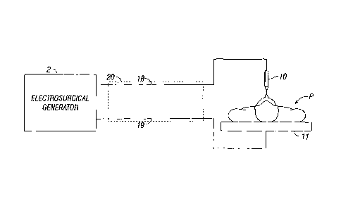
Fig. 2 is a schematic illustration of an electrosurgical system according to
the present
disclosure. The system is a monopolar electrosurgical system that includes an
electrosurgical
instrument 10 having one or more electrodes for treating tissue of a patient
P. Electrosurgical RF
energy is supplied to the instrument 10 by a generator 2 via a supply line 18,
which is operatively
connected to an active output terminal, allowing the instrument 10 to
coagulate, seal and/or
otherwise treat tissue. Energy is returned to the generator 2 through a return
electrode 11 and
transmitted through a return line 19, which is operatively connected to a
return output terminal.
The supply and return lines 18, 19 are enclosed within a cable 20.
System may include a plurality of return electrodes 11, which is believed to
minimize the
chances of damaged tissue by maximizing the overall contact area with the
patient P. In addition,
the generator 2 and the return electrode 11 may be configured for monitoring
so called "tissue-to-
patient" contact to insure that sufficient contact exists therebetween to
further minimize chances
of tissue damage. The generator 2 may include a plurality of supply and return
terminals and
corresponding number of transmission cables (e.g., two of each).
Fig. 3 shows an electrosurgical system 3 according to the present disclosure.
The system
3 is a bipolar electrosurgical system that includes an electrosurgical forceps
12 having opposing
jaw members. The forceps 12 includes one or more shaft members 13 having an
end effector
assembly 100 disposed at the distal end. The end effector assembly 100
includes two jaw
members 110, 120 movable from a first position wherein the jaw members are
spaced relative to
6
CA 02603461 2007-09-19
on another to a closed position wherein the jaw members 110 and 120 cooperate
to grasp tissue
therebetween. Each of the jaw members includes an electrically conductive
sealing plate
connected to an energy source (e.g., a generator 2) that communicates
electrosurgical energy
through the tissue held therebetween. Electrosurgical RF energy is supplied to
the forceps 12 by
generator 2 via the supply line 18 operatively connected to the active
electrode and returned
through the return line 19 operatively connected to the return electrode. The
supply and return
lines 18, 19 are enclosed within cable 20.
As shown in Fig. 3, the forceps 12 is an endoscopic vessel sealing bipolar
forceps. The
forceps 12 is configured to support the effector assembly 100. Those skilled
in the art will
understand that the invention according to the present disclosure may be
adapted for use with
either an endoscopic instrument or an open instrument. More particularly,
forceps 12 generally
includes a housing 21, a handle assembly 42, a rotating assembly 80, and a
trigger assembly 70,
which mutually cooperate with the end effector assembly 100 to grasp and treat
tissue. The
forceps 12 also includes a shaft 13, which has a distal end 14 that
mechanically engages the end
effector assembly 100 and a proximal end 16 that mechanically engages the
housing 21
proximate the rotating assembly 80. Handle assembly 42 includes a fixed handle
50 and a
movable handle 40. Handle 40 moves relative to the fixed handle 50 to actuate
the end effector
assembly 100 and enable a user to grasp and manipulate tissue as shown in Fig.
3.
Referring to Figs. 3 and 4, the end effector assembly 100 includes opposing
jaw
members 110 and 120 having electrically conductive sealing plate 112 and 122,
respectively,
attached thereto for conducting electrosurgical energy through tissue. More
particularly, the jaw
members 110 and 120 move in response to movement of the handle 40 from an open
position to a
closed position. In open position the sealing plates 112 and 122 are disposed
in spaced relation
7
CA 02603461 2014-11-25
relative to one another. In a clamping or closed position the sealing plates
112 and 122 cooperate
to grasp tissue and apply electrosurgical energy thereto. Further details
relating to one
envisioned endoscopic forceps is disclosed in commonly-owned U.S. Patent No.
7,090,673
entitled "VESSEL SEALER AND DIVIDER."
The jaw members 110 and 120 are activated using a drive assembly (not shown)
enclosed
within the housing 21. The drive assembly cooperates with the movable handle
40 to impart
movement of the jaw members 110 and 120 from the open position to the clamping
or closed
position. Examples of a handle assemblies are shown and described in the above
identified
application as well as commonly-owned U.S. Patent Publication No.
US2003/0229344 entitled
"VESSEL SEALER AND DIVIDER AND METHOD MANUFACTURING SAME" and
commonly owned U.S. Patent No. 7,156,846 entitled "VESSEL SEALER AND
DIVIDER FOR USE WITH SMALL TROCARS AND CANNULAS."
Jaw members 110 and 120 also include insulators 116 and 126, which together
with the
outer, non-conductive plates of the jaw members 110 and 120 are configured to
limit and/or
reduce many of the known undesirable effects related to tissue sealing, e.g.,
flashover, thermal
spread and stray current dissipation.
In addition, the handle assembly 42 of this particular disclosure includes a
four-bar
mechanical linkage that provides a unique mechanical advantage when sealing
tissue between the
jaw members 110 and 120. For example, once the desired position for the
sealing site is
determined and the jaw members 110 and 120 are properly positioned, handle 40
may be
compressed fully to lock the electrically conductive sealing plates 112 and
122 in a closed
position against the tissue. The details relating to the inter-cooperative
relationships of the inner-
working components of forceps 12 are disclosed in the above-cited commonly-
owned U.S.
8
CA 02603461 2014-11-25
=
Patent Publication No, US2003/0229344. Another example of an endoscopic handle
assembly
which discloses an off-axis, lever-like handle assembly, is disclosed in the
above-cited U.S.
Patent No. 7,156,846.
The forceps 12 also includes a rotating assembly 80 mechanically associated
with the
shaft 12 and the drive assembly (not shown). Movement of the rotating assembly
80 imparts
similar rotational movement to the shaft 12 which, in turn, rotates the end
effector assembly 100.
Various features along with various electrical configurations for the
transference of
electrosurgical energy through the handle assembly 20 and the rotating
assembly 80 are described
in more detail in the above-mentioned- commonly-owned U.S. Patent Publication
No. 1JS2003/0229344 and US Patent No. 7,156,846.
As best seen with respect to Figs. 3 and 4, the end effector assembly 10(}
attaches to the
distal end 14 of shaft 12. The jaw members 110 and 120 are pivotable about a
pivot 160 from the
open to closed positions upon relative reciprocation, i.e., longitudinal
movement, of the drive
assembly (not shown). Again, mechanical and cooperative relationships with
respect to the
various moving elements of the end effector assembly 100 are further described
by example with
respect to the above-mentioned commonly-owned U.S. Patent Publication No.
US2003/0229344
and U.S. Patent No. 7,156,846.
The forceps 12 may be designed such that it is fully or partially disposable
depending
upon a particular purpose or to achieve a particular result. For example, end
effector assembly
100 may be selectively and releasably engageable with the distal end 14 of the
shaft 12 and/or the
proximal end 16 of the shaft 12 may be selectively and releasably engageable
with the housing
21 and handle assembly 42. In either of these two instances, the forceps 12
may be either
partially disposable or reposable, such as where a new or different end
effector assembly 100 or
9
CA 02603461 2007-09-19
end effector assembly 100 and shaft 12 are used to selectively replace the old
end effector
assembly 100 as needed.
With reference to Figs. 2, 3 and 5, the generator 2 includes suitable input
controls (e.g.,
buttons, activators, switches, touch screen, etc.) for controlling the
generator 2. In addition, the
generator 2 may include one or more suitable display screens for providing the
surgeon with
variety of output information (e.g., intensity settings, treatment complete
indicators, etc.). The
controls allow the surgeon to adjust power of the RF energy, waveform, and
other suitable
parameters to achieve the desired waveform suitable for a particular task
(e.g., coagulating, tissue
sealing, intensity setting, etc.). The instrument 10 and/or forceps 12 may
also include a plurality
of input controls that may be redundant with certain input controls of the
generator 2. Placing the
input controls at the instrument 10 and/or forceps 12 allows for easier and
faster modification of
RF energy parameters during the surgical procedure without requiring
interaction with the
generator 2.
Fig. 5 shows a schematic block diagram of the generator 2 having a controller
4, a high
voltage DC power supply 7 ("HVPS") and an RF output stage 8. The DC power
supply 7
provides DC power to the RF output stage 8, which then converts DC power into
RF energy and
delivers the RF energy to the instrument 10 or forceps 20. The controller 4
includes a
microprocessor 5 operatively connected to a memory 6 which may be volatile
type memory (e.g.,
RAM) and/or non-volatile type memory (e.g., flash media, disk media, etc.).
The microprocessor
5 includes an output port that is operatively connected to the HVPS 7 and/or
RF output stage 8
allowing the microprocessor 5 to control the output of the generator 2
according to either open
and/or closed control loop schemes. A closed loop control scheme may be a
feedback control
loop wherein the sensor circuitry 11, which may include a plurality of sensing
mechanisms (e.g.,
CA 02603461 2007-09-19
tissue impedance, tissue temperature, output current and/or voltage, etc.),
provides feedback to
the controller 4. The controller 4 then signals the HVPS 7 and/or RF output
stage 8, which then
adjusts DC and/or RF power supply, respectively. The controller 4 also
receives input signals
from the input controls of the generator 2 and/or instrument 10. The
controller 4 utilizes the
input signals to adjust power outputted by the generator 2 and/or performs
other suitable control
functions thereon.
Fig. 6 shows a cross-sectional view of the cable 20. The cable 20 includes the
supply
and return lines 18, 19. The supply and return lines 18, 19 are operatively
connected to the
generator 2 via connectors 31, 32 respectively. Connectors 31, 32 may be
either of fixed or
detachable type allowing for the usage of multiple instruments and return
electrode pads with the
generator 2. The generator 2 and the connectors 31, 32 may also include
identification means
(e.g., bar codes or other codes disposed on the connectors and scanners
operatively connected to
the generator, etc.) that identify the device operatively connected to the
connectors 31, 32. Upon
connection of the connectors 31, 32, the generator 2 identifies the instrument
and performs
particular preprogrammed operations (e.g., initialize procedure, set operating
parameters, adjust
power settings, etc.).
The supply and return lines 18, 19 may be insulated. Various types of
insulating
materials may be used, which are within the purview of those skilled in the
art. The supply and
return lines 18, 19 extend from the connectors 31, 32 respectively for a
distance A, which is
optimally controlled by the location of connectors 31, 32 and is between from
about 0.1 inches to
about 6 inches. The lines 18, 19 are then helix wound in a wound portion 35,
which be about 7
feet or more depending upon a desired cable inductance and capacitance.
Alternatively, the
11
CA 02603461 2007-09-19
wound portion 35 may extend from the connectors 31, 32 without extending the
supply and
return lines 18, 19 for the distance A.
The wound portion 35, along cable length B, can be of any length depending on
geometric configuration and physical properties (e.g., tensile strength,
flexibility, etc.) of
materials used in manufacturing of cable components. More specifically the
lines 18, 19 are
oriented in a double helix which includes two congruent helixes with the same
axis, differing by
a translation along the axis. The lines 18, 19 may be oriented in a plurality
of other arrangements
which wrap the lines 18, 19 around themselves. The arrangement of the lines
18, 19 in a double
helix orients the opposing electrical fields generated by the electrosurgical
RF energy passing
therethrough to mitigate and/or cancel out thereby minimizing the amount of
lost stray electrical
RF energy.
The lines 18, 19 are wound within the cable 20 around a dielectric insulator
37, which
provides support for the lines 18, 19, an insulative sheath 39 covers the
lines 18, 19. The
insulator 37 and the sheath 39 may be of the same type. The lines 18, 19 may
comprise wire that
has an inductance rating at 473 kHz of 7.371.1H and A, capacitance at 1 MHz of
32.0 PF to yield
a cable self resonance of 10.4 MHz. The wire may be 26 gauge and 15 kV rated.
With reference to Fig. 6 and the portion 35, the distance D, which represents
the distance
between one apex of one helix and a nearest apex of another helix, may be
about 1/2. The distance
E, which is the distance between two apexes of the same helix may be about 1
inch. The outer
diameter F of the cable 20 may be about 3/8 of an inch.
Cable 20 as illustrated in Fig. 6, provides a transmission medium to deliver
RF energy
from the generator 20 to a tissue site. The cable 20 represents one example of
a preferred
embodiment for the RF transmission medium, which reduces the radiated RF
electrical field and
12
CA 02603461 2007-09-19
maximizes the applied clinical treatment energy delivered to the tissue site.
The dimensions A,
B, C, D, E and F of Fig. 6 form a unique proximal geometric relationship in
three dimensional
space to control the electrical field coupling between the active and return
output terminals of the
generator 20 to significantly reduce the Volts per meter electrical field
radiation by field
cancellation.
The physical dimensions A, B, C, D, E and F are interdependent and optimized
to provide
a low loss inductive and capacitive transmission medium, which in addition to
controlling the
electrical field, reduces uncontrolled capacitive coupling caused by stray RF
radiation. In
particular the following equations (1) and (2) illustrate the interdependent
relationship of
dimensions A, B, C, D, E and F with respect to inductive and capacitive
properties of the cable
20.
(1) Inductance = B (10.16 x 10^-9) Ln [(2 x D)/d)] + 2 (A+C)(01/in. for
specified wire)
(2) Capacitance = [(B x (0.7065 x 10^-12)) / Ln[(2 x D)/d]Jer
In equations (1) and (2) d denotes diameter of the wire (e.g., supply and
return lines 18, 19), er
denotes the dielectric constant of the wire insulator. Further, E = 2 x D, the
ratio of E to D allows
to establish a continuum of the helix configuration and F = k x D, where k is
a constant from
about 0.5 to about 1.5.
At the distal end of the portion 35, the lines 18, 19 are unwound and are
operatively
connected to device connectors 33, 34 respectively. The lines 18, 19 extend a
distance C from
the portion 35 to the connectors 33, 34 in an unwound state for approximately
2.5 feet. The
initial length A of the lines and the unwound state length C are maintained
relatively consistent
with varying lengths of wire with length of the wound portion 35 varying for
different overall
lengths.
13
CA 02603461 2007-09-19
In bipolar surgery, the connectors 33, 34 may be situated on the forceps 20.
In monopolar
surgery, the connector 33 is operatively connected to the instrument 10 and
the connector 34 is
connected to the return electrode 11. As discussed above, in situations where
a plurality of return
electrodes are used, the return line 19 may split into corresponding number of
leads to
operatively connect all of the return electrodes 11 to the generator 2. With
monopolar surgery
the length C for line 18 may lengthen greater than 2.5 feet with a
corresponding decrease in line
19 to accommodate manipulation of surgical instrument in the operating site.
The cable 20 according to the present disclosure orients the supply and return
lines 18, 19
so that the electrical fields generated therethrough are canceled, thereby
reducing the amount of
leaked stray RF energy. More specifically, placement and orientation of the
lines 18, 19 in the
manner discussed above provides for close proximity of electrical fields
generated during
transmission of electrosurgical RF energy and maximizes amount of energy
delivered to the
treatment site. Reducing the electrical fields also increases safety of
personnel and the patient.
Reduced RF radiation decreases capacitive and RF field leakage and improves RF
control
of the delivered energy. Reduced RF radiation also decreases RF transmission
loss and improves
efficiency of the generator 2 by reducing RF harmonic component, minimizing
corruption of the
RF source and reducing peripheral conductive and radiative emissions. Further,
reducing RF
radiation also decreases the RF noise to additional equipment found in the
room, such as patient
monitoring equipment.
While several embodiments of the disclosure have been shown in the drawings
and/or
discussed herein, it is not intended that the disclosure be limited thereto,
as it is intended that the
disclosure be as broad in scope as the art will allow and that the
specification be read likewise.
Therefore, the above description should not be construed as limiting, but
merely as
14
CA 02603461 2014-11-25
exemplifications of particular embodiments. The scope of the claims should not
be limited by the
preferred embodiments set forth herein, but should be given the broadest
interpretation consistent with
the description as a whole.