Note: Descriptions are shown in the official language in which they were submitted.
CA 02603544 2013-02-06
=
THROMBUS REMOVAL SYSTEM HAVING A MACROCOIL
BACKGROUND OF THE INVENTION
1. FIELD OF THE INVENTION
The present invention pertains to intravascular medical devices. More
particularly, the present invention pertains to devices for capturing and
removing blood
clots from a blood vessel. This same system may also be used to remove
material from
other cavities of the body, for example, stones from the urinary or the
biliary tract.
2. BACKGROUND OF THE ART
The present invention pertains generally to thrombus collection and removal.
Blood thrombus, may form a clot in a patient vasculature. Sometimes such clots
are
harmlessly dissolved in the blood stream. At other times, however, such clots
may lodge
in a blood vessel or embolize a distal blood vessel where they can partially
or completely
occlude the flow of blood. If the partially or completely occluded vessel
feeds blood to
sensitive tissue such as, the brain, lungs or heart, for example, serious
tissue damage may
result. =
When symptoms of an occlusion are apparent, such as an occlusion resulting in
a
stroke, immediate action should be taken to reduce or eliminate resultant
tissue damage.
One approach is to treat a patient with clot dissolving drugs. These drugs,
however, do
not immediately dissolve the clot from the patient.
Published U.S Patent Application 2005/0038447 describes A medical device for
removing clots from a blood vessel, comprising: a first longitudinally-
oriented spine
having a distal end; a pushing member coupled to the proximal end of the first
longitudinally-oriented spine and extending proximally therefrom; and a clot-
grabbing
basket generally disposed between and coupled to the first longitudinally-
oriented spine.
Published U.S. Patent Application 2004/0138692 discloses an embolus extractor,
comprising: an elongated shaft having a proximal end and a distal end; first
and second
struts, each strut having a proximal end and a distal end coupled to the
distal end of the
shaft; the first and second struts having a first position and a second
position, wherein in
CA 02603544 2007-09-28
WO 2006/107641
PCT/US2006/011158
the first position, the distal ends and the proximal ends of the struts are
spaced at a first
distance, and in the second position the distal ends and the proximal ends of
the struts are
spaced at a second distance, the second distance being less than the first
distance; and
third and fourth struts, each strut coupled to one of the first and second
struts via a
proximal end and distal end.
Published U.S. Patent Application 2004/0098023 discloses a vaso-occlusive
device, comprising: a core member; and a fibrous structure carried by the core
member,
the fibrous structure comprises one or more strands of nanofibers. The vaso-
occlusive
device may provide the fibrous structure in a product generated at least in
part by an
electrospinning process comprises the steps of: supplying a polymer solution
through a
needle; electrostatically charging the needle; electrostatically charging a
metal plate that
is placed at a distance from the needle, the metal plate having a charge that
is opposite
that of the needle, thereby sending a jet of the polymer solution towards the
metal plate;
and collecting the fibrous structure from the metal plate.
Published U.S. Patent Application 2004/0039435 discloses a self-expanding,
pseudo-braided device embodying a high expansion ratio and flexibility as well
as
comformability and improved radial force. The pseudo-braided device is
particularly
suited for advancement through and deployment within highly tortuous and very
distal
vasculature. Various forms of the pseudo-braided device are adapted for the
repair of
aneurysms and stenoses as well as for use in thrombectomies and embolic
protection
therapy.
There are a variety of ways of discharging shaped coils and linear coils into
a
body cavity. In addition to those patents that describe physically pushing a
coil out of the
catheter into the body cavity (e.g., Ritchart et al.), there are a number of
other ways to
release the coil at a specifically chosen time and site. U.S. Patent No.
5,354,295 and its
parent, U.S. Patent No. 5,122,136, both to Guglielmi et al., describe an
electrolytically
detachable embolic device.
A variety of mechanically detachable devices are also known. Various examples
of these devices are described in U.S. Patent No. 5,234,437, to Sepetka, U.S.
Patent No.
5,250,071 to Palermo, U.S. Patent No. 5,261,916, to Engelson, U.S. Patent No.
2
CA 02603544 2007-09-28
WO 2006/107641
PCT/US2006/011158
5,304,195, to Twyford et al., U.S. Patent No. 5,312,415, to Palermo, and U.S.
Patent No.
5,350,397, to Palermo et al.
Various configurations have been used to remove calculi from the biliary or
urinary system. See, for instance, U.S. Patent No. 5,064,428. Additionally,
devices
having various configurations have been used to remove objects from the
vasculature.
For example, surgical devices comprising one or more expandable and
collapsible
baskets have been described for removing or piercing a thrombus in the
vasculature. See,
U.S. Patent No. 6,066,149. U.S. Patent No. 5,868,754 describes a three prong-
shaped
device for capturing and removing bodies or articles from within a vessel.
Published U.S. Patent Application 2004/0225229 describes a device comprising a
core wire having a distal end and a proximal end; a catheter shaft having a
proximal
catheter end, a distal catheter end and a lumen through which the core wire is
passed such
that the distal end of the core wire extends beyond the distal catheter end; a
retrieval
element disposed at the distal end of the core wire, the retrieval element
movable from a
radially contracted position to a radially expanded position; and a first stop
element
attached to the core wire, the first stop element configured to prevent over-
expansion of
the retrieval element.
Among commercial thrombus-removal systems are at least the following:
1) The MERCI system of Concentric Medical that has a form of a corkscrew or
helix
spring. In this system, which may use a large 0.018 F microcatheter, the
microcatheter tip is first positioned across the thrombus with the help of a
guidewire. Then the guidewire is exchanged with the system which is deployed
distal and into the thrombus. The shape of the system allows it to get
inserted into
the thrombus. Then the thrombus is retrieved out of the artery into a large 9
French working catheter, and then out of the body.
2) The In-Time system of Boston Scientific which is a sort of a clam-shell
guide,
that once placed through the thrombus divides itself into 4 strings that form
an
oval, as with a rugby balloon. The system is pulled back to carry out the
thrombus. This is similar to the disclosed structure in Published US
Application
2004/0138692.
3
CA 02603544 2008-02-21
3) Another system is what is called a lasso, which is a simple catheter with a
wire
attached to its end. This wire makes a loop and enters back into the catheter
(e.g., a large 0.018 F microcatheter). The operator changes the aspect of the
loop by pulling on the wire. This system was originally conceived to catch
foreign bodies.
4) The Catch system of Bait is a stent closed on one end and forming a basket
that is deployed distal to the thrombus. The operator then pulls the system
and
retrieves the thrombus. This is similar to the structure in Figure 7 of U.S.
Patent No. 6,805,684.
The above systems may have various disadvantages, such as either to slide on
the
thrombus, either to fractionate, to be difficult to guide or deploy or to be
traumatic to the
artery while some of them are quite expensive. In addition, all these system
are bulky and
cannot be used in small caliber arteries.
SUMMARY OF THE INVENTION
A device capable of capturing and assisting in the removal of a thrombus in
arteries, and even in small arteries uses a soft coil mesh to engage the
surface of a
thrombus, and a guidewire is used to retract the soft coil mesh with the
captured thrombus.
The soft coil is formed by an elongated microcoil element that forms the
helical elements
of a macrocoil element. The microcoil element provides a relatively elastic
effect to the
helical element forming the macrocoil and allows for control of gripping
forces on the
thrombus while reducing non-rigid contact of the device with arterial walls.
In one aspect, the present invention provides a medical device for removing a
thrombus from a blood vessel, comprising a macrocoil thrombus engaging
component
having a length with a proximal end and a distal end, the length of the
macrocoil
comprising microcoils that allow the length of the macrocoil to be extendable;
a first wire
external to the microcoils capable of providing force on the distal end of the
macrocoil; a
second wire external to the microcoils capable of providing force on the
proximal end of
the macrocoil; the macrocoil being capable of retracting to a maximum
thickness of less
than 0.001mm when extended by the first wire and the second wire, and the
microcoil
being expandable in width greater when distended by relative movement between
the first
wire and second wire.
4
CA 02603544 2014-08-11
In yet another aspect, the present invention provides a medical device for
removing a
thrombus from a blood vessel, comprising: a macrocoil thrombus engaging
component
having a length with a proximal end and a distal end, the length of the
macrocoil comprising
microcoils that allow the length of the macrocoil to be extendable; a first
wire external to the
microcoils capable of providing force on the distal end of the macrocoil; a
second wire
external to the microcoils capable of providing force on the proximal end of
the macrocoil;
the macrocoil diameter being capable of retracting to a maximum thickness of
at least
0.001mm when extended by the first wire and the second wire, and the microcoil
being
expandable in width when distended by relative movement between the first wire
and second
wire towards each other.
BRIEF DESCRIPTION OF THE FIGURES
Figure 1 shows the microcoil/macrocoil structure of the soft coil capture
device
described herein.
Figure 2 shows a soft coil capture device in an insertion position within an
artery.
Figure 3A shows a soft coil capture device in a pre-capture position within an
artery in
a first mode of soft coil delivery.
Figure 3B shows a soft coil capture device in a thrombus engaged position
within the
first mode of soft coil delivery of Figure 3A.
4a
CA 02603544 2013-02-06
Figure 4A shows a soft coil capture device in a pre-capture position within an
artery in a
second mode of soft coil delivery.
Figure 4B shows a soft coil capture device in a thrombus capture position
within an
artery within the second mode of soft coil delivery of Figure 4A.
Figure 5 shows a soft coil capture device (which may be of larger dimensions
than
parenchymal vasculature delivery devices) midway through deployment.
Figure 6 shows a micr[omicron]catheter delivery system with constrained coils
within
the microcatheter.
DETAILED DESCRIPTION OF THE INVENTION
The following description should be read with reference to the drawings
wherein like
reference numerals indicate like elements throughout the several views. The
detailed description
and drawings illustrate example non-limiting specific embodiments of the
generic claimed
invention.
Figure 1 shows a structural material 2 (also referred to herein as material,
coils, coil
material or soft coil material) that can be used as a soft soil capture
element in the practice of the
technology described herein. The material 2 has microcoils or microloops
forming a continuing
chain 6a of microcoils that form the macrcoil or macrohelix 10. The term
'microcoil' as used
herein should not be confused with the RF or MRI responsive coils or
microcoils that are used in
the medical imaging art. These are microcoils in the sense that they are small
coils as compared
to the macrocoils 10 which are large coils. The microcoils are made from
structural material 8
that forms the filaments, threads, fibers, or the like that are used to
provide the microcoils that
build into the macrocoils. The benefits of this material and the structure in
which they perform
will become apparent from the discussion herein.
The microcoils add a significant degree of compliance, effective elasticity
and
cushioning ability to the macrocoil. The microcoils elongate to give the
appearance of elasticity
to the material 2, without providing hard and large abrasive surfaces that
would contact arterial
walls, as would traditional coil or mesh structures.
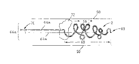
Figure 2 shows the soft coil material 2 within an artery 50. The macrocoils 56
are
shown with the entire length of the coil section 60 of the device being shown
in a slightly
extended position that is useful for insertion of the device 66a. The pusher
wire or
5
CA 02603544 2013-02-06
,
guidewire 62a (also referred to herein as push wire, or push (guide) wire)
stabilizes the insertion
end 68 of the soft coil material 2 while a pull wire 64a (also referred to
herein as pull (guide)
wire) stabilizes the back end 70 of the device 66a and the material 2. The
push wire 62a tends
to be thicker than the pull wire 64a as a matter of course, but they may be of
the same or similar
thicknesses, and the pull wire may be thicker than the push wire 62a. A
thrombus 72 is shown,
with the distended coil section 60 having been pushed past the thrombus 72.
Figure 3 A shows a first mode of delivery of the system 68 wherein the pulling
wire 62
(also referred to herein as pull wire) has been extended from the
microcatheter 66 past the
thrombus 72, and the push wire 64 has been slightly extended beyond the
thrombus, being
carried by the microcatheter 66. The pulling wire 62 and the push wire 64 are
sufficiently close
together so that the entire length of the extended coil 60 is restrained, but
beyond the major mass
of the thrombus 72. FIG. 3B shows the microcatheter 66 having been withdrawn
from past the
thrombus 72, the push wire also pulled rearward of the thrombus 72, and the
end of the pulling
wire 62 being retracted to pull the soft coil material 2 into a tangled
engagement with the
thrombus, engaging the thrombus 72 so that withdrawal of the microcatheter and
the two wires
62 and 64 will withdraw the thrombus 72 while enmeshed in the soft coil
material. The entire
enmeshing length 74b of the soft coil securely entrains the thrombus 72, and
the soft coil
material 2 assists in reducing breakage of the thrombus 72 and damage to
vascular walls.
Figure 4A shows the system 68 delivered in a second delivery mode, without the
microcatheter 66 passing the thrombus 72 mass, where both the push wire 64 and
the pull wire
62 are positioned so that the push wire 64 restrains the soft coil material 2
relatively in front of
the thrombus 72 and the pulling wire 62 has been extended from the
microcatheter 66 to employ
the soft coil material 2. Figure 413 shows that the pulling wire 62 has been
retracted slightly,
causing the soft coil material 2 to engage the thrombus 72 and enmesh the
thrombus 72 within
the soft coil material. By withdrawing the microcatheter 66, and the two wires
62 and 64, the
thrombus can be withdrawn from the vessel 50 with minimal damage to the vessel
50 and
reduced breakage in the thrombus 72. The nature of the mixture of the
microcoils and
macrocoils causes a constriction of the material around the thrombus, without
segmenting
(cutting) the thrombus easily, and without providing a cage surface that is as
potentially
damaging to arterial walls as are other structures used for thrombus retrieval
and capture. The
push
6
CA 02603544 2007-09-28
WO 2006/107641 PCT/US2006/011158
wires and pull wires may be of equal wire dimensions (e.g., diameters) or
different
dimensions, with either one being thicker than the other in different
embodiments.
The system is made of a 3D soft coil such that when the system gets deployed,
it
has the tendency to form a three dimensional cage, with loops of microcoils
extending
across the diameters of the arterial interior to assure that loops will be
able to engage a
thrombus when the loops are retracted. The ends of the coils may be attached
on either
its proximal end to a pusher wire and to its distal end to a very fine wire or
visa versa.
The entire system tends to be able to be provided in a very thin format
(although the size
may vary depending upon the need for fit within particular arterial passages,
and can fit
into a 0.010 Fr microcatheter or smaller. Both wires exit at the proximal end
of the
microcatheter and can be manipulated by the operator. First the microcatheter
is
positioned across the thrombus with the help of a microguidewire. Once the
distal end of
the microcatheter lies beyond the thrombus (usually while it is in a distended
state, fairly
elongate and narrow), the microguidewire is exchanged with the thrombus
retrieval
system. The thrombus retrieval system is activated and deployed so that a
significant
portion of the entire length of coil (e.g., 1/5, 1/4 or one third of the coil)
is positioned distal
to the thrombus. A remaining significant portion of the coil (using, by way of
non-
limiting examples of amounts, with one third distal to or past the thrombus),
such as at
least 1/5, at least 1/4 or one third or more of the coil length is wrapped
around or codistant
with (within the artery) the thrombus and 1/4, 1/5 or one third or more
proximal to the
thrombus. Once the coil is deployed with a significant portion at least at the
distal end of
the thrombus and more desirably a significant portion past the distal end of
the thrombus,
the operator pulls the thin distal wire or pushes the thick proximal wire, so
that the mesh
of coil loops that has formed around the thrombus or expanded beyond the
thrombus
retracts on itself and grabs securely the thrombus. The thrombus now can be
pulled out of
the artery by pulling the microcatheter, the pusher wire and the thin distal
wire on the
same time out of the artery.
One other advantage of the system (in addition to what has been described
already) is its very small size so it can retrieve thrombus from very small
arteries, its
capacity to pull out the thrombus in one piece, and its softness, allowing
manipulation
without trauma to the vessel wall. Larger versions have the advantage of
retrieving a very
7
CA 02603544 2007-09-28
WO 2006/107641
PCT/US2006/011158
large thrombus in one piece. This system may be used in any vessel of the body
for the
retrieval of thrombus or other material like foreign bodies.
The distal end of the soft coil material (where the pulling wire is attached)
may be
limited in its ability to extend away from the proximal end of the soft coil
material (where
the push wire is attached) by using an internal connector, such as a thread,
that attaches to
both ends of the soft coil, and provides a physical limit to how far the coil
may be
distended.
Whatever the consistency of the clot, i.e., soft or hard, once someone has
passed
the clot with the microcatheter, the distal mesh of coils when deployed will
form a
"sponge" or "piston" that should bring back at least a large part of the
thrombus. It is also
likely that the loops of the coil should prevent the loss of parts of the
thrombus if it
breaks into pieces. The tendency of the system to break soft thrombus will
depend on
characteristics such as the soft coil material thickness, the microcoil
thickness the
macrocoil thickness, density of the macrocoil, the 3D configuration of the
macrocoils and
the loop diameter of the coil. Even in the worst case envisioned, one could
only deploy a
distal and a proximal mesh or use a flow reversing system.
For a number of reasons, it may be desirable to capture and/or remove clots
from
the vasculature. The blood vessel can be essentially any vessel or even duct.
The device
may include two or more longitudinal wires, for example a guidewire, a push
wire and a
pull wire, as well as other functional wires (e.g., conductive wires for other
features
provided with the device, such as a resistive wire to enable heating of the
coils, if
conductive/resistive. The basket member or region of soft coils is attached to
or otherwise
coupled with the wires. In general, the device (wires and soft coil material)
can be
advanced through the vasculature to a suitable location, for example adjacent
a clot, and
expanded (when past or adjacent to the clot, so that the clot may be captured
in the soft
coils, upon operator action, and the captured clot can be removed from the
vasculature.
The device may be configured to shift between a first generally collapsed
configuration and a second generally expanded configuration, especially by the
elastic
memory of the coil material, and the guidance imposed by the at least two
wires. In at
least some embodiments, shifting between these configurations includes the
longitudinal
movement of one or both of the wires relative to one another. Movement of the
wires
8
CA 02603544 2007-09-28
WO 2006/107641 PCT/US2006/011158
=
may occur in either the proximal or distal direction and, in the case of both
wires moving,
may be in the same or opposite directions. Shifting may also result in one or
both of the
wires moving somewhat laterally (especially with distally controlled wires on
the coil
material (e.g., with materials that bend when heated, or the like, and a
heating element
attached thereto) so that the wires become closer or move apart one another.
Shifting between the collapsed and expanded configurations may occur in a
number of differing manners. For example, the device or portions thereof may
be made of
a shape-memory material (such as nickel-titanium alloy or oriented coils) that
can assume
a pre-defined shape when unconstrained or when subjected to particular thermal
conditions. According to this embodiment, the device can be manufactured to be
"self
-
expanding" (when the longitudinal distension and restraint by the wires is
removed) so
that it can be delivered in a collapsed configuration then shift to the
expanded
configuration when a constraint is removed (e.g., the distal ends of the two
wires brought
closer together) or when the device is subject to the natural thermal
conditions within
blood vessel. Alternatively, shifting may occur by mechanically moving one or
both of
wires. Moving the wires may occur in a number of different ways such as by
moving one
or other of the wires attached to the distal or proximal end of the coil
material on the
device.
As described above, all or portions of the device (including but not limited
to the
coil materials and the wires) may be manufactured from polymeric, metallic,
natural
(e.g., gut wires), synthetic, or composite materials. Preferred materials tend
to be
polymeric, metallic, composite or mixtures or combinations of these materials.
A
conventional medical structural material such as nickel titanium alloy may be
employed.
However, any suitable material may be used including metals, metal alloys,
polymers,
etc. Some examples of suitable metals and metal alloys include stainless
steel, such as
304V, 304L, and 316L stainless steel; linear-elastic or super-elastic nitinol
or other
nickel-titanium alloys, nickel-chromium alloy, nickel-chromium-iron alloy,
cobalt alloy,
tungsten or tungsten alloys, MP35-N (having a composition of about 35% Ni, 35%
Co,
20% Cr, 9.75% Mo, a maximum 1% Fe, a maximum 1% Ti, a maximum 0.25% C, a
maximum 0.15% Mn, and a maximum 0.15% Si), hastelloy, monel 400, inconel 825,
or
the like; or other suitable material.
9
CA 02603544 2013-02-06
Some examples of suitable polymers may include polytetrafluoroethylene (PTFE),
ethylene tetrafluoroethylene (ETFE), fluorinated ethylene propylene (FEP),
polyoxymethylene
(POM), polybutylene terephthalate (PBT), polyether block ester, polyurethane,
polypropylene
(PP), polyvinylchloride (PVC), polyether-ester (for example a polyether-ester
elastomer such as
ARNITEL available from DSM Engineering Plastics), polyester (for example a
polyester
elastomer such as HYTREL available from DuPont), polyamide (for example,
DURETHAN
available from Bayer or CRISTAMID available from Elf Atochem), elastomeric
polyamides,
block polyamide/ethers, polyether block amide (PEBA, for example available
under the trade
name PEBAX0), silicones, polyethylene (PE), Marlex high-density polyethylene,
Marlex low-
density polyethylene, linear low density polyethylene (for example REXELLt),
polyethylene
terephthalate (PET), polyetheretherketone (PEEK), polyimide (PI),
polyetherimide (PEI),
polyphenylene sulfide (PPS), polyphenylene oxide (PPO), polysulfone, nylon,
perfluoro(propyl
vinyl ether) (PFA), other suitable materials, or mixtures, combinations,
copolymers thereof,
polymer/metal composites, and the like. In some embodiments, portions of or
all of the device
can be blended with a liquid crystal polymer (LCP). For example, the mixture
can contain up to
about 5% LCP.
In some embodiments, a coating, for example a lubricious, a hydrophilic, a
protective, or
other type of coating may be applied over portions or all of the device.
Hydrophobic coatings
such as fluoropolymers provide a dry lubricity which improves device
exchanges. Lubricious
coatings improve steerability and improve lesion crossing capability. Suitable
lubricious
polymers are well known in the art and may include silicone and the like,
hydrophilic polymers
such as polyarylene oxides, polyvinylpyrolidones, polyvinylalcohols, hydroxy
alkyl cellulosics,
algins, saccharides, caprolactones, and the like, and mixtures and
combinations thereof.
Hydrophilic polymers may be blended among themselves or with formulated
amounts of water
insoluble compounds (including some polymers) to yield coatings with suitable
lubricity,
bonding, and solubility. Some other examples of such coatings and materials
and methods used
to create such coatings can be found in U.S. Patent Nos. 6,139,510 and
5,772,609. In some
embodiments, the sheath
CA 02603544 2007-09-28
WO 2006/107641
PCT/US2006/011158
or coating may be applied over basket region. This may provide extra surface
area to
contain clots that might be captured therein.
The sheath or polymeric layer coating may be formed, for example, by coating,
electrophoresis, by extrusion, co-extrusion, interrupted layer co-extrusion
(ILC), or
fusing several segments end-to-end. The layer may have a uniform stiffness or
a gradual
reduction in stiffness from the proximal end to the distal end thereof. The
gradual
reduction in stiffness may be continuous as by ILC or may be stepped as by
fusing
together separate extruded tubular segments. The outer layer may be
impregnated with a
radiopaque filler material to facilitate radiographic visualization. Those
skilled in the art
will recognize that these materials can vary widely without deviating from the
scope of
the present invention.
The device, or portions thereof, may also be coated, plated, wrapped or
surrounded by, doped with, or otherwise include a radiopaque material. For
example, the
wires or coils may be made from a radiopaque material or may include a
radiopaque
marker member or coil coupled thereto. Radiopaque materials are understood to
be
materials capable of producing a relatively bright image on a fluoroscopy
screen or
another imaging technique during a medical procedure. This relatively bright
image aids
the user of the device in determining its location. Some examples of
radiopaque materials
can include, but are not limited to, gold, platinum, palladium, tantalum,
tungsten alloy,
plastic material loaded with a radiopaque filler, and the like.
In some embodiments, a degree of MRI compatibility may be imparted into the
device. For example, to enhance compatibility with Magnetic Resonance Imaging
(MRI)
machines, it may be desirable to make portions of the device, in a manner that
would
impart a degree of MRI compatibility. For example, the device, or portions
thereof, may
be made of a material that does not substantially distort the image and create
substantial
artifacts (artifacts are gaps in the image). Certain ferromagnetic materials,
for example,
may not be suitable because they may create artifacts in an MRI image. The
device, or
portions thereof, may also be made from a material that the MRI machine can
image.
Some materials that exhibit these characteristics include, for example,
tungsten, Elgiloy,
MP35N, nitinol, and the like, and others.
11
CA 02603544 2007-09-28
WO 2006/107641
PCT/US2006/011158
The control wire(s) may be produced from any number of suitable materials
having reasonable strength in tension, e.g., stainless steels, carbon fibers,
engineering
plastics, tungsten alloys, variously in the form of a multi-strand cable or
single strand
thread. Preferably, however, the wire may be made from a "so-called" super-
elastic alloy.
These alloys are characterized by an ability to transform from an austenitic
crystal
structure to a stress-induced martensitic (SIM) structure and to return
elastically to the
austenitic crystal structure (and the original shape) when the stress is
removed. A typical
alloy is nitinol, a nickel-titanium alloy, which is readily commercially
available and
undergoes the austenite-SIM-austenite transformation at a variety of
temperature ranges.
These materials are described, for instance in U.S. Patent Nos. 3,174,851 and
3,351,463.
These alloys are especially suitable because of their capacity to elastically
recover almost
completely to the initial configuration once the stress is removed. Since this
is so, the size
of the actual wire may be made fairly small, e.g., as small as 0.005 inches in
diameter or
smaller, and the resulting device is able to access very small regions of the
body. The
wire may also vary in diameter along its length, for example have a larger
diameter at the
proximal end as compared to the distal end or vice versa.
The wires can have a proximal section and a distal section. The proximal
section
preferably has a uniform diameter of at least about 0.0001 inch, or about
0.005 to 0.025
inches, preferably 0.0010 to 0.018 inches. Commercially available wires with a
microcoil (wire) diameter of 0.008 mm and a macrocoil diameter of lmm are
available as
microcoil materials. Optionally, the distal section may have different (more
or less)
flexibility than the proximal section and extends beyond the catheter.
Typically, both
sections will extend from the distal and proximal ends of the catheter lumen.
The wire
may have a middle section having a diameter intermediate between the diameter
of the
two portions of the wire adjoining the middle section or the middle section
may be
continuously tapered, may have a number of tapered sections or sections of
differing
diameters, or may be of a uniform diameter along its length and be tapered at
or near the
distal section. The entire wire may be between about 50 and 300 cm, typically
between
about 175 to 190 cm in length. The wire may be wrapped to form a coil section
or may be
independently attached to a coil.
12
CA 02603544 2013-02-06
The overall length of the control wire may extend through a catheter and the
wire and
catheter inserted into the vasculature. The catheter and wires (with attached
soft coil may extend
proximal or distal to the site of the clot or the catheter may be positioned
and the wires extend to
the site from the catheter. The configurable soft coil component of the device
is positioned near
the target thrombus site, and the wires position and control the positioning
and attitude of the
soft coil capture components.
Figure 5 shows a soft coil capture device 4 (which may be of larger dimensions
than
parenchymal vasculature delivery devices) midway through deployment. In small
coils, but
particularly with larger coils, greater strength may be built into the elastic
memory of the
material 2 and the macrocoils 6 and the length of remembered coil distribution
80 (also referred
to herein as fully deployed region). The coil material 2 may be delivered
through a catheter 92,
with the elongation of the coils 2 controlled by relative positioning of the
push and pull (guide)
wires 62a and 64a as explained above. One end of the coil material 2 is shown
secured to the
push wire 62a and the distal (leading end) of the coil material 2 is shown
secured to the distal
end of the pull (guide) wire 64a. When in a fully deployed region 80, without
tension or
retension applied by the wires 62a and 64a, a natural distribution (frequency)
of the macrocoils
6 will exist. Points of contact 82 between the coils 6 and the pull (guide)
wire 64a are
preferably not secured to the wires 62a and 64a, but are able to slide freely
against them. If the
contact points were secured, the frequency between the coils would be fixed
before and after
deployment, unlee the pull (guide) wire 64a were able to telescope or
otherwise extend. As
shown in the figure, the macrocoils 6 when in a deploying region 90, without
restraining action
through the connection at the distal connecting point 84 has a greater
frequency (less spacing)
between the macrocoils 6. The macrocoils 6 are shown being deployed out of a
catheter 92.
The microcoils and macrocoils may be manufactured and designed so as to
provide nature
dimensions when tension is released after deployment to fit a range of
dimensions in
vasculature. The selection of the microcoil size, maicrocoil spacing, wire
thickness, wire
material, macrocoil size and macrocoil spacing are used to determine the
frequency, size and
shape of the deployed structure.
Figure 6 shows a microcatheter 66 having the pull wire 62 and the push wire 64
with the
soft coil material 2 completely within the confines of the microcatheter 66.
The
13
CA 02603544 2007-09-28
WO 2006/107641
PCT/US2006/011158
soft coil material will deploy, expanding under its elastic compressive
tension,, to the
limits of its size or the limits of space within the vasculature when the two
wires 62 and
64 force the soft coil material from within the microcatheter 66. In actual
delivery of the
system, the soft coil material may be present within the microcatheter in a
relatively more
linear distribution of the microcoils within the lumen of the catheter, rather
than as the
combination of macrocoils and microcoils shown.
Although the examples show specific dimensions and materials, the examples and
descriptions are not intended to be limiting to the scope of practice and
protection of the
technology described. Rather, any specific statements or values are intended
to be
examples within the generic concepts of the inventions and the disclosure
taught and
provided herein.
=
14