Note: Descriptions are shown in the official language in which they were submitted.
CA 02603642 2011-03-23
WO 2006/107719 PCT/US2006/011754
COMPOSMONS AND METHODS FOR THE INHIBITION OF DISHEVELLED PROTEINS.
FIELD OF THE INVENTION
The present invention relates to .the Dishevelled proteins, which translate
Wnt= =
signals from the transmembrane receptor Frizzled to downstream components in
canonical and= .
non-canonical Wnt signaling pathways. =The invention relates to the field Of
therap.eutic
methods, compositions and uses thereof, in the treatment of various diseases
which are caused by
.Wnt signaling involved in pathogenesis. More particularly, the compositions
and methods are
. = directed to compounds that interrupt the Frizzled-Dishevelled interaction.
The compounds' were =
identified from libraries of compounds using screening inethods. These
compoundimay also.be
Modified to create derivatives or analogues not found in the. libraries or in
nature, which also.
function effectively.
CA 02603642 2011-03-23
WO 2006/107719 PCT/US2006/011754
= BACKGROUND OF THE INVENTION
Wnt signaling pathways play important roles in embryonic and postembryonic
development and have been- implicated in tumorigenesis. In the canonical Wnt-0-
catenin
. pathway, Secreted Wnt glyc.op. roteins bind to seven¨transmembrane=
'domain Frizzled (Fz)
receptors and activate intracellular Dishevelled (Dvl) proteins Activated Dvl
proteins then
inhibit glycogen synthase Itinase-30 (GSK-30); = this inhibition eauses
destabilization of a
. molecular complex formed by GSK-3f3, adenomatous polyposis colt (APC),
axin, and P-catenin
and reduces the .capability of GSK-313 to phosphorylate 0-catenin.
Unphosphorylated P-catenin
proteins escape from ubiqpination and' degradation and accumulate in the
cytoplasm. This .
accumulation leads = to the translocation of 0-catenin into the nucleus, where
it stimulates
transcription of Wnt target genes, such as the gene encoding the T cell
factor/lymphoid enhancer =
factor (Tcf/Lef). Numerous reports address mutations of Wnt¨P-Catenin
signaling pathway
-
components thatare involved in the development of neoplasia.
=
The link between the Wnt pathwa and cancer dates back to the initial
discovery of Wnt
signaling: the first vertebrate Wnt growth faotor was identified is the
product of a cellular
= oncogene (Wnt-1), which is activated by proviral insertion in murine
mammary carcinomas.
Perhaps the most compelling evidence supporting the role of Wnt signaling in
oncogenesis is the
= finding that approximately 85% of colorectal cancers are characterized by
mutations in APC, one
of the key components of the Wnt pathway. Members of the .Wnt signaling
pathway also have
. been implicated in the pathogenesis of various pediatric cancers such as
Burkitt lymphoma,' 4 =
medulloblastoina, -Wilms' tumor, and neuroblastoma. Furthermore, aberrant Wnt
signaling is
involved in other diseases, such as osteoporosis and diabetes. :
= =
Dvl relays the Wnt signals from membrane-bound receptors to downstream
components'
and
and thereby plays an essential role '-in the Wnt signaling pathway. Dvi
proteins are highly =
conserved throughout the animal kingdom. Three Dvl homologs, Dvl-1, -2, and -
3, have been
identified in mamraalian systems. All three human Dvl genes are 'widely
expressed in fetal and
adult tissues = including brain, lung, kidney, skeletal muscle, and heart. The
Dvl proteins are
composed of an N-terminal DDC domain, a central PDZ motif, and a C-terrninal
DEP domain. Of
these three, the PDZ domain appears to play an important role in both the
canonical and non-
2
CA 02603642 2011-03-23
WO 2006/107719 PCMS2006/011754
=
canonical Wnt Pathways. Indeed, the P.DZ domain of Dvl may be inVolved not
only in
. == distinguishing roles between the two = pathways but also in nuclear
localization. Recently, the
interactions between the PDZ domain (residues 247 through 341) of mouse=Dv1-1
(irDv11) and
= its binding partners were hivestigated by using nuclear magnetic
resonance (NMR) spectroscopy.
' The peptide-interacting site of the inl3nr11 PDZ domain interacts
with.various molecules whose
sequences have no Obvious homology. Although it is not a typical PDZ-binding
motif, iine
= peptide that binds to the mDv11 PDZ domain .is the conserved motif
(KTXXXW) 9f Fz, which
= . begins two amino acids after the seventh transinembrane domain. This
finding Showed that there . .
is a direct interaction between Fz and Dvl and revealed a previously unknown
connection
. between the= membrane-bound receptor and downstreani components of the Wnt
signaling ==
pathways. Therefore, an inhibitor of the Dvl PDZ domain is likely to
effectively blonk the Wnt=
= =
signaling pathWay at the Dvl level.
= =
The special role of the Dvl PDZ domain in the Wnt¨p-catenin pathway makes it
an ideal
= pharinaceutical target. Small organic inhibitors of the PDZ domain in Dvl
might be useful in
dissecting molecular mechanisms and formulating pharmaceutical agents that
target tumors or
= other diseases in which the Wnt signaling is involved in pathogenesis. In
light of the structure of =
'the Dv1 ,PDZ domain, virtual ligand screening was used to identify a non-
peptide compound,
NC1668036, that binds to the Dvl PDZ domain. Further MR' experiments validated
that the
= compound binds to the peptide-binding site on the surface of the PDZ
domain:* the binding .
affinity' (dissociation constant, KO of the compound was measured by
rfluorescence
= 'spectroscopy. In addition, we carried out molecular dynamics (MD)
simulations. Of. the .
interaction. between this compound and the PDZ domain as well as that between.
the C:terminal
= region of a known PDZ, domain inhibitor (Dapper) and the PDZ domain, and
we compared the =
. binding free energies of theSe interactions, which were calculated via the
molecular mechanics =
Poisson¨Boltzman surface area (MM-P.BSA) method. =
3
CA 02603642 2011-03-23
WO 2006/107719 PCT/US2006/011754
SUMMARY OF THE INVEN'TI N
=
The present invention is based Off the activation or inactivation .of the
intrecelluler
Dishevelled (Dvl) proteins; or homologs of said proteins, which are involved
in Wnt signaling
pathways.
In 'one aspect, the present invention provided methods for identifying
compounds using
virtual screenings.
= In a preferred embodiment, the present invention pr'ovides methods for
conducting NMR-
.
assisted virtual screening.
In another = as. pect, the present invention provides' compounds which bind to
the
Dishevelled proteins or homologs of said Dishevelled proteins to interrupt the
interaction of
these proteins with Frizzled receptors, or homologs of Frizzled receptors.
In still another aspect, the invention provides compounds which bind to the
PDZ domain= =
of the Dishevelled proteins to interrupt interactions with transmembrane
receptors, such as the
Frizzled receptor.
Other aspects of the present invention will be apparent to one of ordinary
skill in the art=
= from the following detailed description relating to the present
invention.
=
4
CA 02603642 2011-03-23
WO 2006/107719 PCT/US2006/011754
DETAILED DESCRIPTION OF THE INVENTION
Structure-Based Ligand Screening= "
A search was condUcted for potential inhibitors of the PDZ domain of Dvl by
the use of
structure¨based virtual screening. PDZ is a modular protein-interaction=
domain that. has two a
helices and six p sheets. The aB helix and = (34B sheet, together with the
loop that proceeds,
followed' by PB, form .a peptide-binding cleft. In their crystal-coinplex
structure, the Dapper
peptide (derived from one of the binding partners of the Dvl PDZ domain) fomas
hydrogen
bonds with residues Leu265, G1y266,11e267, and 11e269 in the PB sheet of the
PDZ domain.
=
=
=
To 'identify *small organic compounds that can bind to this groove and
interrupt
interactions between the PDZ doniain.and its binding partners, a query was
designed by using the
program UNITYrm, a module in the software package SYBYLTm (Tripos, Inc.). The
query
consisted of two. hydrogen-bond donors (backbone amide nitrogens of G1y266
:and 11e269) and ,
, two hydrogen-bend acceptors (carbonyl exygens of 11e267 and.11e269) on the
PDZ domain,.with
0.3-A tolerances for spatial constraints. The FlexTm search. module of
UNITYThi :was then used to
explore. the three-dimensional (3D) small-molecule database of the National
Cancer Institute
(NCI) to identify compounds that met the. requirements of the query. The 3D
database is
available from. NCI .at no cost, and it includes the coordinates of more than
250,000 drug-like= .
chemical compoimds. The F1ex114 = search option of UNITY Tm considers the
flexibility of
compounds, and it uses the Directed Tweak algorithm to conduct a rapid and
c,onformationally
flexible 3D search. The search yielded 108 organic compounds as the initial
hits.
= These 108 bits then were "docked' hit the binding site of the PDZ domain
using the
= Flex?(Tm program of SYBYLTm. FlexXi'm is energy minimization¨modeling
software that varies
the conformation of the ligand to fit it into the protein-binding site. As a
control, we also -docked
= the Dapper peptide into the PDZ domain. The receptor's binding site was
defined by residues
01y266, 11e269, and Arg325 with a selection radius of 5.9 A, and a core sub-
pocket was defmed
by G1y266 with a selection radius of 5.9 A. Under this condition, the docked
Dapper peptide had
a similar conformation to that found in crystal structure of the complex with
a backbone root .
mean square deviation. (RMSD) of 2.04 A. In particular, the backbone RMSD for
the six C-
terminal amino acids is 1.22 A, indicating that the docking procedure was able
to dock ligand
CA 02603642 2011-03-23
WO 2006/107719 PCT/US2006/011754
spectroscopy experiments by using fiuorophore-labeled PDZ domain (TMR-PDZ). We
followed
= the quenching of fluorescence emiasion of TMR-PDZ at 579 nm (with the
excitation at.552 nm)
as we' ntrated NCI668036 into the TMR-PDZ solution. The fluorescence emission
of TMR was
quenched because of the binding of NC1668036 to the PDZ domain. A double
reciprocal plot of
the fluorescence changes against the 'concentrations of NCI668036 gave a
linear correlation,
Linear fitting using Origin. (Microcal Software,. Inc.) calculated a KD (mean
standard deviatiOn)'
of 237 31 1.1M (Fig.2).
Molecular Dynamics Simulations of the Complex Between the Dvl PDZ Domain and "
= NCI668036
To further itvestigate the' interaction between the PDZ domain and NCI668036,
the .
AMBERTM software 'suite was used to conduct a molecular dynamics (MD)
simulation study of
the NCI668636¨PDZ domain complex. MD simulations were perforMed in explicit
water for 5
ns after equilibration with the particle mesh Ewald (PME) raethod. The MM-PBSA
algorithnt
was then used to calculate the binding free energy of the interaction between
the PDZ dOmain
and NCI668036.
To' aarriple sufficient possible binding 'modes during the MD simulation, we
re-examined
= the entire output of the initial PlexXuA docldng results. were re-
examined. The default settings of
the FlexXTm docking algorithm yielded 30 possible docking conformations (Fig.
3), and the
: 'conformer which had the best docking scores were selected. Although the
conformations of the
= 30 'docked NCI668036 were very similar overall, there were distinct
variations. These 30 bound
Conformers can be clustered into three main groups. Group 'I comprises 5
confonners (in red),
and the RMSDs of all the atoms in NCI668036 are between 0.46 and 0.77 A for
this group of
'conformers; group II has 13 conformers (in yellow) with RMSDs between 1.44
and 1.7 A; and
group III has 12 conformers (in blue) with RMSD between 2.31 to 2.86=A (Fig.
2A). Manual
inspection of these docking conformers led to the selection of 10 conformers
as starting points
for the MD simulations (see Table 1 for the list of the parameters used in the
MD simulations).
Of these 10 conformers, one was. from group I (conformer 6), five were from
=group II
(conformers 4, 7, 10, 14, and 15), and four were from group III (conformers
12, 22, 26, and 27).
During the 10 MD simulation runs, the simulation that started with conformer
22 (group III) had
6
CA 02603642 2011-03-23
the lowest and, most stable binding free energy, suggesting that this
conformer represents the
true PDZ domain-bound conformation of NC1668036 in solution.
Structure of the NCI668036-Bound DvI PDZ Domain
The MD simulation that started with conformer 22 was analyzed in detail.
During the
5-ns MD production run, the total energy of the MD system (waterbox included)
fluctuated
between -44552.6 kcal moil and -44344.2 kcal mo1-1 (mean, -44450.8 kcal mo1-1)
with a root
mean square (rms) of 32.6 kcal moll (Fig. 4A and 4C). The lowest energy occurs
at 4.905 ns;
the structure of mDvIl bound with NCI668036 at this point is shown in Fig. 6A.
In the
complex, NCI668036 formed hydrogen bonds with residues Leu258, G1y259, 11e260,
11e262,
and Arg318 of the DvI PDZ domain (Fig. 5B); close hydrophobic contacts between
the ligand
and the residues in the PDZ domain were also observed. For example, the valyl
group that is
connected to carbon Cl was within 3.5 A of the hydrophobic side chains of
residues Leu258,
11e260, 11e262, Leu317, and Va1314 as well as the CA side chain of Arg318. In
addition, the
CI7 methyl group was within 3.5 A of Phe257, and the "C"-terminal t-butyl
group had
hydrophobic contacts with Va1263 and VaD14 (within 3.5 A of the hydrophobic
side chains of
the two residues).
Bound NCI668036 Adopts a Conformation Similar to That of Bound Dapper Peptide
A comparison between the crystal structure of the PDZ domain bound with the
Dapper peptide and the simulated NCI668036-PDZ domain complex revealed that
both
ligands adopt similar conformations when bound to the PDZ domain (Fig. 4C and
4D). The
mass-weighted backbone RMSD (only the 4 C-terminal amino acids, MTTV, were
included
in the RMSD calculation) for both the PDZ domain-NCI668036 and the PDZ domain-
Dapper
peptide was 1.49 A. The backbone of NC1668036 was defined as the atoms in the
main chain
between and including the carbonyl carbon of the carboxylate group (C) and the
carbonyl
carbon at the other end of NCI668036 (C8), (a total of 13 atoms). The chemical
structure of
NCI668036 was sketched by using ISIS/Draw (MDL Information Systems, Inc.) and
is shown
below. Some atoms (which are mentioned previously) are labeled with the atom
name
assigned by the Antechamber module of AMBER 8(TM).
7
CA 02603642 2011-03-23
07 _c_4 C17
05 0 0 2,1
g.2. II
,r=Lt 1 g_x),,,,,
H
ci 03
To conduct a further detailed comparison, similar to the MD simulation
conducted with the
PDZ domain-NCI668036 complex, we first carried out a 5-ns MD simulation for
the complex
was first carried out which consisted of the PDZ domain and Dapper peptide.
For each
7A
CA 02603642 2011-03-23
W02006/107719 PCT/US2006/011754
MD simulation,. 1000 "snapshots" were saved and analyzed in detail (Fig. 4).
The MD..
. = Simulations allowed the coMparison the hydrogen bonds within the two
'complexes in depth, and
those 'hydrogen bonds, together with their percentage occupancies in the 1000
snapshots, are
listed in Table 5. The most striking difference between the two complexes was
within*. the
hydrogen-bond network between the "CarboxYlate binding loop" formed. by the
conserve.d motif
of Gly-Leu-Gly-Phe (Phe257-Leu258-G1y259-11e260 in the mDvIl. PDZ domain) 'and
the d-
terminal residue of the bound peptide. This hydrogen-bond network is the
hallmark of the
striacture of a C-terminal 'peptide complex of a PDZ. domain; and in the
'structure of the Dapper- =
= PDZ domain complex, the amide groups of Leu258, G1y259, and 11e260
donated hydrogen bonds
to the carboxylate group of the Dapper peptide. In the NC1668036-PDZ domain
complex,
because of:the flexibility of the ether bond, the C-terminal carboxylate group
and *oxygen 03
were in cis conforniation. This confonnation allowed both .oxygen 03 and the C-
tennirial
= carboxylate group to be involved in the "hydrogen netWork"; the amide
groups of 01y259 and
11e260 form hYdrogen bonds with oxygen 93, and the C-terminal carboxylate
group of
NCI668036 foniis a hydrogen bond' with the amide group of Leu258. Outside
the."earboxylate
= binding network", the two bound ligands had very similar hydrogen bonds
and hydrophobit
contacts with the host PDZ domain. Therefore, the increased binding affinity.
of the Dapper.
peptide likely, is dne to the extra length of the peptide¨residues Lys5, Leu6,
and Ser7 of. the
bound Dapper peptide form multiple hydrogen 'bonds and hydrophobic contacts
with the host
,PDZ domain.
= To further compare the binding events of the Dapper peptide and NCI668036
to:the PDZ.
domain,. the binding free energies of the complexes were examined. The
absolute binding free
energies for both systems were calculated by using the MM-PBSA approach in
combination with
the normal mode, analysis. The' binding free energy was -1.88 kcal mai for the
PDZ-
NC1668036 complex and -7.48 kcal- mol-i for the PDZ-Dapper peptide complex
(see Tables 2, 3,
and 4 for all the energy elements obtained from the MM-PBSA free binding
energy,
calculations). The relative ranking of binding free energies was consistent
with experimental
data. Indeed, as the dissociation constants for NCI668036 and the Dapper
peptide were 237 RM
and 10 i.tM, respectively, at 25 C, the binding free energies (G -RTInKn)
were -4.94 kcal
mot.' for NCI668036 and -6.82 kcal mol-'for the Dapper peptide.
8
CA 02603642 2011-03-23
WO 2006/107719 PCT/US2006/011754
Inhibition of the Wnt Signaling Pathway By NCI668036
In an -earlier study, it was demonstrated that the PDZ domain of Dvl interacts
directly
with the conserved sequence that is C terminal to the seventh transmembrane
helix of the Wnt
receptor Fz. This interaction is essential in transduction of the Wnt 'signal
from Fz to the
downstream component of Dvl. Therefore, an inhibitor of the Dvl PDZ domain
should modulate
Wnt signaling by acting as an antagonist. To test Whether NCI668636 can indeed
inhibit Writ
= signaling pathways, NC1668036 was co-injected with various activators of
the canonical Writ
pathway into 'the animal-pole region of Xenopus embryos at the two-cell stage.
RT-PCR was .
= then performed to analyze expression of the Wnt target gene Siamois in
ectodermal explai3ts that
= were dissected from ,blastulae and cultured until their development
reached the early gastrula.
stage. In the RT-PCR experiments, expression of omithine decarboxylase (ODC)
was' used as
the loading control. Although NC1668036 had little effect on Siarnois
expression induced by 13-
catenin, 'a component of Wnt signaline that is downstream of Dvl, NCI668036
inhibited Siamois
= expression induced by Wnt3A (Fig. 6A). These results are consistent with
the notion that
binding of NC1668036 to the PDZ domain of Dvl blocks sierinting in the
canonical Wnt pathway
at the Dvl level. .
. .
Whether NC1668036 affected the well-known ability of Wnt to induce secondary
axis
= formation was then tested. Wnt3A injected into the ventro-vegetal region
of a Xenopus
= ectodermal exPlant induced the formation of a complete secondary axisyr
(Fig. 6B and 6C).
. ' However, When co-injected with Wnt3A, NCI668036 substantially reduced the
secondary aids
= formation induced by Wnt3A (Fig. 6D). This reduction resulted in embryos
with a partial
. secondary axis or only a single axis (see Table 6). Therefore, it may be
:concluded that
NCI668036 specifically blocks signaling in the canonical Wnt pathway.
By using a UNITYTm search for compounds with the potential to bind to the PDZ
domain, FIexXTM docking of candidates into the binding site, Cscorerm ranking
of binding
modes, and chemical-shift perturbation NMR experiments, we identified a non
peptidic small
organic molecule (NC1668036) was identified, which could bind to the mDvil PDZ
domain.
This shows that NMR-assisted virtual ligand screening is a feasible approach
to identify small
molecules that, on the basis of their structural features, are predicted to
bind to the target.
9
CA 02603642 2011-03-23
WO 2006/107719 PCT/US2006/011754
To build the search qUery for the virtual-screening stage, the crystal
structure ofthe PDZ
=
domain ofXenoji'us Dvl bolind .with the Dapper peptide was. used instead of
the NMR solution
structure of the apo-PDZ domain of mOuse Dvl. The two PDZ domains share high
homology,.
especially around the peptide-binding sites; near the binding sites; there is
only a single. amino
acid difference between the two PDZ domains (G1u319 in the PDZ domain of
mDi/11 yersus
Asp326 idthe PDZ domain of Xenbpus Dvl), and the side chain of this residue
pointsaway from
. the peptide-binding cleft. the peptide-binding cavity of the domain is
sMaller in the apo-fonn of
the solution strueture than in the crystal structure of the Dapper-bound PDZ
domain of Xenopus
Dvl.' This difference is consistent with the classic "induce-and-fit"
mechanism, in which, upon
. the binding: of a peptide or a sinall organic molecule, the binding
sites, in the PDZ domain.,
undergo confomiational change to aCconunodate the bound .ligand. However, this
flexibility =
cannot be fully= explored through UNITY' m search and the FlexXtm docking
protocols.
Therefore, although the PDZ domain of Mouse, Dvl was used in the' experimental
studies, the
crystal structure Of the PDZ domain of Xenopus Dvl provides a better template
for the virtual .
= = screening steps. Indeed, the binding free. energies 'calculated from
MID simulation of the PDZ
domain¨NCI668036 and PDZ domain¨Dapper peptide complexes fit well -with' the
experimental
binding data.
=
NCI668036 is a peptide mimetic in which two peptide bonds are substituted by
two ether'
bonds. Therefore NCI668036. is expected to be more stable than the
coFresponding peptide in
. . vivo. Although it binds' the PDZ domain relatively weakly, NCI668036
can be used as a
template for further modifications. Indeed, NCI668036 has a very simple
stnicture; and it is Very
stable and highly Soluble. In 'addition, MD simulation showed. that, compared
with the complex
of the PDZ domain and Dapper peptide, which has higher binding affinity. (Ka =
10 M), the.
coMplex formed by the PDZ domain and NCI668036 does not fully utilize all
possible
= interactions to maximize binding affinity. For example, the binding
affinity is tkpected to ,
= increase if the branching of a hydrophobic group from the backbone of
NCI668038 contacts the'
side chain of Phe257 in the PDZ domain.. =
NCI668036 interacts with the Dvl PDZ domain' specifically. We tested two other
PDZ
domains: the first PDZ domain of PSD-95, PSD95a (PDB code: MO, 11U2), which
belongs to
CA 02603642 2011-03-23
W02006/107719 PC T/US2006/011754
. the class I PDZ doraains, and the PDZ7 domain of the 'glutamate
reCeptor¨interacting protein'
(PDB code: 1M5Z), a memher of class 11 PDZ domains (Fig. 11 shows the
structure-based
sequence alignment of different PDZ dornains). NCI668036 binds to both Of
these PD2 domains
extremebr weakly. The specificity of NC1668036 for the Dvl PDZ domain likely
is due to a:
unique feature of the domain. The Dv1 PDZ domain belongs to neither class I
nor class 4 PDZ
= doinains (Fig. 12). In particular, the Dvl PDZ domain has two loops: one
is between the first and
second j3-strands (the PA-f3B loop), and the other is between the. seCond a-
helix and the last f3-
strand (the f3B-13F loop). These. two loops-of the Dvl PDZ domain are,longer
than that in a typical
PDZ domain. In =the *structure .of a typical PDZ domain bound with a
C.terminal peptide,. the
carboxylate group of the bound peptide is also linked through a bound water
molecule to the
= , guanidiniuni group of an arginine in the 13A-13B loop. The side chain
of the same arginine also
= forms a hydrogen bond with the amide grOund of a glycine in the 1313-13F
loop. However, the Dvl
PDZ domain lacks both the arginine and glycine, and the cavity that holds the
bound .water
. molecule in a typical PDZ domain is much smaller in the Dvl PDZ. Indeed,
there is no bound
water molecule in the crystal structure of the Dvl PDZ domain in a complex
with the Dapper '
peptide.. However, when NCI668036 bOund to the =Dvl PDZ domain, oxygen 03
Participated in
two hydrogen-bond connections with the "carboxylate binding loop". 'of the PDZ
domain, and the.
'carboxylate 'group of the bound NCI668036 was pushed into the empty space and
stayed in the
= . narrow cavity. We speculate that' this binding feature of NC1668036
may explain the specificity
. ,
' = 'of the molecule for the DvI.PDZ domain; in other words, NCI668036
achieves its Specificity by= '
using its unique binding mode. :This notion is supported by result's from one
of our MD
. = simulation studies: In the MD . simulation run, the= starting conformation
of the. PDZ domain¨ =
= NC1668036 complex was created by superimposing NCI668036 over the bound
Dapper peptide,
. so that the carboxylate group of the co.mpOund 'formed all three hydrogen
bonds with the host'
PDZ domain. After a 200-ps production run, the system was no longer stable.
= -
Using the screening methods described, additional compounds were identified
which =
were found to bind to a domain of the Dishevelled proteins; Fig. 7 shows the
structures of
molecular compounds which were all found capable of binding to the Dishevelled
proteins. Fig.
8 and Fig. 9 show structures of compounds that bind to Dishevelled, and they.
also show
compounds which were found to be non-binding. All of the compound structures
in Fig. 10 were
found to bind to the PDZ domain of the Dishevelled protein. These compounds
were NCI
11
CA 02603642 2011-03-23
WO 2006/107719
PCT/US2006/011754
compounds, Sigma Aldrich compounds and Chem Div compounds:
' Considering that Dvl is at the crossroad of the Wnt signaling pathways and
that the
typical binding events in which the molecule is involved are relatively weak
but finely tuned and
well balanced, an effective Dvl antagonist might be very useful in analyses of
Wnt signaling and
in dissecting various pathways. Functional studies of NCI668036
strongli.support this theory.
Besides being a powerful tool for biological studies of Wnt signaling
pathways, a strong
inhibitor of Dv1 serves as a leading compound for further development of
pharmaceutical agents .
useful in the treatment of cancer and Other human diseases in which the Wnt
siinaling pathway =
= has a crucial role in pathogenesis.
12
CA 02603642 2011-03-23
WO 2006/107719 PCT/US2006/011754
=
MATERIALS AND METHODS
=
Purification of "N-labeled mDvIl PDZ Domain. =
The "N-labeled mous. e Dv11 PDZ domain (residue 247 to residue 341 of raDv11)
was
prepared as described previously. To increase the solubility of the protein,
Cys334, which' is
located outside the ligand binding site, was mutated to alanine in the PDZ
domain construct
Preparation of 2-((5(6)-Tetramethylrhodamine)carboxylaMino)ethyl
Methanethiosulfonate
(TMR)-Linked niDvIl PDZ Domain. . .
Wild-type PDZ.domain protein (without the Cys334Ala mutation) was produced
using
the standard procedure. Cys334% is the only cysteine in the protein. Purified
PDZ (40 AM) was..
dialyzed against.100 rnM potassium phosphate buffer (pH 7.5) at 4 C overnight
to remOve DTI',
. which was added during protein purification steps to prevent 'disulfide
bond formation.. We then
dropwise added a 10-fold molar excess of TMR dissolved in.DMS0 to the solution
of the PDZ
domain -while it was being stirred. After 2 hours of reaction at room
temperature, excess TMR
and other reactants were removed by extensive dialysis against 100 mM
potassium phoiphate
buffer pH 7.5) at 4 C.
Structure-based Ligand Screening of Small Compounds Binding to the PDZ Domain.
= The UNITY n4 module of the SYBYLTm software package (Tripos, Inc.) Was
used to
.screen the NCI small-molecule 3D .database for chemical compounds that could
fit into the
= Peptide-binding groove of the Dvl PDZ domain (PDB code: 1L60). the
candidate compounds
= then were docked into the binding groove by Using the FleraTivi module of
SYBYLTm (Tripos,
Inc.). The compounds that displayed the highest conseniiis binding scores were
acquired from
= the Drug Synthesis and Chemistry Branch, Developmental Therapeutics
Program, Division of
Cancer Treatment and Diagnosis, National Cancer Institute for further tests.
=
NAIR Spectroscopy.
= NMR "N-HSQC experiments were performed by using a Varian Inova 600-MHz
NMR
= spectrometer at 25 C. Samples consisted of the Dvl PDZ domain
(concentration, ¨0.3 mM) in
100 mM potassium phosphate buffer (pH 7.5), 10% D20, and 0.5 mM EDTA. NMR
spectra
13
CA 02603642 2011-03-23
WO 2006/107719
= =
PCT/US2006/011754
=
were processed with NMRpipe software and analyzed by Using the program
SParkinvi.
Fluorescence Speetroscopy. =
We used. a Fluorolog-3 spectrofluorometer (Jobin-Yvon, Inc.) was used. .to
obtain the
.fluorescence measurements of the interaction between the IMP-linked PDZ
donitin. and the
NCI668036 compound. Titration experiments .were perforraed at 25 C in '100 mM
potassium
phosphate"buffer (pH 7..5). The solution of NCI668036 '(concentratiOn,=1 mM)
was sequentiallY
= . injected into a fluorescence sample *cell that contained 2 ml 30
1.IM TMR-labeled PDZ doinain in .
100 mM potassium phosphate buffer (pH 7.5). During the fluorescence
.nreaSurement, the
excitation wavelength was 552 nm, and the emission wavelength was .579 nm. The
fluorescence
data were analyzed by using the ORIGIN program (Microcal Software, Inc.). The
KD values .
were determined by using a double reciprocal plot of fluorescence changes
'against increasing
compound concentrations. .
Molecular Dynamics Simulation. =
MD simulation was performed by using the sander program in the software
package
. AMBER 8114 with the parm99 force field. AM1-BCC charges were assigned to
NCI668036 by
using the Anteahamber Module 47 hi AMBER 8. TM The starting structures of
ligand¨protein
complexes were prepared by using the output from the F1exX.114 docking
studies. After
neutralization, complexes were dissolved in a periodic rectangular TIP3P water
box, with each
= side 10 A away from the edge of the system. The components of these MD
systems are
summarized in Table 1 Systems were minimized by 1000-step steepest. desaent
minimization
followed by 9000-Step conjugated gradient minimization. The MD simulations
were performed
with. time step of 2 ps and non-bonded cutoff being set to 9.0 A. Both
constant volume (NTV).
aid constant pressure (NTP) periodic boundary conditions were applied to
gradually relax the
. system. In detail, the MD production run was carried out under the NPT
condition for 5 ns after a
= 50-ps NyT ensemble in which the temperature was increased from 100 K to
300 K, a 50-ps NPT
=ensemble in which solvent density was adjusted, and another 100-ps NPT
ensemble in wlaich
harmonic restraints were gradually reduced from 5.0 kcal mol-i A-2 to O.
Snapshots were saved
every 5 ps during the production run. Other simulation parameters were set
similarly to those
described in the work by Gohlke et al.
14
CA 02603642 2011-03-23
=
WO 2006/107719 PCT/US2006/011754
Binding Free Energy Calculation.
Binding free energy was Calculated by (1) for which the MIVI-PBSA approach was
impleMented by using the mm_pbsa.pl Module of AMBER 8114.
Clad = p Oliva (i)
where
G = TS (2) =
GAMOW, = Gpaerarbelio= G 'gewgaw smigedos (3)
(4)
=
Where gas phase energy, Hgas, is the stun of internal (bond, angle, and
torsion), van..der Weals,
= and, electrostatic energy. in the Molecular mechanical force field with
no cutoff, as calculated by
molecular mechanics: Htranshat is 3RT (R is the gas constant) because of six
translational and= .=
rottional, degrees of freedom. Solvation free energy, salvation, was
calculated by using the PB
model. In PB calculations; the polar salvation energy, G poky salvation , was
obtained by solving the
= PD equation by. with the Delphi software using parse radius, pann94
charges (for the PDZ =
= domain and the Dapper peptide), and AMI-BCC charges (for the compound).
the nonPolar
contribution was calculated by (4). In the equation, A is the solvent
accessible area calculated by
= the Molsurf module. in Amber 8114, and y (surface tension) and b (a
constant) were 0.00542 kcal
rao1-1 A-2 and 9.92 keel mai respectively. All of the above energy terms were
averaged from 150
snapshots extracted every 20 ps, and entropy TS was estimated by normal mode
analysis using
15 snapshots extracted every 200 ps during the last 3,-:ns production run.
CA 02603642 2011-03-23
=
WO 2006/107719
PCT/US2006/011754
. DETAILED DESCRIPTION OF THE FIGURES
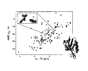
Figure 1. Interaction between the mDvIl PDZ domain and NCI668036.
'5N-HSQC spectra of free NCI668036 (red contour lines) and of NCI668036 bound
to the PDZ
'domain of mDv11 (blue contour lines) are shown, The concentration of the PDZ
domain Was 0.3 ,
= mM. The concentrations of NCI668036 was 7.8 mM (bound form). In the upper
inset, the signals
= froin the same region 'with enlarged spectra were placed in smaller
boxes. The inset also contains
= an additional spectrum (green lines) from a different concentration of
NC1668036 (24 mM). In
the worm representation of the backbone struCture of the mDvIl PDZ domain
(lower inset), the
== thickness of the worm is proportional to the weighted sum (in Hz) of the
'H and shifts upon
binding by iiC1668036; increasing chemical-shift perturbation is shown (blue,
low; red, high).
The figure was prepared by using the software Insight 1JTM (Accelrys, Inc.).
Figure 2. Binding affinity between taDv11 PDZ and NCI668036 as determined
from a
double reciprocal plot of fluorescence intensity quenching (F) against the
concentration of
NCI668036.
, Fluorescence measurements were obtained by titrating NCI668036 into a
solution of the TMIt7
= PDZ domain. The KD value of the complex formed by NCI668036 and the: PDZ
domain of
. mDv11 was 237 31 1.).M as extracted after linear fitting.
=
=
Figure 3. The 30 docking conformations of compound NCI668036 generated by
using
the FJCxXTM program were clustered into three groups.
Group I comprised 5 conformations (red) with RMSDs between 0.46 and 0.77 A,
group II had 13
conformations (yellow) with RMSDs between 1.44 and 1.73 A, and group III had
12
conformations (blue) with RMSDs between 2.31 and 2.86 A.
16
CA 02603642 2011-03-23
WO 2006/107719 PCT/US2006/011754
Figure 4. Backbone root mean square deviations (RMSDs, A) of = the mDv11.
PDZ
domain bound to NCI668036, the mDv11 PDZ domain bound to the Dapper pePtide,
add
the starting structure and total pOteiitial..energies of the MD systems for 5-
ns..explieit
simulations. =
The 200-ps equilibration phase i not included. J I
A.' Backbone RMSDs of the mDv11 PDZ domain (purple) and NCI668036 (green) for
a 5-ni
simulation: =
. B. Backbone RMSDs of the Dvil PDZ domain (purple) and Dapper pePtide (green)
for a 5-ns
simulation.
=. C. The total potential energy (ETOT) oithe mDvIl PDZ domain and
NCI668036 (water
. = molecnles included) during a 5-ns simulation fluctuated between
¨44552.6 kcal mai-land ¨ =
44344.2 kcal molt The total potential energy (mean standard deviation) was
¨44450.8 *32.6
= kcal molt.
D. The total potential energy of the DvIl PDZ dernain (water molecules
included) and Dapper
. . . Peptide during a 5-ns simulation fluctuated between ¨44349.8 kcal mol4
and ¨44122:3 kcal molt =
'The total Potential energy (mean i standard deviation) was ¨.44233.8 i 31.3
kcal mai. =
Figure 5. =Conformation of NCI668036 docked into the PDZ domain and= of the
NCI668036¨mDvIl. PDZ do. main complex.
A. NC1668036 and the Dapper peptide bound to the PDZ 'domain in similar
conformations.
= NCI668036 (blue) was docked into the Dvl PDZ=dotnain (ribbons and tubes
in gray) by using
FIex.XTm (TripoS, Inc.). The Dapper peptide (orange) is in its conformation
determined. by x-ray =
crystallography and is in a complex with the PDZ domain., The difference
between the' backbone
root mean square deviation of compound NCI668036 and that.Of Dapper peptide
(only the 4 C-
terminal amino acids [MITV] backbone atoms were used) was 1:49 A. = , . .
. B. The binding conformation of NCI668036 at 4.905 ns during the 5-ns
simulation. The PDZ
domain is shown as gray ribbons and, tubes. NCI668036 is represented according
to *the bound.
atom (green, carbon; red, oxygen and blue, nitrogen). Residues that formed a
hydrogen bond
with the compound are shown in ball-and-stick format (black, carbon; 'red,
oxygen; blue,
nitrogen); hydrogen bonds are represented by. yellow dashed lines. Residues
within 3.5 A of
isopropyl, methyl (those next to nitrogen atoms), and Autyl groups of compound
are in CPK =
format (gray, carbon; red, oxygen; blue, nitrogen. In addition, Leu258,
11e260, and 11e262 were
17
CA 02603642 2011-03-23
WO 2006/107719 PCT/US2006/011754
within 3.5 A Of the isbpropyl group next. to the carboxylate group. They ire
in ball-and-stick
format for clarity)...
Figure 6. Effect of Na668036 on Canonical Wnt signaling in Xen. opus
embryos.
=A.. NC1668036 inhibited the canonical Wnt Pathway induced by Wnt3A but not by
fl-catenin.
RT-PCR was conducted to analyze the expression of the Xerlopui Wnt target,
gene Siamois in =
ectoderinal explants. Synthetic mRNA corresponding to Wnt3A (1 pg) and.a-
catenin (500 ng)
were injected alone or with NCI668036 (180 ng) into the animal-pole region at
the two-cell
stage, and ectOdermal explants were cUltured until they reached the early
gastrula stage, at which
' time they underwent RT-PCR analysis.
13. A control embryo.that received no injection.
C. An embryo that 'received an injection of Wnt3A mRNA developed a complete
secondary =
, axis.
=
D. An embryo that received coinjections of Wnt3A mRNA and NCI668036 developed
a partial
=
Secondary axis. = =
Figure 7. Molecular structures of NCI & Sigma Aldrich compounds which
were tested
'for their ability to bind to the'Dishevellid protein.
Compounds 221120, 107146045882 and 161613 were found to wealcly bind to Dvl
whereas
compounds 108123, 339938, v8878 and 579270 were found to not bind at all.
Figure 8. Molecular structures of Chem Div compounds which were tested
for their =
ability to bind to the Dishevelled protein. =
Compounds 3237-0565, 3237-0713, 3237-0430, 8006-2560, 0090-0031 and 2372-2393
were =
found to bind to Dvl= whereas 0136-0181 did not.
Figure 9. Molecular structures of Chem Div compounds which were tested for
their
ability to bind to the Dishevelled protein.
Compounds 8004-1312, 3289-8625, 3289-5066, 3237-0719 bound to Dvl. Compounds
8003-
2178, C691-0030, 1748-0253, 1108-0424, 2922-0102, 3379-2274 and 8003-4726 did
not bind to
Dyl.
= =
18
CA 02603642 2011-03-23
WO 2006/107719 PCT/US2006/011754
Figure 10. = Molecular str,ucturet of compounds which were tested for their
ability to
= bind to the Dishevelled pr6tein.
'Compounds 103673, 145882, 3289-5066, 3289-8625, 337837, 7129, 3237-0719,
12,517, pi,
142277, .825.69, 39869, p3; 46893, 661075, 661080, 661086, 661092,
661091,=84123 and 668036
.were all found to bind to Dvl.
Figure 11. StructUre-based alignment of the amino-acid sequences of the PDZ
'domains, of Dvl Homologs and. other proteins.
=
=
Secondary structural elements are indicated above the sequences. Residues at
the gly-his ( H) =
'positions are in boldface type. The asterisk denotes the binding pocket for
.the C
terminUs. Sequence differences among the PDZ domains are indicated by
underlining.
=
. Table 1. Information about atams of simulated systems and dimensions of
water boxes.
= Table 2. Binding free energy components of compound NCI668036 and PDZ
averaged over the
lasi 3 ns ofe 5-ps explicit simulation..
= Table 3. Binding free energy components of the PDZ domain and the Dapper
peptide averaged
.
over the last 3.ns of a 5-ns explicit simulation.
=
Table 4. Binding free energy components of the PDZ domain and NCI4568036 and
' the PDZ domain and the Dapper peptide averaged over the last 3 ns. of the 5-
ns explicit ,
simulatiOnt
Table 5. Hydrogen bonds observed between NCI668036 and the PDZ domain and
between the
Dapper peptide and the PDZ domain during 5-ns explicit simulation-.
= =
Table 6. Effect of NC1668036 on formation of the secondary axis induced by
Wnt3A and B-
catenitr.
aVentro-v-egetal injection of Wnt3A mRNA and P-catenin and of Wnt3A mRNA and
NCI668036
at the two-cell stage. Experimental details are shown in Figure 6B through D.
bDefined as the appearance of a second neural plate on the ventral side of
early neurulae and
19
CA 02603642 2011-03-23
WO 2006/107719 PCT/US2006/011754
ectopic = eyes and cement glands. Percentages indicate the proportion of
embryos that met the
definition.
aotal number of embryos that received injections in two independent
experiments.
=
o
" =Tal)le 1: Atom information of sitnulated systems .and dituensions of
water boxes
Complex = . =PDZ-NCI668036 PDZ-Dapper peptide
No. of atdms inthe ligand 67 = 135
No. of residues in the ligand 1 8 = =. .
0
No. of:atoms in the protein 1348 = = 1348
0
No. of residues in the protein == 90 = 90
No. of Na+ atomi 5 3 =.
= 0
No. of TIP3P molecules 5399 5372 =
L =
=
Total no. of atotns 17617 . 17602 =
=
Box axe = 62Ax67Ax56A 62Ax67Ax56A
cy
r7)
t/I
A
. -
. = =
. . .
, Table 2: Binding free energy coMpcinents of cornpound
NCI668036 and PDZ averaged over the last 3 ns of 5 us
explicitly simulation
.
. .
= 0
.
=
=
_
PDZ-NCI668036. PDZ NCIe68036 =
= Deltab =- =
cm
cf,
-...
Contrib.' Mean" SEC = Mean' . SEcle ' ..
Mean'
SE' . Mean" SE' ,-
cz,
.4
....1
Heice -2726.05 49.15 -2738.88. 52.64
7.31 = . 2.69 .5.52 . 12.57 %D
- - =
H
-306.94 15.67 -272.72 14.71 6.18
2.69 -4039 2.84 vdw
H. = 1832:7927.16 . . 1760.28 25.7 72.51
5.87 0 0
..
_ Hon -1200.2 56.31 -1251.32 59.51 86
6.13 -34.88 12.93 r)
PBsur 31.8 0.5 = 31.9 - 0.5
5.17 = 0.06 -5.27 0.16 0
. - .
- N.,
Mai -1777.12 47.65 -1675.18 51.38 -118.57 2.4
= 16.63 12.7Ef 0,
n)
0
w
0,
PBsoi 474532 47.41 -1643.28 51.13 413.4 =
2.42 . 11.36 12.71 0.
-
_ . .
PBtot -2945.52 21.48 . *-28.94.6 27.13 -27.4
5.38 -23.52 3.36 0"
_
1-,
TS. 16.03 0 15.99 0 13.27 0
-13.23 0
1
. , 0
.=
w
TSrot. - 15.83 0.01 = 15.79 0.01 113
0.21 =
-11.25 0.2
1
N.,
. _ . ,=
.
w
TSvn, 1022.07 - = 4.96 =973.56 4.65 = .
45:67= 1.62 2.84 4.96 .
. _
TS. 1053.93 4.96 1005.34 4.65 = 70.24
1.83 -21.64 = 5.02 .
_ _
.
. 11G
- =
-1.88 - lotat
- . . .
-=oe
=
. Ö
.
0.-.3
All energies in kcal mort.= = -
b
CT, Contribution (PDZ-NCI668036)- Contribution (PDZ) --Contribution
(NCI668036).
'II
, coulombic energy;.11 van der Waals energy; H.,
'internal energy; Hr... = H.. + Hwy, i- Ikt; PB.., non-polar contribution
c,
o.
=-._
for salvation free energy; Pfica, polar contribution fro salvation free
energy; 158. = Pt. -..1- P13.4; PB. = H + P13.1; TS./ TS./
i-
,-.
TS,,,ib, translational/rotational/vibrational entropY; TS.= TS. + TS. +
TSLIGto. = PB. + Hiniõshat -TS10, =-=4
CA
'Average over 150 snapshots. and 15 snapshots for entropy contributions. 0.
'Standard error of mean values. . . =
... ,
.
= =
.
Table 3: Binding free energy components of the PDZ domain and Dapper peptide
averaged over the last 3ns
of 5 ns explicitly simulation'
0
. N
.
0
.
= 0
, r
.
CA
PDZ-Dapper peptide PDZ =
Dapper_peptide Delta - -
o
õ
.-1
= =
. . Mean . Std _ . Mean Std. Mean
Std Mean Std = _. =,..1
1..k
.. , -. . ... .
. .
,
Hocc -3076.24 56.04 -2759.74 50.83 " -127.92
10.73 . -188.58 = 22.76
. . . _
,
, Hydw -315-8 17.33 -268.01 16.27 5.66.
3.81 -53.46 3.51 =
_.... _ . =
_ Hint 1926.1. 25.44 1774.73 . 25.03 151.37
7.34 0. - 0 .
_
Hgs -1465.94 - 57.68 -1253.02 51.63 29.11 .
12.13 -24203 . 23.04 i
, . ,
(-)
_
=
. PB= 34.03 F 0.6 32.83 0.57 =8.21
0.18 . - -7.02 0.1a Stlf . . 0
.. ...
IV
r \ ) Pik., -1764.06 55.33 -1660.76 - 47.57 -.318.15
10.32 214.85 22.79 _ 0,
(7)
.
.
w
PB,,,, -1730.03 55.1 = -1627.93 47.34 -309.94
10.3 207.83 22.73 0,
0.
,
L__ N.,
* PB. -3195.97 25.91 -28a0.94 25.17 -280.83 =
7.24 -34.2 4.13 N.)
0
_
1-,
16.07 0 15.99 0 13.86 0
-13.78 - 0 . =
,
= -
. 0
_
=
TS - _ ., 15.9 .3 0.02
15.79 0.01 12.54 0.05 . -12.42 0.05 =
T
w
=
1
.
N.,
_
w
Svib .7 - 1069 5.22 = 969.69 3.62 "
.100.55 0.69 -0751 : 6.37 _
. .
"
, TS,,,, 1101.7 5.23 . 1001.47 3.63 126.95
0.71 = = = -26.72. == 6.37.
..
/Wt.., . .
- -748 -
7. = . =.
.
en
. . .
-i
= = = = -
=(3
t4
&Abbreviations and equations are the same as thosedefmed for Supplemental
Table 2.. c=
==.
=
c.
o
...
.-
-4
CA
A.
.. - .,. .
, . .
.
o
. Table 4: Binding free energy components of the PDZ domain and NCI668036, the
PDZ and
Dapper peptide averaged over the last 3 ns of 5 ns explicitly simulation
=
=
=
=
Contrib.b. AHdec
Alivdvi Arcs 111:13.e APB. APBs., APB., TS
AGtotal '
NC1668036 5.52 = -40.39 0 16.63 -5.27 11.36 -23.52 -21.64 -1.88
= Dapper peptide -188.58 -53.46 = Q 214.85 -
7.02 207.83 -34.20 -26.72 -7.48 - =
(-)
0
0
=
'All energies are in kcal mort.
0
bContribution (PDZ¨NCI668036) ¨ .Contribution (PDZ) ¨ Contribution (NCI668036)
for NCI668036 and
Contribution (PDZ-Dapper peptide) ¨ Contribution (PDZ) Contribution (Dapper
peptide) for Dapper peptide. 0
Heiec, coulomic energy; 1-1who= van der Waals energy;. Him, internal energy;
AHos, AHeko + /111 +
PB non-polar Contribution for solvation free energy; P13.4, polar
contribution for solvation free energy;
APBsoi = APB + APB; APB = tot AHsa + APB = TAS = TA.Sua + TAS., + TAS,,ib;
AGIõI = AP11tot + s
c)
=
=
- .
Table 5: H-bonds observed between coinpound NCI668036 and PDZ, Dapper peptide
and PDZ during 5 ris explicitly simuLationa 0
t.I
0
.
0
-
" .....
.
i.,
. , . . =
0
=
NCI668036 - PDZ = . Dapper pepti.de
- PDZ = --.1
-.1
=
,.. )...
NCI668036 PDZ , - Occupincyb Dapper peptide
' PDZ .. . Occupancyb , =
0 Leu258N/H 13.5 N./!k100)C1*
Leu258N/H 27.7.
r
01 ' Leu258N/H 85.1 = . Va100
Leu258N/H 98.0
. .
03 Gly259N/H 91.6 = ValOOXT
_Gly259N/H 98.4
. .
' =
03 11e260N/H 32.6 - ., ValOOXT
Ile./60N/H 82.3
.
= = r)
_ .
N/H2 , 11826QN/H 99.8
VaION/H Ile260IN/.1-1 99.1 0
1..)
06 11e262N/H 99.5 . Thr-20 , ,
IIe262N/H 99.8
N) \)
0,
0
.
w
0-1 N1/H5 11e2620 65.1 . Met-3N/H
11e2620 .992 = 0,
0.
0 Arg318 112
= 1..)
o
.. Lys-50 -
G1y264N/H 99.4 . 1-,
1-,
,
1
=
Lys-5N/H
Gly2640 . = 86.9 = .= 0
.
w
.. .
- Ser-70
=Ser266N/H r 85.3 1.)i
. .
w
=
. .
-
=
'The length and angle cutoffs for H-bond are 3.5 A and 129' respectively.
'Occupancy is in the units of percentage. .
iz
g
t.
c,
....
...
-4
(A
ra.
.
.
.
.
b.1
1===
=
-Table 6 Effect of the compound. NCI668036 On the formation of secondary
= axis induced by Wnt3A and 8-catenin =
Double axis)) Single axis
Total!'
No injection = 100%
83
=
Wnt3A 77% . 23%
75 0
r.)
cs)0
Wiat3A/NCI668306 55% = 45% =
78
. ..
11-catenin = 51% 49% =
78
0
=
B-catenin/NCI668306 49% = - 51% =
= 76 0
=
=
aVentro-vegetal injections of Wnt3A naRNA and p-catenin, and NCI668036
at two cell Stage. Experimental details are shown in Figures 7B-7D.
i'Defined as the appearance of a second neural plate on the ventral sida of -
early-neurulae and ectopic eyes and cement glands. Percentages indicate the
proportion of embryos that met the definition.=
.
cTotal numbdr:of enibryos that received.injections in two independent
cf;
experiments
cf.