Language selection

Search

Patent 2603927 Summary

Third-party information liability

Some of the information on this Web page has been provided by external sources. The Government of Canada is not responsible for the accuracy, reliability or currency of the information supplied by external sources. Users wishing to rely upon this information should consult directly with the source of the information. Content provided by external sources is not subject to official languages, privacy and accessibility requirements.

Claims and Abstract availability

Any discrepancies in the text and image of the Claims and Abstract are due to differing posting times. Text of the Claims and Abstract are posted:

  • At the time the application is open to public inspection;
  • At the time of issue of the patent (grant).
(12) Patent Application: (11) CA 2603927
(54) English Title: SYSTEM AND METHOD FOR CHEMICAL IMAGING OF MICROARRAYS
(54) French Title: SYSTEME ET PROCEDE D'IMAGERIE CHIMIQUE DES MICRORESEAUX
Status: Deemed Abandoned and Beyond the Period of Reinstatement - Pending Response to Notice of Disregarded Communication
Bibliographic Data
(51) International Patent Classification (IPC):
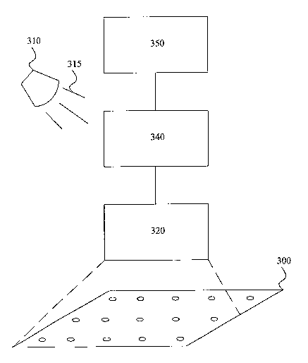
  • G01J 3/00 (2006.01)
  • G01J 3/12 (2006.01)
  • G01J 3/44 (2006.01)
(72) Inventors :
  • TUSCHEL, DAVID (United States of America)
  • VOIGT, THOMAS C. (United States of America)
(73) Owners :
  • CHEMIMAGE CORPORATION
(71) Applicants :
  • CHEMIMAGE CORPORATION (United States of America)
(74) Agent: DIMOCK STRATTON LLP
(74) Associate agent:
(45) Issued:
(86) PCT Filing Date: 2005-04-08
(87) Open to Public Inspection: 2006-10-19
Examination requested: 2008-01-24
Availability of licence: N/A
Dedicated to the Public: N/A
(25) Language of filing: English

Patent Cooperation Treaty (PCT): Yes
(86) PCT Filing Number: PCT/US2005/011922
(87) International Publication Number: WO 2006110135
(85) National Entry: 2007-10-05

(30) Application Priority Data: None

Abstracts

English Abstract


The disclosure relates to systems and method for chemical imaging of
microarrays. In one embodiment, the disclosure relates to a system for
simultaneous spectral imaging of a plurality of samples arranged on an array
(300). The system includes an illumination source (310) for providing
illuminating photons to said plurality of samples, the illuminating photons
interacting with each of the plurality of samples to emit interacted photons;
an array (300) for receiving said plurality of samples, the array (300) having
an external dimension such that the samples are within a simultaneous field of
view of the optical device (320); an optical device (320) for collecting the
interacted photons and directing the photons to an imaging device (350), the
imaging device (350) simultaneously forming a plurality of images
corresponding to each of the plurality of samples.


French Abstract

La présente invention concerne des systèmes et procédés pour imagerie chimique de micro réseaux. Dans un mode de réalisation, l~invention concerne un système pour imagerie spectrale simultanée d~une pluralité d~échantillons agencés sur un réseau (300). Le système comprend une source d~éclairage (310) pour fournir des photons éclairants à ladite pluralité d~échantillons, les photons éclairants interagissant avec chacun de la pluralité des échantillons pour émettre des photons ayant interagi ; un réseau (300) pour recevoir ladite pluralité d~échantillons, le réseau (300) ayant une dimension externe telle que les échantillons sont à l~intérieur d~un champ d~observation simultané du dispositif optique (320); un dispositif optique (320) pour collecter les photons ayant interagi et les diriger vers un dispositif d~imagerie (350), le dispositif d~imagerie (350) formant simultanément une pluralité d~images correspondant à chacun de la pluralité des échantillons.

Claims

Note: Claims are shown in the official language in which they were submitted.


What is claimed is:
1. In a method for providing spectral image of an array of samples provided on
a
substrate by illuminating each sample independently of other samples, the
improvement
comprising the steps of:
(i) ~illuminating all samples with photons simultaneously to produce emitted
photons from each sample;
(ii) ~simultaneously receiving emitted photons from all samples at a tunable
filter;
and;
(iii) ~forming a spectral image from the emitted photons, the spectral image
including a plurality of sub-images, each sub-image corresponding to a sample
in the
array of samples.
2. The method of claim 1, wherein the emitted photons further comprise Raman,
absorption, fluorescence, reflection and transmitted photons.
3. The method of claim 1, wherein the tunable filter is selected from the
group
consisting of LCTF and AOTF.
4. The method of claim 1, further comprising the step of: (iv) selecting a sub-
image
from the plurality of sub-images; and (v) forming an independent image of said
sample.
8

5. A method for simultaneously obtaining a spectral image of plural samples of
an
array, comprising:
(a) ~simultaneously illuminating each of the plurality of samples with
illuminating photons, said illuminating photons interacting with each sample
to produce
interacted photons from each sample;
(b) ~collecting the interacted photons from each sample simultaneously; and
(c) ~forming a spectral image from the collected photons for each of the
plurality of sample simultaneously.
6. The method of claim 5, filtering detected photons through a tunable filter.
7. The method of claim 5, wherein interacted photons include Raman,
Absorption,
fluorescence and reflection.
8. The method of claim 5, further comprising providing an array having a
plurality of
wells.
9. The method of claim 5, further comprising providing an array defined by a
substrate having a plurality of samples.
10. The method of claim 5, further comprising providing an array defined by a
substrate having a plurality of samples, said array having a dimension not
larger than 6
mm2.
11. The method of claim 5, further comprising providing an array defined by a
substrate having a plurality of samples, said array having a dimension not
larger than 3
mm2.
12. The method of claim 5, wherein the spectral image of the plurality of
samples is
spatially-resolved.
9

13. A method for simultaneous spectroscopical imaging of samples comprising:
providing an array defined by at least two samples,
illuminating each of the two samples with illuminating photons, said
illuminating
photons interacting with each sample to produce interacted photons from each
sample;
collecting the interacted photons from each sample simultaneously with an
optical
device; and
forming a spectral image from the collected photons for each sample
simultaneously with a processing apparatus;
wherein the array has an external dimension such that the samples are within a
simultaneous field of view of the optical device.
14. The method of claim 13, wherein illuminating each of the two samples is
simultaneous.
15. The method of claim 13, wherein the processing apparatus is a tunable
filter.
16. The method of claim 13, wherein the optical device further comprises an
optical
lens.
17. The method of claim 13, filtering detected photons through a tunable
filter.
18. The method of claim 13, wherein interacted photons include Raman,
Absorption,
fluorescence and reflection.
19. The method of claim 13, further comprising providing an array having a
plurality
of wells.
20. The method of claim 13, wherein the array is not larger than 6 mm2.
21. The method of claim 13, wherein the array is not larger than 3 mm2.

22. A system for simultaneous spectral imaging of a plurality of samples
arranged on
an array, comprising:
an illumination source for providing illuminating photons to said plurality of
samples, the illuminating photons interacting with each of the plurality of
samples to emit
interacted photons;
an array for receiving said plurality of samples, the array having an external
dimension such that the samples are within a simultaneous field of view of the
optical
device;
an optical device for collecting the interacted photons and directing the
photons to
an imaging device, the imaging device simultaneously forming a plurality of
images
corresponding to each of the plurality of samples;
wherein each of the plurality of samples is spatially-resolved.
23. The system of claim 22, wherein the spectral imaging further comprises
Raman
imaging and Fluorescent imaging.
24. The system of claim 23, wherein the interacted photons include Raman,
fluorescence, illuminating, emitted reflection, absorption and transmission
photons.
25. The system of claim 22, wherein the illumination source is a laser light
source.
26. The system of claim 22, wherein the illumination source is a spot-laser.
27. The system of claim 22, wherein the optical device further comprises a
lens.
28. The system of claim 22, wherein the optical device further comprises an
optical
filter.
29. The system of claim 22, wherein the imaging device further comprises a
tunable
filter and a charge-coupled device.
11

30. The system of claim 22, wherein the array further comprises a substrate
having a
dimension of less than 1 cm2.
31. The system of claim 22, wherein the array further comprises a substrate
having a
dimension of less than 3 mm X 4 mm.
12

Description

Note: Descriptions are shown in the official language in which they were submitted.


CA 02603927 2007-10-05
WO 2006/110135 PCT/US2005/011922
System and Method for Chemical Imaging of Microarrays
Background
[0001] Spectroscopic imaging combines digital imaging and molecular
spectroscopy techniques, which can include Raman scattering, fluorescence,
photoluminescence, ultraviolet, visible and infrared absorption
spectroscopies. When
applied to the chemical analysis of materials, spectroscopic imaging is
commonly
referred to as chemical imaging. Instruments for performing spectroscopic
(i.e., chemical)
imaging typically comprise image gathering optics, focal plane array imaging
detectors
and imaging spectrometers.
[0002] In general, the sample size determines the choice of image gathering
optic.
For example, a microscope is typically employed for the analysis of sub micron
to
millimeter spatial dimension samples. For larger objects, in the range of
millimeter to
meter dimensions, macro lens optics are appropriate. For samples located
within
relatively inaccessible environments, flexible fiberscopes or rigid borescopes
can be
employed. For very large scale objects, such as planetary objects, telescopes
are
appropriate image gathering optics.
[0003] Often the array under study includes multiple samples arranged on an
array
card. Conventional arrays include 4in X 6in well plates having typically 96
wells for
receiving samples. The samples in each well can include similar or dissimilar
substances.
Thus, for example, a conventional array can include as many as 96 different
samples. To
obtain a spectral image or spectra(um) for each sample with a conventional
micro-Raman
instrument an excitation in the form of a spot laser beam of desired
wavelength (killum) is
directed to one of the 96 samples. After a suitable image or spectra(um) of
the first
sample is procured, the illumination source is directed to the subsequent well
and the
process is repeated. The conventional method of serially imaging each sample
is time-
consuming and labor intensive.

CA 02603927 2007-10-05
WO 2006/110135 PCT/US2005/011922
Summary
[0004] In one embodiment, the disclosure relates to a method for
simultaneously
obtaining a spectral image of plural samples of an array. Each image is
spatially
resolved. In one embodiment, the method may include (a) simultaneously
illuminating
each of the plurality of samples with illuminating photons, said illuminating
photons
interacting with each sample to produce interacted photons from each sample;
(b)
collecting the interacted photons from each sample simultaneously; and (c)
forming a
spectral image from the collected photons for each of the plurality of sample
simultaneously.
[00051 In another embodiment, the disclosure relates to a method for
simultaneous
spectroscopic imaging of samples. The method includes providing an array
defined by at
least two samples, illuminating each of the two samples with illuminating
photons, said
illuminating photons interacting with each sample to produce interacted
photons from
each sample; collecting the interacted photons from each sample simultaneously
with an
optical device; and forming a spectral image from the collected photons for
each sample
simultaneously with a processing apparatus. In one embodiment, the array has
an
external dimension such that the samples are within a simultaneous field of
view of the
optical device.
[0006] In still another embodiment, the disclosure relates to a system for
simultaneous spectral imaging of a plurality of samples arranged on an array.
The system
includes an illumination source for providing illuminating photons to said
plurality of
samples, the illuminating photons interacting with each of the plurality of
samples to emit
interacted photons; an array for receiving said plurality of samples, the
array having an
external dimension such that the samples are within a simultaneous field of
view of the
optical device; an optical device for collecting the interacted photons and
directing the
photons to an imaging device, the imaging device simultaneously forming a
plurality of
2

CA 02603927 2007-10-05
WO 2006/110135 PCT/US2005/011922
images corresponding to each of the plurality of samples. Each image is
spatially
resolved.
Brief Description of the Drawings
[00071 Fig. 1 is a schematic representation of a conventional array having
multiple
sample wells;
[0008] Fig. 2 is a schematic representation of a system for spectral imaging
of the
samples in the array of Fig. 1; and
[0009] Fig. 3 is a schematic drawing of an array according to one embodiment
of
the disclosure.
3

CA 02603927 2007-10-05
WO 2006/110135 PCT/US2005/011922
Detailed Description
[0010] Fig. 1 is a schematic representation of a conventional array having
multiple
sample wells. Specifically, Fig. 1 shows array card 100 having wells 110.
Wells 110 are
adapted to each receive a sample (not shown). The sample can be a biological
sample, an
organic sample or an inorganic sample. Array 100 can be seen as having a
dimensions of
a x b. A typical array card has a dimensions of 3in x 4in with a total of 96
wells. The
size of the array card makes it improbable, if not impossible, to fit all of
the samples in
each well (e.g., 96 samples) within the field of view of the imaging device.
In other
words, the field of view of the imaging device is too small to allow
simultaneous spectral
imaging of more than one sample at a time. Consequently, each sample must be
imaged
individually.
[0011] Fig. 2 is a schematic representation of a system for spectral imaging
of the
samples in the array of Fig. 1. System 200 includes array card 100 having
wells 110 with
each well containing a sample. Illumination source 210 directs illuminating
photons 215
having illuminating wavelength (X;ii,,,,,.) to a targeted sample contained in
well 110. The
illuminating photons interact with the sample and emit interacted photons 217
from the
target well. To form an image from emitted photons 217, the target well must
be within
the field of view of optical device 220. Optical device 220 gathers and
directs interacted
photons to imaging system 203 which forms an image of the sample contained in
target
well 110.
[0012] Because of diffraction limitations of the optical device 220 and the
size of
array 100, the field of view of the entire system is limited to one sample and
any image of
the multiple samples will not be spatially resolved. It can be readily seen
from the
schematic representation of Fig. 2, that optical device 220 is limited to the
field of view
of the well immediately before it. Consequently, the wells have to be sampled
one at a
4

CA 02603927 2007-10-05
WO 2006/110135 PCT/US2005/011922
time in serial fashion. To this end, the operator must move array 100 in X
and/or Y
directions sequentially to obtain a spectral image of one sample at-a-time.
[0013] According to one embodiment of the disclosure, the shape, size or form
of
array 100 is configured such that all of the samples fall within the field of
view of the
optical gathering device. For example, a system according to one embodiment of
the
disclosure includes an illumination source for providing illuminating photons
to said
plurality of samples. The illumination can be positioned above, below or in an
oblique
angle with respect to the array. The illuminating photons interact with each
of the
plurality of samples, substantially simultaneously, to emit interacted
photons. An optical
device then collects interacted photons and directs the photons to an imaging
device for
simultaneously forming a plurality of images corresponding to each of the
plurality of
samples.
[0014] An array according to one embodiment of the disclosure can have
external
dimensions such that all or a number of the samples fall within the field of
view of the
optical device or the gathering optics. Because the field of view of a
gathering optic is a
function of the optic's diffraction capability as well as its distance from
the sample, the
array dimensions can be adapted to enable substantially all of the samples to
be within
the field of view of the gathering optics. Thus, according to one embodiment,
the
dimensions of the array are defined as a function of the distance between the
array and
the optics' focal point. According to another embodiment, the dimensions of
the array are
defined such that a portion or all of the samples fall within the field of
view of the
gathering optics. Thus, a spectral image of all of the samples can be formed
simultaneously without having to move the array.
[0015] Fig. 3 is a schematic drawing of an array according to one embodiment
of
the disclosure. Specifically, array 300 is shown with respect to an imaging
system for
capturing multiple images of the samples simultaneously. The samples are
arranged on

CA 02603927 2007-10-05
WO 2006/110135 PCT/US2005/011922
array card 300 and the array card 300 is sized such that all of the samples
fall within the
field of view of gathering optics 320. Gathering optics 320 can include, for
example, one
or more of objective lenses and optical filters.
[0016] Illumination source 310 can be any suitable emission source adapted to
produce photons having the desired excitation wavelength. While illumination
source
3 10 is positioned at an oblique angle with respect to array 300, the
disclosure is not
limited thereto. For example, illumination source 310 can be positioned below
array 300
or above array 300 concentric with gathering optics 320 such that excitation
photons 315
can reach well 300.
[0017] Gathering optics 320 simultaneously collects all, or nearly all of
interacted
photons emitted by the samples. The interacted photons may include Raman,
Fluorescence, absorption, reflection and transmitted photons. The interacted
photons can
be formed by the interaction of illuminating photons with each sample.
Gathering optics
320 focuses the collected photons (not shown) and directs an image formed
therefrom to
tunable filter 340. Tunable filter 340 may include a liquid crystal tunable
filter
("LCTF"), a filter wheel or an acousto-optical tunable filter. Tunable filter
340 can
generate a plurality of spectra (filtered wavelengths) corresponding to each
of the
samples in array 300. An image forming device such as a charge-coupled device
("CCD") or a CMOS image sensor can receive filtered wavelengths from tunable
filter
340 and simultaneously form an image from multiple wells. Since several, if
not all, of
the samples are within the field of view of gathering optics 320, the images
formed by
device 350 includes an image for each of the corresponding samples in the
array.
[0018] According to one embodiment of the disclosure, a method for
simultaneously obtaining spectral images of a plurality of samples in an array
includes:
(a) simultaneously illuminating each of the plurality of samples with
illuminating photons
to produce interacted photons from each of the samples; (b) collecting the
interacted
6

CA 02603927 2007-10-05
WO 2006/110135 PCT/US2005/011922
photons from each sample simultaneously; and (c) forming a spectral image from
the
collected photons for all samples simultaneously. The interacted photons can
include
Raman, Absorption, fluorescence and reflection photons.
[0019] In still another embodiment, the array is designed to have outside
dimensions of about 1- 100 mm2, for example, 3 mm2, 4 mmZ, 6 mm2, 10 mm2.
Other
exemplary dimensions include 2X4 mm2 , 4X4 mm2, 6X4 mm2 and 6X8 mmZ. The
dimensions and the positions of the wells can be adapted to accommodate the
outside
dimensions of the array. Moreover, the number of wells can be increased or
decreased
depending on the desired application. The array dimensions provided herein are
exemplary in nature. Other array dimensions not specifically disclosed are
deemed
within the scope of the principles disclosed herein.
[0020] In another embodiment a magnification lens, or other optical device,
can be
interposed between array 300 and optical device 320 to optically reduce the
size of the
array card such that multiples samples can fit in the view of optical device
320. The
magnification lens can be a stand-alone peripheral unit or can be combined
with
gathering optics 320.
[0021] While the principles of the disclosure have been disclosed in relation
to
specific exemplary embodiments, it is noted that the principles of the
invention are not
limited thereto and include all modification and variation to the specific
embodiments
disclosed herein.
7
Representative Drawing
A single figure which represents the drawing illustrating the invention.
Administrative Status

2024-08-01:As part of the Next Generation Patents (NGP) transition, the Canadian Patents Database (CPD) now contains a more detailed Event History, which replicates the Event Log of our new back-office solution.

Please note that "Inactive:" events refers to events no longer in use in our new back-office solution.

For a clearer understanding of the status of the application/patent presented on this page, the site Disclaimer , as well as the definitions for Patent , Event History , Maintenance Fee  and Payment History  should be consulted.

Event History

Description Date
Application Not Reinstated by Deadline 2010-04-08
Time Limit for Reversal Expired 2010-04-08
Inactive: Abandoned - No reply to Office letter 2009-04-16
Deemed Abandoned - Failure to Respond to Maintenance Fee Notice 2009-04-08
Inactive: Office letter 2009-01-16
Letter Sent 2008-03-04
Request for Examination Received 2008-01-24
All Requirements for Examination Determined Compliant 2008-01-24
Request for Examination Requirements Determined Compliant 2008-01-24
Inactive: Cover page published 2007-12-27
Inactive: Declaration of entitlement/transfer requested - Formalities 2007-12-27
Inactive: Delete abandonment 2007-12-21
Inactive: Notice - National entry - No RFE 2007-12-19
Inactive: First IPC assigned 2007-11-06
Application Received - PCT 2007-11-05
National Entry Requirements Determined Compliant 2007-10-05
Deemed Abandoned - Failure to Respond to Maintenance Fee Notice 2007-04-10
Application Published (Open to Public Inspection) 2006-10-19

Abandonment History

Abandonment Date Reason Reinstatement Date
2009-04-08
2007-04-10

Maintenance Fee

The last payment was received on 2008-01-24

Note : If the full payment has not been received on or before the date indicated, a further fee may be required which may be one of the following

  • the reinstatement fee;
  • the late payment fee; or
  • additional fee to reverse deemed expiry.

Please refer to the CIPO Patent Fees web page to see all current fee amounts.

Fee History

Fee Type Anniversary Year Due Date Paid Date
MF (application, 2nd anniv.) - standard 02 2007-04-10 2007-10-05
Basic national fee - standard 2007-10-05
MF (application, 3rd anniv.) - standard 03 2008-04-08 2008-01-24
Request for examination - standard 2008-01-24
Owners on Record

Note: Records showing the ownership history in alphabetical order.

Current Owners on Record
CHEMIMAGE CORPORATION
Past Owners on Record
DAVID TUSCHEL
THOMAS C. VOIGT
Past Owners that do not appear in the "Owners on Record" listing will appear in other documentation within the application.
Documents

To view selected files, please enter reCAPTCHA code :


To view images, click a link in the Document Description column. To download the documents, select one or more checkboxes in the first column and then click the "Download Selected in PDF format (Zip Archive)" or the "Download Selected as Single PDF" button.

List of published and non-published patent-specific documents on the CPD .

If you have any difficulty accessing content, you can call the Client Service Centre at 1-866-997-1936 or send them an e-mail at CIPO Client Service Centre.


Document
Description 
Date
(yyyy-mm-dd) 
Number of pages   Size of Image (KB) 
Description 2007-10-05 7 323
Representative drawing 2007-10-05 1 6
Claims 2007-10-05 5 148
Drawings 2007-10-05 2 21
Abstract 2007-10-05 2 67
Cover Page 2007-12-27 2 42
Reminder of maintenance fee due 2007-12-19 1 112
Notice of National Entry 2007-12-19 1 194
Acknowledgement of Request for Examination 2008-03-04 1 177
Courtesy - Abandonment Letter (Maintenance Fee) 2009-06-03 1 172
Courtesy - Abandonment Letter (Office letter) 2009-07-09 1 165
PCT 2007-10-05 2 59
Correspondence 2007-12-19 1 27
Fees 2008-01-24 1 31
Correspondence 2009-01-16 1 19