Note: Descriptions are shown in the official language in which they were submitted.
CA 02603963 2012-06-14
f
SURGICAL FIXATION PIN
BACKGROUND OF THE INVENTION
FIELD OF THE INVENTION
[0001] The present invention relates to a pin for use in fractured bones in
arms
or other parts of the body, to bring about support of the fractured bone while
it is
healing.
DESCRIPTION OF THE RELATED ART
10002] It is generally known within the health care field that in the
treatment of
fractured bones means are used to allow reinforcement and support to the area
surrounding a fracture in the bone in question.
[0003] Fractures of the distal forearm (wrist fractures) are the most common
of
all fractures (annual incidence about 3,00011,000,000 inhabitants in the
industrialized
world) and constitutes by it's abundance a major therapeutic problem. Simple
fractures are treated with just a bandage while more complex fractures often
require
open reduction and plate fixation. For a large number of the intermediate
complex
fractures the choice of treatment is less obvious; while plate fixation may be
a too
extensive and expensive procedure bandage immobilization may be insufficient
to hold
fracture fragments in the desired position while the fracture heals. Other
therapeutic
modalities also have their drawbacks: classic external fixation immobilizes
the wrist
joint during treatment and wrist stiffness may ensue. To avoid that, the
external
-1-
CA 02603963 2012-06-14
2009
fixation is often removed before the fracture is consolidated, which may lead
to
secondary displacement of the fracture.
[0004] Another method is to use straight fine wires (1.5- 2.0 mm in diameter)
drilled into the fragments or introduced through the fracture site. While that
is a simple
and minimally invasive procedure it requires power tools. Also, the wires,
being left
protruding through the skin, have to be removed before the patient is able to
mobilize
her wrist.
[0005] The second bone of the forearm, the ulna, is notoriously difficult to
fix,
and fractures of the distal ulna are therefore often neglected. Pins have been
used,
among others, to hold together the bone fragments, or they have been inserted
in the
bone's inner canal. The pin has then either been allowed to remain in place
after the
fractured bone has healed, or it has been extracted afterwards. Plates similar
to angle
iron have also been used and are attached with screws to the bone by the
fracture.
Examples of such aids are shown in International Publication No. WO 01/56452
A2.
[0006] It has been difficult to operate in such supports by the fracture area,
and
even more difficult to remove them. Neither have those supports constituted an
especially good pin in themselves, i.e., they have not interacted with the
bone to
achieve contact against the same during simultaneous tensioning of the pin.
The
introduction of the pin in the bone has not been facilitated with similarly
known pins,
and neither has the screwing of them into the bone been proven to be easy to
achieve.
[0007] It is difficult to insert pins in the bone, and sometimes a power drill
is
required. It is also difficult to securely anchor the pin in the bone, which
is why they
frequently slide out. In addition, they are frequently left protruding from
the skin, with
-2-
CA 02603963 2012-06-14
2009
the risk of both inflammation around the pin as well as infection, which can
spread to
the bone and develop into osteomyelitis.
[0008] An object of the present invention is thus primarily to produce a pin
that
is suitable for use in the healing of fractured bones in the arms or other
parts of the
body, and which solves, among others, the problems identified above, and which
is
also easy and cost effective to manufacture.
SUMMARY OF THE INVENTION
[0009] The object is achieved by means of a pin in accordance with the present
invention, which pin is essentially characterized in that on opposite ends of
the pin, it
includes an angled, rounded front section and one double bent extra cortical
equipped
with an anchor eye, and is formed of spring material.
[0010] Previously disclosed in published French application FR 2,728,155.A1 is
a so-called intramedullary nail.
[0011] The present invention is intended for fixation of moderately complex
fractures. The present implants are pins specifically designed for fixation of
wrist
fractures, but with modifications it can be extended to other fractures. The
implant is
made of 1.6 mm wire (but other materials or dimensions can be used) with
mechanical
properties suitable for this particular use. The larger part of the implant is
introduced
through the fracture line into the intramedullary canal of the main body of
the fractured
bone and thus becomes stabilized. The lesser part of the implant, anatomically
shaped to lie flush against the outer cortex of the distal fragment,
stabilizes the
fracture by serving as a support. Since the implant is introduced through the
fracture
-3-
CA 02603963 2012-06-14
2009
line and into the intramedullary canal no power tools are needed. The implant
has a
low profile and is anatomically shaped and therefore does not normally require
removal - a second procedure is thereby avoided, and the fracture is supported
during
the consolidation period even while normal activity with the wrist and hand is
resumed.
[00121 The form of the implants is as follows:
[0013] Implants for the radius
[0014] The implants meant to fix fractures of the radius have the same basic
configuration and functions; small variations in the design are made with
respect to
variations the anatomy of the specific site where they are used.
[0015] The intramedullary portion is straight but has a curved tip with a
rounded
end to facilitate its introduction into the intramedullary canal.
[0016] The extramedullary portion is shaped to follow the anatomy of the outer
cortex of the radius. It is constituted of a double wire connected by a 180
distal bend
made into the shape of a hoop. That design enlarges the supporting interface
between implant and bone.
10017] The extramedullary and intramedullary parts are roughly parallel but
not
coaxial - they are connected by an intermediate part at about a 90 angle to
each of
the other parts. The length of the intermediate part corresponds to the
thickness of the
cortical wall of the radius at the fracture site. The transverse part prevents
the implant
from sliding out of place.
[0018] One of the double wires is extended beyond the connecting part to form
a fork with the intramedullary part. That is intended to stabilize the implant
against the
outer wall of the main fracture body.
-4-
CA 02603963 2012-06-14
2009
[0019] The hoop formed at the distal end of the implant is shaped to fit a
screw,
which can optionally be used to stabilize the distal fragment.
[0020] Implants for the ulna
[0021] The ulnar implant differs from the radial implant in the respect that
it is
intended to be fixed in position with one or two screws.
[0022] The intramedullary portion is straight and has a pointed tip to allow
its
introduction through the distal part of the ulna into the intramedullary
canal;
[0023] The extramedullary portion has two hoops, the proximal one for screw
fixation of the implant itself to the shaft of the ulna, the distal one for
optional screw
fixation of the distal fragment; and
[0024] The distal fragment is fixed in position by being sandwiched between
the
intramedullary and extramedullary portions of the implant.
BRIEF DESCRIPTION OF THE DRAWINGS
[0025] The invention is described below in terms of a number of preferred
embodiments, whereby reference is made to the appended drawings, in which:
[0026] Fig. 1 schematically shows a pin in accordance with the present
invention implanted in a forearm,
[0027] Fig. 2 shows different views of a pin in accordance with a first
preferred
embodiment of the present invention,
[0028] Figs. 3 and 4 show different plan views of the first pin embodiment,
[0029] Fig. 5 shows different views of a pin in accordance with a second
preferred embodiment of the present invention,
-5-
CA 02603963 2012-06-14
2009
[0030] Figs. 6-10 show different plan views of the second pin embodiment,
[0031] Figs. 11-14 show different examples of the assembly in a bone of
different types of pins in accordance with the present invention,
[0032] Fig. 15 shows different views of a bone fixation screw in accordance
with
the invention, and
[0033] Fig. 16 schematically shows a sketch of how a pin is applied in a bone
fracture and thereafter inserted in position.
DESCRIPTION OF THE PREFERRED EMBODIMENTS
[0034] Referring to Fig. 1, a surgical fixation pin 1 in accordance with the
present invention is included in a simple and effective system to be used as
an aid
when treating irregular distal radius and ulna fractures 2. The pin 1 is
arranged to
achieve maximum stability by using the smallest possible operation and
implant. Pin 1
is further arranged to make it easy to insert through the fracture 2 and will
be
distinctive when inserted against the edge of the fracture in the proximal
bone
fragment in position in bone 3. A fixing device in the form of bone screw 9
(see Fig.
14) is ideal to be utilized together with a fixation pin of the type described
herein to
further achieve the stability of the fracture site. The insertion of the pin 1
is done
through a small incision, as is shown in Fig. 1.
[0035] A pin I (Figs. 1-4); 101 (Figs. 5-10); 201 (Fig. 14) that is ideal for
use
with fracture 2 on bone 3 in arms 14 or other body parts, to achieve support
and
stability to the bone 3 when healing the fracture 2 in question, is formed in
accordance
with the present invention principally of three different designs. The
different pins 1;
-6-
CA 02603963 2012-06-14
2009
101; 201 are called "Radius Contour Pin," "Radius dorsal Pin," and "Ulna Pin,"
respectively.
[0036] In accordance with the invention, the characteristic for all of those
pins is
that at opposite ends 1A, 1B (see Fig. 2); 101A, 101B (see Fig. 5), and 201A
and
201 B (see Fig. 14) the pins have an angled, rounded front part 4; 104; and
204,
respectively, and a double bend with anchor eye 5; 105; 205 at a rear support
part 6;
106; 206; respectively. Pin sections 7A, 7B; 107A, 107B; 207A, 207B lie
parallel and
in close contact with each other. Each of pins 1; 101; 201 is formed of a
spring
material, preferably spring steel.
[0037] The rear support parts 6; 106; 206 of the pins are formed by a bend,
from the center sections 1 C; 101 C; 201 C, of the pins with respective blunt
angle bent
sections 8; 108; 208, and from each of the bent sections a respective
extending end
section, which is double bent with each pair of pin end sections 7A, 7B; 107A,
107B;
207A, 207B lying in tight contact with each other.
[0038] Both pin sections 7A, 7B; 107A, 107B; 207A, 207B can be straight
(see Fig. 5 and Fig. 14) or curved (see Fig. 2).
[0039] Fig. 14 shows a variant where the pin 201 is bent back at the end 201 B
through an angle of about 180 and is especially suitable for anchorage with
screw 9
to the bone 3 in question.
[0040] A hole-shaped outer eye 5; 105; 205, designed to receive therein a
fixation screw 9 or some other fixing device, is located adjacent to the pin
end
sections' outer ends 10; 110; 210, respectively. In that connection, in the
Fig. 14
variant a further hole-shaped inner eye 211 can be arranged, which is designed
to
-7-
CA 02603963 2012-06-14
2009
receive therein a further fixation screw 9A, or some other fixing device, and
which is
located at a distance A from the first-mentioned outer eye 205.
[0041] The rear support part 206 of the pin 201 that includes the above-
mentioned screw eyes 205, 211 is bent back at an angle of about 180 so that
the
reformed section 212 is located parallel with the remaining section 213 of the
pin 201
and is kept at the distance B from it, as shown in Fig. 14. On the other pins
1; 101 the
above-mentioned blunt angle 8, 108, respectively, is essentially right-angled.
The
function of the bending back is for the pin to be kept constantly in position
at fracture 2,
with the pin's transverse section extending across the linear extension of
bone 3, and
with the pin's both parallel pin sections 207A, 207B arranged to extend
internally within
and externally on the outer surface of the bone, respectively.
[0042] The above-mentioned angled, rounded end sections 4; 104; 204 and the
above-mentioned bent sections 6; 106; 206 extend along each plane 15,16 (see
Fig.
4), which are essentially arranged at right angles in relation to each other.
[0043] The fixation pin is principally of a flat design, with a principally
circular
cross section, formed from materials such as spring steel, titanium, stainless
steel,
plastic, resorbing material, or a composite plastic.
[0044] Screw 9, which is arranged to secure pins 1; 101; 201 in position, is
shown in Fig 15. The screw includes threads 17 from the outer end 18 of the
screw up
to a plain, unthreaded section 19 that is located next to or at a distance
from the screw
head 20. A ring-shaped receiving section 21 is thereby arranged on the screw 9
to be
able to be surrounded by the ring shaped eyes 5; 105; 205 in the secured
position and
-8-
CA 02603963 2012-06-14
2009
clicked in position therein. Depending upon the appearance of the bone
fracture 2, a
screw that is suitable for each type of fracture is used.
[0045] A variant of the screw includes threads right up to the screw head 20.
The above-mentioned threads are designed to work together with the pin's round
cross
section part.
[0046] As shown for example in Fig. 7, angled, rounded front sections 4; 104;
204 of the pins can be inclined at an angle Y between 10 -30 to the pin
longitudinal
axis and angled relative to the double bent rear support section 106 that lies
in
common plane 22.
[0047] Depending upon the type of fracture on the bone 3 that has occurred at
the time of the accident, different pins of the type described above are used,
and the
application of the same can vary. As shown in Fig. 12A, the pin I in
accordance with
the examples shown in Figs. 1-4 is a so-called "Radius Contour Pin" 1 and that
type of
pin is inserted through the actual radial fracture 2, as represented by
position I of Fig.
12A. As shown in Fig. 12B, bent rear support section 6 of the pin 1 when it is
in
position II of Fig. 12B will lie tight against the bone 3 and thereby minimize
irritation of
nerves and tendons. The extended section, located outside of bone 3, of the
rear
support section 6 produces good support against the close lying fragment of
the bone
3. The possibility of additional fixation with screw 9 also exists. The
angled, rounded
front part 4 facilitates sliding of the pin against the inside wall of the
cortex of bone 3.
[0048] The pin in accordance with the example as shown in Figs. 5-10 is a so-
called "Dorsal pin" 101. Such a pin is arranged to be inserted through a
"dorsal"
fracture line and is arranged to support the bone fragment with the support
section 106
-9-
CA 02603963 2012-06-14
2009
located at the rear of the pin 101. The double rear pin sections 107A, 107B on
the
above-mentioned rear support section 106 distribute the pressure across a
large area
and reduce the risk of cutting into the patient's brittle bone. The
possibility of extra
bone fixation with screws also exists with pin 101. The dorsal shank on the
pin
provides good support to the distal fragment, and the transverse bend back
prevents
the pin from starting to move. The extra cortical shank, i.e., the section of
the pin
which is arranged to come outside of the bone cortex on the bone, exists in
different
lengths in order to be varied depending upon different types of fracture
positions.
Even with that pin it is possible to anchor it with screws to further increase
stability.
[0049] Effective contact and support against bone 3 is achieved with the above-
mentioned protruding pin end section through the free end 7C; 107C of one pin
end
section 7B; 107B of both pin end sections 7A, 7B; 107A, 107B that extends past
the
bent sections 8; 108 of the pins 1; 101.
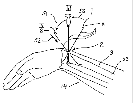
[0050] Fig. 16 shows how the pin 1 in position I is first inserted through the
fracture site 2 in a forearm 14 across the longitudinal direction of bone 3.
After
insertion the pin 1 is tilted in the direction of arrow 50 to an intermediate
position III at
which the pin is rotated about 180 , whereupon continued tilting of pin I
occurs in the
direction of arrow 51 to the final position IV. Pin I is then pressed inward
with its front
section in the direction of arrow 52 internally into the bone-marrow 53 within
the bone
3 a distance so that the angled section 8 on the pin 1 extends across at the
fracture
site 2 and continues with its rear support part 6 to rest against the outside
of the bone
3, as shown in Fig. 12B at the final position II. If the fracture is simple,
it can be
-10-
CA 02603963 2012-06-14
2009
sufficient for one pin to be used, but otherwise two or more pins must be used
to
effectively hold together the bone sections at the fracture site 2.
[0051] Finally, in Fig. 14 examples of a so-called "Ulna Pin" 201 are shown,
which is used with extremely complex fractures with many small bone fragments,
such
as a malpositioned fracture, sometimes with several fragments, on the lower
part of
the elbow bone. The pin 201 is inserted through the fracture's distal section
into the
medullary canal of the ulna. It is then secured to the bone 3 with fixation
screws 9, 9A
which are received in the screw eyes 205, 211 on the back bent reformed
section 212
of the pin 201 to hold the bone fragments relative to the part of the bone
that has not
been fractured. The pin can be left in the bone in the form of an implant.
[0052] On account of the pin's spring properties, the pin stabilizes the
fragment
of the fracture through the tension between the distal cortex and the proximal
medullary canal. The pin is inserted until the transverse section of the pin
snaps into
the fracture at the fracture line.
[0053] An additional advantage offered by the present invention is that the
pin in
accordance with the invention is anatomically designed so that it lies tight
against the
bone, and thereby effectively increases the contact surface against the
contact section
of the bone.
[0054] Consequently, the pin is ideal to be utilized both to position the
fracture
into he right position and to hold together the bone fragments.
[0055] French published patent application FR 2,728,155 Al describes a typical
so-called intermedullary nail, i.e., a device designed to stabilize a broken
tubular bone
through insertion in the medullary canal on each side of the fracture. The
insertion in
-11-
CA 02603963 2012-06-14
2009
the medullary canal takes place through an artificial channel effected into
one end in
one of the fragments.
[0056] The present invention's radial contour pin and dorsal radius pin are
inserted into the fracture site through the existing fracture gap.
Accordingly, the pin is
only inserted into the medullary canal of only one of the fracture fragments,
which
secures the pin. In turn, the pin provides support to the other fragment by
resting
against that fragment's outer area.
[0057] The bending (7) of the device described in the French publication has
as
an object to permit the insertion of the medullary nail into the medullary
canal via an
(artificial) opening in the bone that does not lie in the medullary canal's
longitudinal
direction.
[0058] The present invention's principle of operation involves the external
shank
to be connected to the existing anatomy/topography of the outside of the
distal
fragment, which is why the bend connects to the existing anatomy. In addition,
the pin
in accordance with the present invention is double bent, with a parallel
offset of the
extramedullary and the intramedullary parts to create an intermediate shank
(8, 108,
208) (intrafocal shank) whose length corresponds to the thickness of the
cortical bone
at the fracture gap.
[0059] The device in the French publication has two parallel intramedullary
shanks.
[0060] The invention has one intramedullary shank, but different forms on the
extramedullary shank, which are chosen depending upon how the fracture
appears,
and where the pin is to be inserted.
-12-
CA 02603963 2012-06-14
2009
[0061] On the device disclosed in the French publication the extra cortical
curve .
is the necessary connection between the two intramedullary shanks.
[0062] In the present invention, the extra cortical part is designed to give
the
greatest possible contact against the distal fragments surface and by doing so
to
achieve stability in the same plane as the short shank. That has been achieved
through the anatomically designed hooks, and thus an even larger contact
surface is
achieved extra cortically through doubling and by the ring form of the
external shank.
[0063] The device disclosed in the French publication has been made securable
through different fastening devices that can be connected to the
extramedullary curve.
That has been done to prevent the device from sliding out of the medullary
canal, a
problem that is known for straight and slightly angled intramedullary nails.
[0064] The pin in accordance with the present invention is stabilized in the
skeleton through the intra focal shank (8), i.e., the connection shank between
the intra
and the extramedullary parts of the pin, which is perpendicular to the
fracture line and
will hook into the fracture line. Further fixation in the proximal fragment is
obtained
through the extra cortical shank's returning section being so long that it
passes the
fracture gap and with that runs in parallel to the intramedullary shank on the
way from
the fracture gap to the extramedullary shank's tip. This produces a "fork"
that
straddles the cortex of the proximal fragment. The eye in the pin in
accordance with
the present invention can also be used to fix the distal fragment to the
device with a
special screw, through which stability of the distal fragment is increased.
The screw is
thus not intended to prevent the pin from sliding out of the medullary canal.
-13-
CA 02603963 2012-06-14
r r
2009
[0065] A pure intramedullary device like that disclosed in the French
publication
can not safely stabilize a fractured wrist. The device must be partly inserted
through
the joint surface, or very close to the joint surface, which could injure the
wrist.
Further, the contact surface between the device and the distal bone fragment
would be
so-small that stability would be insufficient.
[0066] The present invention includes different forms on the extramedullary
shank, which are chosen depending upon how the fracture appears and where the
pin
is to be inserted. In general, at least two pins are used, but sometimes three
different
pins are used, inserted through separate openings, so that the fracture is
stabilized on
different planes. The anatomical 'design of the pin's extra cortical section
is a
prerequisite for the stabilization of the brittle (osteoporotic) bone, which
is usually the
cause of fractured wrists in the elderly. The pin's design utilizes the curved
form of the
cortical bone in the fragment close to the distal joint, so that with a small
amount of
foreign material a large contact surface is provided, which distributes the
pressure
from the pin over a greater part of the distal fragment.
[0067] The angled front section (3, 4), which is shown in the French
publication,
is a well-known design solution for all implants that are inserted in the
medullary canal.
That hook can be said to make up a "ski tip," which allows the implant to
slide down
despite the fact that the implant is held angled to the medullary canal's
longitudinal
axis. The top of that "ski tip" is sharp in order to able to penetrate the
cortex during
insertion.
[0068] The pin in accordance with the present invention has a top with a more
rounded design in order to allow maneuvering inside the medullary canal via
the
-14-
CA 02603963 2012-06-14
r
2009
fracture gap, without catching against the opposite side of the medullary
canal or
penetrating the opposite cortex. That characteristic is only utilized at the
start of the
insertion. Further inside the medullary canal the pin can be rotated when the
bend
close to the rounded tip is utilized to coax the pin past any unevenness in
the
medullary canal. That step is impossible with the pin disclosed in the French
publication, because due to its design it can not be rotated.
[0069] In summary the above shows that, the device disclosed in the French
publication and the pin in accordance with the present invention have
different
designs, different functions, and different areas of application.
[0070] There is therefore no motivation for one skilled in the art to invent
the
structural design in accordance with the present invention.
[0071] The present invention is naturally not limited to the above description
or
to the designs shown in the appended drawings. Modifications are possible,
especially with regard to the character of the different parts, or through the
use of
equivalent technology, without deviating from the restricted area for the
invention, such
as it is defined in the claims.
-15-