Note: Descriptions are shown in the official language in which they were submitted.
CA 02604635 2007-10-12
WO 2006/138461 PCT/US2006/023305
Treatment and diagnostic catheters with hydrogel electrodes
BACKGROUND OF THE INVENTION
a. Field of the Invention
[0001] The instant invention is directed toward hydrogel electrode catheters
for
treatment and diagnosis of tissue. More specifically, the instant invention
relates to
treatment and diagnostic catheters with hydrogel virtual and sensing
electrodes.
b. Background Art [0002] Catlieters have been in use for medical procedures
for many years. Catheters
can be used for medical procedures to examine, diagnose, and treat tissue
while positioned
at a specific location within the body that is otherwise inaccessible without
more invasive
procedures (e.g., medical procedures involving the human heart). During these
procedures
a catheter is inserted into a vessel located near the surface of a human body
(e.g., an artery
or vein in the leg, neck, or arm of the patient) and is guided or threaded
through the
vessels, sometimes with the aid of a guidewire or introducer, to a specific
location within
the body for examination, diagnosis, and treatment. For example, one procedure
often
referred to as "ablation" utilizes a catheter to convey energy (e.g.,
electrical or thermal) or
a chemical to a selected location within the human body to create necrosis,
which cuts off
the path for stray or improper electrical signals. Another procedure often
referred to as
"mapping" utilizes a catheter with one or more sensing electrodes to monitor
various forms
of electrical activity in the human body.
[0003] It is well known that benefits may be gained by forming lesions in
tissue
during catheter ablation if the depth and location of the lesions being formed
can be
controlled. In particular, it can be desirable to elevate tissue temperature
to around 50 C
until lesions are formed via coagulation necrosis, which changes the
electrical properties of
the tissue. When sufficiently deep lesions are formed at specific locations in
cardiac tissue
via coagulation necrosis, undesirable atrial fibrillations may be lessened or
eliminated.
"Sufficiently deep" lesions means transmural lesions in some cardiac
applications.
[0004] Several difficulties may be encountered, however, when attempting to
form
adequately-deep lesions at specific locations using some existing ablation
electrodes. For
example, when forming lesions with radiofrequency (RF) energy, high
temperature
1
CA 02604635 2007-10-12
WO 2006/138461 PCT/US2006/023305
gradients are often encountered in the vicinity of the electrode. At the edges
of some
existing electrodes are regions of very high current density, leading to large
temperature
gradients and hot spots. These "edge effects" may result in the formation of
undesirable
coagulum and charring of the surface tissue. For example, undesirable coagulum
may
begin to form when blood reaches around 80 C for an appreciable length of
time, and
undesirable tissue charring and desiccation may be seen when tissue reaches
around 100 C
for an appreciable length of time. There are two main types of undesirable
coagulum:
coagulum that adheres to and damages the medical device; and coagulum blood
clots or
curds that may enter a patient's bloodstream, possibly resulting in other
health problems
for the patient. Charring of the surface tissue may also have deleterious
effects on a
patient.
[0005] During RF ablation, as the temperature of the electrode is increased,
the
contact time required to form an adequately-deep lesion decreases, but the
likelihood of
charring surface tissue and forming undesirable coagulum increases. As the
temperature of
the electrode is decreased, the contact time required to form an adequately-
deep lesion
increases, but the likelihood of charring surface tissue and forming
undesirable coagulum
decreases. It is, therefore, a balancing act trying to ensure that tissue
temperatures are
adequately high for long enough to create deep lesions, while still preventing
or
minimizing coagulum formation and/or charring of the surface tissue. Active
temperature
control may help, but the placement of thermocouples, for example, is tricky
and setting
the RF generator for a certain temperature becomes an empirical exercise as
actual tissue
temperatures are generally different from those recorded next to the electrode
due to
factors such as convection and catheter design.
[0006] Conventional mapping catheters may include, for example, a plurality of
adjacent ring electrodes constructed from platinum or some other metal. Since
mapping
catheters are desirably disposable, incorporation of relatively expensive
platinum
electrodes may be disadvantageous.
[0007] Another difficulty encountered with existing ablation catheters and
mapping
catheters is how to ensure adequate tissue contact. For example, current
techniques for
creating linear lesions (the term "linear lesion" as used herein means an
elongated,
continuous or uninterrupted lesion, whether straight or curved and whether
comprising a
2
CA 02604635 2007-10-12
WO 2006/138461 PCT/US2006/023305
single line of ablation or a series of connected points or lines of ablation
forming a track,
that blocks electrical conduction) in endocardial applications may include
dragging a
conventional catheter on the tissue, using an array electrode, or using pre-
formed
electrodes. All of these devices comprise rigid electrodes that do not always
conform to
the tissue surface, especially when sharp gradients and undulations are
present, such as at
the ostium of the pulmonary vein in the left atrium and the isth.mus of the
right atrium.
Consequently, continuous linear lesions are difficult to achieve. Whether
forming lesions
or mapping in a heart, the beating of the heart, especially if erratic or
irregular, further
conlplicates matters, making it difficult to keep adequate contact between
electrodes and
tissue for a sufficient length of time. For example, with a rigid electrode,
it can be quite
difficult to maintain sufficient contact pressure during lesion formation
until an adequate
lesion has been formed. These problems are exacerbated on contoured or
trabeculated
surfaces. If the contact between electrodes and tissue cannot be properly
maintained,
quality lesions or accurate mapping are unlikely to result.
[0008] Catheters based upon a virtual electrode that deliver RF energy via
conductive
fluid flowing into the patient's body address some of the difficulties with
ablation
catheters, but these ablation catheters often require high flow rates of the
conductive fluid
(e.g., typically around 70 milliliters per minute) to maintain effective
cooling for high-
power RF applications. The introduction of a large amount of conductive fluid
into a
patient's bloodstream may have detrimental effects on the patient.
[0009] Thus, there remains a need for ablation catheters and mapping catheters
that
address these issues with the existing designs.
BRIEF SUMMARY OF THE INVENTION
[0010] Accordingly, it is an object of the disclosed invention to provide
improved
treatment and diagnostic catheters.
[0011] In one form, the present invention comprises a catheter for treatment
of tissue,
the catheter comprising at least one conductive hydrogel virtual electrode
adapted to
contact the tissue to be treated. In this form, the catheter includes a distal
portion that
comprises a straight section; a hoop-shaped section; an offset that joins the
straight section
to the hoop-shaped section; an active region along the hoop-shaped section;
and a hydrogel
3
CA 02604635 2007-10-12
WO 2006/138461 PCT/US2006/023305
delivery feature along the active region, wherein the hydrogel delivery
feature is adapted to
be placed against the tissue to be treated. The hoop-shaped section may defme
a
distally-facing surface, and the hydrogel delivery feature may be on that
distally-facing
surface. Alternatively, the hoop-shaped section may define a radially outer
peripheral wall
that includes an outwardly-facing surface, and the hydrogel delivery feature
may be on that
outwardly-facing surface. The hydrogel delivery feature comprises at least one
opening
extending through the distally-facing surface or the outwardly-facing surface.
The at least
one opening may comprise, for example, a single row of hydrogel portholes, a
plurality of
rows of hydrogel portholes radially, a single hydrogel slot, or a plurality of
hydrogel slots.
The at least one opening my be centered about a radial apex of the distally-
facing surface
or of the outwardly-facing surface.
[0012] In another form, the present invention again comprises a catheter for
treatment
of tissue, the catheter comprising at least one conductive hydrogel virtual
electrode adapted
to contact the tissue to be treated. In this form, the catheter includes a
distal portion that
comprises a straight active region, the straight active region extending
parallel to a catheter
longitudinal axis; and a hydrogel delivery feature along the straight active
region, the
hydrogel delivery feature being adapted to be placed against the tissue to be
treated. The
straight active region defines an outer peripheral wall, wherein the outer
peripheral wall
defines an outwardly-facing surface, wherein the hydrogel delivery feature is
on the
outwardly-facing surface. The hydrogel delivery feature comprises at least one
opening
extending through the outer peripheral wall and its outwardly-facing surface.
The at least
one opening may comprise, for example, a single row of hydrogel portholes, a
plurality of
rows of hydrogel portholes radially, a single hydrogel slot, or a plurality of
hydrogel slots.
The at least one opening may be centered about a radial apex of the outwardly-
facing
surface.
[0013] In yet another form, the present invention comprises a catheter for
treatment of
tissue, the catheter comprising at least one conductive hydrogel virtual
electrode, wherein
the at least one conductive hydrogel virtual electrode is contained within a
permeable or
semi-permeable containment membrane adapted to contact the tissue to be
treated. The
membrane may comprise a shaped membrane adapted to take a predetermined
configuration when filled with conductive hydrogel. For example, the
containment
4
CA 02604635 2007-10-12
WO 2006/138461 PCT/US2006/023305
membrane, when filled with conductive hydrogel, may be adapted to form a
protuberance
having a conformable surface to contact the tissue to be treated. This
protuberance may
take the shape of a hemisphere, a knob, a flattened gob, a hook, or a hoop.
[0014] In another form, the present invention comprises a drug delivery
catheter for
treatment of cardiac arrhythmias. In this embodiment, the catheter comprising
a distal
portion having an active region; a lumen extending inside the catheter
adjacent to the
active region; and a hydrogel delivery feature along the active region and in
fluid
communication with the lumen, wherein the hydrogel delivery feature is adapted
to be
placed against arrhythmia-producing, cardiac tissue inside of a heart. A
conductive
hydrogel matrix is present in the lumen, wherein the conductive hydrogel
matrix is loaded
with, for example, a water-soluble and ionic dispensable drug formulation. The
hydrogel
delivery feature may comprise, for example, a plurality of hydrogel portholes;
and a
permeable membrane attached at the plurality of hydrogel portholes and adapted
to be
alternatingly extendable out of and retractable back into the plurality of
hydrogel portholes,
wherein the membrane is adapted to contain the conductive hydrogel matrix,
wherein the
membrane is adapted to make contact with the cardiac tissue, and wherein the
membrane is
adapted to be traversable by the drug formulation.
[0015] In still another form, the present invention comprises a drug delivery
system
for treatment of cardiac arrhythmias. The system comprises a catheter having a
distal
portion. The distal portion of the catheter comprises an active region; a
lumen extending
adjacent to the active region, the lumen being adapted to contain a conductive
hydrogel
matrix loaded with, for example, a water-soluble and ionic dispensable drug
formulation;
and a hydrogel delivery feature. The hydrogel delivery feature comprises an
opening
through the active region, the opening being in fluid communication with the
lumen and
being adapted to be placed against arrhythmia-producing, cardiac tissue; and a
permeable
membrane attached at the opening and adapted to be alternatingly extendable
out of and
retractable back into the opening, wherein the membrane is adapted to contain
the
conductive hydrogel matrix, wherein the membrane is adapted to make contact
with the
cardiac tissue, and wherein the membrane is adapted to be traversable by the
ionic
dispensable drug formulation. In this embodiment, the system also comprises a
current
supply adapted to deliver low-intensity direct current to the conductive
hydrogel matrix.
CA 02604635 2007-10-12
WO 2006/138461 PCT/US2006/023305
The opening through the sidewall of the catheter may comprise, for example, at
least one
hydrogel porthole or at least one hydrogel slot.
[0016] In a nother form, the present invention comprises a diagnostic catheter
for
diagnosing cardiac tissue, the catheter comprising at least one conductive
hydrogel sensing
electrode. The at least one conductive hydrogel sensing electrode may comprise
a plurality
of isolated, conductive hydrogel disks that are electrically separated by
nonconductive
hydrogel disks. These conductive and nonconductive hydrogel disks may be
constructed
from, for example, high-viscosity, rigid hydrogel that is substantially
unaffected by
moisture. The conductive hydrogel disks may be adhered to the nonconductive
hydrogel
disks. In this embodiment, each of the plurality of conductive hydrogel disks
is electrically
connected with a separate electrical lead (e.g., a silver or silver-chloride
coated wire) for
transmitting electrical signals from the treatment site to instrumentation
outside of a
patient's body.
[0017] In yet another form, the present invention comprises a method of
treating
cardiac tissue. The method comprises the steps of guiding an ablation catheter
having at
least one conductive hydrogel virtual electrode to the cardiac tissue to be
treated;
introducing the at least one conductive hydrogel virtual electrode against the
cardiac tissue;
and directing ablative energy to the cardiac tissue via the at least one
conductive hydrogel
virtual electrode.
[0018] In still another form, the present invention comprises a method of
treating
cardiac tissue, the method comprising the steps of filling at least a distal
portion of a
catheter lumen with conductive hydrogel, the catheter lumen extending adjacent
to a
catheter active region on a catheter outer surface; guiding the catheter
active region into
contact with the cardiac tissue to be treated; activating a hydrogel
displacement device to
advance the conductive hydrogel toward the active region until the conductive
hydrogel
broaches the catheter outer surface to thereby introduce at least one
conductive hydrogel
virtual electrode against the cardiac tissue; directing ablative energy
through the at least
one conductive hydrogel virtual electrode and into the cardiac tissue; and
activating the
hydrogel displacement device to retract the conductive hydrogel and thus the
at least one
conductive hydrogel virtual electrode from contact with the cardiac tissue and
back into the
catheter lumen.
6
CA 02604635 2007-10-12
WO 2006/138461 PCT/US2006/023305
[0019] The foregoing and other aspects, features, details, utilities, and
advantages of
the present invention will be apparent from reading the following description
and claims,
and from reviewing the accompanying drawings.
BRIEF DESCRIPTION OF THE DRAWINGS
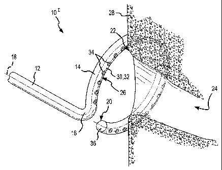
[0020] Fig. 1 is fragmentary, isometric view of the distal portion of an
ablation
catheter according to a first embodiment of the present invention adjacent to
the ostium of
a pulmonary vein.
[0021] Fig. 2 is a fragmentary, isometric view of the distal portion of an
ablation
catheter according to a second embodiment of the present invention.
[0022] Fig. 3 is a fragmentary, isometric view of the distal portion of an
ablation
catheter according to a third embodiment of the present invention depicted
next to the
ostium of a pulmonary vein.
[0023] Fig. 4 is a fragmentary, isometric view of the distal portion of an
ablation
catheter according to a fourth embodiment of the present invention.
[0024] Fig. 5 is a fragmentary, top view of the distal portion of an ablation
catheter
according to a fifth embodiment of the present invention.
[0025] Fig. 6 is a fragmentary, end view (looking distally) of the ablation
catheter
depicted in Fig. 5, shown with at least partially deployed conductive hydrogel
protruding
from the hydrogel portholes.
[0026] Fig. 7 is a fragmentary, side view of the ablation catheter depicted in
Figs. 5
and 6, shown with the conductive hydrogel retracted into the catheter.
[0027] Fig. 8 is a fragmentary, top view of the distal portion of an ablation
catheter
according to a sixth embodiment of the present invention.
[0028] Fig. 9 is a fragmentary, top view of the distal portion of an ablation
catheter
according to a seventh embodiment of the present invention.
[0029] Fig. 10 is a fragmentary, top view of the distal portion of an ablation
catheter
according to an eighth embodiment of the present invention.
[0030] Fig. 11 is an enlarged, fragmentary view of the portion that is circled
in Fig.
10.
7
CA 02604635 2007-10-12
WO 2006/138461 PCT/US2006/023305
[0031] Fig. 12 is a fragmentary, cross-sectional view taken along line 12-12
of Fig.
10, shown with the conductive hydrogel poised at the hydrogel porthole exits
prior to being
forced to protrude from the portholes.
[0032] Fig. 13 is similar to Fig. 12, but depicts the conductive hydrogel in
its deployed
configuration, protruding from the portholes against the tissue to be treated.
[0033] Fig. 14 is a fragmentary, cross-sectional view taken along line 14-14
of Fig. 13
and depicts ablative energy being transferred to the tissue through the
conductive hydrogel.
[0034] Fig. 15 is a fragmentary, cross-sectional view of the distal portion of
an
ablation catheter according to a ninth embodiment and depicts a membrane
containing the
protruding conductive hydrogel.
[0035] Fig. 16 is a fragmentary, top view of the distal portion of an ablation
catheter
according to a tenth embodiment of the present invention prior to deployment
of the
conductive hydrogel.
[0036] Figs. 17, 18, and 19 are fragmentary, top views of the distal portion
of an
ablation catheter according to a first variant, a second variant, and a third
variant,
respectively, of the tenth embodiment of the present invention.
[0037] Fig. 20 is a fragmentary, cross-sectional view of the distal portion of
a
hydrogel drug delivery catheter according to an eleventh embodiment of the
present
invention.
[0038] Fig. 21 is a fragmentary, cross-sectional view of the distal portion of
a
hydrogel drug delivery catheter according to a twelfth embodiment of the
present
invention.
[0039] Fig. 22 is a fragmentary, top view of the distal portion of a
diagnostic catheter
according to a thirteenth embodiment of the present invention.
[0040] Fig. 23 is a fragmentary, cross-sectional view taken along line 23-23
of Fig.
22.
[0041] Fig. 24 is a fragmentary, end view (looking distally) of the distal
portion of a
diagnostic catheter according to a fourteenth embodiment of the present
invention.
8
CA 02604635 2007-10-12
WO 2006/138461 PCT/US2006/023305
DETAILED DESCRIPTION OF THE INVENTION
[0042] The present invention comprises a variety of catheters with hydrogel
virtual
electrodes for treatment and diagnosis of tissue (e.g., human cardiac tissue).
In particular,
Figs. 1-19 depict a number of different configurations for hydrogel virtual
electrode
ablation catheters, Figs. 20 and 21 depict hydrogel drug delivery catheters,
and Figs. 22-24
depict hydrogel diagnostic catheters. Whenever there may be contact between
the
hydrogel and a patient's blood, each of the catheters depicted in Figs. 1-24
uses
hemocompatible hydrogel that may or may not be radiopaque. Viscoelastic
hydrogel, for
example, may be used in the treatment catheters depicted in Figs. 1-21; and a
high-
viscosity, rigid hydrogel that is substantially unaffected by moisture (e.g.,
a hydrogel that
does not swell in the presence of moisture) may be used in the diagnostic
catheters
depicted in Figs. 22-24. In all of the embodiments depicted and described
herein, the
hydrogel does not enter a patient's bloodstream in any appreciable amounts.
The
portholes, slots, and openings depicted in Figs. 1-21 are adapted to allow the
hydrogel to
be alternatingly forced from and retracted back into the catheter using a
hydrogel
displacement device such as a plunger, a pump, or a syringe, none of which are
shown in
the drawings. For example, a screw-, gear-, or piston-pump may be used to move
the
hydrogel under whatever pressure is required (e.g., 500 psi).
[0043] Fig. 1 is a fragmentary, isometric view of the distal portion 101 of an
ablation
catheter according to a first embodiment of the present invention. In this
embodiment, the
distal portion 101 of the ablation catheter comprises a straight section 12
and a curved or
hoop-shaped section 14 that are joined at a bend or offset 16. A longitudinal
axis 18
extends through both the straight section 12 and the curved section 14. As
used herein, the
term "longitudinal axis" refers to the longitudinal axis extending through the
straight
section 12 and through the curved section 14 of the ablation catheter, from
the proximal
end (not shown) of the catheter to the distal end 20 of the ablation catheter.
The curved or
hoop-shaped section 14 is C-shaped as shown, but may define a completely
circular
configuration rather than the open, C-shape depicted in Fig. 1. The bend or
offset 16 may
be formed or configured as shown in Fig. 1, wherein the offset displaces the
straight
section 12 of the catheter to the side, causing the straight section 12 to
meet the cutved
section 14 of the catheter along the perimeter of the hoop-shaped curved
section 14 (i.e.,
9
CA 02604635 2007-10-12
WO 2006/138461 PCT/US2006/023305
substantially perpendicularly to the plane containing the C-shaped curved
section 14 and
along the imaginary cylindrical surface formed by sliding the C-shaped curved
section
parallel to the longitudinal axis 18 of the straight section 20 to create a
substantially
cylindrical surface). Alternatively, the offset 78 (e.g., Fogs. 5-7) may be
configured so that
the straight section 84 approaches the plane containing the C-shaped or hoop-
shaped
curved section 78 near the center of the "C" or hoop (see, e.g., Fig. 6). The
"straight
section" 84 of the catheter shaft is "straight" relative to the C-shaped or
hoop-shaped
section 78, but remains flexible enough to be navigated through a patient's
vasculature to a
treatment site (e.g., the ostium 22 of a pulmonary vein 24 as shown in Fig.
1).
[0044] The curved section 14 of the ablation catheter defines a distally-
facing surface
26. As shown in Fig. 1, the distally-facing surface 26 is placed against the
tissue 28 to be
treated (e.g., the ostium 22 of a pulmonary vein 24 as shown in Fig. 1). In
the embodiment
depicted in Fig. 1, the distally-facing surface 26 defines a distally-facing
radial apex 30.
The distally-facing radial apex is the most distal surface of the curved
section 14 of the
ablation catheter. In Fig. 1, the distally-facing radial apex 30 defmes a C-
shaped line
which, in the embodiment depicted in Fig. 1, overlies a porthole centerline 32
for a
plurality of distally-facing hydrogel portholes 34. In particular, the
ablation catheter
depicted in Fig. 1 includes a hydrogel deployment feature comprising a single
row of
hydrogel portholes 34 centered along the porthole centerline 32 on the radial
apex 30 of the
distally-facing surface 26. In the configuration depicted in Fig. 1, the
conductive hydrogel
used to treat the tissue remains inside the distal portion 101 of the ablation
catheter and has
not yet been forced to protrude through the hydrogel portholes 34 into contact
with the
tissue 28 to be treated. As shown in Fig. 1, the ablation catheter may also
include a
rounded tip 36, which may or may not be conductive.
[0045] Fig 2 is a fragmentary, isometric view of the distal portion 101I of an
ablation
catheter according to a second embodiment of the present invention. Similar to
the
embodiment 101 depicted in Fig. 1, the ablation catheter depicted in Fig. 2
comprises a
straight section 12 and a curved section 38 joined by a bend or offset 16. In
the
embodiment 101I depicted in Fig. 2, the hydrogel deployment feature comprises
concentric
arcs of staggered hydrogel portholes, including a first plurality of hydrogel
portholes 40
along an outer arc and a second plurality of hydrogel portholes 42 along an
inner arc.
CA 02604635 2007-10-12
WO 2006/138461 PCT/US2006/023305
Thus, in the embodiment depicted in Fig. 2, the hydrogel deployment feature is
again on
the distally-facing surface 44 of the distal portion 10u of the ablation
catheter. In the
configuration depicted in Fig. 2, the conductive hydrogel 46 has been pushed
distally in the
catheter until it is flush with the outer surface of the curved section where
each hydrogel
porthole 40,42 broaches the outer surface of the catheter. Thus, the
conductive hydrogel
46, if forced distally any further, will protrude from the hydrogel portholes
40,42, distally
away from the distally-facing surface 44 of the ablation catheter, as
discussed further
below.
[0046] The concentric arcs of staggered hydrogel portholes comprise a
plurality of
hydrogel portholes on alternating sides of a porthole centerline 48, thereby
forming a
zigzagging row of hydrogel portholes 40,42. In general, the hydrogel porthole
configuration depicted in Fig. 2 may be used to make a wider arcuate, linear
lesion than the
lesion that may be formed by the single row of hydrogel portholes 3 4 depicted
in Fig. 1
without greatly changing the size of each individual porthole. By staggering
the portholes
40 of the outer arc of hydrogel portholes relative to the portholes 42 of the
inner arc of
hydrogel portholes, it is possible to reduce opportunities for gaps to exist
in the lesion
formed during treatment. Lesion formation is discussed further below.
[0047] Fig. 3 is a fragmentary, isometric view of the distal portion 10III of
an ablation
catheter according to a third embodiment of the present invention. Similar to
what is
depicted in Fig. 1, Fig. 3 depicts the distally-facing surface 50 of the
distal portion 10ui of
the ablation catheter at the ostium 22 of a pulmonary vein 24. In this
embodiment, the
distal portion 101II of the ablation catheter again includes a straight
section 12 and a curved
section 52 joined by a bend or offset 16. Similar to the embodiments depicted
in Figs. 1
and 2, the embodiment of Fig. 3 also comprises a hydrogel deployment feature
on the
distally-facing surface 50 of the curved section 52 of the catheter. In the
third
embodiment, the hydrogel portholes 34, 40, 42 of Figs. 1 and 2 have been
replaced by a
longitudinally-extending hydrogel slot 54 that straddles a slot centerline 56
on the radial
apex of the distally-facing surface 50. Again, as was shown in Fig. 2, in the
configuration
depicted in Fig. 3, the conductive hydrogel 46 fills the longitudinally-
extending hydrogel
slot 54, flush with the distally-facing surface 50 of the ablation catheter,
but does not yet
protrude outwardly through the hydrogel slot 54. If the tissue 28 to be
treated has a
11
CA 02604635 2007-10-12
WO 2006/138461 PCT/US2006/023305
relatively flat surface, ablative energy may be applied to the tissue while
the conductive
hydroge146 is in this flush, non-protruding configuration. As discussed
further below,
however, if the tissue 28 to be ablated comprises trabeculations or
undulations, the column
or segment of conductive hydrogel in the catheter may be forced distally until
the
conductive hydroge146 actually protrudes from the longitudinally-extending
hydrogel slot
54 so that the conductive hydrogel 46 has an opportunity to conform to the
trabeculated
tissue surface (see, e.g., Figs. 13 and 14).
[0048] Fig. 4 is a fragmentary, isometric view of the distal portion 101V of
an ablation
catheter according to a fourth embodiment of the present invention. The
embodiment
depicted in Fig. 4 is similar to the embodiments depicted in Figs. 1-3, except
for the
hydrogel deployment feature. In Fig. 4, the conductive hydrogel 46 is deployed
or
delivered through the catheter and against the tissue 28 being ablated via a
plurality of
laterally-extending or transversely-extending hydrogel slots 58. These
laterally-extending
hydrogel slots 58 extend substantially perpendicularly to the arc or line
defining a slot
centerline 60 along the radial apex of the distally-facing surface 62 of the
curved section
64 of the ablation catheter. The transverse length 66 of each hydrogel slot 58
may be
adjusted to obtain the desired lesion width. The longitudinal width 68 of each
hydrogel
slot 58 as well as the separation distance 70 between adjacent slots may be
adjusted to
control potential gaps in the arcuate lesion formed during use of the ablation
catheter
depicted in Fig. 4. Similar to what is depicted in Figs. 2 and 3, the
conductive hydrogel 46
depicted in Fig. 4 has been advanced distally until the hydroge146 is flush
with the
distally-facing surface 62 of the ablation catheter where the laterally-
extending hydrogel
slots 58 pierce or broach the outer surface of the curved section 64 of the
ablation catheter.
[0049] Figs. 5-7 are fragmentary views of the distal portion l Ov of an
ablation catheter
according to a fifth embodiment of the present invention. In the embodiment
depicted in
Figs. 5-7, the hydrogel deployment feature comprises a plurality of hydrogel
portholes 72
arranged along a single row, similar to the plurality of portholes 34 depicted
in Fig. 1. In
the embodiment of Figs. 5-7, however, the single row of hydrogel portholes 72
is present
along a porthole centerline 74 on the radial apex of an outer peripheral wall
76 of the
curved section 78 rather than being on the radial apex 30 of the distally-
facing surface 26
as shown in Fig. 1. In other words, the ablation catheter depicted in Figs. 5-
7 comprises an
12
CA 02604635 2007-10-12
WO 2006/138461 PCT/US2006/023305
inner peripheral wall 80 and an outer peripheral wall 76 on the hoop-shaped or
curved
section 78, and the portholes 72 extend substantially radially through the
outer peripheral
wal176 of this C-shaped or hoop-shaped curved section 78 of the ablation
catheter.
[0050] Fig. 6 is a fragmentary, end view (looking distally) at the distal
portion 10v of
the ablation catheter depicted in Fig. 5, shown with at least partially
deployed conductive
hydrogel 46 protruding from the hydrogel portholes 72; and Fig. 7 is a
fragmentary, side
view of the ablation catheter depicted in Figs. 5 and 6, shown with the
conductive hydrogel
46 retracted into the catheter. As depicted to best advantage in Figs. 6 and
7, this fifth
embodiment of the ablation catheter also includes an offset 82 that is
slightly different
from the offset 16 depicted in Figs. 1-4. In particular, the offset 82
depicted in Figs. 5-7
places the straight section 84 of the catheter shaft so that, if extended
distally, the distal end
of the straight section 84 would pass through a plane containing the C-shaped
or hoop-
shaped curved section 78 of the distal portion 10v of the ablation catheter at
nearly the
center of the C-shaped or hoop-shaped curved section 78. Since the hydrogel
portholes 72
of this embodiment pass through the outer peripheral wall 76, this version of
the ablation
catheter may be inserted inside of a pulmonary vein 24, for example, rather
than being
placed at the ostium 22 of a pulmonary vein 24 as depicted in Figs. 1 and 3.
Since this
version 10v of the ablation catheter may be placed inside of a pulmonary vein
24,
configuring the offset 82 to displace the straight section 84 toward the
center of the
C-shaped curved section 78 results in a configuration that places the straight
section 84 of
the catheter shaft away from the wall of, for example, a pulmonary vein 24
into which the
ablation catheter has been inserted to treat tissue 28.
[0051] In Fig. 5, the conductive hydrogel is undeployed. In Fig. 6, on the
other hand,
the conductive hydrogel 46 has been at least partially deployed and protrudes
from each of
the hydrogel portholes 72. Ablative energy (e.g., RF energy) may be applied to
the
hydrogel 46 in its at least partially deployed configuration depicted in Fig.
6. If desired,
additional hydrogel may be deployed from the hydrogel portholes 72 until the
protruding
portions of hydrogel 46 touch any adjacent protruding portions of hydrogel 46
thereby
eliminating gaps 86. By thus controlling the amount of conductive hydrogel 46
protruding
from the hydrogel portholes 72, it is possible to control potential gaps in a
linear lesion
formed by the ablative energy passing through the protruding conductive
hydrogel 46. As
13
CA 02604635 2007-10-12
WO 2006/138461 PCT/US2006/023305
shown in Fig. 6, the conductive hydroge146 itself may come into contact with
the tissue 28
(see, e.g., Figs. 1 and 3) to be treated. Alternatively, as described below in
connection
with, for example, Fig. 15, the conductive hydrogel 46, in all of the
embodiments, may be
contained within a permeable or semi-permeable containment bag or liner or
membrane
88. In these latter configurations, the contauun.ent membrane 88 makes the
actual contact
with the tissue 28 to be treated rather than the conductive hydrogel 46
itself.
[0052] Fig. 8 is a fragmentary, top view of the distal portion l OvI of an
ablation
catheter according to a sixth embodiment of the present invention. This
embodiment is
similar to the embodiment depicted in Figs. 5-7, but the plurality of hydrogel
portholes 72
have been replaced with a longitudinally-extending hydrogel slot 90 as the
hydrogel
deployment feature. This longitudinally-extending hydrogel slot 90 straddles a
slot
centerline 92 along the radial apex of the outer peripheral wa1194 of the
curved section 96
of the distal portion 10vi of the ablation catheter. The longitudinally-
extending hydrogel
slot 90 is present between a distal slot edge 98 and a proximal slot edge 100.
The
longitudinally-extending hydrogel slot 90 depicted in Fig. 8 is similar to the
longitudinally-extending hydrogel slot 54 depicted in Fig. 3; however, the
slot 90 depicted
in Fig. 8 extends through the outer peripheral wall 94 of the curved section
96 rather than
through the distally-facing surface 50 of the curved section 52 (Fig. 3).
Thus, the ablation
catheter depicted in Fig. 8 is again configured for use inside, for example, a
pulmonary
vein 24 so that the conductive hydroge146 extending into or through the
longitudinally-
extending hydrogel slot 90 would come into contact with the tissue 28 to be
treated. With
this type of target use, the ablation catheter depicted in Fig. 8 may again
comprise an offset
82 that places the straight section 84 of the catheter shaft central to the
curved, C-shaped or
hoop-shaped section 96 as discussed in connection with Figs. 5-7.
[0053] Fig. 9 is a fragmentary, top view of the distal portion 10vII of an
ablation
catheter according to a seventh embodiment of the present invention. In this
embodiment,
the hydroge146 is delivered adjacent to or against the tissue 28 to be ablated
via a hydrogel
deployment feature comprising a first plurality of hydrogel portholes 102
arranged in a
distal arc and a second plurality of hydrogel portholes 104 arranged in a
proximal arc.
These arcs of portholes symmetrically straddle a porthole centerline 106 along
the radial
apex of the outer peripheral wall 108 of the curved section 110 of the distal
portion l Ovu of
14
CA 02604635 2007-10-12
WO 2006/138461 PCT/US2006/023305
the ablation catheter, and, in the specific configuration depicted in Fig. 9,
each hydrogel
porthole 102 of the distal arc has a corresponding hydrogel porthole 104 along
the
proximal arc. These two arcs of portholes could be offset or staggered,
similar to what is
shown in Fig. 2. In the embodiment of Fig. 9, however, the portholes 102, 104
extend
through the outer peripheral wall 108 of the curved section 110 of the distal
portion 10vII
of the ablation catheter rather than through the distally-facing surface 44 of
the distal
portion 10n of the ablation catheter as shown in Fig. 2. Also, more than two
arcs of
hydrogel portholes may be present. For example, a third, intermediate arc of
hydrogel
portholes (not shown) may be present between the hydrogel portholes 102 of the
distal arc
and the hydrogel portholes 104 of the proximal arc depicted in Fig. 9.
[0054] Fig. 10 is a fragmentary, top view of the distal portion 10vIIi of an
ablation
catheter according to an eighth embodiment of the present invention. The
embodiment
l OvIii depicted in Fig. 10 is similar to the fifth embodiment l Ov depicted
in Figs. 5-7. In
Fig. 10, however, the portion of the catheter comprising the hydrogel
deployment feature
(i.e., the plurality of hydrogel portholes along the active region 112 of the
catheter) is
relatively straight and not C-shaped or hoop-shaped. The plurality of hydrogel
portholes
includes a most distal porthole 114, a most proximal porthole 116, and at
least one
intermediate porthole 118 arranged along a porthole centerline 120. These
portholes 114,
116, 118 extend through an outer peripheral wall 122 of the distal portion
10vIn of the
ablation catheter, substantially perpendicularly to the longitudinal axis 124
of the catheter.
[0055] Fig. 11 is an enlarged, fragmentary view of the portion that is circled
by a
dashed line in Fig. 10. As shown in Fig. 11, a bridge 126 is present between
adjacent
portholes (e.g., 114, 118, in Fig. 11). The width of the bridge is the
distance between a
distal trailing edge 128 of one porthole 118 and the proximal leading edge 130
of an
adjacent porthole 114. Adjusting the distance 132 between adjacent portholes
clearly
affects the size of the bridge 126 between portholes. By adjusting the size of
the bridges
126 and the size of the portholes 114, 116, 118 themselves, it is possible to
attain a
configuration for the ablation catheter to produce a linear lesion of a
predetermined depth
and length, and a lesion with or without gaps in it. Similar adjustments could
be made to
the hydrogel portholes depicted in any of the other figures.
CA 02604635 2007-10-12
WO 2006/138461 PCT/US2006/023305
[0056] Fig. 12 is a fragmentary, cross-sectional view taken along line 12-12
of Fig.
10. Visible for the first time in this figure is one possible cross-sectional
configuration for
the catheter shaft for all of the embodiments. In this configuration, the
catheter shaft
includes a first lumen 134 through which the conductive hydrogel 46 moves and
a second
lumen 136 containing a shape memory wire or a steering wire 138 used to
position the
hydrogel 46 deployment feature adjacent to the tissue 28 to be treated. In
Fig. 12, the
conductive hydrogel 46 is poised for deployment. In other words, the hydrogel
46 has
been pushed distally in the catheter until the conductive hydroge146 is flush
with the outer
surface 122 of the ablation catheter. The conductive hydrogel remains within
the hydrogel
portholes 114, 116, 118, but may be placed adjacent to the tissue to be
treated. Thus, as
mentioned above, with the hydrogel thereby poised for deployment, if the
active region
112 (Fig. 10) of the ablation catheter (i.e., the hydrogel portholes in the
depicted
embodiment) were placed against tissue to be treated, and if that tissue
comprised a
relatively flat surface, ablative energy may be transmitted to the tissue with
the conductive
hydrogel positioned as shown in Fig. 12. As previously mentioned, the rounded
tip 36 of
the catheter may or may not be conductive. If the rounded tip is
nonconductive, it may
comprise, for example, a sphere or "plug" of adhesive or polymer 140 that
seals the end of
the catheter lumen.
[0057] Fig. 13 is similar to Fig. 12, but depicts the conductive hydrogel 46
in its
deployed configuration, protruding from the hydrogel portholes 114, 116, 118
against the
tissue 28 to be treated. In order to facilitate better contact with the tissue
28 to be ablated,
particularly when the surface 142 of the tissue 28 is trabeculated or
undulated as shown in
Fig. 13, and to help eliminate potential gaps in the lesion that is formed by
the ablative
energy delivered through the conductive hydrogel 46, the conductive hydrogel
may be
forced distally through the first lumen 134 (i.e., in the direction of arrow
144 in Fig. 13) of
the catheter shaft until the portions of hydrogel protruding through each
hydrogel porthole
contact 114, 116, 118 adjacent portions of hydrogel as shown in Fig. 13. In
the
embodiment depicted in this figure, no containment bag or membrane or liner 88
is present
(compare what is shown in Fig. 15, which includes a membrane 88); and the
conductive
hydrogel 46 itself directly contacts the tissue 28 being treated. Again, as
previously
mentioned, after the tissue treatment has been completed, the conductive
hydrogel 46 is
16
CA 02604635 2007-10-12
WO 2006/138461 PCT/US2006/023305
pulled or pumped back into the shaft of the ablation catheter (i.e., in the
direction of arrow
146 in Fig. 13) before the catheter is extracted from the patient. Thus, very
little, if any,
conductive hydrogel 46 remains in the patient's body after the treatment is
completed.
[0058] Fig. 14 is a fragmentary, cross-sectional view taken along line 14-14
of Fig. 13
and depicts ablative energy 148 being transferred to the tissue 28 through the
conductive
hydrogel 46. This figure depicts additional details about one possible
configuration for the
catheter shaft. In this depicted configuration, the first lumen 134, through
which the
conductive hydrogel 46 is moved, comprises a nearly-circular subportion 150
and a
rounded-rectangular subportion 152. The rounded-rectangular subportion 152 may
be used
to retain an electrode 154 that delivers ablative energy 148 (e.g., RF energy)
through the
conductive hydrogel 46 to the tissue 28 being treated. The second lumen 136,
when
present, may contain the shape memory wire or steering wire 138 used to
position the
hydrogel deployment feature of the ablation catheter adjacent to the tissue
being treated
and may permit the physician to manipulate the shape of the distal portion l
OvIIi of the
ablation catheter to better conform to the tissue being treated. In the
embodiment depicted
in Fig. 14, the second lumen 136 is adjacent to an inner peripheral wall 156
of the distal
portion l OvIii of the catheter.
[0059] Fig. 15 is a fragmentary, cross-sectional view of the distal portion
10IX of an
ablation catheter according to a ninth embodiment of the present invention.
This
cross-sectional view is similar to the cross-sectional view of Fig. 13, but
depicts a hydrogel
deployment feature comprising a longitudinally-extending hydrogel slot 158
(compare slot
54 in Fig. 3 and slot 90 in Fig. 8) and a flexible, permeable or semi-
permeable membrane
88 cooperating to deliver the conductive hydrogel 46 to the tissue 28 being
treated. In the
particular configuration depicted in Fig. 15, the protruding conductive
hydrogel is
contained within the flexible, permeable or semi-permeable membrane 88; and it
is this
membrane 88 that makes contact with the surface 142 of the tissue 28 being
treated. This
membrane may be used, for example, to facilitate hydrogel containment and/or
to ensure
that the conductive hydrogel 46 protruding from the distal portion of the
ablation catheter
takes a desired configuration as explained further below in connection with
Figs. 16-19.
[0060] Figs. 16-19 are fragmentary, isometric top views of the distal portion
10x of an
ablation catheter according to a tenth embodiment of the present invention.
Fig. 16 is a
17
CA 02604635 2007-10-12
WO 2006/138461 PCT/US2006/023305
fragmentary, top view of the distal portion of the ablation catheter prior to
deployment of
the conductive hydrogel. In this figure, an opening 160 is present at the
extreme distal end
162 of the distal portion l OX of the ablation catheter, and the conductive
hydrogel remains
within the catheter shaft, behind a shaped, containment membrane 164. In
particular, in
Fig. 16, the conductive hydroge146 has not yet been forced distally in the
catheter shaft to
"inflate" or "fill" the shaped, containment membrane 164. Although the opening
160
depicted in Figs. 16-19 is shown as circular, the opening may have a shape
other than
circular, if desired. The opening 160 and the containment membrane 164
together
comprise the hydrogel deployment feature in the tenth embodiment.
[0061] Figs. 17, 18, and 19 are fragmentary, top views of the distal portion
of an
ablation catheter according to a first variant 10Xa, a second variant l OXb,
and a third variant
10X0, respectively, of the tenth embodiment of the present invention.
Referring first to Fig.
17, which depicts the first variant 10Xa of the tenth embodiment, the
conductive hydrogel
has been forced longitudinally, distally (i.e., in the direction of arrow 166)
within the
catheter shaft and has now filled the shaped, containment membrane 168. In
this variant,
the filled membrane 168 forms a protuberance having a hemispherical
configuration. With
the conductive hydroge146 thus deployed in the coritainment membrane 168 of
this
configuration, the distal tip of the ablation catheter may be used to make
point or spot
ablations 170 or drag burns. In the second variant 10Xb, which is depicted in
Fig. 18, the
shaped, containment membrane 172 has a deployed shape that is slightly
different from the
deployed shape of the containment membrane 168 of Fig. 17. In particular, in
the variant
10X' of Fig. 18, the filled containment membrane 172 forms a knoblike
protuberance that
bulges slightly more adjacent to the surface 142 of the tissue 28 than does
the
hemispherical protuberance of Fig. 17. Thus, the ablation catheter with the
containment
membrane 172 of Fig. 18 may be used to make somewhat larger point or spot
ablations
170' than the catheter having the containment membrane of 168 Fig. 17.
[0062] In Fig. 19, the containment membrane 174 has yet another deployed
configuration. In this third variant 10X of the tenth embodiment of the
present invention,
the filled containment membrane 174 forms a protuberance at the distal end 162
of the
ablation catheter in the shape of a flattened gob that contacts more of the
surface 142 of the
tissue 28 than is contacted using the membrane shapes 168, 172 respectively
depicted in
18
CA 02604635 2007-10-12
WO 2006/138461 PCT/US2006/023305
Figs. 17 and 18. In all of the variants 10xa, 10x', l OX0 of the tenth
embodiment, the
protuberance created by the filled containment membrane forms a"conformable
surface"
that contacts the surface 142 of the tissue 28 to be treated. By adjusting the
specific
configuration of the shaped, containment membrane, thp size and shape of the
resulting
lesion may be adjusted. The containment membrane also may be hoop-shaped when
filled
with conductive hydrogel. With such a hoop-shaped or hook-shaped containment
membrane, it would be possible to vary the radius of curvature of the
resulting, filled,
containment membrane by increasing or decreasing the pressure on the hydrogel
filling the
containment membrane. The ultimate membrane design, configuration, or shape is
dictated by the intended ultimate use for the virtual electrode.
[0063] Figs. 20 and 21 depict hydrogel drug delivery catheters. Fig. 20 is a
fragmentary, cross-sectional view of the distal portion 1761 of a hydrogel
drug delivery
catheter according to an eleventh embodiment of the present invention; and
Fig. 21 is a
fragmentary, cross-sectional view of the distal portion 176II of a hydrogel
drug delivery
catheter according to a twelfth embodiment of the present invention. In these
embodiments, a "loaded" conductive hydrogel matrix 178 is depicted in the
first lumen 134
at the distal portion of the catheter. In particular, a dispensable drug
formulation or other
beneficial, chemotherapeutic agent 180 is "loaded into" the hydrogel 178 for
delivery to
the tissue 28. The dispensable drug or other beneficial agent 1801oaded into
the hydrogel
178 may be water soluble and ionic (either positive or negative). For example,
ionic botox
or ionic paxitaxol may be loaded into the hydrogel 178. The dispensable drug
or other
beneficial agent may be used for the treatment of, for example, cardiac
arrhythmias. These
catheters may, for example, deliver drugs directly to an area of the heart
that is producing
arrhythmias to control or eliminate those arrhythmias. The delivered substance
may cause
a linear lesion or a spot lesion similar to the lesions that are caused by the
ablative energy
(e.g., RF energy) in the embodiments depicted in Figs. 1-19.
[0064] In the hydrogel drug delivery catheters of Figs. 20 and 21, the
hydrogel
deployment feature comprises a permeable or semi-permeable membrane 88 to
contain the
loaded conductive hydrogel matrix 178 and to thereby minimize the amount of
hydrogel
178 potentially entering the patient's bloodstream. Although this membrane is
not
required, when the membrane is present, the dispensable drug or other
beneficial agent 180
19
CA 02604635 2007-10-12
WO 2006/138461 PCT/US2006/023305
permeates the membrane, whereas the hydrogel 178 remains substantially (if not
completely) contained inside of the membrane 88.
[0065] The embodiment 1761 of Fig. 20 is similar to the embodiment 10vu of
Figs.
10-14. During use of the catheter depicted in Fig. 20, however, loaded
hydrogel 178 is
used and a different type of energy is delivered to that hydrogel via the
electrode than is
delivered during use of the embodiment of Figs. 10-14. Rather than delivering,
for
example, RF energy to the tissue 28 (see ablative energy lines 148 in Fig.
14), direct
current emanating from the electrode is delivered to the tissue. This
embodiment thereby
actively delivers the ionic chemotherapeutic substance 180 to the tissue 28.
The
low-intensity direct current may be used to drive the ionic agent into the
tissue by, for
example, iontophoresis. Fig. 21 is similar to Fig. 20, but in the twelfth
embodiment 176u
the hydrogel deployment feature comprises a hydrogel slot 158 and membrane 88,
similar
to what is depicted in the ninth embodiment of Fig. 15.
[0066] Figs. 22-24 depict multi-purpose, multi-electrode hydrogel diagnostic
catheters. Fig. 22 is a fragmentary, top view of the distal portion 1821 of a
diagnostic
catheter according to a thirteenth embodiment of the present invention. The
distal portion
1821 comprises a plurality (e.g., 2 to 50) of isolated, conductive hydrogel
disks 184 (or
"electrodes") separated by nonconductive hydrogel disks 186. The conductive
hydrogel
disks 184 and the nonconductive hydrogel disks 186 are adhered together to
form the
"stack" depicted in, for example, Fig. 22.
[0067] Fig. 23 is a fragmentary, cross-sectional view taken along line 23-23
of Fig.
22. Fig. 23 clearly shows that at least one conductive lead 188 is
operatively/electrically
connected with each conductive hydrogel disk 184. These conductive leads 188,
which
may be, for example, silver or silver-chloride coated wires, transmit
electrical signals to
and from the conductive hydrogel disks 184. In this manner, the conductive
hydrogel disks
may be connected to monitoring equipment outside of the patient, and the
catheters
depicted in Figs. 22-24 may be used as diagnostic devices to map the
endocardial tissue of
the heart at various locations.
[0068] Fig. 24 is a fragmentary, end view (looking distally) of the distal
portion 182u
of a diagnostic catheter according to a fourteenth embodiment of the present
invention. In
this embodiment, the distal portion of the catheter comprises a curved or C-
shaped section
CA 02604635 2007-10-12
WO 2006/138461 PCT/US2006/023305
190, and the stacked conductive hydrogel disks 184' and nonconductive hydrogel
disks
186' are present along an active region of the curved section 190 of the
distal portion 182u
of the diagnostic catheter. The distal portion of the catheter need not be C-
shaped and may
be formed into any desired shape and configured to any desired size required
for a
particular application.
[0069] The hydrogel diagnostic catheters depicted in Figs. 22-24 may include
shape
memory wires or steering wires like those depicted in, for example, Figs. 12-
14 to permit a
physician to guide and shape the distal portion of the catheter.
[0070] As previously mentioned, the hydrogel used to form the conductive and
nonconductive hydrogel disks depicted in the embodiments of Figs. 22-24 is
substantially
unaffected by moisture. Therefore, these diagnostic catheters can be placed
in, for
example, the heart for long periods of time without changing shape. Also, the
hydrogel
matrix is hydrophilic and, therefore, lubricious, making it easy to move
through the
patient's vasculature.
[0071] Although several embodiments of this invention have been described
above
with a certain degree of particularity, those skilled in the art could make
numerous
alterations to the disclosed embodiments without departing from the spirit or
scope of this
invention. For example, although each of the treatment and diagnostic
catheters is
depicted in the figures with a circular transverse cross section, the present
invention does
not require this circular cross section. An important feature in this
invention is that
hydrogel is used to treat or diagnose tissue. The conductive hydrogel used in
the different
embodiments described above comprises a desired hydrogel matrix, whether
commercially
available or specially designed, and includes additives that result in desired
electrical
and/or chemical properties. For example, the hydrogel matrix may be adjusted
to achieve a
desired electrical resistance for the conductive hydrogel to minimize, if
desired, heating of
the hydrogel itself during ablation. In other words, the hydrogel matrix may
be adjusted so
that most of the ablative energy is delivered to the tissue rather than merely
heating up the
conductive hydrogel itself. Further, although the devices depicted and
described are all
uni-polar and, thus, a dispersive electrode (e.g., a grounding pad) may be
placed on the
patient during use of these devices, certain bi-polar devices that use
hydrogel virtual
electrodes may also fall within the scope of the present invention. All
directional
21
CA 02604635 2007-10-12
WO 2006/138461 PCT/US2006/023305
references (e.g., upper, lower, upward, downward, left, right, leftward,
rightward, top,
bottom, above, below, vertical, horizontal, clockwise, and counterclockwise)
are only used
for identification purposes to aid the reader's understanding of the present
invention, and
do not create limitations, particularly as to the position, orientation, or
use of the invention.
It is intended that all matter contained in the above description or shown in
the
accompanying drawings shall be interpreted as illustrative only and not
limiting. Changes
in detail or structure may be made without departing from the spirit of the
invention as
defined in the appended claims.
22