Note: Descriptions are shown in the official language in which they were submitted.
CA 02605324 2007-10-17
WO 2006/121579 PCT/US2006/014391
TARGETED GENE ADDITION IN STEM CELLS
CROSS REFERENCE TO RELATED APPLICATION
This application claims the benefit of U.S. application Serial Number
60/672,617 filed April 18, 2005, the disclosure of which is incorporated
herein by
reference.
GOVERNMENT SUPPORT
Portions of this work were supported by grant numbers GM62234-
03S1, R01GM071023 and P20GM075019 awarded by the National Institute of
General Medical Sciences of the National Institutes of Health. The United
States
government may have certain rights in this invention.
BACKGROUND OF THE INVENTION
Wild type adeno-associated virus (wtAAV) has met the ultimate
challenge of maintaining a capacity to propagate its genome without
threatening the
health of the host organism by adopting the strategy of two alternative
pathways
during the viral life cycle. First, AAV replicates, killing the host cell,
only in the
presence of helper factors, which are by themselves deleterious to the host
cell.
Among those helper functions identified to date are super- or co-infection
with viruses
like adenovirus and herpes viruses. In the absence of helper functions, wtAAV
enters
the latent pathway by integrating its DNA site-specifically into the human
genome. In
this integrated state AAV can stay dormant for many passages with no
deleterious
effects. The observations regarding the absence of phenotypic changes are
based on
studies in tissue culture since, as yet, no suitable animal model has been
available. To
conclude the life cycle, when the host cell is challenged with super-infection
by a
helper virus, AAV can be rescued from its latent state, initiating replication
and
highly efficient propagation of the virus, leading to host cell deatli. This
strategy of
alternative pathways distinguishes AAV from the autonomous parvoviruses and
has
led to the establishment of an independent genus within the family of the
Parvoviridae, the dependoviruses. Berns (1996) in Fields Virology (eds. Fields
et al.)
2173-2197, Lippencourt-Raven, Philadelphia.
CA 02605324 2007-10-17
WO 2006/121579 PCT/US2006/014391
wtAAV has a 4.7 kb linear single-stranded genome containing two
open reading frames (ORF), flanked by inverted terminal repeats (ITRs).
Srivastava
et al. (1983) J. Virol. 45:555-564. The right ORF encodes the three capsid
proteins,
and the left ORF encodes the four non-structural proteins (Rep proteins) that
are
involved in regulating all aspects of the viral life style. The 145-nt ITRs
are the only
viral sequences required in cis for DNA replication, packaging of the viral
genome
into the capsid, and site-specific integration. Within the ITRs, a Rep binding
site
(RBS) allows for specific recruitment of the large Rep proteins (i.e. Rep 68
and Rep
78) to the origin of replication. Chiorini et al. (1995) J. Virol 69:73334-8.
A Rep-
specific endonuclease site (terminal resolution site, TRS) is separated from
the RBS
by a 13 nt- spacer. Brister et al. (1999) J. Virol. 73:9325-36. Together, RBS
and TRS
can act as a minimal origin for Rep-mediated DNA replication. Smith et al.
(1999) J.
Virol. 73:2930-7.
It has been shown that site specificity in targeted AAV DNA
integration is determined by cellular sequences (Giraud et al. (1994) Proc.
Natl. Acad.
Sci. USA 91:10039-43) and that a 33-nt sequence is necessary and sufficient
for this
targeted nonhomologous recombination event to occur. Linden et al (1996) Proc.
Natl. Acad. Sci. USA 93:7966-72. This 33-nt chromosomal sequence is similar to
the
minimal viral origin of DNA replication, consisting of an RBS and TRS,
suggesting
that Rep-mediated DNA replication is involved in the integration mechanism.
Complementing this idea was the observation that the viral Rep proteins are
required
for site-specific integration. Surosky et al. (1997) J. Virol. 7:7951-9.
Biochemical assays have further shown that the Rep proteins can
specifically interact with the viral and cellular RBS and TRS motifs to
mediate
replication and potentially targeted integration of AAV into AAVSI. Kotin
(1994)
Hum. Gene Ther. 5:793-801. Although all of several isolated viral cellular
junctions
contain AA VS1 sequences, the immediate transitions from virar to cellular
sequences
are scattered over a range of approximately 1,000 nucleotides downstream of
the
TRS-RBS motif withinAAVSl. Samulslci et al (1991) Embo. J. 10:3941-50. These
observations are in agreement with the hypothesis that limited cellular
replication is
involved in the initial steps of the mechanism underlying AAV site-specific
integration.
2
CA 02605324 2007-10-17
WO 2006/121579 PCT/US2006/014391
The target sequence for AAV site-specific integration is closely linked
to the muscle-specific genes TNNT1 (encoding slow skeletal muscle troponin T)
and
TNNI3 (encoding cardiac troponin I). In addition, site-specific AAV DNA
integration
can result in the formation of TIVNTI AAV junctions. Dutheil et al. (2000)
Proc. Natl.
Acad. Sci. USA 97:4862-6. It has recently been reported that the AAVSI RBS is
located 17-nt upstream from the translation initiation site of the protein
phosphatase 1
regulatory inhibitor subunit 12C gene (PPPIR12C), also called MBS85, that
encodes
the Myosin Binding Subunit 85 protein. Tan et al. (2001) J. Biol. Chem.
276:21209-
1.
As discussed hereinabove, wtAAV has evolved a unique mechanism
for integrating its genome site-specifically into human chromosome 19 at
AAVSI. In
the context of AAV-based strategies for gene delivery, such targeted
integration may
diminish concerns about mutagenesis due to random integration. However, the
question remains whether integration into the AAVS1 site is safe and
beneficial. The
potential consequences of insertional mutagenesis are of particular concern in
fast-
dividing embryonic stem (ES) cells.
ES cells are continuously growing stem cell lines of embryonic origin
which may be derived from the inner cell mass of developing mammalian
blastocysts, and which were initially derived from the mouse blastocyst. Evans
et al.
(1981) Nature 292:154-6. The distinguishing features of ES cells are the
capacity to
be maintained and expanded in an undifferentiated state indefinitely in
culture while
retaining the potential to participate fully in fetal development when
reintroduced into
the embryo. Bradly et al. (1981) Nature 309:255-6. Maintenance of the
pluripotent
stem cell phenotype is not cell-autonomous. Embryonic feeder layers or
leulcemia
inhibitory factor (LIF), in the presence of serum, may be used to sustain self-
renewal
in mouse ES cells. Williams et al. (1988) Nature 336:684-7; Smith et al.
(1988)
Nature 336:688-90. In serum-free cultures, bone morphogenetic proteins (BMPs)
and
LIF are needed for ES cell self-renewal. Ying et al. (2003) Cell 115:281-292.
Since
their establishment in 1981, ES cells have been widely used to create mice
with
specific genetic deletions, since mutations introduced in mouse ES cells by
homologous reconlbination may be carried into the gemz line. Capecchi (1989)
Science 244:1288-92. Human ES cells may be maintained in an undifferentiated
state
3
CA 02605324 2007-10-17
WO 2006/121579 PCT/US2006/014391
by culturing with fibroblast feeder layers in the presence of serum or under
serum-free
conditions using serum replacement supplemented with basic fibroblast growth
factor
(bFGF). Culture systems may be based on human feeder layers. Amit et al.
(2003)
Biol. Re-prod. 68:2150-2156._Human ES cells may also be maintained on matrigel
or
laminin in medium conditioned by mouse embryonic fibroblast feeders (Xu et al.
(2001) Nat. Biotechnol. 19:971-974) or in unconditioned medium with bFGF and a
BMP antagonist (Xu et al. (2005) Nature Methods 2:185-190.).
ES cells have the unique ability to spontaneously differentiate and to
generate a wide range of well-defined cell types under appropriate conditions
in
culture. Smith (2001) Annu. Rev. Cell Dev. Biol. 17:435-62. The model system
for
ES cell in vitro differentiation is based on the formation of three-
dimensional
structures known as embryoid bodies that contain developing cell populations
presenting derivatives of all three germ cell layers. Id. Culture conditions
have been
defined for the in vitro generation of cell types found in the blood, heart,
muscle,
blood vessels, brain, bone and reproductive system. As a result of this multi-
lineage
differentiation capacity, ES cells have been widely recognized as a valuable
model
system for studying the mechanisms underlying lineage specification during the
early
stages of mamnialian development. Odorico et al. (2001) Stem Cells 19:193-204.
The potential of genetic engineering of ES cells has long been
recognized. The first reports demonstrated that vectors derived from
retroviruses
could infect ES cells and that the integrated virus was transmitted through
the germ
line. Robertson et al. (1986) Nature 323:445-8. However, later analyses
revealed that
expression from the viral long terminal repeats (LTR) was not active due to
transcriptional silencing attributed to trans-acting factors binding to the
viral
promoters in the LTRs and methylation of the proviral genome and flanking host
DNA sequences. A more successful strategy to genetically modify ES cells was
found to be homologous recombination between an incoming DNA and its cognate
DNA. Wong et al. (1986) Somat. Cell Mol. Genet. 12:63-72. This method has
allowed investigators to create knock-out, knoclc-in, subtle and even
conditional
mutations in ES cells. Since genome engineering via homologous recombination
is
quite time-consuming, the search for alternative methods to deliver foreign
genes into
ES cells has continued. Two recent studies have shown that transgenes
delivered to
4
CA 02605324 2007-10-17
WO 2006/121579 PCT/US2006/014391
ES cells by lentiviral vectors were not shut off during differentiation and
that the
transgene was expressed in inultiple tissues of chimeric animals generated by
transfer
of lentiviral vector-transduced ES cells in blastocysts. However, in both
studies
transgene expression was related to the number of proviral copies; in some
clones up
to twelve copies were observed. Hamaguchi et al. (2000) J. Virol. 74:10778-84.
Data in the literature on the infectivity of ES cells with AAV is
discouraging, showing at best minimal infection efficiency with one serotype,
and a
lack of stable transgene expression (Smith-Arica et al. (2003) Cloning tein
Cells
5:51-62) or random transgene integration (Wei et al. (2004) Preclinica 2:262-
266).
Random integration, particularly of multiple copies, is a concern in the
development
of ES cell-based cell replacement therapies. Random integration by
retrovirally
delivered transgenes implies that the chromosomal context and thus expression
of a
transgene will vary between vector-transduced cells. Many of these studies
have
indeed been hampered by shutdown of transgene expression as soon as
differentiation
is initiated. A second consideration concerning random integration by
retrovirally
delivered transgenes is the risk of insertional mutagenesis. While in
differentiated
cells the potential risk associated with insertional mutagenesis is apparently
negligible, in ES cells, which could be expanded, differentiated and
ultimately used as
a source for transplantation, this aspect has not heretofore been addressed.
The
autogenesis potential of rapidly dividing stem cells has now been tragically
documented in humans by the emergence of leukemia as a result of retrovirally
mediated gene therapy of X-linked SCID in an otherwise highly successful
clinical
trial. Therefore, a need exists to develop an efficient and safe method to
genetically
modify stem cells.
SUMMARY OF THE INVENTION
The present invention provides a method for site-specific integration
of a transgene into the genome of an embryonic stem (ES) cell comprising
introducing into the ES cell an adeno-associated virus (AAV) vector containing
the
transgene and a Rep protein or a nucleic acid encoding a Rep protein.
The present invention further provides a method for site-specific
integration of a transgene into the genome of an adult stem cell coinprising
5
CA 02605324 2007-10-17
WO 2006/121579 PCT/US2006/014391
contacting the adult stem cell with an AAV vector comprising the transgene and
a
Rep protein or a nucleic acid encoding a Rep protein.
In another embodiment, the present invention provides a stem cell
having a transgene integrated into the genome of the stem cell by the method
of the
present invention. Differentiated cells and tissues generated from such stem
cells are
also provided.
An animal modified to have a stem cell produced by the method of the
invention introduced therein, or a differentiated cell or tissue derived from
said stem
cell introduced therein is also provided.
In another embodiment, the present invention provides an in vivo assay
system comprising a non-human animal having introduced therein a cell modified
by
the method of the present invention.
The present invention further provides a transgenic non-human animal
and progeny thereof wherein said transgenic animal comprises a transgene
integrated
into AASV1.
BRIEF DESCRIPTION OF THE DRAWINGS
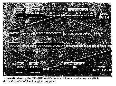
Fig. 1 is a schematic showing the TRS/RBS motifs present in human
and mouse AAVS1 in the context of Mbs85 and neighboring genes.
DETAILED DESCRIPTION OF THE INVENTION
The present invention provides a method for site-specific integration of
a transgene into the genoine of a mammalian ES cell comprising introducing
into the
ES cell an AAV vector containing the transgene and a Rep protein or a nucleic
acid
encoding a Rep protein. In particular, the present invention provides an
efficient
method for the site-specific integration of a transgene into the genome of an
ES cell.
In a preferred embodiment, the ES cell is a human or a mouse ES cell.
ES cells may be obtained commercially or isolated from blastocysts by methods
lcnown in the art, as described for example by U.S. Patent No. 5,843,780;
Thompson
et al. (1998) Science 282:1145-1147; U.S. Patent No. 6,492,575; Evans et al.
(1981)
Nature 292:154-156; and Reubinoff et al. (2000) Nature Biotech. 18:399.
6
CA 02605324 2007-10-17
WO 2006/121579 PCT/US2006/014391
The method described herein may also be used to deliver a transgene to
an adult, i.e. somatic, stem cell. Adult stem cells include, for example,
hematopoietic
stem cells, bone marrow stromal stem cells, adipose derived adult stem cells,
olfactory adult stem cells, neuronal stem cells, skin stem cells, and so on.
Adult stem
cells have a similar ability as ES cells to give rise to many different cell
types, but
have the advantage that they can be harvested from an adult.
The AAV vector containing the transgene comprises a pair of AAV
inverted terminal repeats (ITRs) which flank at least one cassette comprising
a
transgene under the control of a promoter. Transgene in this context refers to
any
nucleotide sequence which is not native to AAV. The AAV ITRs, in combination
with a Rep protein, confer infectivity and site-specific integration without
toxicity.
The ITRs may be derived from any AAV serotype, including AAVl-9. A preferred
embodiment utilizes serotype 2. The AAV ITRs and methods of obtaining the ITRs
are well-known in the art and disclosed, for example, in U.S. Patent No.
5,252,479.
The vectors may further contain sequence elements which facilitate expression
and
cloning, for example enhancers and selectable markers. Recoinbinant AAV
vectors
for noncytotoxic gene transfer and methods for making such vectors are known
in the
art and disclosed for example in U.S. Patent Nos. 6,632,670; 5,252,479;
5,173,414
and Kotin et al. (1994) Human Gene Therapy 5:793-801. Methods for producing
stocks of recombinant AAV are known in the art and disclosed for example by
Zolotukhin et al. (2002) Methods 28:158-167; Zolotukhin et al. (1999) Gene
Ther.
6:973-985; and Grimm et al. (1998) Hum. Gene Ther. 9:2745-60 and reviewed by
Zolotukhin (2005) Hum. Gene Ther. 16:551-557. Viral vector systems having
hybrid
serotypes and custom AAV capsids are also included in the present invention
and
disclosed for example by Choi et al. (2005) Curr. Gene Ther. 5:299-3 10; Gas
et al.
(2005) Curr. Gene Ther. 5:285-297; Muzyczlca et al. (2005) Hum. Gene Ther.
16:408-
416; and Buning et al. (2004) Cells Tissues Organs 177:139-150. The AAV vector
may comprise an AAV capsid comprising capsid proteins from any of the AAV
serotypes, or combinations thereof. Pseudotyped vectors comprising the AAV
ITRs
from one serotype and capsid proteins from a different serotype are included
herein.
The transgene is a nucleic acid sequence that is heterologous to AAV.
For example, the transgene may encode a marlcer or reporter molecule, protein,
7
CA 02605324 2007-10-17
WO 2006/121579 PCT/US2006/014391
peptide, antisense nucleic acid, or catalytic RNA. The transgene may encode a
naturally or non-naturally occurring molecule, including for example a
chimeric or
hybrid polypeptide. In a preferred embodiment, the transgene encodes a product
that
is useful for the treatment of a disease or disorder.
A Rep protein or nucleic acid encoding a Rep protein used in the
present method mediates the site-specific integration of the transgene. The
Rep
protein may be any AAV Rep protein or combination of AAV Rep proteins or a Rep
protein variant or fragment that is sufficient to mediate site-specific
integration. The
term Rep protein as used herein also includes Rep-like proteins such as the
human
herpes virus 6(HHV-6) Rep (Thompson et al. (1994) ViroloQV 204:304-311) and
goose parvovirus (GPV) Rep 1 (Smith et al. (1999) J. Virol. 72:2930-2937) and
fragments thereof that are sufficient to mediate site-specific integration.
Hybrids of
Rep proteins or fragments thereof with Rep-like proteins or fragments thereof
are also
included and disclosed for example by Yoon et al. (2001) J. Virol. 75:3230-
3239.
The Rep protein may be derived from any AAV serotype, and includes native,
variant
and cliimeric forms of a Rep protein. Variants that maintain the function of
mediating
integration are well-known in the art (see, e.g. Yoon et al. (2001) J. Virol.
75:3230-
3239) or can be ascertained by mutational analyses. In a preferred embodiment
the
Rep protein is Rep 68 or Rep 78 or a fragment thereof that is sufficient to
mediate
site-specific integration. In a preferred embodiment the Rep protein comprises
the
amino-terminal 208 amino acids of Rep 78.
In accordance with the present method, a Rep protein or a nucleic acid
encoding a Rep protein is provided to the ES cell. A nucleic acid encoding a
Rep
protein may be provided in trans by co-transfection of the AAV vector with a
Rep-
expressing construct, which may be in the form of a plasmid, phage,
transposon,
cosmid, virus or virion. Such constructs are known in the art and disclosed
for
example in U.S. Patent Nos. 6,632,670; 5,952,221; 5,139,941; Samulski et al.
(1989)
J. Virol. 63:3822-3828 and McCarty et al. (1991) J. Virol. 65:2936-2945. The
ES cell
may be stably transformed by a nucleic acid encoding a Rep protein prior to
introduction of an AAV vector. A nucleic acid encoding a Rep protein may also
be
provided in cis by methods known in the art, for example by a vector that
directs the
delayed expression of the rep sequences as disclosed in U.S. Patent No.
6,294,370 or
8
CA 02605324 2007-10-17
WO 2006/121579 PCT/US2006/014391
a vector in which a rep coding region is sited outside the ITRs, as disclosed
by Linden
et al. (1997) Gene Therapy 4:4-5.
A Rep protein may be provided to an ES cell by methods known to
those of ordinary skill in the art including methods using encapsulating media
such as
cationic lipid reagents, or methods of calcium phosphate precipitation,
electroporation
and microinjection. Additional methods that may be used include protein
transduction methods in which the Rep proteins are conjugated to peptides
known as
protein transduction domain (PTPs) or cell penetrating peptides (CPPs). Such
peptides include, for example, the herpes simplex virus (HSV) type 1 protein
VP22,
the human immunodeficiency virus (HIV-1) transactivator TAT protein,
polyarginine
and polylysine. Methods of protein transduction are known in the art and are
reviewed by Noguchi et al. (2006) Acta Med. Okayama 60:1-11, and Wadia et al.
(2002) Curr. Opiri. Biotechnol. 13:52-56. The peptides may be covalently cross-
linked to the Rep proteins or synthesized as fusions with the Rep proteins.
Other
methods for delivering the Rep proteins into ES cells include a non-covalent
peptide-
based method using an amphipathic peptide as disclosed for example by Morris
et al.
(2001) Nat. Biotechnol. 19:1173-1176 and U.S. Patent No. 6,841,535 and
indirect
polyethylenimine cationization as disclosed for example by Kitazoe et al.
(2005) J.
Biochem. (Tolcyo) 137:643-701.
It has been discovered in accordance with the present invention that
transduction efficiency of stem cells is enhanced by the use of double-
stranded AAV
vectors (dsAAV) in the present method. Such dsAAV is known in the art and
disclosed by Wang et al. (2003) Gene Ther. 10:2105-2111, and has a deletion of
the
terminal resolution site (TRS) in one ITR. As a result, this ITR cannot be
resolved
during replication, leading to the generation of replication intermediates
that are 2x in
length with two complementary single strands that are separated by the
partially
deleted ITR. Accordingly, in one embodiment of the present invention the AAV
vector comprises a pair of ITRs flanlcing a cassette comprising a transgene
under the
control of a promoter, in wllich one of the ITRs has a deletion of the TRS.
In accordance with the present invention, the method is preferably
performed at multiplicities of infection of 103-.10G genomes per cell. The
undifferentiated ES cells are preferably maintained under conditions that
allow
9
CA 02605324 2007-10-17
WO 2006/121579 PCT/US2006/014391
maintenance of healthy colonies in an undifferentiated state. For example.
human ES
cells may be maintained on a feeder layer such as irradiated mouse embryonic
fibroblasts in the presence of serum, or with serum replacement in the
presence of
bFGF, or in medium conditioned by mouse embryonic fibroblasts, or under serum
free conditions using human feeder layers derived from, for example, human
embryonic fibroblasts, fallopian tube epitlielial cells or foreskin.
Mouse ES cells may be maiiitained, for example, on a feeder layer
such as irradiated mouse embryonic fibroblasts in the presence of serum and
LIF, or
on gelatin plates without feeder cells in the presence of LIF and serum.
In another preferred einbodiment, ES cells are maintained on a
solubilized basement membrane preparation such as MatrigelTM (Kleinman et al
(1982) Biochem. 21:6188; Becton Diclcinson Biosciences). Methods for
maintaining
ES cells are known in the art and disclosed for example by Williams et al.
(1988)
Nature 336:684-7; Smitli et al. (1988) Nature 336:688-90; Ying et al. (2003)
Cell
115:281-92; Amit et al. (2003) Biol. Reprod. 68:2150-2156; and Amit et al.
(2000)
Developmental Biolo~y 227:271-278.
The method of the present invention results in site-specific integration
of the transgene at the AAVS 1 locus of the ES cell genome (human chromosome
19
at 19 q 13.4; mouse chromosome 7; 9.0 cM). The ES cells having the integrated
transgene undergo normal embryoid body (EB) development and retain the
capacity
to differentiate into multiple cell types. Expression of the transgene is
maintained
throughout differentiation. Further, the ES cells having the integrated
transgene
maintain the capacity to generate cells of multiple lineages.
Stem cells having a transgene integrated therein as made by the
method of the present invention are useful, inter alia, for generating
transgenic non-
llunian animals, for generating differentiated cells and tissues having a
transgene
integrated therein, for studying differentiation of stem cells, for evaluating
strategies
for safe and effective gene targeting in stem cells, and for targeted
therapeutic gene
transfer.
Methods for generating differentiated cells from stem cells are known
in the art. The model system for ES cell in vitro differentiation is based on
the
foimation of three dimensional structures knowa as embryoid bodies (EBs) that
CA 02605324 2007-10-17
WO 2006/121579 PCT/US2006/014391
contain developing cell populations presenting derivatives of all three germ
layers and
is disclosed in the art, for example by Keller (1995) Curr. Opin. Cell Biol.
7:862-869.
For exainple in one embodiment, prior to differentiation, ES cells are
removed from feeder cells piior to differentiation by subcloning the ES cells
directly
onto a gelatinized culture vessel. Twenty-four to 48 hours prior to the
initiation of EB
generation, ES cells are passaged into IMDM-ES. Following 1-2 days culture in
this
medium, cells are harvested and transferred into liquid medium (IMDM, 15% FBS,
glutamine, transferrin, ascorbic acid, monothioglycerol and protein free
hybridoma
medium II) in Petri-grade dishes. Under these conditions, ES cells are unable
to
adhere to the surface of the culture dish, and will generate EBs.
Culture conditions are known in the art for the differentiation to cell
types found in blood (Wiles et al. (1991) Development 111:259-67), heart
(Maltsev et
al. (1993) Mech. Dev. 44:41-50), inuscle (Rohwedel et al. (1994) Dev. Biol.
164:87-
101), blood vessels (Yamashita et al. (2000) Nature 408:92-96), brain (Bain et
al.
(1995) Dev. Biol. 168:342), bone (Buttery et al. (2001) Tissue Eng. 7:89-99)
and
reproductive system (Toyooka et al. (2003) Proc. Natl. Acad. Sci. USA
100:11457-
11462).
The differentiated cells and/or tissue generated therefrom may be
introduced in an animal for therapeutic purposes. Accordingly, in another
embodiment the present invention provides an animal comprising differentiated
cells
having a transgene integrated into the AAVS1 locus thereof, or comprising a
tissue
generated from such cells. In a preferred embodiment the differentiated cell
is a
hemotopoietic cell, endothelial cell, cardiomyocyte, skeletal muscle cell or
neuronal
cell. The cells or tissues may be transplanted into the animal by methods
lrnown in
the art.
The present invention also provides a transgenic non-human animal
and progeny thereof wherein said transgenic animal comprises a transgene
integrated
into AAVS 1. In a preferred embodiment, the animal is a mouse. Such transgenic
animals provide an in vivo system for studying the consequences of disruption
of the
AAVS1-associated gene cluster, and for assessing the safety, efEcacy and
regulatability of AAV-mediated delivery of transgenes. Transgenic mice having
a
marker gene such as the gene encoding GFP are particularly useful for testing
site-
11
CA 02605324 2007-10-17
WO 2006/121579 PCT/US2006/014391
specific integration of a transgene, since successful integration results in
loss of the
marker due to disruption of the marker gene.
Methods for producing transgenic mice are well-known in the art. For
example, the transgenic mouse may be obtained by injecting ES cells having a
transgene integrated therein into blastocysts, which are then implanted into
pseudopregnant females and allowed to develop to term. Recipient mouse strains
having a different fur color then the strain from which the ES cell is derived
may be
used to facilitate the identification of chimeric mice. The inclusion of a
marker gene
as a transgene facilitates the identification of donor ES cell derived cells
in tissues
other than the fur, e.g., blood.
All references cited herein are incorporated herein in their entirety.
The following examples serve to further illustrate the present
invention.
EXAMPLE 1
CHARACTERIZATION OF MOUSE AAVS 1 ORTHOLOG
The nonpathogenic human adeno-associated virus (AAV) has
developed a mechanism to integrate its genome into human chromosome 19 at
19q13.4 (termedAAVSl), thereby establishing latency. U.S. Patent No.
5,580,703;
Dutheil et al. (2000) Proc. Natl. Acad. Sci. USA 97:4862-66. This example
demonstrates that the chromosomal signals required for site-specific
integration are
conserved in the mouse genome proximal to the recently identified Mbs85 gene.
These sequence motifs can be specifically nicked by the viral Rep protein
required for
the initiation of site-specific AAV DNA integration. Furthermore, these
signals can
serve as a minimal origin for Rep-dependent DNA replication. In addition, the
mouse
Mbs85 proximal promoter was isolated and transcriptional activity was shown in
three mouse cell lines.
By using MBS85 (myosin binding subunit 85) exon sequences, the
National Center for Biotechnology Information mouse database was analyzed for
siinilarities to the human AAVSI locus as described by Altschul et al. (1997)
Nucleic
Acids Res. 25:3389-3402. This analysis revealed a homology of 90% between the
5'
end of the human MBS85 cDNA and the 969-nt mouse cDNA clone AK010836,
12
CA 02605324 2007-10-17
WO 2006/121579 PCT/US2006/014391
which contains a sequence homologous to the human TRS-RBS motifs as well as
the
Mbs85 initiation codon (separated by 25 nt). Fig. 1. A simian AAVSI locus
containing the corresponding upstream region and a TRS-RBS motif has recently
been isolated from the African green monkey genome by Amiss et al. (2001)
Methods
Mol. Biol. 175:455-469. AAVS1 is located 14.9 and 36 kb centromeric to the
slow
skeletal troponin T (TNNT1) and cardiac troponin I(TNNI3) genes, respectively.
Dutlieil et al. (2000) Proc. Natl. Acad. Sci. USA 97:4862-4866. The mouse
Tnni3
and Tnntl genes are located on chromosome 7, in a region previously shown to
be
syntenic to the human chromosome 19 region that contains AAYS1. Blake et al.
(2000) Nucleic Acids Res. 28:108-111. The Celera discovery system was used to
search the Celera mouse genome assenlbly with the mouse Tnni3 and Tnntl genes,
the
AK010836 cDNA, and the human MBS85 genomic sequence. All of these sequences
specifically matched the same scaffold (500 kb) in the Celera database. The
mouse
Mbs85 is located on chromosome 7 and is separated by only 2.5 and 16 kb from
the
Tizntl and Tnni3 genes, respectively. The Celera map revealed a gene 3.1 kb
downstream of MBS85, designated DRC3, the mouse homolog of which is located
2.1
lcb downstream of the Mbs85 gene.
Three mouse expressed sequence tag clones (AA021750, AW911639,
and BE847281) containing Mbs85 were sequenced and assembled. The resulting 3.1-
kb mouse cDNA was 77% identical to the human MBS85 cDNA. The mouse Mbs85
gene spans 20 kb of genomic sequences, and the 2.3-kb predicted open reading
frame
is composed of 22 coding exons. Thus, the mouse and the human homologs of
MBS85 display the same overall genomic organization. The deduced mouse Mbs85
protein sequences is 781 amino acids in length and is 86% identical to its
human
counterpart. Tan et al. (2001) J. Biol. Chem. 276:21209-21216.
To access the distribution of Mbs85 mRNAs, a mouse poly(A)
multiple tissue Northern blot (Clontech, Palo Alto, Calif.) was hybridized to
a mouse
Mb"s85 eDNA probe consisting of exons 5 to 22. As is observed in a human
multiple
tissue Northern blot (Tan et al. (2001) J. Biol. Chem. 276:21209-21216), a
single
mRNA of approximately 3.1 kb is highly expressed in heart and testis, and to a
lesser
extent in lcidney, brain, liver, and lung.
13
CA 02605324 2007-10-17
WO 2006/121579 PCT/US2006/014391
To determine if Rep68 can specifically nick the putative mouse TRS,
double-stranded and partially single-stranded 5' end-labeled origin substrates
were
incubated with purified His-tagged Rep68 proteins in a cell-free endonuclease
assay
as described by Yoon et al. (2001) J. Virol. 75:3230-3239. Rep68 nicked the
AAV,
human, and mouse TRS substrates releasing an expected 14-nt labeled fragment.
Nicking is Rep68 dependent since no cleavage of the AAV, human, or mouse
origin
substrates is observed when an endonuclease-negative mutant is used
(Rep68Y15617).
Smith et al. (2000) J. Virol. 74:3122-3129; Yoon et al. (2001) J. Virol.
75:3230-3239.
Substitution of the two thymidine residues within the mouse TRS sequence
resulted in
an expected loss of specific Rep-mediated cleavage.
Origin interactions by Rep are thought to represent the initiating steps
of integration. Ward et al. (2001) J. Virol. 75:10250-10258. To test whether
the
mouse TRS-RBS sequence could also serve a similar function, cell-free DNA
replication assays were performed as described by Ward et al. (1994) J. Virol.
68:6029-6037. Linearized substrates containing the AAV, human, or putative
mouse
origin in a pBluescript backbone were incubated with HeLa cell extracts in the
presence or the absence of purified His-tagged Rep68 (75 ng) and [a32P]dCTP.
Rep68 initiated replication on templates containing the AAV, human, or mouse
origin
but not on the vector DNA alone. In all cases, replication was Rep dependent.
The
human and mouse 5' untranslated regions were further compared. It has been
reported that the hunlan AAVS1 fragment located 74 to 426 upstream of the
translation
initiation codon is sufficient to drive the expression of a reporter gene
following
transient transfections in both 293 and HeLa cells. Lamartina et al. (2000) J.
Virol.
74:7671-7677.
Alignment of the huinan and mouse sequences upstream of the ATG
revealed an overa1162% identity in the putative promoter region. Several
conserved
putative cis-acting DNA elements (i.e., Spl, CRE/ATF) indicate the presence of
a
TATA-less promoter and common regulatory mechanisms for the expression of the
human and mouse MBS85 genes.
Mouse cell lines expressing Mbs8S were identified. Total RNAs were
extracted from C2C 12, NIH 3T3, and N2A cell lines (Tel-Test, Friendswood,
Tex.)
14
CA 02605324 2007-10-17
WO 2006/121579 PCT/US2006/014391
Northern blots hybridized to the mouse Mbs85 ex5-22 cDNA probe revealed a
unique
3.1-kb transcript in all three cell lines.
To test the 324-bp NaeI fragment containing the RBS and TRS motifs
for transcriptional activity, it was cloned into the pDsRed2.1 promoterless
red
fluorescent protein vector (Clon-tech) in both the sense and antisense
orientation.
C2C 12, N2A, and NIH 3T3 cells were transfected and fixed 45 hours
posttransfection
with 3.7% paraformaldehyde, and the slides were mounted in vectashield
mounting
medium with DAPI (4',6'-diamidino-2-phenylindole) (Vector Laboratories,
Burlingame, Calif.). The sense, but not the antisense, construct shows
transcriptional
activity in all three cell lines. These results were conflrmed by fluorescence-
activated
cell sorter analysis.
This example demonstrates that the target for AAV site-specific
integration is not restricted to primates but is also present in the mouse
genome in a
region that is syntenic to the human chromosome 19 region containing AA VS1.
Currently, Rep interactions with a minimal origin are defined by
specific binding to the RBS followed by site- and strand-specific nicking at
the TRS.
This example demonstrates that the TRS and RBS motifs present in the 5'
untranslated region of the mouse Mbs85 gene can act as a substrate for Rep-
mediated
nicking and as a functional Rep-dependent origin.
It also demonstrates that a region containing the TRS-RBS motif
upstream of the mouse Mbs85 ATG contains regulatory elements sufficient to
drive
the expression of a reporter gene in vitro.
The following materials and methods were used in the foregoing
example.
To determine tissue distribution of Mbs85 mRNAs in the mouse, a
multiple tissue Northern blot derived from mouse tissues was hybridized with a
cDNA probe consisting of exons 5 to 22 (ex5-22) of the mouse Mbs85 cDNA. The
ex5-22 probe was generated by digestion of clone BF540586 with EcoRUHindIIl.
For cell-free endonuclease assays, fully double-stranded and partially
single-stranded origin substrates (as a specificity control) (5fino1)
containing the
AAV, human (his), mouse (mS 1), and mouse origin mutant (mSlmut) sequences
were
incubated for 45 min at 37 C in eitlier the absence or presence of 1 pmol of
AAV
CA 02605324 2007-10-17
WO 2006/121579 PCT/US2006/014391
Rep68 protein or 1 pmol of AAV Rep68 endonuclease mutant (Y156F). 5' end-
labeled marker oligonucleotides corresponding in sequence and length to the
expected
reaction product (14nt) were used. Synthetic oligonucleotide substrates were
used in
the nicking and replication assays. The TRS-containing strand was first kinase
labeled and then annealed to its complementary strand.
In the cell-free DNA replication assay, the AAV, human (hSl) and
mouse (mS1) origins (consisting of the TRS and RBS sequences) were cloned into
pBluescript via XbaI and SaII sites. Prior to the assay, plasmids were
linearized with
XmnI. Each linear, origin containing substrate was incubated in the presence
or
absence of AAV Rep68 protein.
Expression of Mbs85 was determined by Northern blot analyses. The
Northern blot of C2C12, N2A, and NIH 3T3 cells was hybridized with a cDNA
probe
consisting of exons 5 to 22. The blot was stripped and rehybridized with a(3-
actin
cDNA probe. Transcriptional activity of the mouse Mbs85 proximal promoter was
determined as follows. Plasmid pDsRed2-NI (red fluorescent protein under the
cytomegalovirus promoter; Clontech) and the sense and antisense plasmids were
transfected into C2C12, NIH 3T3, and N2A cells. Forty-five hours
posttransfection,
the cells were visualized for redfluorescent protein expression and DAPI
staining by
using an epifluorescent microscope (Leica DMRA2) and a Hamamatsu digital
camera.
The foregoing results are published as Dutheil et al. (2004) J. Virol.
78:8917-8921, the disclosure of which is incorporated herein in its entirety.
EXAMPLE 2
MATERIALS AND METHODS
The following materials and methods were used in subsequent
examples.
Plasmid constructs. The conventional rAAV-GFP vector plasmid
(pTRUF1 1) is described by Zolotuldiin et al. (1996) J. Virol. 70:4646-4654
and
Zolotukhin et al. (1994) Gene Tlier. 6:973-985. It carries the humanized green
fluorescent protein (hGFP) sequence under the control of the hybrid CMVie
16
CA 02605324 2007-10-17
WO 2006/121579 PCT/US2006/014391
enhancer/chicken 0-actin promoter (CBA) flanked by the ITRs of AAV2. Plasmid
pAV2, used to produce wild type AAV2 virus, is described by Laughlin et al.
(1983)
Gene 23:65-73. Plasmids pXYZ1, pDG, pXYZ5, pDG-AAV8, pDG-AAV9 were
used as helper to produce AAV serotypes 1, 2, 5, 8 and 9 respectively. These
plasmids
were all derived from pDG (Grimm et al. (1998) Human Gene Ther. 10:2745-2760)
and carry the genes required for rAAV packaging. pXYZ1 and pXYZ5 are described
by Zolotukliin et al. (2002) Methods 28:158-167. pDG-AAV8 and pDG-AAV9 were
constructed using the AAV8 capsid sequence isolated from non-human primates in
the laboratory of K. R. Clarlc. The AAV9 capsid sequence is described by Gao
et al.
(2004) J. Virol. 78:6381-6388.
Wt and recombinant adeno-associated virus production.
The rAAV production and purification schemes were based on the
protocol described by Zolothukin et al. (1999) supra. Briefly, 293-T cells
(ATCC,
Manassas, VA) were cotransfected with pTRiTF11 together with the helper
plasmid.
After 72 hours, the virus was purified from cell crude lysates over a density
gradient
made of iodixanol (Optiprep, Greiner Bio-One Inc., Longwood, FL). Serotype 2
virus
stocks were additionally purified by affinity chromatography using heparin-
agarose
type I (Sigma-Aldrich Inc., St-Louis, MO) as a matrix. Virus samples were next
concentrated and formulated into lactated Ringer's solution (Baxter Healthcare
Corporation, Deerfield, IL) using a Vivaspin 20 Centrifugal concentrators 50K
MWCO (Vivascience Inc., Carlsbad, CA).
Wild-type AAV2 was produced following the same protocol, using
pAV2 instead of pTRUFl 1.
Maintenance ayzd irafectiosa of ES cells. Mouse ES cells (CCE and
E14) were maintained in 6-well plates on irradiated mouse embryonic feeder
cells in
DMEM medium (DMEM-ES) containing 1% L-Glutamine, 2.5% Hepes buffer, 15%
fetal bovine serum (FBS, pretested for maintenance of ES cells), 1% Leukemia
Inllibitory Factor (LIF - medium conditioned by CHO-LIF cells), and
monothioglycerol (1.5 x10⁴M). Cultures were monitored daily and cells were
passaged every 2-3 days. For passaging, ES cells were trypsinized (0.25%
trypsin,
17
CA 02605324 2007-10-17
WO 2006/121579 PCT/US2006/014391
0.1 % EDTA), washed and approximately 10% of the cells were replated on fresh
feeder cells. Cells were maintained in 37 C incubators at 5% COZ.
For feeder depletion prior to infection, ES cells were cultured for 1
passage in wells of 6-well plates coated with a 0.1% solution of gelatin and
containing
DMEM-ES medium.
Cells were harvested from this culture vessel, counted and seeded in
gelatin-coated, DMEM-ES-containing 96-well plates at a density of
approximately
10,000 cells per well.
Twenty four hours later, cells from a couple of representative wells
were counted in order to calculate the amount of virus needed to infect every
well at a
multiplicity of infection of 106. ES cells were then infected with single or
double
strand recoinbinant AAV2-GFP viruses, resuspended in 30 O1 of DMEM-ES medium.
Infections were performed at 37 C; plates were shaken by hand every 15
minutes.
After 1 hour, 70 ~ 1 of fresh medium was added and plates were placed back in
the
incubator. ES cells were incubated for 48 hours without removing the virus-
containing medium.
Geszef=ation of EBs from ES cells. The capacity of ES cells to
differentiate into multiple cell lineages can be reproduced in culture where
ES cells
can produce a wide range of well-defined cell types. The model system for ES
cell in
vitro differentiation is based on the formation of three-dimensional
structures known
as embryoid bodies that contain developing cell populations presenting
derivatives of
all three genn cell layers. Keller et al. (1995) Curr. Opin. Cell Biol. 7: 862-
869.
Prior to differentiation, ES cells were removed from the feeder cells by
subcloning the ES cells directly onto a gelatinized culture vessel. Twenty-
four to 48
hours prior to the initiation of EB generation, ES cells were passaged into
IMDM-ES.
Following 1-2 days culture in this medium, cells were harvested and
transferred into
liquid medium (IMDM, 15% FBS, glutamine, transferrin, ascorbic acid,
monothioglycerol and protein free hybridoma medium II) in Petri-grade dishes.
Under
these conditions, ES cells are unable to adhere to the surface of the culture
dish, and
will generate EBs. Keller et al. in Hematopoietic Stem Cell Protocols (eds.
Klug et
al.) 209-230, Humana Press, Inc., Totowa.
18
CA 02605324 2007-10-17
WO 2006/121579 PCT/US2006/014391
Generation of heinatopoietic cells, endotlzelial cells, cardiomyocytes,
skeletal muscle and neuronal cells from EBs. Developing hematopoietic
precursors
within EBs can be identified and studied in a standard colony-forming cell
(CFC)
assay. After harvest and dissociation (trypsin or collagenase treatment,
depending on
the duration of EB development), cells were mixed into the methylcellulose-
containing medium with specific hematopoietic cytokines, and aliquots were
plated in
35x10 mm Petri-grade dishes, which were incubated at 37 C for various periods
of
time. Colonies that developed from the hematopoietic precursors were scored
between
5-10 days following the initiation of culture. The types of precursors present
depend
on the age of the EBs. The changing precursor populations provide the basis
for
defining the three different stages of EB hematopoietic development. The
earliest
stage, the hemangioblast stage, contains the blast-CFC able to generate both
endothelial and hematopoietic progeny. EBs at the next stage contain primitive
erythroid (Ep), defmitive erythroid (Ed), macrophage, bipotential Ed/Mac,
bipotential
Ed/megakaryocyte (Mega), and multipotential precursors. The multilineage
defmitive
stage EBs contain Ed, bipotential Ed/mast cell (Mast), Mast, bipotential
Ed/Mega,
Mega, bipotential Ed/Mac, Mac, neutrophil (Neut), bipotential Mac/Neut, and
multipotential precursors.
To assess the vascular potential of the developing EBs, Flk-1+ cells
isolated from day 3- EB differentiation cultures were cultured in collagen
gels and
analyzed 10 days later. The cells formed vascular sprouts that expressed PECAM-
1
(CD3 1).
Cardiomyocyte potential was analyzed by moving EBs from serum-
containing to serum-free medium. Cultures were monitored over a 2- to 7-day
period
for the development of beating masses. To confirm that the cells were of the
cardiomyocyte lineage, aggregates were analyzed for expression of the cardiac
specific form of Troponin T. Cells within the masses expressed- this marker.
EBs generated in the absence of serum were cultured on gelatin coated
six-well-plates and monitored for neurite outgrowth, indicative of
neurectoderm
differentiation. EBs with visible neurites were transferred to glass cover-
slips and
stained for B-III tubulin expression. The neurites expressed abundant levels
of B-III
tubulin demonstrating their neuronal nature. Using conditions described by
Rohwedel
19
CA 02605324 2007-10-17
WO 2006/121579 PCT/US2006/014391
et al., supra, it was shown that cells with skeletal muscle morphology also
develop in
these cultures.
Establishment of clotziazg teclzzziques in tzzouse ES cells. Since
clonality is a prerequisite to analyze AAV-mediated integration events, a
cloning
technique was developed that would allow for the isolation of clean single-
cell
derived ES cell clones. A cost-effective way to do this was to generate GFP-
expressing ES cells based on transfection. ES cells were transfected, grown on
neor
MEF at 50-70% confluency, with pTRUF1, a plasmid that contains the "humanized"
GFP (hGFP) gene (Zolotukhin et al. (1996) J. Virol. 4646-4654) and a neomycin
resistance cassette flanked by the AAV terminal repeats. Forty-eight hours
after
transfection, cells were trypsinized and analyzed with flow cytometry (FACS)
for
transfection efficiencies (A: 90% of the cells were GFP-positive when
transfections
were executed with Lipofectamin 2000). Part of the cells were seeded onto
fresh neo'
feeders and G418 selection was started. Since ES cells could not be single
cell sorted,
ES colonies were aspirated. These colonies originate from a single cell and
can thus
be considered clonal. For short periods of G418 selection (e.g. three days),
resistant
ES colonies were well-spread and could easily be aspirated. For longer
selection
periods (e.g. two weeks) in which selective colonies were expanded and
passaged,
single ES colonies were aspirated, trypsinized and seeded in one well of a 24-
well
plate. The newly developing colonies were now well-spread and single clones
could
easily be aspirated. GFP-positive colonies, established with this cloning
technique,
showed homogeneous GFP-expression profiles when analyzed with FACS.
Infectivity of mouse ES cells.
Gefzeratiorz of fAAV. Recombinant viruses of the AAV serotypes 1, 2,
4, 5, 8 and 9 were generated using transfection methods in either triple
flasks or ten-
layered cell factories. Recombinant AAV contains the marker genes neomycin and
GFP flanked by the AAV-ITRS. The serotypes 1, 4, 5, 8 and 9 were generated
using
the "pseudotyping" approach in which the reconibinant genome is flanlced by
the
AAV2 ITRs and the different serotype capsids are packaged by the AAV2 REP.
Typically, approximately 2x10¹³ genome containing particles (gcp) per triple
flask
were produced.
CA 02605324 2007-10-17
WO 2006/121579 PCT/US2006/014391
A"double-strand" or dsAAV (in this case containing CMV-EGFP)
was produced which is different from traditional viruses in that it has a
deletion of the
TRS in one ITR. As a result, this ITR cannot be resolved during replication,
which
leads to the generation of replication intermediates that are 2x in length
with two
complementary single strands that are separated by the partially deleted ITR.
If the
total length of these intermediates does not exceed the full length of wtAAV
they can
be packaged similarly to traditional recombinant viruses. However, when this
DNA
enters the nucleus it is hypothesized that the complementary strands can
anneal,
resulting in DNA structures that can directly be transcribed. It is thought
that this
strategy circumvents the rate-limiting step of second-strand synthesis of
traditional
recombinant AAV DNA that is believed to underlie the delayed onset of
transduction
by AAV. Impressively, when these viruses are used for in vivo transduction
assays,
the expression of the transgene was accelerated and transduction was
significantly
enhanced (e.g. in contrast to 5% of hepatocytes transduced with traditional
viruses,
dsAAV infection led to 80-90% of transduced hepatocytes).
EXAMPLE 3
INFECTION OF MOUSE ES CELLS WITH AAV
In this example, infection experiments were performed using
recombinant AAV1, 2 and 5 GFP viruses to infect CCE and E14 cells. Infections
at
different MOIs were performed on small ES colonies, cultured on gelatin. Flow
cytometric analysis of GFP was used to determine transduction efficiencies.
Transduction efficiency was measured as the number of GFP-expressing cells
present
in the cultures 48 hours post-infection. Infections with rAAV2 at a
multiplicity of
infection (MOI-gcp/cell) of 106 resulted in GFP-expressing ES cells. Infection
of ES
cells by the other serotypes was not detectable. Experiments were expanded
with
rAAV2 and transduction efficiencies of single strand (ss) versus ds virus were
compared. As shown in Table 1, infections with ss AAV2 consistently yielded
about
1% GFP-positive cells and infections with ds AAV2 significantly increased the
nunzber of transduced cells. These data indicate that a sufficient number of
cells are
infected but that the onset of transduction is delayed when ss virus is used.
Thus, this
example shows that mouse ES cells can be infected with AAV2. C-kit expression
and
21
CA 02605324 2007-10-17
WO 2006/121579 PCT/US2006/014391
alkaline phosphatase staining were similar in control and infected cell
populations,
indicating that ES cells could withstand these relatively high multiplicities
of
infection. These markers indicate that the cultures consisted of
undifferentiated, self-
renewing ES cells. Similar results were obtained for E14 cells.
Table 1
Mouse ssAAV2-GFP dsAAV2-GFP
CCE 0.87 0.23 (n=7) 12.92 1.16 (n=6)
E14 0.97 0.13 (n=4) 7.66 0.92 (n=4)
Transduction of embryonic stem cells is indicated in %
GFP-positive cells. Cells were infected at an M.O.I. of 106.
The transduction efficiency was determined by FACS
analysis performed 48 hours post-infection.
Subsequently, these experiments were expanded to include additional
serotypes, AAV8 and AAV9. As with the previous serotypes, AAV8 and AAV9 were
"pseudotyped", i.e. these vectors contain the AAV2 ITRs and the identical
transgene
as used earlier. These genomes were packaged into the AAV8 and AAV9 capsids,
respectively. In these experiments both single-stranded (ss) as well as double-
stranded
(ds) vectors of serotypes, AAV1, AAV2, AAV5, AAV8 and AAV9 were used.
Infections at an MOI of 106 were performed on small mouse ES
colonies (CCE), cultured on gelatin. Transduction efficiency was determined as
the
number of GFP expressing cells present in the cultures 48 hours post-
infection. As
can be seen in Table 2, with the exception of AAV5 (ss and ds), infections of
mouse
ES cells with ds vectors of all serotypes resulted in significant
transduction.
22
CA 02605324 2007-10-17
WO 2006/121579 PCT/US2006/014391
Table 2
Transduction efficiencies: percentage of GFP-expressing cells as determined by
flow
cytometry
Single-stranded Double-stranded
AAV1 0.11% 46.37%
AAV2 7.11% 16.58%
AAV5 0.20% 0.23%
AAV8 0.59% 10.70%
AAV9 0.20% 7.42%
EXAMPLE 4
TARGETING OF TRANSGENES TO
AAVS1 IN MOUSE ES CELLS
The foregoing observation that ES cells could be infected with AAV2
prompted the initiation of infection-based integration essays. The transgenes
to be
integrated, GFP and the neomycin resistance gene, were provided by recombinant
single strand AAV2, whereas Rep, responsible for targeting the transgenes, was
provided in trans by means of co-infection with wtAAV2. In brief, CCE mouse ES
cells cultured on gelatin were co-infected with wt AAV and recombinant AAV2 at
an
MOI of 106. Cells were passaged onto fresh neomycin-resistant feeders 48 hours
after
infection, and G418 selection (300 mg/ml) was started. Five days after the
start of
selection, G418-resistant colonies were aspirated and expanded. Finally, cells
were
harvested for FACS analysis and genomic DNA extraction. In this experiment,
infections were performed in 96-well plates, and 6 clones were generated of
which 3
were GFP-positive. Transgene integration analysis was focused on clone 4, as
FACS
analysis of this clone showed a single population of GFP-expressing cells.
Direct PCR and an unbiased linker-mediated PCR technique (Schroder
et al. (2002) Cell 110: 521-529; Wu et al. (2003) Science 300: 1749-1751) were
used
to detect where the transgene had integrated. Both strategies showed that the
transgene in clone 4 was targeted to AAVS 1, 8429 bp downstream of the TRS/RBS
23
CA 02605324 2007-10-17
WO 2006/121579 PCT/US2006/014391
motifs. PCR results indicated that wt AAV sequences are absent in the genome
of
this targeted clone.
Southern blot analysis showed disruption of Mbs85, the gene that is
embedded in AAVS 1. A different blot indicated that rAAV2 only integrated in
AA.YS1, since hybridization with a GFP probe resulted in a single band that
cohybridized with the disrupted Mb85 band. Control DNA hybridized against a
genomic MBS85 probe revealed the about 6.5kb undisrupted AAVS1 fragment. After
removal of the MBS85 probe and hybridization to an rAAV-specific probe, the
Southern blot indicated a single rAAV integration event with a vector genome
fragment that co-migrates with the disrupted MBS85 fragment.
AAVS1-targeted mouse ES cells show normal in vitro
differentiation capacities and continue to express GFP throughout
differentiation. Clone 4 ES cells were grown on gelatin for two passages in
order to
deplete feeders, trypsinized and cultured in non-adherent conditions to allow
for the
formation of EBs. It was found that EB differentiation occurred normally while
GFP
expression remains unchanged. At day 4, EBs expressed Flk- 1 and c-kit
profiles
indicative of normal differentiation.
The following Differentiation assays were performed on targeted
mouse ES cells.
1. Blast colony-forming assay
This assay supports the growth of the hemangioblast, a precursor witli
the potential to generate both hematopoietic and endothelial lineages. These
bipotential precursors represent a transient population that develops between
day 3.0
and day 3.25 of differentiation and persists for 12-18 hours. These times can
vary by
3-6 hours, depending on the batch of FCS and on the ES cell line used. The
embroyoid body (EB)-derived hemangioblasts grow in response to VEGF and
generate colonies consisting of cells with undifferentiated blast-cell
morphology
(Keller G.M., Webb S., and Kennedy M. in Metlaods in Molecular Medicine, vol.
63:
Heinatopoietic Stein Cell Protocols)
Targeted ES cells were differentiated in standard serum-containing
conditions, EBs were harvested and dissociated at day 3.5 and added to a blast-
methylcellulose (MEC: 1%, D4T (embryonic endothelial cell line) conditioned
24
CA 02605324 2007-10-17
WO 2006/121579 PCT/US2006/014391
medium 25%, FCS 10%, Glutamine 1%, transferrin 300 g/ml, ascorbic acid 25
g/ml, monothioglycerol 4x10⁻⁴ M, VEGF 5 ng/ml, 11-6 10 ng/ml, IMDM up to
100%) assay.
Blast colonies detected 3 days after initiation of the assay had typical
morphology and maintained uniform GFP expression.
The assay is described by Kouskoff et al. (2005) Proc. Natl. Acad. Sci.
102:13170-5.
2. Cardiomyocyte assay
Targeted ES cells were differentiated in standard serum-containing
conditions, EBs were harvested and dissociated at day 4 and re-aggregated for
20
hours in serum-free conditions (StemPro34, L-Glutamine 2 mM, transferrin 200
g/m1, ascorbic acid 0.5 mM, monothioglycero14.5 x10-4 M, VEGF 5 ng/ml, bFGF
(30 ng/ml). Aggregates were transferred to gelatin-coated dishes containing
StemPro34, L-Glutamine 2 mM, VEGF 5 ng/ml, bFGF (30 ng/ml). Three days later,
beating cardiac clusters were observed. These clusters maintained uniform GFP
expression.
3. Neuronal differentiation assay
Targeted ES cells were first depleted of feeders in N2B27 medium.
After the second round of feeder depletion, cells were harvested and
transferred to
gelatin-coated dishes containing N2B27 medium and 0.3% MTG. Medium was
changed daily. Neuron-like cell types were visible after 12 days of culture.
Neuronal
morphology was confirmed by immunohistochemistry using the neuron-specific
marker Tuj 1(anti-tubulin bIII). Uniform GFP expression was observed in
tubulin
bIII-expressing neurons. The assay is adapted from Ying et al. (2003) Methods
En iz~o1.365:327-41.
Ifztegratzon assays pesforfrzed oii CCE aitd Hela cells. The first clone
analyzed for site-speciric integration carried the transgenes in AAVS 1. In
order to
address the issue of frequency, pools of CCE cells that were either infected
with
single-stranded wt and reconzbinant AAV2, with single-stranded rAAV2 alone or
with single-stranded wtAAV2 alone were generated. For the cells that were
infected
with rAAV2, both in the absence and presence of wtAAV2, the population was
split
up in cells that were selected witli G418 and cells that were not selected
with G418.
CA 02605324 2007-10-17
WO 2006/121579 PCT/US2006/014391
Respectively, 11 and 6 clones were aspirated from the pools of rAAV2- and wt +
rAAV2-infected cells. One of those clones, 'clone 4 (r+wt)' was analyzed.
Since it
was important to compare the obtained integration frequencies with those
obtained
from a human cell line that previously had been shown to support AAV-mediated
site-specific integration, an integration assay was perfonned in Hela cells
using the
same conditions as used for CCE cells. The number of GFP-positive cells in the
pools
that were infected with rAAV2 alone or with wt and rAAV2 are in the same range
for
both mouse ES cells and human Hela cells (see table 3).
Table 3 CCE Hela
rAAV2 - no selection 0.15% 0.21%
r+wtAAV2 - no selection 0.08% 0.35%
rAAV2 - G418 selection 26.09% 2.11%
r+wtAAV2 - G418selection 81.72% 77.41 %
This table shows the number of GFP-positive cells,
determined by FACS analysis respectively 4 and 5 passages
after infection, in the absence and presence of selection.
As can be seen in Table 3, the number of GFP-positive cells increased
dramatically when cells are coinfected with wtAAV2. Non-selected CCE cells are
an
exception.
The foregoing example demonstrates that a) AAV-mediated targeted
gene delivery can be achieved into the mouse AAVS1 ortholog, b) targeted gene
delivery to this locus is feasible in ES cells, c) as determined to date,
disruption of
AAVS1 does not interfere with multilineage in vitro differentiation of ES
cells and d)
that transgene expression is maintained throughout differentiation.
EXAMPLE 5
INFECTION OF HUMAN ES CELLS WITH AAV
Maintenance and it fection of hES cells. Human ES cells (WAO1)
were maintained on irradiated mouse embryonic feeder cells in DMEM-F12 medium
(L-Glutamine 1mM, non-essential amino acids 1%) containing 20% serum
replacement (Knockout-Invitrogen), 4ng/ml basic Fibroblast Growth Factor, and
beta
26
CA 02605324 2007-10-17
WO 2006/121579 PCT/US2006/014391
mercaptoethanol (0.1mM). Cultures were monitored daily and cells were passaged
every 4-5 days. For passaging, ES cells were trypsinized (0.25% trypsin, 0.1%
EDTA) for 3 minutes; the trypsin removed and replaced with medium containing
50%
FBS and 50% F12 medium and MatrigelTM (0.2%). Then, cells were resuspended and
washed. Approximately 25% of the cells were replated on fresh feeder cells.
Cells
were maintained in 37 C incubators at 5% COZ. Using this protocol healthy hES
colonies that are alkaline phosphatase and c-kit positive were obtained.
Minimal cell
death occurred during the passaging process.
In addition, in order to determine transduction efficiencies that were
not influenced by the presence of mouse feeder cells, growtli conditions on
MatrigelTM were established. Using this approach the colonies were maintained
for
several passages without significant differentiation.
For feeder depletion prior to infection, ES cells were cultured for 1
passage in wells of 6-well plates coated with MatrigelTM (Becton Dickinson-
growth
factor-reduced, diluted 1:1 in DMEM).
Cells were harvested from this culture vessel, counted and seeded in
MatrigelTM-coated, serum-free medium-containing 96-well plates at a density of
approximately 10,000 cells per well.
24-48 hours later, cells from a couple of representative wells were
coi.inted in order to calculate the amount of virus needed to infect every
well at a
multiplicity of infection of 106. ES cells were then infected with single or
double
strand recoinbinant AAV-GFP viruses, resuspended in 30 ~1 of serum-free F12
medium. Infections were performed at 37 C in the presence or absence of
adenovirus;
plates were shaken by hand every 15 minutes. Adenovirus was included in these
experiments in order to first assess virus uptake without the contribution of
downstream roadblocks as for example second-strand synthesis that has
previously
been shown to influence transduction rate. After 1 hour, 70 ~ l of fresh
medium was
added and plates were placed back in the incubator. ES cells were incubated
for 72
hours while daily replacing 75% of the medium with fresh medium.
72 hours post-infection cells were harvested for flow cytometry and the
number of GFP-positive cells was determined.
27
CA 02605324 2007-10-17
WO 2006/121579 PCT/US2006/014391
Table 4 shows the results of these experiments.
Table 4. Infection of WA01 cells by AAV serotypes
1000 1000 10,000 10,000 100,000 100,000
-Ad +Ad -Ad +Ad -Ad +Ad
rAAV1 0 0 0 0 0.19 0.31
rAAV2 0 0.38 0.18 0.65 0.79 3.27
rAAV5 0 0 0 0 0 0
Table 4. Small hES colonies that were seeded on mouse feeder cells were
infected by
AAV serotypes 1, 2 and 5 and, where indicated, superinfected by adenovirus
(MOI:
500) at multiplicities of 103 to 105 genomes per cell. The data are given as
percent
GFP positive cells that were gated on a population enriched for hES cells.
FACS
analysis was performed 72h post infection.
Based on these initial data that highlighted the preference of AAV2 for
infection of WA01 cells under these conditions, further optimization of the
procedures
utilized this AAV serotype.
Optifiaization of ir fectiofa coraditioyas. In further infection studies,
adenovirus was excluded from the infection mixture. Multiplicity of infection
was
increased to 106 genomes per cell. When infection was performed in the
presence of
mouse feeder cells, c-kit labeling was included in the FACS analysis in order
to
exclude contributions by the mouse cells. In addition, the following
conditions were
tested: WA01 cells were infected either on a mouse feeder layer, on MatrigelTM
or in
suspension (Table 5). Subsequent to infection the suspension cells were plated
on
mouse feeder cells. Further analysis of the cells that had been infected in
suspension
showed a significant change in morphology, as also confirmed by
forward/sideward
scatter in FACS analysis. In this analysis it also became apparent that c-kit
had been
down-regulated as a result of this particular condition. These changes could
not be
observed in cells that were infected on feeders or on MatrigelTM. Based on
these
observations and the results shown in Table 5 further infection experiments
were
performed on cells that were maintained on Matrigel.
28
CA 02605324 2007-10-17
WO 2006/121579 PCT/US2006/014391
Table 5. Transduction of WA01
Single Double strand AAV2
strand
AAV2
H1 on 5.58- 0.51 n.d.
feeders (n=4)
Hl in 14.89:L2.40 n.d.
suspension (n=4)
Hl on 17.98f1.86 42.82:L4.04
Matrigel (n=4) (n=4)
WA01 (H1) cells were infected by dsAAV and ssAAV2 based
viruses using three different conditions for cell growth and
maintenance.
An additional variable shown in Table 5 was the inclusion of double-
strand (or self-complementary) AAV2 that in a number of previous studies had
shown
enhanced transduction efficiencies. As can be seen, on average nearly 43% of
WA01
cells can be transduced on MatrigelTM when dsAAV2 is used. The FACS analysis
of
WA01 cells infected with dsAAV2 was performed. Also c-kit expression levels of
mock-infected and dsAAV2-infected cells were compared. The c-kit expression
levels
were comparable in both conditions. In addition, cells infected with dsAAV2
were
passaged onto fresh feeder cells and continued to show normal growth
characteristics.
EXAMPLE 6
INFECTION OF HUMAN ES CELLS WITH AAV
Human embryonic stem cells (HES2 cells) were maintained on mouse
embryonic feeders using the same protocol as described for WA01 cells
hereinabove.
HES2 cells were transduced with recombinant AAV1, 2, 5, 8 and 9,
respectively. The viruses were "pseudotyped", i.e. these vectors contain the
AAV2
ITRs and the identical transgene as used hereinabove. These genomes were
packaged
into the AAVI, 2, 5, 8 and 9 capsids, respectively. In these experiments both
single-
stranded (ss) as well as double-stranded (ds) vectors were used. Infections at
an MOI
of 106 were performed on small human ES colonies (HES2), cultured on
MatrigelTM.
Transduction efficiency was determined as the nuniber of GFP expressing cells
present in the cultures 48 hours post-infection. As can be seen in Table 6,
with the
29
CA 02605324 2007-10-17
WO 2006/121579 PCT/US2006/014391
exception of AAV5 (ss and ds), infections of HES2 cells with ds vectors of all
serotypes resulted in significant transduction.
Table 6
Tt=aiasdtcctioya efficiencies oiz HES2 cells: perceiatage of GFP-expressing
cells as
detersfzisaed by floiv cytonaetfy
Single-stranded Double-stranded
AAV1 0.16% 14.15%
AAV2 8.31% 45.91%
AAV5 0.00% 0.03%
AAV8 0.45% 11.7%
AAV9 0.06% 2.21%
EXAMPLE 7
TARGETING OF TRANSGENES TO AAVS1 IN HUMAN ES CELLS
WAO1 cells were grown on MatrigelTM and co-infected with single-
stranded wt AAV and recombinant AAV2, containing the hGFP gene and a neomycin
resistance cassette, flanked by the AAV terminal repeats (MOI 106). Cells were
passaged onto fresh feeders 48 hours after infection, and G418 selection was
started.
It had previously been determined that G418 selection at 50 g/ml left the
feeder cells
undisturbed, but killed off mock-infected hES. Mouse embryonic fibroblasts
(feeder
cells) grown in serum-free medium do not tolerate higher concentrations of
G418,
which they do when grown in serum-containing medium. Two weeks after the start
of
selection, healthy-looking G418-resistant colonies had developed. Aspiration
teclmiques that were used to isolate mouse ES clones also worked for liES as
in an
independent experiment 15 wt AAV-infected hES clones were generated.
CA 02605324 2007-10-17
WO 2006/121579 PCT/US2006/014391
Forty G418-resistant clones were aspirated and expanded on
MatrigelTM in order to isolate genomic DNA. This DNA was then digested with
EcoRI or HindIII and run on a Southern blot. Hybridization with a hAAVS 1-
specific
probe showed disruption of the target site in 2 out of the 28 thus far
analyzed WAO 1
clones, indicating that the AAVS 1 locus has been targeted.
EXAMPLE 8
GENERATION OF CHIMERIC ANIMALS
Three wells of a six-well plate were coated with gelatin and irradiated
mouse embryonic fibroblasts. 2-3 days before the blastocyst injections, one
frozen
vial of amplified targeted ES cells was thawed and plated into the earlier
prepared
three wells. On the day of injection, the medium was changed to medium without
LIF, 1-2 hours before the cells were used. The cells were then trypsinized,
pelleted
and resuspended in 10 ml of DMEM supplemented with 20 mM HEPES (pH 7.3) and
10% FCS.
Blastocysts were obtained from immature (4 week old) B6D2F1
female mice which had been superovulated with PMS and HCG, followed by matings
with C57B1/6 males. Three days after plugs were identified in these,females,
the
mice were sacrificed by COz overdose. The uterus was isolated from each
animal,
and blastocysts were flushed from each uterus. Isolated blastocysts were then
injected
with targeted ES cells. These injected blastocysts were reimplanted into the
uterus of
pseudopregnant females and mated two days before the day of blastocyst
microinjection. Generally, 12-15 microinjected blastocysts were reimplanted
into
each host female. The reimplantation surgeries were done under avertin
anesthesia,
and topical 1% lidocaine was administered immediately after the surgery. After
the
animals recovered from the surgery, they were returned to the aninlal room
where
most of them delivered their pups 17 days after the reimplantation of embryos.
Approximately 1 week after delivery of pups derived from microinjected
blastocysts,
the coat color becomes apparent on the pups, and it is this coat color which
is used to
determine the relative success of the experiment. The ES cells used for these
studies
were derived from a mouse strain (129) which has agouti coat color, while the
donor
blastocysts were obtained from black niice (C57B1/6). The experiment is judged
31
CA 02605324 2007-10-17
WO 2006/121579 PCT/US2006/014391
successful if coat color chimeras are observed in which the agouti color
(dominant to
black) makes up to at least 50% of the animal's coat color. In this example, 6
chimeric animals (2 males and 4 females) were born; based on coat color, the
percentage of chimerism was estimated at 30-50%.
In order to assess transgene expression from AAVS1 in vivo, blood
was collected from female mice and labeled with a pan-leukocyte marker (CD
45.2).
CD 45.2 positive cells were analyzed for GFP expression using FACS analysis. 4-
8.5% of the leukocytes expressed GFP, demonstrating that transgenes were
expressed
from AAVS 1 throughout differentiation in vivo. Furthermore, no apparent
deleterious =
effects of integration into AAVS 1 and the resulting genomic disruption were
observed.
32