Note: Descriptions are shown in the official language in which they were submitted.
CA 02605604 2013-07-31
-1-
METHODS AND SYSTEMS FOR LASER TREATMENT USING
NON-UNIFORM OUTPUT BEAM
BACKGROUND OF THE INVENTION
Plastic surgeons, dermatologists and their patients continually search for new
and improved methods for treating the effects of an aging skin. One common
procedure for rejuvenating the appearance of aged or photodamaged skin is
laser
skin resurfacing using a carbon dioxide laser. The carbon dioxide laser energy
is
absorbed by tissue water causing vaporization of the outer skin layer. Carbon
dioxide lasers have been utilized for approximately three decades. However it
has
only been the past few years that these lasers have been arranged to remove
only thin
tissue layers with minimal heat damage to the surrounding skin. While carbon
dioxide lasers may remove about 150 microns of skin, that skin may take a
month or
more to heal under such a procedure.
Er:YAG lasers have been utilized to ablate even thinner layers of tissue than
carbon dioxide layers. However they lack the coagulation characteristics and
thus
allow more bleeding than a carbon dioxide laser during use.
Non-ablative skin rejuvenation is a methodology which does not take the top
layer of skin off, but which uses a deep-penetrating laser to treat the layers
of skin
beneath the outer epidermal layer, treating unsightly vascular and pigmented
lesions,
and shrinking and modifying the underlying collagen, tightening the skin and
reducing wrinkles to provide a more youthful appearance. This methodology
however, has a low efficiency, and an aggressive cooling method Must be used
on to
CA 02605604 2013-07-31
-2-
the skin so as to minimize damaging the top or upper layer thereof and also to
minimize pain generation. The "fluence" or energy density used is greater than
10
joules per square centimeter and to be more effective this fluence often
reaches 30
Joules per square centimeter. This level of energy often causes pain and
epidermal
damage.
United States Published Patent Application No. 2002/0161357 Al, by
Anderson et al., discusses a method and apparatus for performing therapeutic
treatment on a patient's skin by using focused radiation beams to create
"islands" of
treatment/damage within untreated portions of the patient's skin. However, the
parameters of the treatment beam in this method are not optimal for skin
rejuvenation treatment.
Yet another treatment method is disclosed in U.S. Patent No. 6,077,294 to
Cho et al. This patent describes a system and methodology for noninvasive skin
treatment that utilizes a pulsed dye laser having a wavelength of about 585
nanometers (nm), and an energy of less than 5 Joules per square cm. In
contrast to
earlier techniques which used higher-energy pulses to damage and "shrink" the -
collagen below the epidermis, the relatively lower energies of the beams in
the '294
patent are designed to stimulate the collagen to regenerate and "fill in"
valleys of the
skin for a younger more clearer skin.
SUMMARY OF THE INVENTION
The present invention relates to methods and apparatus for treatment using
non-uniform laser radiation. Preferably, the invention is used for skin
rejuvenation
treatment, in which a high-intensity portion of the laser radiation causes
collagen
destruction and shrinkage within select portions of the treatment area, while
a lower-
intensity portion of the radiation causes fibroblast stimulation leading to
collagen
production across other portions of the treatment area.
Preferably, the method and system of the invention utilize a solid-state laser
source, such as an Nd:YAG laser. The output beam from the laser source is
coupled
into an optical system that modifies the beam to provide a large-diameter beam
CA 02605604 2007-10-19
WO 2006/116141
PCT/US2006/015180
-3-
having a non-uniform energy profile, comprised of a plurality of high-
intensity zones
surrounded by lower-intensity zones within the treatment beam. The higher-
intensity zones heat select portions of the target tissue to temperatures
sufficient for
a first treatment (e.g. collagen shrinkage), while the lower-intensity zones
provide
sufficient energy to the surrounding tissue for a second treatment (e.g.
stimulated
collagen production). Thus, a large area of tissue, preferably 7-10 mm in
diameter,
can be treated simultaneously, while minimizing the risk of burning or other
damage
to the skin.
In one embodiment, the invention uses a fiber bundle to provide a non-
uniform energy output beam. In another embodiment, the invention uses a
diffractive lens array to produce the non-uniform output beam.
A method of treating human skin in accordance with one aspect of the
invention comprises generating an output beam from a laser source, such as an
Nd:YAG laser; coupling the beam into an optical system that modifies the beam
to
provide a treatment beam having a non-uniform energy profile, the treatment
beam
comprised of a plurality of high-intensity zones surrounded by low-intensity
zones
within the treatment beam; and directing the treatment beam to a target tissue
area
such that the high-intensity zones heat select portions of the target tissue
to
temperatures sufficient for a first treatment, while the lower-intensity zones
provide
sufficient energy to the surrounding tissue for a second treatment.
Preferably, the
first treatment comprises collagen shrinkage and the second treatment
comprises
collagen stimulation. The output beam can have a wavelength between about 1.3
to
1.6 microns, and preferably between about 1.41 and 1.44 microns, and a pulse
duration between 0.1 and 100 milliseconds, and preferably between about 1 and
5
milliseconds. The average fluence of the treatment beam can be less than about
10
J/cm2. Generally, the average fluence of the treatment beam is between about 5-
6
J/cm2. The average fluence in the lower-intensity zones is generally on the
order of
2-3 Pcm2.
The optical system can comprise a fiber bundle, having 1000 to 2000
separate fibers, for instance, and a focusing lens for coupling the beam into
the fiber
bundle. An optical window, preferably between 1 and 5 mm thick, can be located
at
CA 02605604 2007-10-19
WO 2006/116141
PCT/US2006/015180
-4-
the distal end of the bundle, the optical window permitting the beams emitted
from
each fiber in the bundle to diverge and partially overlap with one another
before they
reach the target tissue. In certain embodiments, a transport fiber can carry
the output
beam from the laser source to the fiber bundle, and the fiber bundle can be
located in
a handpiece.
In another embodiment, the optical system can comprise a diffractive lens
array, preferably comprised of about 2000 or less lenses, arranged in an
optical path
between a laser source and the treatment area, such that each lens in the
array
provides a high-intensity zone surrounded by a low intensity zone of
radiation. Each
lens in the array can have a diameter of between about 150 and 450 microns,
and the
entire lens array can have a diameter of between about 7 and 10 mm.
Preferably, the
average fluence of the laser output beam is less than about 10 Jr/cm2.
In another embodiment, a laser system of the invention comprises a laser
source that generates an output beam; and an optical system that modifies the
output
_ beam to provide a treatment beam having a non-uniform energy profile, the
treatment beam being comprised of a plurality of high-intensity zones
surrounded by
low-intensity zones within the treatment beam, such that the high-intensity
zones
heat select portions of a target tissue to temperatures sufficient for a first
treatment,
while the lower-intensity zones provide sufficient energy to the surrounding
tissue
for a second treatment. The laser source can be an Nd:YAG laser, and generally
produces an output beam having a wavelength between about 1.3 to 1.6 microns,
and
preferably between about 1.41 and 1.44 microns, and a pulse duration between
0.1
and 100 milliseconds, preferably between about 1 and 5 milliseconds. The
optical
system can comprise a fiber bundle, preferably with an optical window between
the
distal end of the bundle and the target tissue. Alternatively, the optical
system can
include a diffractive lens array in the optical path between the source and
the
treatment area, such that each lens in the array provides a high-intensity
zone
surrounded by a low intensity zone of radiation.
According to another embodiment, a laser system comprises a laser source
that generates an output beam; a fiber bundle comprising a plurality of
individual
fibers, the fiber bundle having a proximal end and a distal end; a focusing
lens for
CA 02605604 2007-10-19
WO 2006/116141
PCT/US2006/015180
-5-
coupling the output beam into a proximal end of the fiber bundle; and an
optical
window at the distal end of the fiber bundle, the optical window permitting
the
beams emitted from each fiber in the bundle to diverge as the beam passes
through
the optical window so that each beam partially overlaps with the beam(s) from
adjacent fibers in the bundle. The optical window can comprise a transparent
material, such as glass, or could comprise a spacer having an empty space
between
the distal end of the fiber bundle and the treatment area.
According to yet another embodiment, a laser system comprises a laser
source that generates an output beam; and a diffractive lens array arranged in
an
optical path between a laser source and a treatment area, such that each lens
in the
array provides a high-intensity zone surrounded by a low intensity zone of
radiation.
In certain embodiments, a laser system and method of the invention
comprises a tip housing that contains the optical system for providing a
treatment
beam having a non-uniform energy profile, a distal end of the tip housing
being
adapted to contact against the target tissue area of the patient; and a
conduit that
carries cooled air to the tip housing, the conduit comprising an outlet that
is angled
to direct cooled air onto the distal end of the tip housing.
In further embodiments, a laser system of the invention further comprises a
pulse light system, such as a flashlamp system, integrated with the laser
system, for
treating pigmented lesions.
The present invention provides a laser treatment which covers a large area of
the patient, is characterized by high-absorption of the laser radiation and
lower peak
energies, which results in minimal risk of skin damage. In one aspect, the
present
invention advantageously accomplishes stimulated collagen production as well
as
collagen shrinkage simultaneously in a single treatment area. In addition to
skin
rejuvenation treatment, the principles of the invention can also be extended
for use
in other types of optical radiation treatments, including, without limitation,
treatment
of acne, hair removal, and treatment of vascular or pigmented lesions.
CA 02605604 2007-10-19
WO 2006/116141
PCT/US2006/015180
-6-
BRIEF DESCRIPTION OF THE DRAWINGS
The foregoing and other objects, features and advantages of the invention
will be apparent from the following more particular description of preferred
embodiments of the invention, as illustrated in the accompanying drawings in
which
like reference characters refer to the same parts throughout the different
views. The
drawings are not necessarily to scale, emphasis instead being placed upon
illustrating
the principles of the invention.
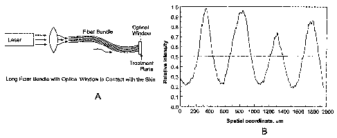
Fig. 1A illustrates a laser treatment system comprising a fiber bundle and
optical window;
Fig. 1B is a plot of the beam profile on the skin for the laser treatment
system
of Fig. 1A;
Fig. 2 illustrates a laser treatment system comprising a short fiber bundle
with expanded distal face;
Fig. 3 shows a diffractive lens having four levels;
Fig. 4 shows a diffractive lens having two levels;
Fig. 5 shows a diffractive lens with eight levels;
Fig. 6 shows a diffractive lens array having a hexagonal pattern;
Fig. 7 shows a diffractive lens array having an elongated hexagonal pattern;
Fig. 8 shows a treatment beam profile for a diffractive lens array;
Fig. 9 shows a plot of the relative hot area fluence factor, FI/Faõ as a
function of the relative diameter of the central hot area, d/D for a
diffractive lens
array in accordance with one aspect of the invention;
Fig. 10 shows the temperature profile of skin treated with a non-uniform
output beam from a diffractive lens array;
Fig. 11 shows a tip of a laser treatment handpiece having a cooling
mechanism; and
Fig. 12 shows an integrated laser and pulse light system for skin rejuvenation
treatment.
DETAILED DESCRIPTION OF THE INVENTION
A description of preferred embodiments of the invention follows.
CA 02605604 2007-10-19
WO 2006/116141
PCT/US2006/015180
-7-
As shown in Fig. 1A, the apparatus includes a laser source that emits an
output beam. The beam is coupled into a bundle of optical fibers using one or
more
focusing lenses. The bundle preferably contains between 1000 and 2000 separate
fibers. Typically, each fiber has a diameter of about 100-200 microns. The
output
laser beam is thus directed to 1000-2000 smaller beams, each of which
traverses the
length of the fiber bundle in individual optical fibers. The fiber bundle
terminates at
its distal end at an optical window that can be held in direct contact with
the
patient's skin. The window is approximately 1-5 mm thick, and protects the
output
face of the fiber bundle from contamination, and also permits the beam emitted
from
each fiber to diverge before it reaches the patient's skin, preferably so that
each
beam partially overlaps with the beam(s) from adjacent fibers in the bundle.
The fibers in the bundle can be packed together tightly, or can be spaced
apart from each other using mechanical spacers. The use of mechanical spacers
at
the distal end of the bundle spreads the energy from the bundle over a larger
area,
and helps to reduce the pain sensation for the patient. In general, the
combined spot
size on the skin from all the fibers in the fiber bundle is between
approximately 7
and 10 mm in diameter.
In a preferred treatment method for the embodiment of Fig. 1A, the laser
source, which is preferably an Nd:YAG laser, produces an output laser pulse
having
a wavelength of between 1.3 and 1.6, preferably between about 1.40 and 1.44
microns, and a pulse duration of between 0.1 and 100 milliseconds, preferably
between about 1 and 5 milliseconds. Because the laser operates at wavelengths
that
are well-absorbed by the skin, the laser can operate at relatively low
energies, and
minimize the risk of burning or damage to the skin.
In operation, the optical window is held against the skin of the patient, and
the laser source is energized to produce a pulse of laser light that travels
from the
source through the fiber bundle and the optical window, and penetrates into
the
patient's skin. Since the optical window is approximately 1-5 mm thick, the
window
also serves as a spacer between the output end of the fiber bundle and the
skin of the
patient. Thus, as the laser light is emitted from each fiber in the bundle,
the light is
permitted to diverge as it travels through the window to the patient's skin.
In a
CA 02605604 2007-10-19
WO 2006/116141
PCT/US2006/015180
-8-
preferred embodiment, the fibers are approximately 100-200 microns in
diameter,
and the beam emitted from each fiber, after passing through the window,
produces a
spot between 150-900 microns in diameter on the patient's skin. Because of the
diverging nature of light emitted from an optical fiber, the light at the
center of each
spot will be relatively high-energy light, while the light at the periphery of
each spot
will have significantly lower energy. Thus, over a combined spot size of 7 to
10 mm
for the entire fiber bundle, there are approximately 1000 to 2000 smaller
treatment
spots, generally about 150-900 microns in diameter, each consisting of a
higher-
fluence "hot spot" at the center of the spot surrounded by a lower-fluence
"cooler
zone" of radiation. The energy at the central "hot spot" is sufficient to
shrink the
underlying tissue, damage the collagen and produce collagen shrinkage. In
general,
the energy at the high-intensity zones, or "hot spots" is sufficient to raise
the
temperature of the target tissue to 70 C or higher. However, the radiation in
"cooler
zone" surrounding the hot spot is generally not sufficient to damage the
tissue and
cause collagen shrinkage in the tissue underlying these areas. In these lower-
intensity "cooler zones," the energy provided will only raise the temperature
of the
skin by a few degrees (or perhaps result in no appreciable temperature rise),
and thus
will not damage or even "shock" the tissue. However, this lower-intensity
radiation
is generally more appropriate or preferred to stimulate the fibroblasts in the
tissue to
produce collagen and "fill in" the skin for a younger more clearer skin
In a preferred embodiment, the fibers in the bundle are arranged so that the
spot sizes of radiation from each fiber abut or partially overlap with the
spots from
the adjacent fibers in the bundle on the patient's skin. In this way, the
invention can
simultaneously provide two modes of skin rejuvenation treatment: higher-energy
collagen shrinkage treatment in the "hot spots" at the center of each output
spot from
the fiber bundle, and overall stimulated collagen production throughout the
entire
area of the combined fiber-bundle output beam.
An example of a laser treatment method using a fiber bundle delivery system
is illustrated in Fig. 1B, which is a plot of the relative intensity on the
skin as a
function of location on the skin for four fibers in the bundle. In practice,
the fiber
bundle will consist of 1000-2000 individual fibers, in a regularly-spaced
CA 02605604 2007-10-19
WO 2006/116141
PCT/US2006/015180
-9-
arrangement to form a bundle. In this embodiment, the center-to-center
distance
between adjacent fibers in the bundle is approximately 500 microns. The
diameter
of each fiber is approximately 200 microns, and the numerical aperture (NA) of
the
fibers is approximately 0.2. The total diameter of the fiber bundle is
approximately
9 millimeters. The laser energy emitted from each fiber diverges as it passes
through
the transparent window, so that the spot size on the skin from each fiber is
at least
about 250 microns in diameter. Thus, the spots from each fiber generally abut
or
partially overlap with the spots from the adjacent fibers in the bundle. This
is shown
in Fig. 1B, where it can be seen that the whole area is treated with at least
a low-
intensity pulse, while the areas at the center of each spot receive a
significantly
higher dose of energy. The dotted line represents the average intensity
throughout
the treatment area. In this example, the peak fluence on the skin at the
center of each
spot is approximately 9 J/cm2, while the fluence at the periphery of each spot
is
approximately 2 J/cm2. The total area fluence is approximately 5 J/cm2.
The fluence(s) received at various portions of the treatment area can be
varied and controlled by, for instance, raising or lowering the total energy
output
from the laser source, changing the center-to-center distances between fibers
in the
bundle, using different diameter fibers, using fibers with a different NA to
change
the divergence of the beam and/or altering the thickness of the optical window
to
allow for a greater or lesser amount of beam divergence. The beam profile can
thus
be optimized for a variety of different conditions and laser treatment
methods.
Fig. 2 shows yet another embodiment that is similar to the embodiment of
Fig. 1, except that instead of a long-fiber bundle coupling the laser output
beam from
the source to the optical window, this embodiment uses a single transport
fiber to
carry the laser energy from the laser source to a handpiece containing a
shorter fiber
bundle. At the handpiece, the output laser pulse from the single fiber is
coupled into
the short fiber bundle. As in the prior embodiment, the short fiber bundle is
comprised of a plurality of separate optical fibers, preferably 1000 to 2000
fibers.
The short fiber bundle has a smaller bundle diameter at its proximal end to
allow the
output light from the single transport fiber to efficiently couple into the
bundle. The
fiber bundle "fans out" from its proximal end to its distal end, using, for
example,
CA 02605604 2007-10-19
WO 2006/116141
PCT/US2006/015180
-10-
mechanical spacers, to provide an expanded face at it's output. Preferably,
the
expanded face has a diameter of between approximately 7 to 10 mm, and is
coupled
to an optical window, as in the embodiment of Fig. 1. The embodiment of Fig. 2
preferably uses the same treatment parameters as those described in connection
with
Fig. 1.
Turning now to Figs. 3-8, yet another embodiment of the invention is
illustrated which uses a diffractive lens array to provide non-uniform heating
in the
target tissue. A multilevel diffractive lens consists of a number of
concentric rings
made of optically transparent material with variable thicknesses. The top
surface of
each concentric ring is flat so the refractive effects are negligible. The
variable-
thickness rings give rise to a spatial phase delay pattern on a propagating
incident
optical beam. The propagating optical beam carries the spatial phase delay
pattern
past the plane of the diffractive lens and produces an illumination pattern of
spatially
variable optical intensity. The optical intensity is high at geometrical
points that
meet the conditions for constructive interference and low at the points that
meet the
conditions for destructive interference. In general the design of a
diffractive lens is
optimized so that the principal diffi ________________________________ action
maximum (or minimum) would be on the
optical axis at a distance f from the plane of the lens. The distancefis the
focal
length of the lens. In general the goal of the diffractive lens design is to
increase the
fraction of the incident power in the principal diffraction maximum. However,
that
fraction is always less than 1 depending on the number of levels, the F-number
of
the lens and other design parameters. In fact, it is possible to design the
diffractive
lens pattern so that any fraction (less than 1) of the incident power would be
in the
principal maximum and the rest of the power would be distributed in the
secondary
maxima.
Various examples of multi-level diffractive lenses are shown in cross-
sectional views in Figs. 3-5. Fig. 3 shows a diffractive lens having four
levels; Fig.
4 shows a diffractive lens having two levels; and Fig. 5 shows a diffractive
lense
with eight levels.
In one embodiment of the present invention, a laser treatment apparatus and
method utilizes plurality of diffractive lenses that are arranged in an array
to produce
CA 02605604 2007-10-19
WO 2006/116141
PCT/US2006/015180
-Il-
an output beam having a non-uniform energy profile. More specifically, the
diffractive lens array is arranged in an optical path between a laser source
and the
treatment area, such that each lens in the array provides for an area of
higher-fluence
"hot spots" surrounded by lower-fluence regions of radiation. In a skin
rejuvenation
treatment, for example, the higher-energy areas provide sufficient heating to
damage
and shrink collagen in the "hot spots," while the lower-intensity radiation
regions
outside of these hot spots overlap and combine to stimulate collagen regrowth
over
the entire treatment area.
In this embodiment, the laser source preferably produces a pulse of radiation
having a wavelength between approximately 1.3 and 1.6 microns, preferably
between 1.40 and 1.44 microns, and a pulse duration of between about 0.1 and
100
milliseconds, preferably between 1 and 5 milliseconds. The laser source can be
an
Nd:YAG laser, for example. An optical system carries the beam from the laser
source to the treatment area. The diffractive lens array is preferably
arranged at the
distal end of the optical system, adjacent to the patient's skin. The array
comprises a
plurality of separate diffractive lenses adjacent to one another. In general,
there are
2000 or less lenses in an array, and preferably about 1800 lenses. Each lens
is
between about 150 and 450 microns in diameter, and is preferably about 250
microns in diameter. The entire array of diffractive lenses is generally about
7 to 10
mm in diameter. The array directs the input beam from the laser source (which
is
preferably also about 7-10 mm in diameter) into a plurality of higher-
intensity "hot
spots," corresponding to the central portion of each individual lens in the
array, and
lower intensity regions surrounding each hot spot. The combined effect in the
patient's tissue is to produce a plurality of higher-intensity zones in the
skin
corresponding to the center of each diffractive lens surrounded by areas of
lower-
intensity radiation. This is shown in the treatment beam profile of Fig. 8. As
can be
seen in this graph, the entire treatment area receives at least a low level of
treatment
radiation, with certain spaced-apart portions receiving a higher dose of laser
radiation. In the case of skin rejuvenation, for example, the laser energy
penetrates
deep into the collagen layer, where the collagen is heated to shrinkage
temperatures
in the "hot spots," while the entire treatment area is treated to effect
collagen
CA 02605604 2007-10-19
WO 2006/116141
PCT/US2006/015180
-12-
regeneration. In addition to skin rejuvenation treatment, the diffractive lens
array
can be optimized for use in other applications, such as treatment of acne and
hair
removal. A different beam profile from the diffractive lens array can be used
for
different applications.
The diffractive lens is considered to be irradiated by an average uniform
fluence, Fa,,, determined by the laser fluence setting selected by the user.
In general,
the average fluence of the laser in this embodiment is less than about 10
J/cm2, and
is preferably about 9 J/cm2. For purposes of illustration, each diffractive
lens with
diameter D is assumed to have a simplified design so that it produces a hot
area with
diameter, d, assumed to have uniform fluence, F1, and a periphery having a
uniform
fluence, F2. The lens design is assumed to produce a fluence ratio, p, of the
hot area
versus the periphery, p= F.,/F2. Under these simplifying assumptions, is it
possible
to derive a simple formula to approximate the hot area fluence, F1:
Fi
(Eq. 1)
Fav 'd"2 1
¨ +¨ ¨
\D) ,8 \D)
Fig. 9 shows a plot of the relative hot area fluence factor, F//Fa,,, as a
function of the
relative diameter of the central hot area, d/D. As an example, if the
diffractive lens
is designed to have /8= 5, with diameter D = 250 p.m, hot area diameter d= 100
m,
and the laser is selected to have average fluence Fay= 9 J/cm2, then the hot
area
fluence is F1= 3.05 x 9 J/cm2 = 27.4 J/cm2.
As a second example, if the diffractive lens is designed to have fi= 5, with
diameter D = 350 p,m, hot area diameter d= 200 p,m, and the laser is selected
to
have average fluence Fay= 9 J/cm2, then the hot area fluence is Fl= 2.17 x 9
J/cm2=
19.5 J/cm2.
Figs. 6 and 7 illustrate two exemplary embodiments of a diffractive lens
array according to the invention. In Fig. 6, the diffractive lenses are
arranged in a
CA 02605604 2007-10-19
WO 2006/116141
PCT/US2006/015180
-13-
hexagonal pattern. In Fig. 7, the lenses are arranged in an elongated
hexagonal
pattern.
Fig. 10 shows the peak tissue temperature distribution for a portion of skin
irradiated with a 1,440 nm laser through a diffractive lens array. As can be
seen from
the graph, a first dial active lens is centered at about 200 m, and a second
diffractive lens is centered at about 600 i.tm on the horizontal axis. As can
be seen
from this graph, there is an area of tissue about 200 pm wide centered on each
of the
, diffractive lenses that is heated to relatively high peak temperatures
(e.g., 70 C or
higher). This high-temperature zone extends from essentially the surface of
the skin
to a depth of about 350 p,m. As discussed above in connection with the fiber-
bundle
embodiment of Figs. 1 A and 1B, these temperatures are sufficient to cause
collagen
shrinkage. Outside of these high-temperature treatment zones, the peak
temperatures quickly drop off For example, in the area between about 300 rn
and
500 pm on the horizontal axis, the peak skin temperatures are generally
between 35
C (or less) and 50 C, and are generally less than about 40 C. As previously
discussed, these lower intensity zones provide collagen stimulation treatment.
Fig. 11 is a cross-sectional view of a tip 10 of a laser treatment apparatus
having a diffractive lens array for providing an output beam having a non-
uniform
energy profile. The operator applies the tip 10 directly against the patient's
skin 30.
A laser source (not shown) is energized to produce an output beam 23, and the
output beam is carried to the tip 10 by an optical fiber 20. The output beam
23 is
emitted from the end of optical fiber 20, and is directed to diffractive lens
array 61.
Adjacent to the diffractive lens array 61 is an optical window 60 that
directly
contacts the patient's skin 30. The optical window 60 is similar to the
optical
window described in connection with Fig. 1, and functions as a spacer between
the
output end of the fiber bundle and the skin of the patient. The optical window
60
can be integral with the diffractive lens array 61. Preferably, the window is
made of
a good thermal conductive material, such as glass. The optical fiber 20, lens
array
61, and optical window 60 are all enclosed in a tip housing 40, which is
preferably a
cylindrically-shaped housing. The tip housing 40 can be made of plastic.
Outside
the tip housing 40 is a cooling mechanism 11. Preferably, the cooling
mechanism 11
CA 02605604 2007-10-19
WO 2006/116141
PCT/US2006/015180
-14-
comprises a conduit 50 that carries cooled air 51 from a cooled air source
(not
shown) to the tip 10 of the treatment apparatus. The conduit 50 preferably
includes
an outlet that is angled with respect to the tip housing 40, so that cooled
air 51 is
directed at the distal end of the tip housing 40 (i.e. where the tip 10
interfaces with
the patient's skin 30). This arrangement provides effective cooling of the
skin
during laser treatment. Although the tip 10 and cooling mechanism 11 are shown
here in connection with the diffractive lens array embodiment of Figs. 3-8, it
will be
understood that this design may also be employed with a laser apparatus having
a
fiber bundle, such as shown and described in connection with Figs. 1 and 2.
Fig. 12 shows an integrated laser and pulse light system for skin rejuvenation
treatment, according to one aspect of the invention. As shown, the system 100
includes a housing 101 containing a laser source 103, preferably a solid-state
laser,
such as an Nd:YAG laser operating at about 1.4 microns wavelength and about 3
msec pulse width. Light from the laser source 103 is coupled into an optical
fiber
delivery system 20, which extends from the housing 101 to a first handpiece
105.
The first handpiece 105 includes an optical system for producing a beam with a
non-
uniform energy profile, in accordance with any of the embodiments previously
described herein. The handpiece 105 can include a tip 10 as previously
described in
connection with Fig. 11. The system can also employ a cooling system as
described
in connection with Fig. 11.
The integrated system 100 also includes a pulse light portion, that preferably
includes a flashlamp light source 115. In a preferred embodiment, the
flashlamp
source comprises a Xenon flashlamp that produces treatment pulses having
wavelengths between 560 and 950 nm and pulse widths between 5 and 35
milliseconds. The flashlamp 115 is located in a second handpiece 113 connected
to
the housing 101 by a high-voltage cable 111 that provides power to the
flashlamp
115 from a high-voltage source 109 located within the housing 101. The pulse
portion preferably also includes a water circulating system (not shown), as is
conventionally known, for cooling the flashlamp. The pulse light system can
also
employ a cooling system as described in connection with Fig. 11. In one
embodiment, an air cooler and conduit carry cold air to handpiece 115. The tip
of
CA 02605604 2007-10-19
WO 2006/116141
PCT/US2006/015180
-15-
handpiece 115 includes a sapphire window, The edge of the proximal side of the
sapphire window (i.e. the side closest to the flashlamp source) is cooled by
the cold
air from the cooling system. The distal surface of the sapphire window
contacts the
patient's skin for treatment.
In operation, the second handpiece 113 is held proximate to the patient's
skin, and the flashlamp 115 is energized to provide a treatment pulse. The
spot size
of the pulse light portion is generally larger than the laser portion, and is
generally
around 11 x 55 min (or 6 cm2). The pulse light portion is thus able to treat
large
areas of the patient's skin in.a relatively short time period. The maximum
fluence of
the pulse light portion is typically around 20 J/cm2.
The pulse light portion of the integrated system is well-suited to treat
pigmented and certain vascular lesions. The pulse light portion effectively
treats, for
example, dischromia, a common condition associated with aging skin, as well as
superficial pigmented lesions, veins, and the blush of rosacea associated with
sun-
damaged skin. The laser portion of the system is effective for stimulation of
collagen production and skin tightening, as previously discussed. The
combination
of laser treatment and pulse light treatment in an integrated system provides
a
complete and efficient system for facial rejuvenation treatment. The laser and
pulse
light system(s) are integrated in a common housing, and preferably use a
common
control system 117, and can even use the same electronic drive circuit 119 for
driving both the laser source 103 and the flashlamp source 115.
While this invention has been particularly shown and described with
references to preferred embodiments thereof, it will be understood by those
skilled in
the art that various changes in form and details may be made therein without
departing from the scope of the invention encompassed by the appended claims.