Note: Descriptions are shown in the official language in which they were submitted.
CA 02606502 2013-07-11
WO 2006/118984
PCT/C1S2006/1116146
INSTRUMENT FOR DIRECT LARYNCOSCOPY WITH A
RIGID BLADE AND FLEXIBLE FIBEROPTICS
FIELD OF THE INVENTION
[0001] The present invention concerns a medical instrument that allows for
both direct
laryngoscopy with a rigid blade and indirect flexible fiberoptics in the same
instrument. More
particularly the present invention concerns a laryngoscope having a rigid
blade to retract
oropharyngee tissue and a flexible fiberoptic scope with guidcwire systems, to
permit
examination of the larynx during laryngoscopy of a patient as well as a means
to intubate
patient.
10002]
BACKGROUND OF THE INVENTION
10003] The intuhation of a patient in need of medical care is a frequent
occurrence in operating
theaters, emergency rooms and other medical situations. The insertion of a
tube, through which
life saving and sustzining oxygen may be provided, must be made through the
natural defenses
provided in the human body. Namely, an endo-tracheal tube must be fed through
the mouth and
throat, past the epiglottis, larynx and then into the trachea. The epiglottis
is generally a flap of
tissue that overlies the glottic opening into the larynx to prevent passage of
food into the trachea
during eating. In order to intubate a patient, the epligottis must be moved
aside in order TO
provide visualization and access to the larynx and the trachea where
intubation is made. It is
CA 02606502 2007-10-29
WO 2006/118984
PCT/US2006/016146
generally agreed that laryngoscopy of many patients, with the state of prior
art technology is at
times difficult, concomitantly making intubation difficult. In addition, once
visualization of the
vocal cords is accomplished, actual passage of an endotracheal tube between
the vocal cords with
state of prior art may also be difficult.
[0004] There are a number of laryngeal devices presently available for use in
intubating patients.
In most cases these devices comprise a first means to displace the epiglottis
and a separate
means to insert an endotracheal tube past the epiglottis and into the trachea.
In patients assessed
and considered difficult to intubate, the doctor, or other medical personnel,
typically requires
assistance to intubate patients with difficult anatomy when using almost all
of the devices of the
prior art. An extra hand is needed to maintain the patient in an appropriate
position, with mouth
open, for the insertion of the blade to displace the epiglottis, while a
second medical practitioner
manipulates the mandible, tongue or larynx externally, or inserts an
intubation device in the
patient and pushes an endotracheal tube into the patient's trachea.
[0005] While the cooperation of medical personnel is generally available such
that intubation is
almost never denied, however, it is desirable to provide medical personnel
with the tools that
allow for excellent care of patients while permitting additional medical
personnel, previously
needed to assist in intubation, to attend to other tasks and/or patients.
SUMMARY OF THE INVENTION
[0006] In accordance with the present invention, a rigid laryngoscope and
flexible-viewing
device with intubation means as one instrument is provided. The device of the
present invention
=
2
CA 02606502 2007-10-29
WO 2006/118984
PCT/US2006/016146
comprises a handle operationally attached to a viewing member and a direct
laryngoscope blade,
the viewing device comprising a flexible fiberoptic scope and rigid extension
housing. The
flexible fiberoptic scope further has a means for providing a variable angle
view of the interior of
a patient's anatomy; the flexible fiberoptic scope is operationally attached
to the viewing member
and is carried within a rigid C-shaped housing channel along the length of and
adjacent to the
rigid laryngoscope blade. In one embodiment of the present invention, there
are means to extend
the tip of the flexible fiberoptic scope beyond the tip of the rigid blade.
The extended or
retracted position can be locked in place with an available clip or other
means. The device
further has a means to cause, when desired, the distal end of the flexible
fiberoptic scope to pivot
to any angle in a range from a linear position to a first pivoted position and
back through the
linear position to a second pivoted position opposite the first pivoted
position. In this manner a
panoramic view of the patient's oral anatomy is provided. In one embodiment
the means to pivot
the end of the flexible fiberoptic scope through its arc of movement includes
a ratcheting means,
which can also be provided with means to lock the flexible tip scope in a
desired position.
[0007] The laryngoscope blade of the present invention comprises a rigid
element for effecting
the opening of a passageway for an intubation device, in a manner known in the
art with the use
of a typical laryngoscopic blade. Typical laryngbscopic blades may be
available in a variety of
shapes and sizes, including straight or curved and such as Miller or Macintosh
designs. The
device of the present invention further comprises a means to carry an
intubation device adjacent
to the flexible tube such that the user can on his own view the interior of
the patient's anatomy,
provide an open passageway and deliver an intubation device into the patient's
trachea.
3
CA 02606502 2007-10-29
WO 2006/118984
PCT/US2006/016146
[0008] In one preferred embodiment of the laryngoscope and flexible-viewing
device of the
present invention the viewing device comprises a fiber optic cable and viewing
element. Further,
the viewing element of the device is a lens. Further; in another embodiment,
the viewing device
is a television-like monitor and the device of the present invention is
operationally attached to the
monitor such that one or more persons can view a patient, on a screen, when
the device is used.
[0009] In one preferred embodiment of the device of the present invention, the
handle and
viewing device are rotationally attached together for storage and also to
provide novel angles,
between the handle and viewer, to assist in direct viewing of the patient's
larynx upon the initial
insertion of the instrument into the patient's oropharynx. In several
embodiments of the device
of the present invention, the viewing member comprises an extension tube
attached to the rigid
blade, in which a flexible tube member is maintained, and a lens or monitor
connection defined
at its proximal end, operationally connected to the flexible tube. The
extension tube of the
viewing member is provided with means for telescopically extending, and
subsequently
retracting the member, so as to assist the user in viewing the patient's
anatomy through the lens
and flexible tube, by permitting closer inspection within the patient's
larynx. The extended or
retracted position of the flexible tube can be secured with a clip or other
available means.
[0010] In the present invention the intubation device is a rigid laryngoscope
and the flexible tube
includes means to carry a guidewire with which to assist the guidance of the
intubation device
within the patient's throat. In a preferred embodiment, the means to carry the
guidewire includes
a slit defined within the flexible tube within which the wire is positioned.
In the preferred
embodiment the preferred guidewire is of a type having a pivotal end piece,
located at its
4
=
CA 02606502 2007-10-29
WO 2006/118984
PCT/US2006/016146
proximal end, designed to pivot from a first position parallel to the
guidewire to a second
position perpendicular to the guidewire, much in the way that a cufflink is
kept within a shirt
sleeve. The slit in the flexible tube can be made in a number of ways without
departing from the
novel scope of the present invention and in a preferred embodiment has a
diameter slightly larger
than the cross-sectional diameter of the guidewire. In a preferred embodiment,
in its initial
position the guidewire is housed within a hollow channel, having a size
greater than 3 times the
guidewire diameter, along the length of the flexible fiberoptic scope. The
fiberoptic scope, in
this embodiment lays in a rigid C- shaped channel housing located under the
rigid blade.
[0011] The present invention further includes a method of utilizing the above
described device.
The method includes insertion of the distal end of the device within the
patient's mouth, utilizing
the viewing means to provide better hand eye coordination in finding and
displacing the
epiglottis and subsequently visualizing their vocal cords. Lifting the
epiglottis with the blade
portion of the device, visualizing the vocal cords directly, or through the
flexible fiberoptic
scope, and then guiding an endo-tracheal tube into the patient's trachea
directly. Alternatively,
the endo-tracheal tube may be guided utilizing the guidewire systems held in
the flexible tube
member of one embodiment of the present invention. In addition, in one
embodiment an
umbilical-type detachable lasso cord is attached to the distal tip of the
enclosed guidewire that is
to be advanced into the trachea. The endotracheal tube is threaded over the
lasso cord and then
guided into the trachea. In this embodiment, the lasso cord is then detached
from the guidewire,
by operation of the device. The lasso cord of the present embodiment can also
be provided with
a pivotal proximal end piece, such as used in one embodiment of the guide wire
(noted above).
CA 02606502 2007-10-29
WO 2006/118984
PCT/US2006/016146
[0012] A more detailed explanation of the invention is provided in the
following description and
claims and is illustrated in the accompanying drawings.
BRIEF DESCRIPTION OF THE DRAWINGS
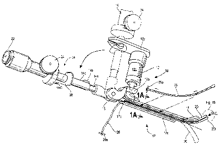
[0013] Figure 1 is a perspective view of a device made in accordance with the
teachings of the
present invention.
[0014] Figure lA is a cross-section of the device of Figure 1, taken along the
plane of line 1A-
1A of Figure 1.
[0015] Figure 1B is an exploded perspective view of element Fig. 1B of Figure
1.
[0016] Figure 1C is an partially cut-away exploded perspective view of element
Fig. 1B of
Figure 1.
[0017] Figure 2A is a schematic representation of one embodiment of the
ratcheting mechanism
of the device of Figure 1.
[0018] Figure 2B is a second schematic representation of one embodiment of the
ratcheting
mechanism of the device of Figure 1.
[0019] Figure 3A is an elevational view, partially broken away, of the
laryngological device of
the present invention in place in a patient's body, with a flexible tube in a
first position.
[0020] Figure 3B is a second elevational view, partially broken away, of the
device of Figure 3A
in place in a patient's body, with the flexible tube in an advanced position,
showing possible
movement in phantom.
6
CA 02606502 2007-10-29
WO 2006/118984
PCT/US2006/016146
[0021] Figure 4A is an elevational view, partially broken away, of the device
of the present
invention.
[0022] Figure 4B is another elevational view, partially broken away, of the
device of the present
invention.
[0023] Figure 5 is a perspective view, partially broken away, of the device of
the present
invention in a first position at the opening of the larynx with an
endotracheal tube in a first
carried position.
[0024] Figure 5A is a partial cross sectional view and partial perspective
view of the device of
Figure 5, taken along the plane of line 5A-5A and projected forward therefrom.
[0025] Figure 6 is a perspective view, partially broken away, of the device of
the present
invention in a first position at the opening of the larynx with an
endotracheal tube in place for
intubation.
[0026] Figure 6A is a partial cross sectional view and partial perspective
view of the device of
Figure 6, taken along the plane of line 6A-6A and projected forward therefrom.
[0027] Figure 7 is a perspective view, partially broken away, of another
embodiment of the
device of the present invention in a first position at the opening of the
larynx with an
endotracheal tube in a first carried position.
[0028] Figure 7A is an exploded perspective view of element labeled "Fig. 7A"
of Figure 7.
[0029] Figure 8 is a perspective view, partially broken away, of the
embodiment of Figure 7 of
the device of the present invention in a first position at the opening of the
larynx with an
endotracheal tube in place for intubation.
7
CA 02606502 2007-10-29
WO 2006/118984
PCT/US2006/016146
[0030] Figure 8A is an exploded perspective view of element labeled "Fig. 8A"
of Figure 8.
DETAILED DESCRIPTION OF THE
ILLUSTRATIVE EMBODIMENT
[0031] While the present invention is susceptible of embodiment in various
forms, there is
shown in the drawings a number of presently preferred embodiments that are
discussed in greater
detail hereafter. It should be understood that the present disclosure is to be
considered as an
exemplification of the present invention, and is not intended to limit the
invention to the specific
embodiments illustrated. It should be further understood that the title of
this section of this
application ("Detailed Description of the Illustrative Embodiment") relates to
a requirement of
the United States Patent Office, and should not be found to limit the subject
matter disclosed
herein.
[0032] Referring to the figures, a laryngeal device or laryngoscope 10 having
a handle 12 and a
viewing member 14 is provided. Viewing member 14, in a preferred embodiment is
made so that
it can telescope between a first extended position 14a (Fig. 4A) and a second
contracted position
14b (Fig. 4B). Telescoping portion 14, in the present embodiment, is attached
at hinge 16 to a
blade portion 17, including a blade 18. Telescoping portion 14 is also, in one
embodiment,
hingedly attached, near handle 12, such that telescoping portion 14 can be
placed in a first folded
position parallel to handle 12 when in a first stored position and during the
initial insertion into
the patient's mouth (see phantom lines). It will be understood that while the
connection between
the major components of the device of the present invention is shown as a
hinged the elements,
8
CA 02606502 2007-10-29
WO 2006/118984
PCT/US2006/016146
of the device, can be attached together in different manners, all well known
to persons having
ordinary skill in the art, without departing from the novel scope of the
present invention. As
noted above, viewing member 14, further, can be placed in a first contracted
position or in
extended position and can be placed in an infinite number of positions between
the first extended
position and the second contracted position, so that the user can place the
device in an optimal
viewing position. The uses for such telescopic action will be discussed in
greater detail below.
[0033] Referring again to Fig. 1, it will be seen that the present invention
of the device includes a
flexible tubular member 20, adjacent to the blade portion 18, an eyepiece 22
and ratcheting
member 24, both operationally attached to the flexible tubular member 20.
Flexible tube
member 20, in a preferred embodiment, can be made to advance forward so that
tip 20t of
member 20 is distal of the end of blade 18 within the rigid C-channel housing
17c along the
underside of the blade, to provide a better view of the patient's anatomy.
Tubular member 20, as
shown in Fig. 1B can include a number of useful implements including an
optical view member
20a, comprised in a preferred embodiment of a fiber optic scope; an
illumination means 20b,
which in a preferred embodiment comprises a second fiber optic cable attached,
at a more
proximal end to an illumination source such as an LED lamp; a suction entry
point 20e, which in
a preferred embodiment is attached to a vacuum port 28 (which in turn can be
connected to a
suctioning device external to the laryngoscope 10). Tubular member 20 further
includes a
transversal opening 20d defined generally along its entire length. Opening 20d
is defined in
member 20 so as to accommodate a guidewire 26. Opening 20d is generally a slit
having a
dimension slightly larger than that of the preferred guidewire 26 at its
opening, with its full
9
CA 02606502 2007-10-29
WO 2006/118984
PCT/US2006/016146
length channel being about 3 times the diameter of the guidewire, that will be
carried therein,
such that the guidewire 26 can be peeled out from tubular member 26 when
desired, as explained
in greater detail below. It will be understood by persons having ordinary
skill in the art that the
distal tip opening 20d will be larger than the diameter of channel 20d, so as
to allow for the full
and complete retraction of the distal tip of guidewire 26 into the distal tip
of flexible tube 20,
which is shown having a specialized shape and diameter.
[0034] In a preferred embodiment of the present invention, guidewire 26
includes a novel
retention member 26a for use in association with the laryngoscope 10. Further,
in one
embodiment, a guidewire 26 having a distal tip holding member 26d is provided
to assist in the
carrying of a specialized cord so that intubation into the trachea below the
vocal cords is
facilitated, this will be explained in greater detail below. A more detailed
explanation of the use
of the guidewire 26 and the retention member 26a will be made below. It will
be understood by
persons having ordinary skill in the art that while a number of desirable
elements of flexible
member 20 are shown other elements can be substituted and some of those shown
can be
eliminated by persons having ordinary skill in the art, from flexible tube 20,
as desired or
desirable, without departing from the novel scope of the present invention.
[0035] Handle 12 of the laryngoscope 10 provides a location for gasping the
device so that its
= use within a patient can be facilitated. Handle 12, in a preferred
embodiment can house a power
source (not shown) such as a battery that can provide power to an illumination
means located at
the distal end of the rigid blade 18 as in conventional laryngoscopes. In such
a situation an LED
lamp (not shown) can be housed in the handle 12 and a fiber optic cable 12a
can deliver light
CA 02606502 2007-10-29
WO 2006/118984
PCT/US2006/016146
from the LED lamp at the end of the rigid blade as is common in the field of
the present
invention. Handle 12 can in a preferred embodiment includes a holding means
12b, having
means to grasp and hold viewing member 14. While a particular type of holding
means 12b is
shown, it will be understood by persons having ordinary skill in the art, that
any means capable
of holding viewing member 14 in its stored position can be utilized without
departing from the
novel scope of the present invention. It will be understood that other types
and means to hold the
device of the present invention, similar to or different from handle 12 can be
used without
departing from the novel scope of the present invention. In the illustrative
embodiments, handle
12 is shown with a knurled surface to facilitate holding and manipulating the
device of the
present invention in typical wet environment in which the device is used.
However, it will be
understood by persons having ordinary skill in the art, that handle 12 can be
configured
differently, as needed, to facilitate its use in whatever environment the user
is in, without
departing from the novel scope of the present invention. In one embodiment of
the handle 12 a
power source, not shown, such as a battery and/or a rechargeable battery can
be encased to
provide power to a light source. In a further embodiment a separate light
source entry point 46
(Figs. 4A and 4B) is provided, a light element (not shown) can be attached to
the entry point 46
to provide light via the viewing element's fiberoptics to the distal tip of
20b when positioned
within the patient's larynx. As is known to persons having skill in the art
any manner of light
source can be attached thereto, such as a portable AC powered lamp source or a
battery-held
lamp source, without departing from the novel scope of the present invention.
[0036] Blade portion 17 is provided with a laryngeal blade 18 having a typical
straight or curved
11
CA 02606502 2007-10-29
WO 2006/118984
PCT/US2006/016146
size and shape, as conventionally known to the art, that permits the user to
easily insert the
device in a persons throat and move the glottis such that an endotracheal
device (described
below) can be inserted within the trachea. The use of a typical laryngoscope
is well known to
medical personnel. Blade portion 17 further includes means to hold the
flexible tube portion 20,
which includes, as shown in Fig. 1B, a fiber optic viewing device 20a, an LED
light source 20b,
a suction point 20c (attached to a vacuum means, as described in greater
detail below) and a
guidewire carrier means 20d. In a preferred embodiment of the present
invention, a generally
"C"-shaped rigid channel 17c is provided to act as a means to hold flexible
tube 20 and to act as
a loose guide for the flexible tube when it is advanced through the device. In
a preferred
embodiment of the present invention, channel 17c has a length approximately
the length of blade
18 less approximately 2 cm proximally and 3 cm distally, to allow for ease of
movement of the
flexible member. It will be understood by persons having ordinary skill in the
art that channel
17c can be made of different lengths and have a different cross-sectional
shape without departing
from the novel scope of the present invention.
[0037] The device 10 of the present invention includes means to peer into the
throat from a
viewer 22, such as a lens or, in one embodiment, a television monitor attached
to the viewing
means in a manner well known in the art. The device 10 further includes, in
the illustrative
embodiment, a ratcheting means 24 which when rotated causes the flexible tube
20 to pivot
alternatively up from an axis 20x parallel to the axis of the device 10 and
down from that axis
20x. In this manner the user can achieve a generally panoramic view of the
interior of the
patient's throat, so as to place (the blade 18 in the appropriate place so
that) an endo-tracheal
12
CA 02606502 2007-10-29
WO 2006/118984
PCT/US2006/016146
tube 40 (Figs. 5-8) can be inserted into the trachea 42 (Fig. 3B). Further, as
explained above and
illustrated in Figs. 3B, 4A and 4B viewing member 14 can be moved from a first
contracted
position (Fig. 4B) to an extended position (Fig. 4A), or any position there
within, to the effect
that flexible tube member 20 is, when viewing member 14 is moved to the
extended position, is
thrust forward, as shown in Fig. 3B. Movements within the viewing member 14,
in a preferred
embodiment, are produced by providing telescoping segments, inner segment 14a
and outer
segment 14b. It will be understood that outer segment 14b will slide over
inner segment 14a
permitting movement of flexible tube 20 which is operationally attached within
inner segment
14a to outer segment 14b. The distal end of 14b will retain a locking
mechanism 14s, such as a
pin in hole system (see Fig. 1), where 14b carries a rigid pivoting pin, which
will be placed
within one of a series of holes located within the rigid wall of 14a when the
distal tip of 20 has
been satisfactorily positioned in the larynx. This or any other locking
mechanism will prevent
any unwanted extension or retraction of the viewing element while passing the
guidewire or
endotracheal tube during the use of the device of the present invention. When
segment 14b is
pushed forward inner segment 14a slides there within and flexible tube 20 is
pushed forward,
when it is desired to retract flexible tube 20, outer segment 14b is pulled
back along inner
segment 14a.
[0038] Referring again to Fig. 1, the guidewire 26 in a preferred embodiment
is provided with a
holding means 26a as noted above. It will be understood by persons having
ordinary skill in the
art that the main body of guidewire 26 can be made in any manner typically
associated with
medical guidewire technology without departing from the novel scope of the
present invention.
13
CA 02606502 2007-10-29
WO 2006/118984
PCT/US2006/016146
In the illustrative embodiments a twisted cable member is show, however, any
method of making
such guidewire and any type of guidewire found useful to the present invention
can be used
without departing from the novel scope of the present invention. The holding
means 26a of the
illustrated guidewire 26 is pivotal, along an axis of rotation adjacent its
connection to the
guidewire 26. In this manner, the long axis of holding means 26a can be
brought parallel to the
main axis of the guidewire, such that guidewire 26 can be slid through
cannular openings in the
manner of an ordinary wire. However, because of the pivoting feature, when the
holding means
is moved from a parallel configuration it provides a means to retain the
endotracheal tube on the
guidewire before positioning and to stop the complete insertion of the wire
beyond the proximal
end of the endotracheal tube. This is particularly useful in its use in the
present invention, where
the loss of a guidewire in a patient's throat would likely cause choking and
possibly death. As
the device of the present invention can be used by one person alone, the means
to assure the safe
maintenance of the guidewire within elements of the present device, provides
added security to a
single health care provider.
[0039] Slit 20d, as described above, permits the carrying of a guidewire into
the patient's throat
to assist in placement of the endotracheal tube 40, as described below. The
slit 20d in flexible
tube 20 can be created in any number of ways, including by cutting, pre-
molding in place or
extrusion means, without departing from the novel scope of the present
invention. In a preferred
embodiment the slit is provided of a size such that the diameter of the
guidewire to be inserted
therein is slightly smaller than the slit. While this will permit the easier
removal of the
guidewire, the properties of the flexible tube 20 will hold the guidewire 26
in place until an
14
CA 02606502 2007-10-29
WO 2006/118984
PCT/US2006/016146
appropriate force, as described below, is applied to the guidewire during
intubation. It will be
understood by persons having skill in the art that the dimensions of the slit
in flexible tube 20 can
be varied, in manners which will cause an effective holding force, without
departing from the
novel scope of the present invention. In a preferred embodiment, the housing
channel 20d (length
of channel 20 within the mouth) should have a diameter approximately 3 times
the size of the slit
opening or of the guidewire diameter.
[0040] In the use of the device of the present invention, a user will place an
intubation device,
preferably an endotracheal tube 40 onto holding means in device 10,
specifically in the area
below blade portion 17. A guidewire 26 is placed partially within a flexible
tube 20 in a channel
20d and its proximal end is threaded through the channel 40a of the
endotracheal tube 40.
Initially, medical personnel can insert the device merely by eyeing the
general location of the
anatomy with respect to the instrument, as shown in Fig. 3B, while the viewing
element 14 is in
its "stored" or up position, as is typically done with standard rigid blades
in the art. The device 10
is then guided such that the blade 18 is placed adjacent the glottis 46 such
that the blade 18 can
hold the epiglottis 48 open allowing access to the trachea 42 for the
guidewire 26, flexible tube
20 and endotracheal tube 40. Once placed in the appropriate location for
proceeding with
intubation, the user then checks the position of the blade element 17 and
blade 18 inside the
patient by activating a lighting means, such as light element 20b, and peering
through viewing
element 22 into the mouth of the patient. The device 10, in combination with
endotracheal tube
40, is then extended or contracted as necessary (see Figs 4A and 4B). The user
manipulates the
viewing tube 20 using ratcheting means 24 such that the distal end of the
viewing tube moves
CA 02606502 2007-10-29
WO 2006/118984
PCT/US2006/016146
alternatively upward and downward from a line parallel to the central axis x
of the device, so as
to provide a panoramic view of the throat 3. At this point in the procedure,
once the vocal cords
are visualized, the user may place an endotracheal tube into the trachea
without the use of a
guidewire or the user may choose to use one of the guidewire systems of the
device of the present
invention as explained herein. In the use of a preferred guidewire system, the
guidewire 26 is
pushed distally into the larynx past the vocal cords and into the trachea
while remaining partially
within channel 20d. Then the endotracheal tube 40 is advanced, peeling the
guidewire 26 out of
its channel 20d; endotracheal tube 40 is then advanced into and down the
trachea 42 to intubate
the patient. The guidewire 26 is then removed from the trachea and
endotracheal tube.
[0041] The proximal end 40a of the endotracheal tube 40 can then be attached
to a source of
oxygen and other gases as required for treatment. In a preferred embodiment,
the guidewire 26 is
provided with holding means 26a (Fig.1) such that as the guidewire is advanced
it is safely held
within device 10 so that it cannot be lost in the patient's mouth or throat.
[0042] In one embodiment of the device of the present invention, as shown in
Figures 7 through
8A, a special umbilical tie 60, to assist in correctly placing the
endotracheal tube 40, is shown.
The umbilical tie 60 comprises a thin filament cord of a type well known in
the art, having an
elastic and/or detachable lasso element 62 comprising a means to grasp a
guidewire 126. In the
present embodiment, the device 10 and endotracheal tube 40 are assembled as
noted above, with
the exception that the umbilical tie 60 is fed into the endotracheal tube 40
such that its lasso end
62 emerges from the distal end to the endotracheal tube 40 and is attached to
the guidewire 126.
Guidewire 126 comprises a special head member 126a having a configuration
similar to a
16
CA 02606502 2013-07-11
OA 02606502 2007-10-29
WO 2006/118984
PCT/US2006/016146
bowling pin, such that first and second pseudo-spherical members are attached
so that a "waist
line" 126b is formed there between. Umbilical tie 60 is threaded within the
endotracheal tube 40
such that lasso element 62 emerges from the distal end of endotracheal tube 40
and umbilical
member 60 is extended so that the lasso element 62 loops over guidewire head
126a, resting
within waist line 126b. The device 10 and endotracheal tube 40 so configured
is then placed
within the patient, in much the same manner as described above. However, tic
60 is now can-led
into the trachea by guidewire 126 while guidewire 126 remains in its channel
20d, along its
length as described above, and the endotracheal tube is threaded over cord 60
below the vocal
cords into the trachea. The user then releases cord 60 from guidewire 126,
leaving the
endotrucheal tube in place. It will be understood by persons having skill in
the art that tie 60 may
be created in any manner known, including molding, braiding, extruding and
others, without
departing from the novel scope of the present invention. Further, while a
lasso element is shown
at the distal end of tie 60, it will be understood that other means for
grasping and holding
guidewire 26, including means molded or otherwise formed or more permanently
attached to the
distal end of the guidewire, can be used without departing from the novel
scope of the present
invention, In a preferred embodiment, lasso cord 60 is also provided with a
holding element 26a
on it proximal end, so as to hold and retain an endotracheal tube 40 over the
cord 60 while in the
procedure preparatory stage as well as to prevent loss of the cord 60 in the
trachea after
endotracheal intubation.
17