Note: Descriptions are shown in the official language in which they were submitted.
CA 02607036 20122-11
LEG ALIGNMENT FOR SURGICAL PARAMETER MEASUREMENT
IN HIP REPLACEMENT SURGERY
FIELD OF THE INVENTION
The present invention generally relates to
computer-assisted hip replacement surgery and, more
precisely, to surgical parameter measurement and
adjustment in hip replacement surgery.
BACKGROUND OF THE INVENTION
Total hip replacement surgery involves the
introduction of an artificial hip joint in a patient.
The artificial hip joint typically consists of a pelvic
implant and a femoral implant. The pelvic implant is a
cup received in the acetabulum. The femoral
implant
consists of a spherical portion received at an end of a
longitudinal implant portion, or a femoral implant
secured to the resurfaced femoral head. In the first
case, the longitudinal implant portion is introduced into
the intramedullary canal of the resected femur, with the
spherical portion being generally centered with respect
to the previous position of the femoral head. Therefore,
the femoral head (i.e., spherical portion of the femoral
implant) and the cup (i.e., pelvic implant) coact to
create the artificial hip joint.
CA 02607036 2007-10-31
WO 2006/128301
PCT/CA2006/000905
Different output values are of concern in hip
replacement surgery. In
order to reproduce a natural
and/or improved gait and range of motion to a patient,
the position and orientation of the implants, the medio-
lateral offset of the femur and the limb length
discrepancy must be considered during surgery. The work
of the surgeon during hip replacement surgery will have
a direct effect on these output values, and a successful
surgery will relieve pain, provide motion with stability
and correct deformities.
There is no precise definition of the
intraoperative limb length discrepancy (hereinafter
"intraop-LLD") and intraoperative medio-lateral offset
(hereinafter "intraop-MLO"). On the
preoperative
X-rays, surgeons usually measure preoperative limb
length discrepancy (hereinafter "preop-LLD") along the
vertical axis of the body as a relation between the
interischial line of the pelvis and the lesser
trochanter of the femur. Intraoperatively, in order to
obtain reasonable measurements that are then possible to
validate with X-ray measurements, the surgeons have to
align the leg along the vertical axis of the body. This
alignment is highly dependent on the surgeon skills and
experience.
Changes in adduction/abduction of the leg
will significantly alter the measurement and introduce
measurements errors.
The accuracy of the measurements rests heavily
on the surgeon's ability to reposition the leg
accurately before each measurement. Therefore, in order
to obtain an accurate intraop-LLD and intraop-MLO
measurement, the leg, after the implant reduction, must
be realigned in the same orientation as before the
dislocation. Again,
changes in adduction/abduction,
flexion/extension and rotation of the leg will
significantly alter the measurement.
-2-
CA 02607036 2007-10-31
WO 2006/128301
PCT/CA2006/000905
Failure to provide a robust and accurate
method for leg length and offset measurement
intraoperatively might lead to the postoperative leg
length inequality. This in turn might lead to patient
dissatisfaction and/or discomfort, functional impairment
(low back pain, static nerve palsy, abductor weakness,
dysfunctional gait), unstable hip joint, early
mechanical loosening.
SUMMARY OF THE INVENTION
It is an aim of the present invention to
address the issues pertaining to the prior art.
It is a further aim of the present invention
to provide a novel method for guiding an operator in
measuring surgical parameters such as limb length
discrepancy and medio-lateral offset intraoperatively in
computer-assisted surgery.
Therefore, in accordance with the present
invention, there is provided a method of measuring
surgical parameters in computer-assisted surgery so as
to guide an operator in inserting a hip joint implant in
a femur, comprising the steps of: i) digitizing a frame
of reference of the pelvis, the frame of reference of
the pelvis being trackable in space for position and
orientation; ii) digitizing a first frame of reference
of the femur as a function of the frame of reference of
the pelvis; iii) obtaining a reference orientation for
the frame of reference of the femur with respect to the
frame of reference of the pelvis; iv) digitizing a
second frame of reference of the femur with respect to
said reference orientation as a function of the frame of
reference of the pelvis, after initiation of implant
reduction; whereby surgical parameters associating the
-3-
CA 02607036 2007-10-31
WO 2006/128301
PCT/CA2006/000905
femur to the pelvis are measured as a difference between
the first and second frames of reference of the femur.
Further in accordance with the present
invention, there is provided a CAS system for measuring
surgical parameters during hip replacement surgery to
guide an operator in inserting a hip joint implant in a
femur, comprising: at least a first trackable reference
in fixed relation with the pelvis, the first trackable
reference being trackable to form a pelvic frame of
reference; a registration tool being trackable; a sensor
apparatus for tracking at least the first trackable
reference and the registration tool; and a controller
unit connected to the sensor apparatus so as to receive
tracking data for at least the first trackable reference
and the registration tool, the controller unit having: a
position and orientation calculator to calculate from
the tracking data a position and orientation of at least
the pelvic trackable reference to track the pelvic frame
of reference, and of the registration tool to produce a
femoral frame of reference at two sequential operative
steps; a reference orientation adjustor connected to the
position and orientation calculator so as to receive
tracking data for the pelvic frame of reference, and the
femoral frame of reference associated with at least the
first trackable reference, to orient the femoral frame
of reference in a reference orientation with respect to
the pelvic frame of reference, and to produce a
reference adjustment value as a function of the
reference orientation; and a surgical parameter
calculator receiving tracking data from the registration
tool to calculate surgical parameters as a function of
the reference adjustment value, the surgical parameters
at the two sequential operative steps being related by
the reference orientation.
- 4 -
CA 02607036 2007-10-31
WO 2006/128301
PCT/CA2006/000905
BRIEF DESCRIPTION OF THE DRAWINGS
These and other features, aspects and
advantages of the present invention will become better
understood with regard to the following description and
accompanying drawings wherein:
FIG. 1 is a front elevation view of leg bones
involved in a hip replacement method in accordance with
the present invention;
FIG. 2 is a block diagram of a computer-
assisted surgery system in accordance with the present
invention; and
FIG. 3 is a flow chart of a method of hip
replacement surgery in accordance with the present
invention;
Fig. 4 is a block diagram of a controller
device of the computer-assisted surgery system of
Fig. 2.
DESCRIPTION OF THE PREFERRED EMBODIMENT
According to the drawings, and more
particularly to Fig. 1, bones of the leg that are
involved in the hip replacement surgery are generally
shown at 1. Fig. 1
is provided as reference for the
description of the steps of surgical parameter
measurements associated with the hip replacement surgery
method described herein. The bones are the pelvis 10,
the femur 20, the tibia 30 and the fibula 40.
Hereinafter, parts of these bones will each be
referenced by numerals from the same numeric decade.
For instance, parts of the pelvis (e.g., the acetabulum
11) will bear reference numerals between 11 and 19.
Referring to Fig. 2, a
computer-assisted
surgery system is generally shown at 50 (hereinafter CAS
system 50) and generally consists of a CAS controller 52
-5-
CA 02607036 2007-10-31
WO 2006/128301
PCT/CA2006/000905
connected to sensor apparatus 54. The sensor apparatus
54 tracks for position and orientation tools 56, to be
described with the description of the parameter
measurement method. The controller 52 is typically a PC
unit that has user interfaces by which a surgeon will
receive or send information that will guide him during
the hip replacement surgery. For
instance, monitors
(e.g., touch-screen monitor), keyboard, mouse, and foot
pedals are a few of the user interfaces that can be
provided with the controller 52. A
database of the
controller 52 is illustrated separately as database 58,
and is typically the hard disk drive of the
controller 52.
Referring to Fig. 3, a
method for hip
replacement surgery incorporating surgical parameter
measurements in accordance with the present invention is
generally shown at 100. It is
pointed out that the
method 100 is a hip replacement method incorporating
additional surgical parameter measurement steps to
provide the operator with supplemental information.
Accordingly, the method 100 is associated with existing
hip replacement methods, such as the method described in
United States Publication No. 2004/0230199 by Jansen
et al., published November 18, 2004. Moreover, although
the method 100 is described with a given sequence of
steps, some digitizing steps may be suitably switched
with surgical steps in accordance with the surgical
method chosen by the operator. Although the method 100
is referred to in the singular, various choices of
procedure will be given to the surgeon, as will be set
forth in the forthcoming description, according to the
preferences of the surgeon. A plurality of methods can
be derived from the method 100 according to the
decisions of the surgeon.
-6-
CA 02607036 2007-10-31
WO 2006/128301
PCT/CA2006/000905
It is pointed out that the following
definitions will be used in this document: pre-operative
refers to the pre-dislocation period, intraoperative
refers to the post-reduction period and postoperative
refers to the post surgery period.
In Step 102, preparative steps for surgery are
effected.
Namely, general patient information can be
entered into the CAS system 50 (Fig. 2) for opening a
patient file. For
instance, a general patient profile
can be entered, that can consist of the name, birth
date, identification number, sex and the like, as well
as more specific data pertaining to the surgery, such as
preoperative leg length discrepancy (with the
identification of the longer leg), if applicable. For
instance, the preoperative leg length discrepancy is
measured using X-rays of the hip joint. More precisely,
the leg length discrepancy is measured from the vertical
comparison between the trochanters. These
X-rays are
typically taken during the diagnostic stages leading to
surgery, so they are usually available for hip joint
surgery. The calibration of the various surgical tools
to be used is done. For
instance, a calibration base
and method, as set forth in International Publication
No. WO 01/67979 Al by Jutras et al., can be used for the
calibration. Also, correspondence between the tracking
of the tools 56 and the display on the CAS controller 52
can be verified in further calibration steps included in
Step 102.
It is pointed out that the general patient
information can be entered preoperatively. Moreover,
the entering of the general patient information is
straightforward such that the surgeon need not be
involved.
However, in order to minimize the
preoperative procedures, all steps of method 100 can be
-7-
CA 02607036 2007-10-31
WO 2006/128301
PCT/CA2006/000905
performed at the beginning of the surgical session,
during a short time span preceding the surgery.
Surgery is initiated between Step 102 and
subsequent Step 104, by the surgeon exposing the hip
joint. No computer assistance is required thereat.
In Step 104, a tracking reference (included in
the tools 56) is secured to the pelvis 10. Therefore,
the pelvis 10 can be tracked for position and
orientation in space as a function of the tracking
reference, by the CAS system 50 of Fig. 2.
In Step 106, another tracking reference is
secured to the femur 20, for the tracking thereof for
position and orientation. In
order to reduce the
invasiveness of the surgery, the use of a femoral
tracking reference is optional, hence Step 106 is
optional, as is illustrated in Fig. 3. The
tracking
references will remain anchored to their respective
bones (if applicable) throughout the computer-assisted
steps of surgery. The
CAS system 50 must thus be
adapted to track at least two tracking references
simultaneously, and in real time. An
interrelation
between the two tracking references is preferably
digitized at a given position of the leg. For instance,
it is suggested to align the leg along the longitudinal
axis of the body, and bend the knee at 90 degrees, to
then digitize a relation between the trackable
references.
Step 108 consists in the digitization of the
acetabular and preoperative femoral coordinate systems,
i.e., the acetabular frame of reference and the
preoperative femoral frame of reference.
The acetabular coordinate system is digitized
with a registration pointer from the tools 56. Various
methods can be used to define an acetabular coordinate
system. In one
contemplated embodiment, three points
-8-
CA 02607036 2007-10-31
WO 2006/128301
PCT/CA2006/000905
are taken on the pelvis 10 to create the acetabular
coordinate system.
Referring to Fig. 1, there is one
point on the iliac crest 12 of the operated side, one
point on the contra lateral iliac crest 13, and one
point on one of the two pubic tubercles 14 of the pelvis
10. To be
generally aligned, the points digitized on
the iliac crests 12 and 13 are taken at the outermost
anterior point of the iliac crests 12 and 13. The
points digitized on the iliac crests 12 and 13 are
preferably taken directly on the soft tissue covering
the bone pelvis on the iliac crests, as the soft tissue
is relatively thin thereon. The
point on the pubic
tubercle 14 completes a first plane, the frontal plane
(a.k.a. the coronal plane). A
second plane, the
transverse plane (a.k.a. the horizontal plane), is
perpendicular to the frontal plane and includes the
points on the iliac crests. A third plane, the sagittal
plane, is perpendicular to the frontal and transverse
planes.
Supplemental information regarding the frontal
plane can be obtained for various postures of a patient,
as described in International
Publication
No. WO 2004/030559 by Jansen et al., published on
April 15, 2004. For instance, trackable references can
be used to gather information about sitting, standing
and walking postures. This information can be used to
adjust the orientation of the frontal plane, as these
postures can provide information not available from the
typical lying posture in which a patient is during
surgery. This information can influence the anteversion
positioning of the implants.
Also in Step 108, the preoperative femoral
coordinate system is digitized. Various methods can be
used to define the femoral coordinate system, and this
-9-
CA 02607036 2007-10-31
WO 2006/128301
PCT/CA2006/000905
will be dependent on whether a trackable reference is
used for the femur (i.e., optional Step 106).
In one contemplated embodiment, the
preoperative femoral coordinate system is defined by
obtaining an anatomical axis, a mechanical axis and
various planes for the femur 20. It is
considered to
provide five points of reference on the leg to the CAS
controller 52, which is equipped with software that will
create the femoral coordinate system.
Referring to Fig. 1, a first point is taken on
the tip of the greater trochanter 23 of the femur 20,
and will be defined as a starting point of an anatomical
axis of the femur 20. Thereafter, points are taken on
the medial and lateral epicondyles 24 and 25 of the
femur 20, respectively. A midpoint between the medial
epicondyle and lateral epicondyle points, in alignment
therewith, is defined as an endpoint of the anatomical
axis of the femur.
Alternatively, a point on the
patella can be digitized. The fourth and fifth points
are taken on the medial malleolus 31 of the tibia 30 and
on the lateral malleolus 41 of the fibula 40, with the
leg being bent at the knee.
By having the leg bent at the knee, the tibia
stands on the posterior condyles 26 of the femur 20.
25 Therefore, an assumption is made wherein an aligned
midpoint of the medial and lateral malleoli points is
said to define a plane (i.e., sagittal plane) with the
anatomical axis, with an axis of the knee being normal
to the sagittal plane. The
frontal plane is
30 perpendicular to the sagittal plane, with the anatomical
axis lying therein. The
transverse plane is
perpendicular to the sagittal and frontal planes, and
can be positioned at any height. It is noted that it is
not required to measure two points to obtain a midpoint
of the malleolus region. As this latter point will be
-10-
CA 02607036 2007-10-31
WO 2006/128301
PCT/CA2006/000905
in the sagittal plane, the only requirement is that a
point is taken at a midpoint of the malleolus region,
and may thus be placed approximately by the operator.
Also in Step 108, a registration of the
mechanical axis of the femur, which will become a
femoral reference axis, is performed. The registration
of the mechanical axis will be dependent on whether only
a tracking reference on the pelvis is used, as in Step
104, or whether the femur also supports a tracking
reference, as optionally performed in Step 106.
For the purposes of method 100, the mechanical
axis of the femur 20 passes through a midpoint of the
medial and lateral epicondyles 24 and 25, as described
in Step 108, and a center of rotation of the hip joint.
The digitization of the center of rotation of the hip
joint will be dependent on the number of tracking
references, as exposed above (either one or two tracking
references).
If only one tracking reference is used, namely
with the pelvis, a temporary tracking reference is
positioned in a stable manner to the femur 20, and
rotational movements of the femur 20 with respect to the
pelvis 10 are performed. Accordingly, these movements
will enable the CAS system 50 to calculate a center of
rotation of the hip joint 10, and an assumption is then
made that the center of rotation of the femur 20 is
coincident with the center of rotation of the acetabulum
11. The calculated center of rotation of the hip joint
will then be associated with the tracking reference on
the pelvis. This method for obtaining the center of
rotation of the hip joint 10 can also be performed if a
tracking reference is provided on the femur 20. It is
also contemplated to track a reamer from the tools 56
(Fig. 2) so as to obtain, in view of the geometry of the
-11-
CA 02607036 2007-10-31
WO 2006/128301
PCT/CA2006/000905
reamer, a position for the center of rotation of the
acetabulum 11.
Another method contemplated for obtaining the
center of rotation of the hip joint 10 is to digitize
points in the acetabulum 11 with respect to the tracking
reference on the pelvis 10. This
method also assumes
that the centers of rotation of the femur and the pelvis
are coincident. Some
references, such as U.S.
Publication No. 2004/0230199, have already exposed this
method of obtaining the center of rotation of the hip
joint 10.
If the femur is also provided with a tracking
reference, the center of rotation of the femoral head
can be determined by digitizing points on the surface of
the femoral head, as exposed in U.S. Publication
No. 2004/0230199. The
mechanical axis passes through
the center of rotation and the midpoint of the
epicondyles 24 and 25.
Thereafter, in Step 110, this digitized
mechanical axis must be registered with respect to the
acetabular coordinate system in the femoral reference
orientation in view of subsequent surgical parameter
measurement. The reference orientation of the femur 20
may be defined as a plurality of positions. However, it
has been identified that a reference orientation in
which the mechanical axis is at a 3 orientation with
respect to the vertical axis of the body in the pelvic
frontal plane is well suited to represent a reference
orientation for a standing posture of the patient. This
reference orientation is registered virtually by the CAS
system 50 with respect to the acetabular coordinate
system, once the mechanical axis has been obtained.
Surgical parameter measurements will be based
upon the femoral reference orientation. For
instance,
the point on the greater trochanter, as obtained when
- 12 -
CA 02607036 2007-10-31
WO 2006/128301
PCT/CA2006/000905
defining the anatomical axis of the femur 20 in Step
108, can be used as a landmark for the calculation of
medio-lateral offset and limb length discrepancy from
preoperative, intraoperative, as well as postoperative
data.
In Step 112, the implant reduction is
initiated. As
mentioned previously, the Step 112 of
implant reduction is dependent on the method of surgery
chosen by the operator.
Accordingly, few details are
given herein, but reference is made to U.S. Publication
No. 2004/0230199, in
which a suitable method for
performing the implant reduction is described.
Throughout implant reduction, the operator
will need surgical parameter measurements to validate
the work being performed. The alterations to the femur
and the acetabulum 11 will result in potential
changes to the position of the center of rotation of the
hip joint 10.
It is therefore necessary to redigitize the
20 center of rotation to perform the surgical parameter
measurements with respect to the landmark points (e.g.,
the anatomical axis point on the greater trochanter),
and the leg must not be moved between the digitization
of the landmarks if no trackable reference is provided
on the femur.
Therefore, Step 114 consists in the
digitization of an intraoperative femoral coordinate
system. The
object is to obtain an intraoperative
center of rotation for the hip joint 10 so as to
redefine the mechanical axis to refer this measurement
to the femoral reference orientation acquired in Step
110. The
digitization of intraoperative femoral
coordinate system in Step 114 will be dependent on a
plurality of factors, such as the presence of one or two
- 13 -
CA 02607036 2007-10-31
WO 2006/128301
PCT/CA2006/000905
tracking references, as well as the types of alterations
performed in the implant reduction.
For instance, alterations may be performed to
the acetabulum 11 in addition to the insertion of a
femoral implant to replace the femoral head 21. In both
these cases, the implants will potentially change the
position of the center of rotation of the acetabulum 11
and the femoral head 21.
Therefore, in order to
redigitize the center of rotation of the acetabulum 11
if an acetabular implant is used, points may be
digitized in the acetabular cup or liner implanted in
the acetabulum 11. Alternatively, calibration tools can
be inserted into the implanted hip joint so as to obtain
the center of rotation of the acetabulum 11. One such
calibration tool is described in International
Publication No. WO 2005/023110, by the present assignee.
For the center of rotation of the femur 20,
physical models of femoral implant are often provided to
the operator for the modelization of the center of
rotation of the femur 20. More
specifically, the
physical models represent different sizes of femoral
implant, and are used to temporarily estimate the leg
length and mediolateral offset.
With such physical models, the femur 20 is
readily digitized, for instance, by digitizing surface
points on the physical model inserted into the femur 20,
or by reproducing a motion of the femur 20 with respect
to the pelvis, with a tracking reference secured or
positioned on the femur 20.
It is pointed out that the presence of a
tracking reference on the femur 20 has an effect on the
intraoperative step of digitizing a center of rotation
for the femoral implant (i.e. ball head) 21.
Calibration tools can be placed on the femoral implant
(physical model if used) so as to obtain the center of
- 14 -
CA 02607036 2007-10-31
WO 2006/128301
PCT/CA2006/000905
rotation of the femoral implant. Alternatively, surface
points may be digitized on the surface of the inserted
implant. It is
noted that in these cases the implant
reduction is not required for subsequent calculation of
the limb length discrepancy and medio-lateral offset, as
the system simulates implant reduction by superimposing
the acetabular implant COR and femoral implant COR.
It is therefore required to have a COR for the
acetabulum, assumed to be the hip joint and possibly
acquired with the calibration tool while the joint is
dislocated, which COR will be used subsequently in the
alignment of the femur in a selected orientation.
If no tracking reference is secured to the
femur 20, the leg is reduced with its implant, and at
least two points on the femur 20, excluding the femoral
implant (i.e. ball head) center, must be digitized in
Step 114 so as to complete the intraoperative femoral
coordinate system. It is contemplated to mark points on
the bone during the digitization of the preoperative
femoral coordinate system in Step 108, at which points
the registration pointer from the tools 56 (Fig. 2) will
be used to digitize known points. It is
pointed out
that it is important to have the femur 20 immobilized
when taking these points. Once
these points are
confirmed, they will be related to the same points as
measured in Step 108, whereby the intraoperative femoral
coordinate system will be completed.
With the intraoperative center of rotation of
the hip joint 10 obtained by digitizing the center of
rotation of the altered acetabulum with respect to the
pelvic trackable reference, the intraoperative
mechanical axis (i.e., from the intraoperative center of
rotation to the midpoint of the epicondyles) is
realigned digitally in the frontal plane with respect to
the femoral reference orientation defined previously.
- 15 -
CA 02607036 2007-10-31
WO 2006/128301
PCT/CA2006/000905
As the alignment of the preoperative mechanical axis was
calculated previously, the intraoperative and
postoperative greater trochanter points can be aligned
in the frontal plane with respect to the pelvic
trackable reference.
Therefore, in order to perform the realignment
procedure without any femoral trackable reference, the
starting point of the anatomical axis obtained in Step
108 (i.e., on the greater trochanter) is redigitized
with the reduced femur, whereby the limb length
discrepancy can be calculated on the acetabular frontal
plane as the vertical spacing between the preoperative
and the intraoperative or postoperative landmark (e.g.,
greater trochanter).
Similarly, the media-lateral
offset can be calculated as the difference between the
horizontal position of the landmarks in the frontal
plane.
Accordingly, information will be provided to
the operator, so as to guide the operator in the
alterations to be performed on the femur 20 in view of
the calculated surgical parameters.
In Decision 118, the limb length discrepancy
and the medio-lateral offset calculated in Step 116 may
prompt adjustment in the implant reduction 112.
Ultimately, acceptable limb length discrepancy and
media-lateral offset will lead to Step 120 with the
completion of the implant reduction.
Steps 122 and 124 relate to the calculation of
postoperative surgical parameters.
Following the
description of Steps 114 and 116 respectively, Steps 122
and 124 are performed to obtain limb length discrepancy
and media-lateral offset from final measurements taken
on the implants.
Various parameters considered during the
method 100 are described below. The target leg length
-16-
CA 02607036 2007-10-31
WO 2006/128301
PCT/CA2006/000905
is a desired position for the femoral center of
rotation, and is calculated as follows:
(target leg length) = ALL x-ray adjustment value
where (ALL x-ray )is the initially acquired limb
length discrepancy from the preoperative X-rays as
described previously. The adjustment value is any value
selected by the operator to correct the target leg
length in view of the initially acquired limb length
discrepancy.
Another guiding parameter to be provided to
the surgeon is the current leg length discrepancy. The
current leg length discrepancy, (current ALL), is
calculated as follows:
(current ALL) = (grintraop) - (grpreop) (target leg length),
where (GT;
.ntraop) is the intraoperative Y value
of the greater trochanter point following the
realignment procedure, (grpreop) is the preoperative Y
value of the greater trochanter point following the
realignment procedure, and where
(target leg length)
has been calculated previously. The current leg length
discrepancy can be displayed by the CAS system 50 as an
overall leg length, or as a relative value between leg
lengths, with the value 0 representing legs of equal
length.
Another guiding parameter to be provided to
the surgeon is the current medio-lateral offset. The
current medio-lateral offset, (current AMLO), is
calculated as follows:
(current AMLO) = (GT intraop) (arpreop)
where (GTintraop) is the intraoperative X value
of the greater trochanter point following the
realignment procedure (Step 116),
(GTpreop) is the
preoperative X value of the greater trochanter point
following the realignment procedure.
-17-
CA 02607036 2007-10-31
WO 2006/128301 PCT/CA2006/000905
The anteversion of the femoral implant is
represented by the angle between the intersection of the
frontal plane and the transverse plane and a projection
of the neck axis (anticipated for the femoral implant)
onto the transverse plane (Step 108).
Another guiding parameter to be provided to
the surgeon is the varus/valgus angle of the femoral
implant, which is equivalent to the varus/valgus angle
of the tracked rasp. The angle is measured between the
projection of the intramedullary canal axis and the
projection of the longitudinal rasp axis onto the
femoral frontal plane (Step 108), and is displayed to
the surgeon in degrees.
Another guiding parameter to be provided to
the surgeon is the distance between the previous femoral
center of rotation (i.e., digitized in Step 112) and the
current femoral center of rotation. The current femoral
center of rotation is calculated as a function of the
femoral implant geometry (e.g., the ball head size) and
the tracking of the rasp. The distance can be given in
X, Y and Z values (e.g., in mm) according to the femoral
coordinate system (Step 108).
Now that the method 100 has been described in
detail, the CAS system 50 will be described in
accordance with the preferred embodiment of the present
invention.
Referring to Fig. 2, an operator (e.g.,
surgeon) is illustrated at S and is guided in performing
surgery by the CAS system 50. More specifically, the
operator S interacts with the controller 52 of the CAS
system 50 using the user interfaces of the controller 52
(e.g., mouse, touch-screen display unit, keyboard, sound
emitter). As shown in Fig. 2,
the controller 52 will
provide guiding information on the method 100 to the
operator S throughout CAS. The guiding information is
-18-
CA 02607036 2007-10-31
WO 2006/128301
PCT/CA2006/000905
for instance retrieved by the controller 52 from the
database 58, and will guide the operator S in handling
the tools 56.
The tools 56 are each trackable in space for
position and orientation by the sensing apparatus 54,
such that a position and/or orientation of given
components thereof are calculable. As a general basic
requirement, the tools 56 include the reference tools,
such as the trackable references securable to the bones
(Steps 104 and 106), for the creation of frames of
reference of the bones.
Another one of the required
tools 56 is a registration tool that will enable to
gather surface information about the bones (e.g., Steps
110, 114, 116, etc.). As
mentioned previously, the
registration tool can be a registration pointer, a
tracked photogrammetric sensor, or the like. Finally, a
bone altering tool is included in the required tools 56,
such as a reamer and a rasp, for which uses are
associated with Step 112. Also,
the tools 56 include
the pelvic (impactor) and the femoral implant, that can
be tracked for position and orientation, to guide the
operator during the insertion. It is pointed out that
information relating to the tools (e.g., geometry,
position of tip) is either known by the controller 52
(or retrievable from the database 58) or determinable
using various steps of calibration.
The sensing apparatus 54 is connected to the
controller 52, and transfers position and orientation
tracking to the controller 52. The
position and
orientation trackings are used by the controller 52 to
calculate parameters pertaining to the CAS. More
precisely, the position and orientation trackings of the
reference tool and registration tool are used to create
frames of reference of the pelvis and the femur, as
described in Steps 110, 114 and 116. As shown
in
- 19 -
CA 02607036 2007-10-31
WO 2006/128301
PCT/CA2006/000905
Fig. 2, the frame of reference information is provided
to the operator S, for instance using the display unit
of the controller 52.
For the pelvic implant, an initial center of
rotation is calculated with respect to the frame of
reference, as described in Step 108. The
acetabular
center of rotation will be used with the pelvic frame of
reference as references for the alteration of the
acetabulum in view of the insertion of the pelvic
implant therein. The
database 58 stores information
that is retrieved by the controller 52 to make the
calculation.
The current pelvic and femoral implant
positions and orientations are calculated as a function
of the position and orientation tracking of the bone
altering tools, and of the geometry of the respective
implants. Once more, the controller 52 uses the output
of the sensor apparatus 54 and information stored in the
database 58 for the calculations, that will be displayed
for guiding the operator S.
The CAS system 50 can operate with active or
passive tracking. In a
preferred embodiment of the
present invention, the sensor apparatus 54 is a NDI
Polaris optical tracking apparatus, with appropriate
operating software in the controller 52. With the
Polaris optical tracking apparatus, passive detectable
devices, such as retro-reflective spheres, are used in
patterns to be tracked in space for position and
orientation. Each one of the tools 56 that requires to
be tracked has an own detectable pattern.
The CAS system 50 must guide the surgeon
throughout the method 100, and relevant information is
displayed to ensure the surgeon follows the proper Steps
of operation. For instance, when leg length discrepancy
values are given, the cranial-caudal convention can be
- 20 -
CA 02607036 2007-10-31
WO 2006/128301
PCT/CA2006/000905
displayed to explain the reading obtained. Animations
can be initiated automatically to guide the surgeon, for
example, in taking reference points on the various
bones, such that the reference points are taken in a
given order, or at the right locations.
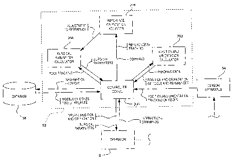
Referring to Fig. 4, the CAS controller 52 is
shown in greater detail. The
CAS controller 52
typically is a processing unit having a controller
device 200 which processes the information. The
controller device 200 is connected to the sensor
apparatus 54 so as to receive position and orientation
tracking data of tools, such as the trackable references
and the registration tool.
A position and orientation calculator 202
receives the tracking data, and calculates position and
orientation of tools, as well as frames of reference.
Therefore, the controller device 200 allows the operator
S to perform the surgery in real-time CAS navigation.
In order to compare points taken pre-
operatively with points taken intra-operatively and
post-operatively according to the method 100, a
reference orientation adjustor 204 is provided in
association with the controller device 200. More
specifically, updates to data associated with the
femoral frame of reference are received by the reference
orientation adjustor 204. When
the information is
complete, the reference orientation adjustor 204
calculates a reference adjustment value that consists in
the realignment of the re-digitized frame of reference
to the reference orientation. As mentioned previously,
this consists in positioning the mechanical axis (with
the intraoperative or post-operative center of rotation)
at a predetermined angle to the vertical axis in the
frontal plane of the pelvic frame of reference, with the
-21-
CA 02607036 2007-10-31
WO 2006/128301
PCT/CA2006/000905
centers of rotation of the femur and acetabulum in a
known relation.
The reference adjustment value is then
provided to a surgical parameter calculator 206, which
will calculate surgical parameters taken into account
the reference adjustment value. Therefore, no physical
alignment is required considering that the CAS
controller 52 performs all alignment virtually.
- 22 -