Note: Descriptions are shown in the official language in which they were submitted.
CA 02607162 2007-11-01
WO 2006/119387 PCT/US2006/017042
1
SYSTEM AND METHOD FOR DETERMINING TIBIAL ROTATION
Cross-Reference to Related Applications
[0001] This application claims the benefit of U.S. Provisional Application No.
60/677,399, filed 2 May 2005.
Statement Regarding Federally Sponsored Research or Development.
[0002] Not Applicable.
Appendix.
[0003] Not Applicable.
Backgronnd of the Invention
1. Field of the Invention
[0004] This invention relates generally to computer assisted surgery and more
particularly to a system for computer assisted surgery utilizing a projected
method for
determining tibial rotation.
2. RelatedArt
[0005] During knee arthroplasty, one or more of the distal surfaces of the
femur are cut
away and replaced with a metal component to simulate the bearing surfaces of
the femur.
Similarly, one or more of the proximal surfaces of the tibia is modified to
provide a metal-
backed plastic bearing surface. The metal femoral component of the new
prosthetic joint
transfers the weight of the patient to the tibial component such that the
joint can support the
patient's weight and provide a near-normal motion of the knee joint.
[0006] Orthopedic surgeons have been struggling with the alignment of knee
arthroplasties since their inception in the early 1970s. Basically, what is
generally necessary is
a 5-7 degree angular resection of the distal femoral condyles as related to
the mechanical axis
of the femur and a perpendicular resection of the proximal tibia as related to
its central axis.
CA 02607162 2007-11-01
WO 2006/119387 PCT/US2006/017042
2
Early on, resections of the distal femur and proximal tibia were made by
visually trying to
match or correct the existing anatomy by eye. Alignment varied considerably
depending on
the skill of the operating surgeon.
[0007] Several studies have indicated that the long term performance of a
prosthetic
knee joint is dependant on how accurately the components of the knee joint are
implanted with
respect to the weight bearing axis of the patient's leg. The most important
parameter in
achieving long term performance is accurate alignment of the components. It
has been proven
that only 4.5 degrees of misalignment causes the components to only load one
side of the knee
joint leading to rapid failure of the implant. The literature strongly
supports the conclusion
that the closer the surgeons approach neutral alignment, the more successful
the implant
system will be with longevity. Misaligned knee arthroplasties tend to get
worse with time
because the abnonnal weight distribution accelerates the wear on the
overloaded side leading
to rapid failure within a few years in the case of the gross malalignment.
[0008] In a correctly functioning knee, the weight bearing axis passes through
the
center of the head of the femur, the center of the knee and the center of the
ankle joint. This
weight bearing axis typically is located by analyzing an X-ray image of the
patient's leg, taken
while the patient is standing. The X-ray image is used to locate the center of
the head of the
femur and to calculate the position of the head relative to selected landmarks
on the femur.
The selected landmarks are then found on the patient's femur during surgery
and the
calculations used to estimate the actual position of the femoral head. These
two pieces of
information are used to determine the correct alignment of the weight bearing
axis for the
femur, commonly referred to as the mechanical axis of the femur. To completely
define the
correct position for the femoral component of the knee prosthesis, the correct
relationship
between the center of the femoral head and the knee joint and the rotation of
the knee joint
CA 02607162 2007-11-01
WO 2006/119387 PCT/US2006/017042
3
about the mechanical axis must be established. This information is determined
from
landmarks on the distal portion of the femur. The correct alignment for the
tibial component
of the knee prosthesis ordinarily is deterinined by finding the center of the
ankle joint and
relating its position to landmarlcs on the tibia. This point and the center of
the proximal tibial
plateau are used to define the weight bearing axis, or mechanical axis, of the
tibia. The correct
relationship between the ankle joint and the knee joint and the rotation of
the knee joint about
the mechanical axis are determined by reference to the distal portion of the
femur and
landniarks on the tibial plateau.
[0009] Presently, doctors commonly determine a desired rotation of the tibia
simply by
placing the knee in full extension and looking at the alignment of the foot.
This method has
several deficiencies. First, any errors that are developed in the
determination of the femur's
rotational axis are projected onto the tibia. Second, this method is much more
susceptible to
anatomic abnormalities and joint instability, which is common in patients
requiring total knee
arthroplasty. Third, a good rotational assessment of the tibia itself is not
accurately
determined, but ratlier, the entire rotation of the limb is being assessed in
aggregate, without
specific knowledge of the rotation of the tibia itself or the tibial
component.
[0010] Other methods currently used to determine the Anterior-Posterior (AP)
axis of
the tibia rely on anatomic landmarks. One common method uses a line drawn from
the medial
1/3 of the tibial tubercle to the center of the tibial plateau. Another method
uses a line drawn
from the anterior cruciate ligament insertion to the posterior cruciate
ligament insertion. Still
another method considers the average of these two or lines drawn from other
landmarks, which
assumes that averaging of these methods adds credence to the result.
Ultimately though,
because these points are all very close to each other in space, these methods
are greatly
affected by very small changes in their perceived location and thus are poorly
reproduceable.
CA 02607162 2007-11-01
WO 2006/119387 PCT/US2006/017042
4
[0011] In yet another method, the rotation of both the femur and tibia is
determined by
developing a kinematic axis in the knee joint. This method requires the limbs
to be moved
with respect to each other, during which software determines the axis about
which the tibia
rotates with respect to the femur. Software then uses this axis for measuring
rotation around
the mechanical axis of the tibia and femur. The problem with this method is
that it is
extremely sensitive to anatomic abnormalities, as well as ligament
instability.
[00121 For some time, computer assisted surgery (also known as "image-guided
surgery," "surgical navigation," or "3-D computer surgery") has been applied
to invasive
surgical procedures, such as knee arthroplasty. Computer assisted surgery,
often abbreviated
CAS, typically includes systems and processes for traclcing anatomy,
implements,
instrumentation, trial implants, implant components and virtual constructs or
references, and
rendering images and data related to them in connection with orthopedic,
surgical and other
operations. CAS allows for the association of anatomical structures,
constructs, and points-in-
space with a fiducial. Fiducial functionality allows the CAS system to sense
and track the
position and orientation of these items. Such structures, items and constructs
can be rendered
onscreen properly positioned and oriented relative to each other using
associated image files,
data files, image input, and other sensory input based on the tracking. The
CAS system,
among other things, allow surgeons to navigate and perform knee arthroplasty
using images
that reveal interior portions of the body combined with computer generated or
transmitted
images that show surgical implements, instruments, trials, implants, and/or
other devices
located and oriented properly relative to the body part. By using the CAS
system, the surgeon
can accurately and effectively resection bones, place and assess trial
implants and joint
performance, and place and assess actual implants and joint performance.
CA 02607162 2007-11-01
WO 2006/119387 PCT/US2006/017042
[0013] There remains a need in the art for computer assisted surgery system
that enables
surgeons to accurately and reliably perform knee arthroplasty. In particular,
there remains a need
in the art for a computer assisted surgery system that allows a user to
identify an angular rotation
of an item, such as a tool, relative to the mechanical axis of a tibia.
5 Summary of the Invention
[0014] It is in view of the above problems that the present invention was
developed. The
invention is a system and method for determining tibial rotation. The
invention has several
advantages over prior devices and techniques. First, the invention has
improved accuracy over
the art. The invention utilizes the mechanical axis of the femur and the
mechanical axis of the
tibia to construct a reference plane. Because the endpoints of each axis are
not in proximity to
each other, small errors in their respective identification do not greatly
affect the determination
of the reference plane. Moreover, anatomic defects are less likely to effect
the rotational
position of the tibia. Second, the simplicity of the invention allows it to be
easily repeatable.
Surgeons are intimately familiar with finding the mechanical axis of the femur
and the tibia
and significant effort is not required to put the axes in 90 degrees of
flexion. The simple and
straightforward character of the invention allows it to be carried out by both
new and
experienced users.
[0015] Thus, in furtherance of the above goals and advantages, the present
invention is,
briefly, a system for performing computer assisted surgery. The system
comprises: a first
fiducial operatively connected to a first part; a second fiducial operatively
connected to a
second part; at least one position and orientation sensor adapted to track
said first fiducial and
said second fiducial; a computer having a memory, a processor, and an
input/output device,
said input/output device adapted to receive data from said at least one
position and orientation
sensor relating to a position and an orientation of said first fiducial and
said second fiducial,
CA 02607162 2007-11-01
WO 2006/119387 PCT/US2006/017042
6
said processor adapted to process said data to identify a first axis of the
first part and a second
axis of the second part, and said processor adapted to construct a reference
plane througli said
second axis and orthogonal to said first axis; and a monitor operatively
connected to said
input/output device of said computer, and wherein said monitor is adapted to
display a
rendering of said reference plane.
[0016] Further features, aspects, and advantages of the present invention, as
well as the
structure and operation of various embodiments of the present invention, are
described in detail
below with reference to the accompanying drawings.
Brief Description of the Drawings
[0017] The accompanying drawings, which are incorporated in and form a part of
the
specification, illustrate the embodiments of the present invention and
together with the
description, serve to explain the principles of the invention. In the
drawings:
[0018] FIG. 1 is a schematic view of a computer assisted surgery system;
[0019] FIG. 2 is a view of a knee prepared for surgery, including a femur and
a tibia, to
which fiducials have been attached;
[0020] FIG. 3 is a view of a portion of a leg prepared for surgery with a C-
arm for
obtaining fluoroscopic images associated with a fiducial;
[0021] FIG. 4 is a fluoroscopic image of free space rendered on a monitor;
[0022] FIG. 5 is a fluoroscopic image of femoral head obtained and rendered;
[0023] FIG. 6 is a fluoroscopic image of a knee obtained and rendered;
[0024] FIG. 7 shows a probe being used to register a surgically related
component for
tracking; ,
[0025] FIG. 8 shows a probe being used to register a cutting block for
tracking;
[0026] FIG. 9 shows a probe being used to register a tibial cutting block for
tracking;
CA 02607162 2007-11-01
WO 2006/119387 PCT/US2006/017042
7
[0027] FIG. 10 shows a probe being used to register a femoral cutting block
for tracking;
[0028] FIG. 11 shows a probe being used to desigiiate landmarks on bone
structure for
tracking;
[0029] FIG. 12 is another view of a probe being used to designate landmarks on
bone
structure for tracking;
[0030] FIG. 13 is another view of a probe being used to designate landmarks on
bone
structure for tracking;
[0031] FIG. 14 is a screen face produced during designation of landmarks to
determine a
feinoral mechanical axis;
[0032] FIG. 15 is a screen face produced during designation of landmarks to
determine
an epicondylar axis;
[0033] FIG. 16 is a screen face produced during designation of landmarks to
determine
an anterior-posterior axis;
[0034] FIG. 17 is a screen face that presents graphic indicia which may be
employed to
help determine reference locations within bone structure;
[0035] FIG. 18 is a screen face showing mechanical and other established axes;
[0036] FIG. 19 is a schematic view of a patient's leg;
[0037] FIG. 20 is an illustration of a screen face displaying degrees of
flexion;
[0038] FIG. 21 is a flowchart illustrating software steps for tracking and
using a tibial
rotation plane;
[0039] FIG. 22 is a schematic front view of a patient's leg;
[0040] FIG. 23 is a schematic medial side view of a patient's leg;
[0041] FIG. 24 is a schematic front view of a femur;
[0042] FIG. 25 is a schematic medial side view of a femur;
CA 02607162 2007-11-01
WO 2006/119387 PCT/US2006/017042
8
[0043] FIG. 26 is a schematic front view of a patient's leg;
[0044] FIG. 27 is a schematic medial side view of a patient's leg;
[0045] FIG. 28 is another screen face showing mechanical and other established
axes;
[0046] FIG. 29 is another screen face showing mechanical and other established
axes;
[0047] FIG. 30 shows navigation and placement of an intramedullary rod;
[0048] FIG. 31 is another view showing navigation and placement of an
intramedullaiy
rod;
[0049] FIG. 32 is a screen face produced which assists in navigation and/or
placement of
an intramedullary rod;
[0050] FIG. 33 is another view of a screen face produced which assists in
navigation
and/or placement of an extramedullary rod.
[00511 FIG. 34 is a view which shows navigation and placement of an alignment
guide;
[0052] FIG. 35 is a screen face which shows a fluoroscopic image of bone in
combination with computer generated images of axes and components;
[0053] FIG. 36 is a view showing placement of a cutting block;
[0054] FIG. 37 is a view showing articulation of trial coniponents during
trial reduction;
and
[0055] FIG. 38 is a screen face which may be used to assist in assessing joint
function.
Detailed Description of tlae Preferred Enzbodiments
[0056] Various positional terms referring to the human anatomy - such as
distal,
proximal, medial, lateral, anterior and posterior - are used in this
application in their customary
and usual manner. The term "distal" refers to the area away from the point of
attachment to the
body, whereas the term "proximal" refers to the area near the point of
attachment the body. The
term "medial" refers to something situated closer to the middle of the body,
while "lateral" refers
CA 02607162 2007-11-01
WO 2006/119387 PCT/US2006/017042
9
to something situated closer to the left side or the right side of the body.
Finally, "anterior" refers
to something situated closer to the front of the body and "posterior" refers
to something situated
closer to the rear of the body.
[0057] Also, the term "mechanical axis" of the femur refers to an imaginary
line drawn
from the center of the femoral head to the center of the distal femur at the
knee, and the term
"anatomic axis" of the femur refers to an imaginary line drawn the middle of
the femoral shaft.
The angle between the mechanical axis and the anatomic axis is generally about
six degrees.
[00581 FIG. 1 is a schematic view showing one embodiment of a system 100 and
one
version of a setting in which surgery on a knee, in this case a Total
Knee'Arthroplasty, may be
performed. The system 100 can track various body parts, such as tibia 10 and
femur 12, to
which fiducials 14 may be iniplanted, attached, or otherwise associated, be it
physically,
virtually, or otherwise. Fiducials 14 are structural frames that can be sensed
by one or more
sensors 16 suitable for sensing, storing, processing and/or outputting data
("tracking") relating
to position and orientation of fiducials 14 and, thus, components, such as
tibia 10 and femur
12, that are attached or otherwise associated with the particular fiducial.
The fiducials 14 may
have active elements, passive elements or both. For example, some fiducials
may include
reflective elements, some may include light emitting diode (LED) active
elements, and some
fiducials include both reflective elements and active LED elements.
Position/orientation
sensor 16 may be any sort of sensor functionality for sensing position and
orientation of
fiducials 14 and, therefore, items that are associated, according to whatever
desired electrical,
magnetic, electromagnetic, sound, physical, radio frequency, or other active
or passive
technique. In the embodiment depicted in FIG. 1, position sensor 16 is a pair
of infrared
sensors or a stereoscopic infrared sensor disposed on the order of about one
meter (sometimes
CA 02607162 2007-11-01
WO 2006/119387 PCT/US2006/017042
more, sometimes less) apart and whose output can be processed in concert to
provide position
and orientation inforination regarding fiducials 14.
[0059] In the embodiment shown in FIG. 1, computing functionality 18 can
include
processing finzctionality 70, memory functionality 72, input/output
functionality 74, whether
5 on a standalone or distributed basis, via any desired standard,
architecture, interface and/or
network topology. Computing functionality 18 may be a stand alone computer, a
networked
computer, a mobile computing device, or similar device. In the case of a
networked computer,
the computing functionality 18 is connected to a network 80. In the depicted
embodiment,
computing functionality 18 is connected to a monitor 24 on which graphics and
data may be
10 presented to the surgeon during surgery. The monitor 24 may have a tactile
interface so that
the surgeon may point and click on screen for tactile screen input in addition
to or instead of, if
desired, keyboard and mouse conventional interfaces. Optionally, a foot pedal
20 or other
convenient interface may be coupled to computing functionality 18 as can any
other wireless
or wired interface to allow the surgeon, nurse or other desired user to
control or direct
functionality 18 in order to, among other things, capture position/orientation
information when
certain components are oriented or aligned properly.
[0060] Item 22, such as trial components and prosthetic devices, instrument
23, or
other devices used in a surgical procedure may be tracked in position and
orientation by the
sensor 16. For example, item 22 and instrument 23 may be tracked relative to
tibia 10 and
femur 12 using fiducials 14. As another example, item 22 and instrument 23 may
be tracked
relative to a global coordinate system.
[0061] Computing functionality 18 can process and store various forms of data.
Further, computing functionality 18 can output data on touch-screen or monitor
24. As an
example, the data may correspond in whole or in part to body parts or
components, such as
CA 02607162 2007-11-01
WO 2006/119387 PCT/US2006/017042
11
tibia 10, femur 12, or item 22. For example, in the embodiment shown in FIG.
1, tibia 10 and
femur 12 are shown in cross-section or at least various internal aspects of
them such as bone
canals and surface structure are shown using fluoroscopic images. These images
may be
obtained using, as an example, a C-arm or imager attached to a fiducial 14.
The body parts,
for example, tibia 10 and femur 12, also have fiducials attached. When the
fluoroscopy
images are obtained using the C-arm with fiducial 14, a position/orientation
sensor 16 "sees"
and tracks the position of the fluoroscopy head as well as the positions and
orientations of the
tibia 10 and femur 12. The computer 18 stores the fluoroscopic images with
this
position/orientation information, thus correlating position and orientation of
the fluoroscopic
image relative to the relevant body part or parts. Thus, when the tibia 10 and
corresponding
fiducial 14 move, the computer 18 automatically and correspondingly senses the
new position
of tibia 10 in space and can correspondingly move implements, instruments,
references, trials
and/or implants on the monitor 24 relative to the image of tibia 10.
Similarly, the image of the
body part can be moved, both the body part and such items may be moved, or the
on-screen
image may otherwise be presented to suit the preferences of the surgeon or
others and carry out
the imaging that is desired. Similarly, when an item 22, such as an
extramedullary rod,
intramedullary rod, or other type of rod, that is being tracked moves, its
image moves on
monitor 24 so that the monitor shows the item 22 in proper position and
orientation on monitor
24 relative to the femur 12. The item 22 can thus appear on the monitor 24 in
proper or
improper alignment with respect to the mechanical axis and other features of
the femur 12, as
if the surgeon were able to see into the body in order to properly navigate
and position item 22.
The computer functionality 18 can also store data relating to configuration,
size and other
properties of items 22, such as implements, instrumentation, trial components,
implant
components and other items used in surgery. When those are introduced into the
field of
CA 02607162 2007-11-01
WO 2006/119387 PCT/US2006/017042
12
position/orientation sensor 16, computer functionality 18 can generate and
display overlaid or
in combination with the fluoroscopic images of the body parts, such as tibia
10 and femur 12,
computer generated images of implements, instrumentation components, trial
components,
implant components and other items for navigation, positioning, assessment and
other uses.
[0062] In some embodiments, the system 100 may include a designator or probe
26.
The probe 26 may be used in conjunction with the computer functionality 18 to
track any point
in a field 17 of the position/orientation sensor 16. One of the fiducials 14
is attached to probe
26 for tracking purposes. The surgeon, nurse, or other user touches the tip of
probe 26 to a
point such as a landmark on bone structure and actuates the foot pedal 20 or
otherwise
instructs the computer 18 to note the landmark position. The
position/orientation sensor 16
"sees" the position and orientation of fiducial 14, "knows" where the tip of
probe 26 is relative
to that fiducial 14, and calculates and stores the point or other position
designated by probe 26
when the foot pedal 20 is hit or other command is given to the computer 18.
The computer 18
can also display on monitor 24 the identified point whenever desired and in
whatever form or
fashion or color. Thus, probe 26 can be used to designate landmarks on bone
structure in order
to allow the computer 18 to store and track, relative to movement of the
fiducial 14, virtual or
logical information, such as mechanical axis 28 of the femur 12,
medial/lateral axis 30 and
anterior/posterior axis 32 of femur 12, tibia 10 and other body parts in
addition to any other
virtual or actual construct or reference.
[0063] FIG. 2 shows a human knee in the surgical field, as well as the
corresponding
femur and tibia, to which fiducials 14 have been rigidly attached. In some
embodiments,
attachment of fiducials 14 is accomplished using structure that withstands
vibration of surgical
saws and other phenomenon which occur during surgery without allowing any
substantial
movement of fiducial 14 relative to body part being tracked by the system 100.
CA 02607162 2007-11-01
WO 2006/119387 PCT/US2006/017042
13
[0064] FIG. 3 shows fluoroscopy images being obtained of the body parts with
fiducials 14 attached. The fiducial 14 on the fluoroscopy head in this
embodiment is a
cylindrically shaped cage which contains LEDs or "active" emitters for
tracking by the sensors
16 (not shown in FIG.3). Fiducials 14 attached to tibia 10 and femur 12 can
also be seen. The
fiducial 14 attached to the femur 12 uses LEDs instead of reflective spheres
and is fed power
by the wire seen extending into the bottom of the image.
[0065] FIGS. 4-6 are fluoroscopic images shown on monitor 24 obtained with
position
and/or orientation information received by, noted and stored within computer
18. FIG. 4 is an
open field with no body part image, but which shows the optical indicia which
may be used to
normalize the image obtained using a spherical fluoroscopy wave front with the
substantially
flat surface of the monitor 24. FIG. 5 shows an image of the femur 12 head.
This image is
taken in order to allow the surgeon to designate the center of rotation of the
femoral head for
purposes of establishing the mechanical axis and other relevant constructs
relating to of the
femur according to which the prosthetic components will ultimately be
positioned. Such
center of rotation can be established by articulating the femur within the
acetabulum or a
prosthesis to capture a number of samples of position and orientation
information and in turn
to allow the computer to calculate the average center of rotation. The center
of rotation can be
established by using the probe and designating a number of points on the
femoral head and
thus allowing the computer to calculate the geometrical center or a center
which corresponds
to the geometry of points collected. Additionally, graphical representations
such as
controllably sized circles displayed on the monitor can be fitted by the
surgeon to the shape of
the femoral head on planar images using tactile input on screen to designate
the centers
according to that graphic, such as are represented by the computer as
intersection of axes of the
circles. Those skilled in the art would understand that other techniques for
determining,
CA 02607162 2007-11-01
WO 2006/119387 PCT/US2006/017042
14
calculating or establishing points or constructs in space, whether or not
corresponding to bone
structure, may be used.
(0066] FIG. 5 shows a fluoroscopic image of the femoral head, while FIG. 6
shows an
anterior/posterior view of the knee which can be used to designate landmarks
and establish
axes or constructs such as the mechanical axis or other rotational axes.
REGISTRATION OF SURGICALLY RELATED ITEMS
(00671 FIGS. 7-10 show designation or registration of items 22 which will be
used in
surgery. Registration simply means, however it is accomplished, ensuring that
the computer
18 knows which body part, item or construct corresponds to which fiducial or
fiducials 14, and
how the position and orientation of the body part, item or construct is
related to the position
and orientation of its corresponding fiducial or a fiducial attached to an
impactor or other
component which is in turn attached to an item. Such registration or
designation can be done
before, after, or instead of registering bone or body parts as discussed with
respect to FIGS. 4-
6. FIG. 7 shows a technician designating with probe 26 an item 22, such as an
instrument
component to which fiducial 14 is attached. The sensor 16 "sees" the position
and orientation
of the fiducial 14 attached to the item 22 and also the position and
orientation of the fiducial
14 attached to the probe 26 whose tip is touching a landinark on the item 22.
The technician
designates onscreen or otherwise the identification of the item and then
activates the foot pedal
or otherwise instructs the computer 18 to correlate the data corresponding to
such
identification, such as data needed to represent a particular cutting block
component for a
particular knee implant product, with the particularly shaped fiducial 14
attached to the
component 22. The computer 18 has then stored identification, position and
orientation
information relating to the fiducial for component or item 22 correlated with
the data such as
configuration and shape data for the item 22 so that upon registration, when
sensor 16 tracks
CA 02607162 2007-11-01
WO 2006/119387 PCT/US2006/017042
the item 22 fiducial 14 in the infrared field, monitor 24 can show the cutting
block component
moving and turning, and properly positioned and oriented relative to the body
part which is
also being tracked. FIGS. 8-10 show similar registration for other
instrumentation components
22.
5 REGISTRATION OF ANATOMY AND CONSTRUCTS
[0068] Similarly, the mechanical axis and other axes or constructs of body
parts 10 and
12 can also be "registered" for tracking by the system 100. As an optional
step, the system 100
may employ a fluoroscope to obtain images of the femoral head, knee and ankle
of the sort
shown in FIGS. 4-6. The system 100 correlates such images with the position
and orientation
10 of the C-arm and the patient anatomy in real time as discussed above with
the use of fiducials
14 placed on the body parts before image acquisition and which remain in
position during the
surgical procedure. Using these images, the surgeon can select and register in
the computer 18
the center of the femoral head and ankle in orthogonal views, usually
anterior/posterior and
lateral, on a touch screen.
15 [0069] Alternatively, the surgeon or other person uses the probe 26 to
select any
desired anatomical landmarks or references to register body parts and related
constructs.
These points are registered in three dimensional space by the system 100 and
are tracked
relative to the fiducials 14 on the patient anatomy which are preferably
placed intraoperatively.
FIG. 11 shows the surgeon using probe 26 to designate or register landmarks on
the condylar
portion of femur 12 using probe 26 in order to feed to the computer 18 the
position of one
point needed to determine, store, and display the epicondylar axis. (See FIG.
16 which shows
the epicondylar axis and the anterior-posterior plane and for lateral plane.)
Although
registering points using actual bone structure such as in FIG. 11 is one way
to establish the
axis, a cloud of points approach by which the probe 26 is used to designate
multiple points on
CA 02607162 2007-11-01
WO 2006/119387 PCT/US2006/017042
16
the surface of the bone structure can be employed, as can moving the body part
and tracking
movement to establish a center of rotation as discussed above. Once the center
of rotation for
the femoral head and the condylar component have been registered, the computer
18 is able to
calculate, store, and render, and otherwise use data for, the mechanical axis
28 of the femur 12.
FIGS. 12 and 13 once again show the probe 26 being used to designate points on
the condylar
component of the femur 12.
[0070] FIG. 14 shows the onscreen images being obtained when the surgeon
registers
certain points on the bone surface using the probe 26 in order to establish
the femoral
mechanical axis 28. Tibial mechanical axis 38 (best seen in FIG. 19) is then
established by
designating points to determine the centers of the proximal and distal ends of
the tibia so that
the mechanical axis can be calculated, stored, and subsequently used by the
computer 18. FIG.
shows designated points for determining the epicondylar axis, both in the
anterior/posterior
and lateral planes, while FIG. 16 shows such determination of the anterior-
posterior axis as
rendered onscreen. The posterior condylar axis is also determined by
designating points or as
15 otherwise desired, as rendered on the computer generated geometric images
overlain or
displayed in combination with the fluoroscopic images, all of which are keyed
to fiducials 14
being tracked by sensors 16.
[0071] FIG. 17 shows an adjustable circle graphic which can be generated and
presented in combination with orthogonal fluoroscopic images of the femoral
head, and
tracked by the computer 18 when the surgeon moves it on screen in order to
establish the
centers of the femoral head in both the anterior-posterior and lateral planes.
[0072] FIG. 18 is an onscreen image showing the anterior-posterior axis,
epicondylar
axis and posterior condylar axis from points which have been designated as
described above.
These constructs are generated by the computer 18 and presented on monitor 24.
Optionally,
CA 02607162 2007-11-01
WO 2006/119387 PCT/US2006/017042
17
the constructs may be presented in combination with the fluoroscopic images of
the femur 12,
correctly positioned and oriented relative thereto as tracked by the system
100. In the
fluoroscopic/computer generated image combination shown at left bottom of FIG.
18, a
"sawbones" knee as shown in certain drawings above which contains radio opaque
materials is
represented fluoroscopically and tracked using sensor 16 while the computer
generates and
displays the mechanical axis 28 of the femur 12, which runs generally
horizontally. The
epicondylar axis runs generally vertically, and the anterior/posterior axis
runs generally
diagonally. The image at bottom right shows similar information in a lateral
view. Here, the
anterior-posterior axis runs generally horizontally while the epicondylar axis
runs generally
diagonally, and the mechanical axis generally vertically.
[0073] FIG. 18, as is the case with a number of screen presentations, also
shows at
center a list of landmarks to be registered in order to generate relevant axes
and constructs
useful in navigation, positioning and assessment during surgery. Textural cues
may also be
presented which suggest to the surgeon next steps in the process of
registering landmarks and
establishing relevant axes. Such instructions may be generated as the computer
18 tracks,
from one step to the next, registration of items 22 and bone locations as well
as other measures
being taken by the surgeon during the surgical operation.
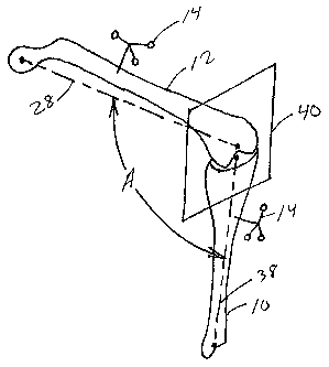
[0074] FIG. 19 is a schematic view of a patient's leg with fiducials 14
associated
therewith. In the embodiment depicted in FIG. 19, the tibia 10 is in flexion
with respect to the
femur 12. The femur 12 has a mechanical axis 28, and the tibia has a
mechanical axis 38.
Because the tibia 10 is in flexion, the femoral mechanical axis 28 is at an
angle A relative to the
tibial mechanical axis 38. In the enlbodiment depicted in FIG. 19, the angle A
is about 90
degrees, plus or minus one degree. By tracking the femoral mechanical axis 28
and the tibial
mechanical axis 38, the computing functionality 18 can identify when the axes
are orthogonal to
CA 02607162 2007-11-01
WO 2006/119387 PCT/US2006/017042
18
one another. The computing functionality 18 can then use this inforn-iation to
construct a tibial
rotational plane 40 that extends through the tibial mechanical axis 38 and is
substantially
perpendicular to femoral mechanical axis 28. Thereafter, computing
functionality 18 can use the
constructed plane 40 to measure the angular rotation of items 22 about tibial
mechanical axis 38.
Alternatively, computing functionality 18 may use the constructed plane 40 to
create a tibial
coordinate system which includes the tibial mechanical axis 38, an
anteroposterior axis and a
medial-lateral axis. The medial-lateral axis, or transverse axis, is co-planar
with the
constructed plane 40 and orthogonal to the tibial mechanical axis 38, and the
anteroposterior
axis is orthogonal to both the constructed plane 40 and the tibial mechanical
axis 38.
Thereafter, the tibial coordinate system can be compared to other fiducials or
a global
coordinate system, and further, the tibial coordinate system can be used to
identify orientation
or position data of a surgical device, such as item 22, or construct, such as
the femoral
mechanical axis 28.
[0075] FIG. 20 illustrates the monitor 24 displaying degrees of flexion. The
monitor 24
includes a first area 42 to display a menu, a second area 44 to display
rendered images, and a
third area 46 to display the amount of flexion between the femur 12 and the
tibia 10. During
construction of the tibial rotational plane, a user moves the tibia 10
relative to the femur 12 until
the third area 46 displays about 90 degrees. Thereafter, the user indicates to
the computer
fiuictionality 18 that the patient's knee is in the required amount of
flexion. This indication may
be accomplished by touching the monitor 24, by holding the knee in flexion for
a predetermined
period of time, through the use of the probe 26, or the through the use of the
foot pedal 20.
[0076] FIG. 21 illustrates the steps taken by the computing functionality 18
to create and
use the tibial rotational plane 40. The computing functionality 18 begins at
step 110. This may
be a result of another software routine or a menu selection by a user. In step
112, a decision is
CA 02607162 2007-11-01
WO 2006/119387 PCT/US2006/017042
19
made whether to start with the femur 12 or with the tibia 10. This step may be
optional as some
embodiments may specify that it is always best to start first with the femur
and the tibia second,
or vice versa. In steps 114 and 120, the femoral mechanical axis 28 is
established. This may be
done kinematically, through the use of fluoroscopic images, through the use of
the probe 26 to
identify landmarks of the femur, or some combination thereof. In steps 116 and
118, the tibial
mechanical axis 38 is established by indicating landmarks of the tibia with
the probe or through
the use of fluoroscopic images. In step 122, the tibia 10 is placed in about
90 degrees of flexion
relative to the femur 12. This places the tibial mechanical axis 38
substantially perpendicular to
the femoral mechanical axis 28. The computing functionality 18 develops the
tibial rotational
plane 40 as extending through the tibial mechanical axis 38 and perpendicular
to the femoral
mechanical axis 28 in step 124. In step 126, the computing functionality 18
identifies the
orientation of the tibial rotational plane 40 relative to fiducials 14 and/or
relative to a global
coordinate system. Computing functionality 18 stores this orientation into
memory in step 128.
Thereafter, computing functionality 18 can use the tibial rotational plane 40
as a reference to
compare the angular rotation, orientation, or position of items 22 relative to
the tibial mechanical
axis 38 or to the tibial coordinate system described above. In FIG. 25,
computing functionality
18 performs the angular comparison in step 130. However, those skilled in the
art would
understand that the steps necessary to establish the reference plane 40 and
the comparison step
130 may be performed separately or together. For example, the reference plane
40 first may be
established and at a later time, such as by menu selection, the comparison
step 130 is performed.
After the reference plane 40 is stored in memory, the routine ends in step
132.
[0077] FIGS. 22 and 23 show in schematic form the relationship of the weight
bearing
axis (WBA) 50 to a left human femur 12 and tibia 10 in normal stance. FIG. 22
is a schematic
in the coronal (medial-lateral) plane of the patient and FIG. 23 is in the
sagital (anterior-
CA 02607162 2007-11-01
WO 2006/119387 PCT/US2006/017042
posterior) plane of the patient. Weight bearing axis 50 is defined to pass
through two points:
the center of the hip joint 52 and the center of the ankle joint 54. Weight
bearing axis 50
normally passes slightly medial to the anatomic center of the knee joint
although this may very
considerably from patient to patient. Hip joint center 52 is defined as the
center of rotation of
5 the hip joint and is generally accepted to be the anatomic center of the
head of the femur.
Ankle joint center 54 is defined as the center of rotation of the ankle joint
and is generally
accepted to lie midway along an axis passing through the malleoli of the lower
limb. Medial
malleolus 56 exists on the distal end of the tibia 10. The lateral malloelus
is a similar structure
on the distal end of the fibula (not shown). Joint line 58 is a plane
perpendicular to weight
10 bearing axis 50 at a point approximating the bearing surface between femur
12 and tibia 10.
[0078] FIGS. 24 and 25 show in schematic forin the motion of femur 12 about
hip joint
center 52 in the patient's coronal and sagital planes respectively. The motion
of femur 12 is
governed by the ball socket hip joint such that, during any movement of femur
12, femoral
registration point 60 fixed with respect to femur 12 will be constrained to
move on the surface
15 of a theoretical sphere with center at hip joint center 52 and radius equal
to the distance
between femoral registration point 60 and hip joint center 52. By measuring
the vectorial
displacement between three or more successive positions of femoral
registration point 60 in a
reference frame in which hip joint center 52 remains stationary as femur 12 is
moved, the
position of hip joint center 52 in that reference frame can be calculated.
Additionally, the
20 location of hip joint center 52 with respect to femoral registration point
60 can also be
calculated. Increasing the number of measured positions of femoral
registration point 60
increases the accuracy of the calculated position of hip joint center 52. By
using the probe 26
to locate registration points 60, the computer 18 can calculate the
geometrical center or a
center which corresponds to the geometry of points collected.
CA 02607162 2007-11-01
WO 2006/119387 PCT/US2006/017042
21
[0079] Other methods may be used to identify the hip joint center 52. For
example,
The femoral head may be located using various scanning techniques, such as
computed
tomography (CT) or magnetic resonance imaging (MRI). Further, the hip joint
center 52 may
be located through laser triangulation. The laser method is similar to
measuring the vectorial
displacement. A laser is mounted, on the distal end of the femur, and the
femur is rotated in the
acetabulum or a prosthesis to capture a number of samples of position and
orientation
information. The laser light indicates the center of rotation on a target,
which is used by the
laser operator to identify the center of the femoral head.
[00801 FIGS. 26 and 27 show in schematic form a simplified representation of
the
motion of tibia 10 with respect to femur 12 in the patient's coronal and
sagital planes
respectively. The motion of tibia 10 with respect to femur 12 is a complex,
six degree-of-
freedom relationship governed by the ligamentous tension and the three bearing
surfaces of the
knee joint. However for the purposes of implant location, a reasonable
approximation of the
motion of tibia 10 can be made assuming the knee joint to be a sliding hinge
in the sagital
plane with limited motion in the coronal plane. Based on these simplifying
assumptions,
movement of tibial registration point 62 fixed with respect to tibia 10 will
be constrained to
move on the surface of a theoretical sphere with instantaneous center within
the locus of knee
joint center 64 and radius equal to the distance between tibial registration
point 62 and knee
joint center 64. Because the bony nature of the human ankle permits
intraoperative estimation
of ankle joint center 54 by palpation, tibial registration point 62 can be
fixed to tibia 10 at a
known vectorial displacement from ankle joint center 64 through the use of a
notched guide or
boot strapped to the lower limb as is commonly known in knee arthroplasty.
Measurement of
the vectorial displacement of tibial registration point 62 with respect to
femoral registration
point 60, previously fixed-relative to femur 12 and at a calculated position
relative to hip joint
CA 02607162 2007-11-01
WO 2006/119387 PCT/US2006/017042
22
center 52, thereby permits the calculation of the vectorial position of ankle
joint center 64 with
respect to hip joint center 52 and the weight bearing axis to be determined.
As with
calculation of the position hip joint center 52, repeated measurements improve
the accuracy of
the determined weigllt bearing axis 50.
[0081] Further, by measuring the vectorial displacement between successive
positions
of tibial registration point 62 in a reference frame in which femoral
registration point 60
remains stationary as tibia 10 is moved, the locus of positions of knee joint
center 64 in that
reference frame can be calculated.
[0082] By identifying the vectorial displacements, the hip joint center 52,
and the anlcle
joint center 54, computing functionality 18 can "learn" and "memorize" the
femoral
mechanical axis 28 and the tibial niechanical axis 38. Thereafter, computing
functionality 18
can construct the tibial reference plane 40.
[0083] FIG. 28 shows mechanical, lateral, anterior-posterior axes for the
tibia
according to points registered by the surgeon. FIG. 29 is another onscreen
image showing the
axes for the femur 12.
MODIFYING BONE
[0084] After the mechanical axis and other rotation axes and constructs
relating to the
femur and tibia are established, instrumentation can be properly oriented to
resect or modify
bone in order to properly fit trial components and implant components.
Instrumentation such
as, for instance, cutting blocks, to which fiducials 14 are mounted, can be
employed. The
system 100 can then track instrumentation as the surgeon manipulates it for
optimum
positioning. In other words, the surgeon can "navigate" the instrumentation
for optimum
positioning using the system and the monitor. In this manner, instrumentation
may be
positioned according to the system of this embodiment in order to align the
ostetomies to the
CA 02607162 2007-11-01
WO 2006/119387 PCT/US2006/017042
23
mechaiiical and rotational axes or reference axes and planes on a rod
(extramedullary,
intramedullary, or other type) that does not violate the canal. The monitor 24
also can then
display the instrument, such as the cutting block and/or the implant relative
to the instrument
and the rod during this process, in order to, among other things, properly
select implant size
and perhaps implant type. As the instrument moves, the varus/valgus,
flexion/extension and
internal/external rotation of the relative component position can be
calculated and shown with
respect to the referenced axes; in some embodiments, this can be done at a
rate of six cycles
per second or faster. The instrument position is then fixed in the computer
and physically, and
the surgeon makes the bone resections.
[0085] FIG. 30 shows orientation of an intramedullary rod to which a fiducial
14 is
attached via item 22, such as an impactor. The surgeon views the monitor 24
which has an
image as shown in FIG. 32 of the rod overlain on or in combination with a
fluoroscopic image
of the femur 12 as the two are actually positioned and oriented relative to
one another in space.
The surgeon then navigates the rod into place preferably along the mechanical
axis of the
femur and drives it home with appropriate mallet or other device. This may
avoid the need to
bore a hole in the metaphysis of the femur and place a reamer or other rod
into the medullary
canal, which can cause fat embolism, hemorrhaging, infection and other
untoward and
undesired effects.
[0086] FIG. 31 also shows the intramedullary rod being located. FIG. 32 shows
fluoroscopic images, both anterior-posterior and lateral, with axes, and with
a computer
generated and tracked image of the rod superposed or in combination with the
fluoroscopic
images of the femur and tibia. FIG. 33 shows the rod superposed on the femoral
fluoroscopic
image similar to what is shown in FIG. 32.
CA 02607162 2007-11-01
WO 2006/119387 PCT/US2006/017042
24
[0087] FIG. 32 also shows other information relevant to the surgeon such as
the name
of the component being overlain on the femur image (new EM nail), suggestions
or
instructions at the lower left, and angle of the rod in varus/valgus and
extension relative to the
axes. Any or all of this information can be used to navigate and position the
rod relative to the
femur. At a point in time during or after placement of the rod, its tracking
may be "handed off'
from the impactor fiducial 14 to the femur fiducal 14 as discussed below.
[0088] Once the extrainedullary rod, intramedullary rod, other type of rod has
been
placed, instrumentation can be positioned as tracked in position and
orientation by sensor 16
and displayed on screen face 24. Thus, a cutting block of the sort used to
establish the
condylar anterior cut, with its fiducial 14 attached, is introduced into the
field and positioned
on the rod. FIG. 34 illustrates a cutting block being positioned. Because the
cutting block
corresponds to a particular implant product and can be adjusted and designated
on screen to
correspond to a particular inlplant size of that product, the computer 18 can
generate and
display a graphic of the cutting block and the femoral component overlain on
the fluoroscopic
image as shown in FIG. 35. The surgeon can thus navigate and position the
cutting block on
screen using not only images of the cutting block on the bone, but also images
of the
corresponding femoral component that ultimately will be installed. The surgeon
can adjust the
positioning of the physical cutting block component and secure it to the rod
in order to resect
the anterior of the condylar portion of the femur in order to optimally fit
and position the
ultimate femoral component being shown on the screen. Other cutting blocks and
other
resections may be positioned and made similarly on the condylar component.
[0089] In a similar fashion, instrumentation may be navigated and positioned
on the
proximal portion of the tibia 10 as shown in FIG. 36 and as tracked by sensor
16 and on screen
by images of the cutting block and the implant component as shown in FIG. 35.
CA 02607162 2007-11-01
WO 2006/119387 PCT/US2006/017042
[0090] In summary, the computer 18 and monitor 24 show femoral component and
tibial component overlays according to certain position and orientation of
cutting
blocks/instrumentation as bone resections are made. The surgeon can thus
visualize where the
implant components will be and can assess fit, and other things if desired,
before resections are
5 made.
NAVIGATION, PLACEMENT AND ASSESSMENT OF TRIALS AND IMPLANTS
[0091] Once resection and modification of bone has been accomplished, implant
trials
can then be installed and tracked by the system 100 in a manner similar to
navigating and
positioning the instrumentation, as displayed on the screen 24. Thus, a
femoral component
10 trial, a tibial plateau trial, and a bearing plate trial may be placed as
navigated on screen using
computer generated overlays corresponding to the trials.
[0092] During the trial installation process, and also during the implant
component
installation process, instrument positioning process or at any otller desired
point in surgical or
other operations, the system 100 can transition or segue from tracking a
component according
15 to a first fiducial to tracking the component according to a second
fiducial. Thus, as shown as
FIG. 37, the trial femoral component is mounted on an impactor to which is
attached a fiducial
14. The trial component is installed and positioned using the impactor. The
computer 18
"knows" the position and orientation of the trial relative to the fiducial on
the impactor (such
as by prior registration of the component attached to the impactor) so that it
can generate and
2 0 display the image of the femoral component trial on screen 24 overlaid on
the fluoroscopic
image of the condylar component. At any desired point in time, before, during
or after the trial
component is properly placed on the condylar component of the femur to align
with
mechanical axis and according to proper orientation relative to other axes,
the system 100 can
be instructed by foot pedal or otherwise to begin tracking the position of the
trial component
CA 02607162 2007-11-01
WO 2006/119387 PCT/US2006/017042
26
using the fiducial attached to the femur rather than the one attached to the
impactor. The
sensor 16 "sees" at this point in time both the fiducials on the impactor and
the femur 12 so
that it already "knows" the position and orientation of the trial component
relative to the
fiducial on the impactor and is thus able to calculate and store for later use
the position and
orientation of the trial component relative to the femur 12 fiducial. Once
this "handoff'
happens, the impactor can be removed and the trial component tracked with the
femur fiducial
14 as part of or moving in concert with the femur 12. Similar handoff
procedures may be used
in any other instance as desired.
[0093] The tibial trial may be placed on the proximal tibia and then
registered using the
probe 26. Probe 26 is used to designate preferably at least three features on
the tibial trial of
known coordinates, such as bone spike holes. As the probe 26 is placed onto
each feature, the
system 100 is prompted to save that coordinate position so that the system 100
can match the
tibial trial's feature's coordinates to the saved coordinates. The system 100
then tracks the
tibial trial relative to the tibial anatomical reference frame.
[0094] Once the trial components are installed, the surgeon can assess
alignment and
stability of the components and the joint. During such assessment, in trial
reduction, the
computer can display on monitor 24 the relative motion between the trial
conlponents to allow
the surgeon to make soft tissue releases and changes in order to improve the
kinematics of the
knee. The system 100 can also apply rules and/or intelligence to make
suggestions based on
the information such as what soft tissue releases to make if the surgeon
desires. The system
100 can also display how the soft tissue releases are to be made.
[0095] FIG. 37 shows the surgeon articulating the knee as he monitors the
screen
which is presenting images such as those shown in FIG. 38 which not only show
movement of
the trial components relative to each other, but also orientation, flexion,
and varus/valgus data.
CA 02607162 2007-11-01
WO 2006/119387 PCT/US2006/017042
27
During this assessment, the surgeon may conduct certain assessment processes
such as
external/internal rotation or rotational laxity testing, varus/valgus tests,
and anterior-posterior
drawer at 0 and 90 degrees and mid range. Thus, in the AP drawer test, the
surgeon can
position the tibia at the first location and press the foot pedal. The surgeon
then positions the
tibia at the second location and once again presses the foot pedal so that the
computer has
registered and stored two locations in order to calculate and display the
drawer and whether it
is acceptable for the patient and the product involved. If not, the computer
can apply rules in
order to generate and display suggestions for releasing ligaments or other
tissue, or using other
component sizes or types. Once the proper tissue releases have been made, if
necessary, and
alignment and stability are acceptable as noted quantitatively on screen about
all axes, the trial
components may be removed and actual components navigated, installed, and
assessed in
performance in a manner similar to that in which the trial components were
navigated,
installed, and assessed.
[0096] At the end of the case, all alignment information can be saved for the
patient
file. This is of great assistance to the surgeon due to the fact that the
outcome of implant
positioning can be seen before any resections have been made to the bone. The
system 100 is
also capable of tracking the patella and resulting placement of cutting guides
and the patellar
trial position. The system 100 then tracks alignment of the patella with the
patellar femoral
groove and will give feedback on issues, such as, patellar tilt.
[0097] The tracking and image information provided by the system 100
facilitate
telemedical techniques because it provides useful images for distribution to
distant geographic
locations where expert surgical or medical specialists may collaborate during
surgery. Thus,
the system can be used in connection with computing functionality 18 which is
networked or
otherwise in communication with computing functionality in other locations,
whether by
CA 02607162 2007-11-01
WO 2006/119387 PCT/US2006/017042
28
public switched telephone networlc (PSTN), information exchange
infrastructures, such as
packet switched networks, including the Internet. Such remote imaging may
occur on
computers, wireless devices, videoconferencing devices or in any otlier mode
or on any other
platform which is now or may in the future be capable of rending images or
parts of them.
Parallel communication links, such as switched or unswitched telephone call
connections, may
also accompany or form part of such telemedical techniques. Distant databases,
such as online
catalogs of implant suppliers or prosthetics buyers or distributors, may form
part of or be
networked with functionality 18 to give the surgeon in real time access to
additional options
for implants which could be procured and used during the surgical operation.
[0098] The invention may include one or more of the following steps. An
optional
first step is to obtain appropriate images, such as fluoroscopy images of
appropriate body parts.
This first step may include tracking the imager via an associated fiducial
whose position and
orientation is tracked by position/orientation sensors, such as stereoscopic
infrared (active or
passive) sensors. A second step is to register tools, instrumentation, trial
components,
prosthetic components, and other items to be used in surgery. The second step
may include
associating the tool, instrument, trial component, prosthetic component, or
other device with a
corresponding fiducial. A third step is to locate and register body structure,
such as
designating points on the femur and tibia using a probe associated with a
fiducial, in order to
provide the processing functionality information relating to the body part,
such as rotational
axes. A fourth step is to navigate and position instrumentation, such as
cutting
instrumentation, in order to modify bone, at least partially using images
generated by the
processing functionality corresponding to what is being tracked and/or has
been tracked,
and/or is predicted by the system, and thereby resecting bone effectively,
efficiently and
accurately. A fifth step is to navigate and position trial components, such as
femoral
CA 02607162 2007-11-01
WO 2006/119387 PCT/US2006/017042
29
components and tibial components, some or all of which may be installed using
impactors with
a fiducial and, if desired, at the appropriate time discontinuing tracking the
position and
orientation of the trial component using the impactor fiducial and starting to
track that position
and orientation using the body part fiducial on which the component is
installed. A sixth step
is to assess alignment and stability of the trial components and joint, both
statically and
dynamically as desired, using images of the body parts in combination with
images of the trial
components while conducting appropriate rotation, anterior-posterior drawer
and
flexion/extension tests and automatically storing and calculating results to
present data or
information which allows the surgeon to assess alignment and stability. A
seventh step
includes the release of tissue, such as ligaments, if necessary and adjusting
trial components as
desired for acceptable alignment and stability. An eighth step includes
installation of implant
components whose positions may be tracked at first via fiducials associated
with impactors for
the components and then tracked via fiducials on the body parts in which the
components are
installed. A ninth step includes assessing alignment and stability of the
implant components
and joint by use of some or all tests mentioned above and/or other tests as
desired, releasing
tissue if desired, adjusting if desired, and otherwise verifying acceptable
alignment, stability
and performance of the prosthesis, both statically and dynamically. Some or
all of these steps
may be used in any total or partial joint repair, reconstruction or
replacement, including knees,
hips, shoulders, elbows, ankles and any other desired joint in the body.
[0099] The system uses computer capacity, including standalone and/or
networked
computer capacity, to store data regarding spatial aspects of surgically
related items and virtual
constructs or references including body parts, implements, instrumentation,
trial components,
prosthetic components and rotational axes of body parts. Any or all of these
may be physically
or virtually connected to or incorporate any desired form of mark, structure,
component, or
CA 02607162 2007-11-01
WO 2006/119387 PCT/US2006/017042
other fiducial or reference device or technique which allows position and/or
orientation of the
item to which it is attached to be sensed and tracked, preferably in three
dimensions of
translation and three degrees of rotation as well as in time if desired. As an
example, such
"fidicuals" are reference frames each containing at least three, preferably
four, sometimes
5 more, reflective elements, such as spheres reflective of lightwave or
infrared energy, or active
elements, such as light emitting diodes (LEDs).
[00100] In one embodiment, orientation of the elements on a particular
fiducial varies
from one fiducial to the next so that sensors may distinguish between various
components to
which the fiducials are attached in order to correlate for display and other
purposes data files or
10 images of the components. The fiducials may be active, passive, or some
combination thereof.
In other words, some fiducials use reflective elements and some use active
elements, both of
which may be tracked by preferably two, sometimes more infrared sensors whose
output may
be processed in concert to geometrically calculate position and orientation of
the item to which
the fiducial is attached.
15 [00101] Position/orientation tracking sensors and fiducials need not be
confined to the
infrared spectrum. Any electromagnetic, electrostatic, light, sound,
radiofrequency or other
desired technique may be used. Alternatively, each item, such as a surgical
implement,
instrumentation component, trial component, implant component or other device
may contain
its own "active" fiducial, such as a microchip with appropriate field sensing
or
20 position/orientation sensing functionality and communications link, such as
spread spectrum
radio frequency (RF) link, in order to report position and orientation of the
item. Such active
fiducials, or hybrid active/passive fiducials, such as transponders, can be
implanted in the body
parts or in any of the surgically related devices mentioned above or
conveniently located at
their surface or otherwise as desired. Fiducials may also take the form of
conventional
CA 02607162 2007-11-01
WO 2006/119387 PCT/US2006/017042
31
structures, such as a screw driven into a bone, or any other three dimensional
item attached to
another item, position and orientation of such three dimensional item able to
be tracked in
order to track position and orientation of body parts and surgically related
items. Hybrid
fiducials may be partly passive, partly active such as inductive components or
transponders
which respond with a certain signal or data set when queried by sensors.
[00102] The system employs a computer to calculate and store reference axes of
body
components, such as in a total knee arthroplasty, for example, the mechanical
axis of the femur
and tibia. From these axes such systems track the position of the
instrumentation and
osteotomy guides so that bone resections will locate the implant position
optimally, usually
aligned with the mechanical axis. Furthermore, during trial reduction of the
knee, the system
provides feedback on the balancing of the ligaments in a range of motion and
under
varus/valgus, anterior/posterior and rotary stresses and can suggest or at
least provide more
accurate information than in the past about which ligaments the surgeon should
release in
order to obtain correct balancing, alignment and stability. The system can
also suggest
modifications to implant size, positioning, and other techniques to achieve
optimal kinematics.
The system can also include databases of information regarding tasks such as
ligament
balancing, in order to provide suggestions to the surgeon based on performance
of test results
as automatically calculated by such systems and processes.
[00103] The invention also includes a computerized method for determining
tibial
rotation within a coordinate system. The method may include one or more of the
following
steps, which are provided in no particular order. A first step of the method
is to provide a
computer having a processor, a memory, and an input/output device. A second
step is to
identify a mechanical axis of a femur. A third step is to identify a
mechanical axis of a tibia.
A fourth step is to place the tibia in about 90 degrees of flexion relative to
the femur. A fifth
CA 02607162 2007-11-01
WO 2006/119387 PCT/US2006/017042
32
step is to construct a plane through the mechanical axis of the tibia and
orthogonal to the
mechanical axis of the femur. The constructed plane may be used to create a
tibial coordinate
system which includes the mechanical axis of the tibia, an anteroposterior
axis and a medial-
lateral axis. A sixth step is to identify an orientation of the plane relative
to other fiducials or a
global coordinate system. A seventh step is to store the orientation of the
plane in the memory
of the computer. An eighth step is to measure an angular rotation of an item
relative to the
plane and the mechanical axis of the tibia or to the tibial coordinate system.
Items may
include, but are not limited to, tools, instruments, trial components, and
prosthetic devices.
The step of identifying a mechanical axis of a femur may include the step of
locating data
points corresponding to structure of the femur. The step of identifying a
mechanical axis of a
tibia may include the step of locating data points corresponding to structure
of the tibia.
[00104] The invention may also include one or more of the following optional
steps.
For example, the method may include the step of storing in the memory the
mechanical axis of
the femur or the step of storing in the memory the mechanical axis of tibia.
The method may
include the step of obtaining images of body parts, the step of registering
items, or the steps of
locating and registering body structure. Finally, the method may include the
step of mounting
a fiducial to a body part or the step of displaying the constructed plane on a
monitor.
[00105] The invention further includes a process for conducting knee surgery
using a
surgical navigation system. The process may include one or more of the
following steps,
which are provided in no particular order. A first step of the method is to
identify a first axis
of a first bone. A second step is to track an orientation of the first axis
relative to the first
bone. A third step is to identify a second axis of a second bone. A fourth
step is to track an
orientation of the second axis relative to the second bone. A fifth step is to
place the second
bone in about 90 degrees of flexion relative to the first bone. A sixth step
is to construct a
CA 02607162 2007-11-01
WO 2006/119387 PCT/US2006/017042
33
plane through the second axis and orthogonal to the first axis. A seventh step
is to track an
orientation of the constructed plane. An eighth step is to expose bones in a
vicinity of a knee
joint. A ninth step is to measure an angular rotation of an item relative to
the constructed
plane and the second axis. Items may include, but are not limited to, tools,
instruments, trial
components, and prosthetic devices. A tenth step is to at least partially
resect the first bone.
An eleventh step is to close the exposed knee. An optional step may be to
attach a surgical
implant to the at least partially resected first bone.
[00106] In view of the foregoing, it will be seen that the several advantages
of the
invention are achieved and attained.
[00107] The embodiments were chosen and described in order to best explain the
principles of the invention and its practical application to thereby enable
others skilled in the art
to best utilize the invention in various embodiments and with various
modifications as are suited
to the particular use contemplated.
[00108] As various modifications could be made in the constructions and
methods herein
described and illustrated without departing fiom the scope of the invention,
it is intended that all
matter contained in the foregoing description or shown in the accompanying
drawings shall be
interpreted as illustrative rather than limiting. For example, while some
embodiments are
illustrated in conjunction with total knee arthroplasty (TKA), those of
ordinary skill in the art
would understand that the invention may equally be applied to unicompartmental
knee
arthroplasty (UKA), bicompartmental knee arthroplasty, or articulating joint
resurfacing. Thus,
the breadth and scope of the present invention should not be limited by any of
the above-
described exemplary embodiments, but should be defined only in accordance with
the following
claims appended hereto and their equivalents.