Note: Descriptions are shown in the official language in which they were submitted.
CA 02608712 2007-11-16
1
ENDOTHELIALISED ARTIFICIAL FIBRIN GEL MATRIX CONSISTING OF A FIBRIN
GEL WHICH IS A SUPERPRODUCER OF PROANGIOGENIC FACTORS
TECHNICAL FIELD
The present invention applies to the field of artificial matrices prepared
from
polymeric substances present in nature, where they are seeded and make cells
grow for
their subsequent use in plastic and reconstructive surgery.
BACKGROUND OF THE INVENTION
In the last few years, the development of microsurgical techniques,
complemented
with improved knowledge of anatomy, has been one of the great advances that
have
benefited Plastic and Reconstructive Surgery. Despite this, when
reconstructions with
flaps are made, there is a variable risk of necrosis of the same, in many
cases due to
vascular disturbances. The flaps are tissues in themselves (consisting of
skin, muscles,
bones or a combination of the same) which can be placed in anatomical areas
where, due
to oncological or traumatic processes, among others, a defect has been
produced that
requires reconstruction. Flaps have to be used when the losses of the
cutaneous
substance or subcutaneous tissue are not suturable or cannot heal
spontaneously. The
purpose of the flap is to close a loss of substance or rebuild an amputated
structure. A
skin flap is a piece of skin and subcutaneous cellular tissue that maintains
autonomous
vascularisation through the pedicle, with which it remains in contact with the
deep
structures. The flap pedicle is the cutaneous bridge that directly irrigates
the same;
sometimes it is reduced and may be represented by an artery or one or two
veins. The
flap is called local when the tissue that it is made from is obtained in an
area near the
defect that is to be repaired, and called a distant flap when the tissues are
obtained from
areas remote from the defect. In this last case, the flap has one artery and
one vein which
have to be anastomosed to another vein and artery, respectively, from the
anatomical
area where it is going to be located.
Thrombotic events, in both the venous and the arterial zone, and even in the
micro-vessels, are the biggest problems that have to be confronted when
performing a
CA 02608712 2007-11-16
2
reconstruction with flaps, their appearance rate being higher in distant
flaps, as they
depend on microsuturing vessels of 2 mm to 5 mm in diameter. In these cases
the pedicle
has to be moved from its site of origin, and has to be resutured to local
vessels near the
area that requires reconstruction, which increases the morbidity of the
process. For this
reason, different methods have been sought to decrease the rate of thrombosis.
There are
clinical and research studies with drugs that reduce the thrombogenic
potential, such as
platelet antiaggregants, anticoagulants or thrombolytic agents.
Angiogenesis is the formation of new capillaries from already existing ones.
It is a
complex process, which may be activated in response to tissue damage. The
factors
involved in its stimulation are called proangiogenic factors; they play a key
role in the
wound healing process, decisively orchestrating the dermal neovascularisation
phase. Of
those, one part appears to be growth factors that are capable of stimulating
the in vivo
proliferation and migration of the cells that take part in the formation and
stabilisation of
blood capillaries. In the field of clinical practice in reconstructive
surgery, growth factors
also have an important role for stimulating healing in deficiency or
complicated states, as
happens in diabetic, oncology, and malnourished patients or those who have
suffered
severe traumas, in those where stress leads to a lack of all the factors that
influence
healing and, also, prolonged bed confinement usually increases the thrombosis
risk due to
their poor general state, as well as their medications. In diabetic patients
in particular,
neuropathy is produced, which changes the functioning of the blood vessels, or
microangiopathy, which obstructs the blood capillaries, leading to a deficit
in tissue
perfusion which then leads to destruction of the tissue that these vessels
nourish, thus the
need for localised factors that accelerate the incorporation of, for example,
a flap at the
site where it is going to be transplanted should help to increase the vascular
connections
between the site and the flap and thus increase the survival rate. The role
growth factors
play in tissue regeneration is also important, such as, for example, when
working with
prefabricated flaps.
Among the growth factors that appear to be involved in the regulation of
angiogenesis, fibroblast growth factors (FGF), platelet derived growth factors
(PDGF),
alpha-transforming growth factor (TGF-alpha) and hepatocyte growth factor
(HGF), can be
mentioned. Also, it has been suggested that a specific endothelial cell growth
factor,
CA 02608712 2007-11-16
3
vascular endothelial growth facture (VEGF) is responsible for the stimulation
of growth
and differentiation of endothelial cells, and certain functions of
differentiated cells.
The existence of FGF (fibroblast growth factor) in the brain and in the
pituitary was
established by Gospodarowicz in 1974. Today, it is known that FGFs represent a
group of
similar proteins that act as powerful mitogens for some mesodermal and
ectodermal cells.
The fibroblasts are more common in connective tissue and are adhesion cells
that
play an important role in aiding the healing process. The stabilisation of
collagen in
healing is promoted by the introduction of FGF in the site of the wounds,
which appears to
help in the viability of the blood vessels and promote fibroblast activity.
The angiogenic effect of FGF has also been shown in other studies. Lu et al
((Lu
WW et al., Br J Plast Surg, 53: 225-229, 2000) observed that there was less
ischaemia
and less changes in the distribution of collagen in wounds treated with FGF,
which led to a
higher ability to support tautness and a higher elasticity of the tissues.
The structure and functions of acidic and basic FGF are known. Basic FGF is
located in the brain, hypophysis, retina, kidneys, corpus luteum, placenta,
prostate,
adrenal cells and macrophages. Acid FGF is found in the brain and retina. Both
stimulate
endothelial cell migration and proliferation. There are studies that
demonstrate the
angiogenic ability and improvement in viability of flaps treated with FGF,
whether the
aforementioned is injected subcutaneously (Im MJ et al., Ann Plast Surg, 28:
242-245,
1992) or if it is repeatedly applied using slow release pellets (Less VC et
al., Br J Plast
Surg 47: 349-359, 1994). In melanomas it is capable of producing angiogenesis
along
with other growth factors (Rofstad EK et al., Cancer Res, 60: 4719-4724, 2000)
and
appears that it could help in survival and the branching of myocardial
arteries (Carmeliet
P, Cir Res, 87: 176.178, 2000).
VEGF, for its part, was initially described as a protein secreted by tumour
cells,
which increased the permeability of the local cells to circulating
macromolecules. It is
produced by different cells in the body, among them, endothelial cells, on
which it
specifically acts. The direct actions of VEGF are numerous and include, among
others, an
CA 02608712 2007-11-16
4
increase in endothelial cell permeability. Compared to histamine, VEGF is
50,000 times
more powerful as far as vascular permeability is concerned. The administration
of topical
VEGF produces fenestrations in the endothelium of the micro-vessels and
capillaries
(Roberts WG et al., J. Cell Sci, 108: 2369-2379, 1995).
During the healing process, the production of VEGF form keratinocytes is
increased. This also happens in the mononuclear cells in the region where
healing is
taking place (Tabu PJ et al, Plas Reconst Surg, 105: 1034-1041, 2000). Under
physiological conditions, its production is induced by the decrease in tissue
oxygen
tension. The half life of VEGF under normal conditions is from 30 to 45
minutes, but under
hypoxia conditions its production is extended to 6-8 hours, depending on its
level of
production by the tissue which is subjected to ischaemia, and the extent of
tissue affected.
Its production can also be increased in several diseases (Akagi K et al., Br J
Can, 83:
887-891, 2000; Philipp W et al., Invest Ophtalmol Vis Sci, 41: 2514-2522,
2000).
In ischaemic areas, the endothelial cells are capable, in response to VEGF
(initially
liberated by inflammatory cells), of synthesising more VEGF, as well as
increasing the
density of the receptors for this factor in their membranes. For this reason,
in an
emergency situation such as ischaemia, the endothelial cells behave as
producers and
targets of VEGF, thus generating a chain and amplified reaction to the factor.
Several experiments have been carried out with VEGF in plastic surgery, with
the
aim of improving tissue perfusion. Padubiri et al (Padubiri A et al., Ann
Plast Surg, 37:
604-611, 1996) injected VEGF (as recombinant protein) into the pedicle of an
abdominal
flap and subsequently produced an ischaemia in the same. After 7 days, the
subjects
treated with VEGF had a flap survival higher than those not treated.
Similarly, Banbury et
al (Banbury J et al., Plast Reconst Surg, 106: 1541-1546, 2000) demonstrated
that it was
possible to improve the perfusion of muscular flaps (cremaster muscle, in
rats) subjected
to ischaemia when these same rats received treatment with a VEGF perfusion in
the sub-
critical phase.
Studies have also been carried out to try to find the best application route
for
growth factors. The Kryger group (Kryger Z et al., Br J Plast Surg, 53: 234-
239, 2000)
CA 02608712 2007-11-16
designed a rat study, with the objective of comparing different application
routes for VEGF
in flaps. They designed six treatment groups, which were distinguished by
being treated
as follows: a single systemic dose of VEGF, multiple doses systemically,
subcutaneously,
subfascially and topically and a final control group, treated with normal
saline. The best
5 results were obtained in the group treated with multiple systemic doses of
VEGF, over 72
hours. The worst result was obtained with the group treated with topically
with VEGF.
VEGF has also been used in prefabricated flaps. This factor appeared to
accelerate the
maturing of these flaps when applied in rats using polyvinyl alcohol gel (Li
QF et al., J
Reconst Microsurg, 16: 45-50, 2000).
Although the results are promising, the use of growth factors such as
recombinant
proteins has a clear limitation, which is its short half life in vivo.
Although growth factors
are only needed temporarily, until the resolution of the defect, it is
fundamental to obtain a
therapeutic effect where the bioavailability of the factor is guaranteed
during this
temporary period. One of the strategies used to overcome this obstacle has
been to resort
to repeated doses of the factor in a fixed period (Kryger Z et al., Br J Plast
Surg, 53: 234-
239, 2000). A probably more efficient alternative would be to apply the growth
factor not
as a protein, but as a gene that is continuously expressed until the process
is complete.
For this reason, the introduction of gene therapy techniques in the field of
reconstructive surgery and in wound healing is of great use. Although the
techniques for
applying gene therapy are diverse and advancing rapidly, they mainly use viral
vectors
and liposome or plasmid complexes (Patterson C et al., Circulation, 102: 940-
942, 2000).
Adenoviruses are among the viruses being studied for use in gene therapy. They
form
part of a group of similar viruses, of which 47 serotypes are known. Serotypes
2 and 5 are
the ones most used in gene therapy. It is a double chain DNA virus, with an
icosahedral
capsid. In its cycle, the viral genome resides in the nucleus, as an episomal
element. They
are capable of infecting a wide variety of cells.
According to Oligino (Oligino TJ et al., Clin Orth, 379S: S17-30, 2000), the
efficiency of the infection by adenovirus is high, compared to the
lentinivirus or adeno-
associated virus, although it is less than the herpes virus. Adenoviruses are
not integrated
in the genome of transduced cells and the duration of the transgene expression
is
CA 02608712 2007-11-16
6
transient, although very high. Large scale production is relatively easy.
Adenovirus
carriers of the VEGF gene have been used to treat patients with ischaemia of
the limbs
(Laitinen M et al., Hum Gene Ther, 9: 1481-86, 1998; Isner JM et a/., Lancet,
348: 370-
374, 1996), with a good tolerance by the patients and with no local
inflammation or
adverse effects. The application route was intra-arterial, although the
presence of
anatomical barriers, such as the lamina interna or arteriosclerosis usually
reduces its
efficacy. The production of growth factors with this technique generally
reaches a peak at
one week after treatment and the effect usually disappears at four weeks (Yla-
Hettuala S,
Curr Opin Lipidol, 8: 72-76, 1997).
Another strategy for using adenovirus as vectors to provide genetic material
to
angiogenesis promoter cells are described in the document WO 02/36131, in
which it
promotes the transfection by two adenoviral vectors, each one of them
containing a
different form of VEGF (VEGF-B167 and VEGF-A), by injecting it in rat ears. As
with the
use of adenovirus VEGF carriers mentioned in the previous paragraph, the
transfection is
produced in vivo, therefore the angiogenesis promoter action, although it
involves
endothelial cells, is really non-specific. Injections of the adenoviruses were
carried out in
the blood vessels of the area to treat; therefore they were able to be
systemically
dispersed, with possible adverse effects. Although there is increased VEGF
synthesis in
the first hours (24-48 hours), the effect is not maintained; therefore
repeated inoculations
of the adenovirus are required over several days, with the subsequent
discomfort to the
hypothetical patient.
It would be worthwhile having an administration method available for
angiogenic
factors coded by virus carriers where the transfection is produced in vitro,
thus permitting
this transfection to be specific for endothelial cells and could avoid
injecting the viruses
into the blood vessels. Also, it would be advantageous if that method would
enable the
liberation of VEGF (or other proangiogenic factor) to be maintained over days
with a
single inoculation of viral vectors, making it possible for the proangiogenic
factor to be
available in sufficient quantities throughout the whole period of time that
would be required
to promote angiogenesis, but without requiring the inconvenience of repeated
doses of
that vector. For this reason, the matrices of the fibrin gel where the cells
are made to
grow are a very suitable vehicle. The fibrin provides a good base for the
growth of both
CA 02608712 2007-11-16
7
dermal and epidermal cells, as this protein has often been used as a support
for culturing
keratinocytes (Ronfard etal., Burns 17:181-184, 1991).
As the fibrin does not interfere with the subsequent development of the
correct
dermal/epidermal binding between a wound site and the cultured keratinocytes,
it has
been widely used as a transport system for the aforementioned keratinocytes
with the
objective of repairing cutaneous lesions (Pellegrini et al., Transplantation
68: 868-879,
1999; Kaiser & Stark, Burns 20: 23-29, 1994).
Fibrin has also been used as a dermal base destined for producing large
surfaces
of cultured skin (Meana et al., Burns 24: 621-630, 1998). The seeded
fibroblasts are able
to grow inside the fibrin gels. At the same time, these fibroblasts behave as
inducers of
keratinocyte growth, therefore, by seeding fibroblasts and a very limited
number of
cultured keratinocytes over a fibrin gel, stratified confluent epithelials,
very similar to
normal human epithelials, are obtained in a few days (Spanish Patent
ES2132027). As
described in the European patent application EP 1375647, the results can be
improved by
using human plasma as a fundamental base for the extra-cellular matrix, which
includes
platelets in its composition, resuspending them in the same dermal fibroblasts
to obtain an
artificial dermis after coagulation of the plasma, a dermis over which
keratinocytes are
seeded which adhere, migrate and grow in such a way that, in a few days, a
tissue
consisting of two parts is obtained, an upper one, consisting of stratified
epithelial cells,
and a lower one, consisting of an extra-cellular matrix densely populated with
fibroblasts.
With the purpose of using skin analogues similar to those described previously
in
transplant processes where it is attempted to replace damage skin with the
aforementioned analogues, it has been proven with different strategies where
there is an
attempt to increase the presence of substances that they take part in
angiogenesis with
the aim of improving the chances of success of the artificial skin transplant.
Thus, for
example, the introduction of microspheres coated with fibroblast growth factor
(FGF) into
artificial dermis has been described (Kawai K et a/., Biomaterials 21: 489-
499, 2000), or
inducing the production of recombinant proteins involved in angiogenesis by
means of the
genetic modification of keratinocytes with retroviral carrier vectors of genes
of, for
example, leptin hormone (WO 03/002154), VEGF (Supp et al., J Invest
CA 02608712 2007-11-16
8
Dermatol 114: 5-13, 2000; Del Rio M et a/., Gene Therapy 6: 1734-1741, 1999)
or FGF
(Erdag G et al., Molecular Therapy, 10: 76-85, 2004) or by the genetic
modification of
fibroblast with carrier vectors of genes that express TGF (WO 02/030443) or
other
angiogenic factors (WO 03/095630). However, there are no cases described where
the
cell modification includes or grows over a fibrin matrix that has been
produced with
adenovirus or cases where the cells are genetically modified and are made to
grow in a
fibrin matrix are endothelial cells. The nearest to this latter case would be
the strategy in
document US5674722, where it describes the transfection of endothelial cells
so that they
might synthesise some non-specific protein, using as vectors, not an
adenovirus, but a
retrovirus. Also, the purpose described for the transfected cells, is not to
culture a fibrin
matrix, but to coat a synthetic material with it that is shaped like a blood
vessel. Except in
this last case, in all the rest of the works mentioned the final purpose of
the matrix with
generated cells is to obtain a skin analogue which could be transplanted as
such in
patients with lesions, without describing, in any case its insertion jointly
with a flap
obtained directly from the anatomy of the patient to treat.
The present invention, however, proposes a different strategy, the development
of
vascularised bridges made from fibrin gel matrices invaded by endothelial
cells, a
superproducer of proangiogenic factors due to having been transfected in vitro
with
adenoviral vectors that contain genes that code them, with the purpose that
the fibrin
matrix that contains the endothelial cells act as a bridge that could be
inserted between
flaps of any composition (skin, muscles, bones or a combination of the same)
and the
anatomical part requiring reconstruction, to improve the success of the
implant process.
By using the aforementioned endothelialised matrix as a vascularised bridge,
it speeds up
the incorporation of the skin, muscle or bone flap, to the receptor site, in
order to increase
angiogenesis in the transplanted tissues, as well as in the receptor site
itself, the latter
being an advantage that is particularly important in subjects with diabetes,
malnourished
subjects, those who have suffered severe traumas or who have been treated with
radiotherapy (for example, mastectomised women, with radiotherapy treatment,
who are
going to receive a reconstruction with a musculo-cutaneous free flap);
subjects in whom
the failure rate in flaps is usually greater, because the tissues are poorly
irrigated. As
explained previously, a medium like the endothelialised fibrin matrix of the
invention which
facilitates the formation of vascular bridges between a flap and the receptor
site being of
CA 02608712 2007-11-16
9
special importance in these cases. On placing the fibrin gel matrix with the
endothelial
cells between the flap and the radiated area, the angiogenesis produced by the
gel should
affect both in the same way and vascular bridges will be established between
the two
tissues in the first few hours after the intervention. Also, the angiogenic
effect is local and
more specific than that obtained by injecting adenovirus carriers of growth
factor coding
genes into the blood vessels, avoiding the risk of systemic dispersal, due to
the
transfection of the endothelial cells with the adenovirus vectors having been
performed in
vitro, before obtaining and implanting the vascularised bridge. The releasing
of the growth
factor, on the other hand, continues for days, and repeat doses are not
necessary, which
is more convenient for the patient. Also, unlike what might happen if the
vectors used
originated from a retrovirus, the use of adenovirus vectors eliminates the
risk that, along
with the inactivated retrovirus generated to act as carriers of the coding
sequences of
interest, non-inactive retrovirus genetic material is packed and, therefore,
with the
potential of being wholly integrated in the host cell blocking genes of
interest or the
blocking of which could give rise to an oncogenic process.
DESCRIPTION OF THE INVENTION
The invention refers to an endothelialised matrix destined to be used as a
vascularised bridge, composed of a fibrin gel, which supports in its interior
endothelial
cells capable of synthesising VEGF and/or FGF under conditions in which its
synthesis
would not be induced under normal conditions, due to having been transfected
in vitro
with adenoviral vectors that carry genes that code the aforementioned
proteins. Due to
those transfected genes, these endothelial cells express higher amounts of
VEGF than
could be produced in the normal angiogenic process that takes place in an
individual
receptor of a flap in a normal transplant process, therefore they have been
labelled as
"superproducers" of angiogenic factors in the present descriptive report.
The objective of its development is to recreate, in the laboratory, in a
highly
efficient way, a situation similar to that which occurs in vivo in an
ischaemic area, as the
aforementioned matrix invaded by endothelial cells would mimic the first
stages of
migration and proliferation that takes place in vivo in an ischaemic area.
Once obtained,
the final purpose of the endothelialised matrix is its insertion as an
intermediate element
CA 02608712 2007-11-16
between a flap used in the reconstruction process and the receptor site that
receives it,
such that, after the transplant, the matrix inserted like a vascular bridge
acts on the
individual receptor as a strong inducer of angiogenesis. To increase the
performance of
the system, the endothelial cells, before being seeded in the matrix, are
converted into
5 superproducers of proangiogenesis factors (VEGF and/or FGF) using a gene
transference
protocol with adenoviral vectors that carry its coding sequence and control
elements that
enables it to be expressed in endothelial cells. The matrix used, a fibrin
gel, acts as an
optimal support for cell proliferation and migration as well as for the
production of the
factors.
This system, which combines the introduction of endothelial cells into the
matrix
with the sustained in situ production (but for a limited time) of
proangiogenic factors,
represents a clear advantage over the topical or systemic administration of
growth factors
such as recombinant proteins or by injecting adenoviral vectors that contain
genes that
code the aforementioned recombinant proteins with the aim of obtaining an in
vivo
transfection process.
Another objective of the present invention is a method for the production of
the
aforementioned superproducer endothelialised fibrin matrix of at least one
proangiogenic
factor due to its endothelial cells having been partly or completely
transfected in vitro with
one or more adenoviral vectors which have at least one gene corresponding to a
proangiogenic factor in their sequence, which consists of the following steps:
a) to obtain individualised endothelial cells after having been isolated from
a
mammal and cultured in vitro;
b) to transfect in vitro a part or all the aforementioned endothelial cells
with one or
more different adenoviral vectors which contain in their sequence at least one
gene corresponding to a proangiogenic factor inserted in such a way that the
gene is able to be expressed in endothelial cells;
c) to mix the medium that contains the endothelial cells transfected in the
previous step with a solution that contains fibrinogen and to stimulate the
gelling of the fibrinogen to form fibrin;
CA 02608712 2007-11-16
11
d) to allow the mixture from the previous step to stand in a suitable
receptacle so
that the formation of the fibrin gel matrix is produced in which the
endothelial
cells transfected with adenoviral vectors have been left to soak.
Similarly, it is an objective of the invention to use the superproducer of
proangiogenic factors endothelialised matrix as a vascularised bridge to
insert between a
flap and a receptor site of the same, to improve the survival of the said
flap.
In a preferred realisation of the invention, the superproducer of
proangiogenic
factors endothelialised fibrin matrix will be designed with the aim that the
receiver
individual for whom it is foreseen would be human.
In a realisation of the invention, the individual from whom the endothelial
cells as
well as the fibrinogen from which the fibrin originates is the same as that
foreseen as the
receiver individual of the endothelialised matrix, the matrix being completely
autologous.
In another realisation of the invention, the matrix is not autologous,
individuals
different from the one foreseen as the receiver of the matrix being possible
as donors of
endothelial cells and/or fibrinogen. The donating and receiving individuals
could even
belong to different species.
SHORT DESCRIPTION OF THE FIGURES
Figure 1 is a schematic representation of a dissected flap. (1): flap; (2):
avascular
site; (3): artery; (4): vein.
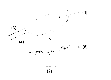
Figure 2 is a schematic representation of the way in which the flap (1) and
the
endothelialised matrix (5) that will act as a vascularised bridge will be
placed in relation to
the receiver site (2). An artery (3) and a vein (4) are again shown in the
flap.
Figure 3 is a photograph showing the flap design, on the dorsal side of the
ear of a
rabbit and with the axis centred in the intermediate caudal vessels. The
endothelialised
matrix being distributed over the cartilage situated below the flap.
CA 02608712 2007-11-16
12
Figure 4 shows immunohistochemical stains of CD32 in treated subjects (part a)
and control subjects (b) at 500 magnification.
Figure 5 shows a photograph of vessels of a receiver individual of a flap,
treated
with endothelialised fibrin gel matrix, with a positive reaction as regards
VEGF in its
endothelial cells.
DETAILED DESCRIPTION OF THE INVENTION
The present invention, therefore, provides a fibrin matrix that has, inside
it,
endothelial cells transfected with adenoviral vectors and for this reason
superproducers of
VEGF and/or FGF, a matrix that is prepared with the purpose of being used as a
connecting vascularised bridge between the receiver site and a flap.
The endothelial cells have been extracted previously from peripheral veins of
an
individual of the same species, which can be the actual subject to treat, and
are cultured
and genetically modified to finally be absorbed into a matrix of fibrin gel
obtained from
plasma, usually also from the actual patient. The genetic modification of the
endothelial
cells is produced with adenovirus carriers of the VEGF and/or FGF genes, in
such a way
that, in vivo, they behave as bioreactors of the aforementioned factors for a
limited time
only, while the aforementioned cells transfected with the adenovirus derived
vector
survive in the fibrin gel. This endothelialised vector and superproducer of
proangiogenic
factors has the purpose of acting as a vascularised bridge to induce the
development of a
functional vascular plexus between the flap and the receptor site, with the
aim of
accelerating the adaptation between both. The fibrin gel matrix provides a
suitable
environment so that growth factors may generate and accumulate in it.
The advantages of using a bridge vascularised by an endothelialised matrix
composed of a matrix of fibrin gel in which endothelial cells that are
superproducers of
proangiogenic cells due to having been transfected in vitro with adenoviral
vectors that
contain genes that code the aforementioned proangiogenic facts are absorbed,
are as
follows:
CA 02608712 2007-11-16
13
- The fibrin gel matrix allows the growth and migration of endothelial cells
within it.
Inside this matrix, genetically modified endothelial cells are capable, in
vitro, of
secreting proangiogenic growth factors (VEGF and FGF) and being organised by
forming micro-capillaries before being transplanted.
- After transplanting this vascularised bridge, placed between the receptor
site and
the flap, the growth factors produced by the genetically modified cells are
also
capable of inducing the proliferation and migration of new vessels in the
receptor
site as well as in the flap itself. The endothelial cells of the vascularised
matrix
should act as a bridge between both, many of them ending up by being included
as part of the vascular stroma which will bind the transplanted tissue to the
receptor site, such that, overall, increases the possibilities of success in
the
reconnection of the flap.
The increase in angiogenesis in the transplanted tissue helps to accelerate
the
incorporation of a flap of skin, muscle, or bone tissue or a combination of
these
tissues to the receptor site.
- The use of this endothelialised tissue as a vascularised bridge is of great
use not
only for reducing thrombotic events that could threaten the survival of the
flaps,
but also for the treatment of flaps that may have to be used to reconstruct
areas
treated with radiotherapy, flaps in diabetic patients or smokers (who usually
have
micro- and/or macrovascular problems), and also to prepare prefabricated
flaps.
- The fact that the genetic modification of the endothelialised cells is
produced by an
in vitro transfection, before the matrix transplant, ensures that the
incorporation of
the genetic material is produced specifically in the cells desired and has a
clear
advantage over the in vivo injection of viral vectors, as it decreases the
risk of
systemic dispersal, allows a continued release effect of growth factors for
days and
does not require repeat doses.
- The use of adenovirus as carrier vectors is also an advantage compared to
the use
of a retrovirus, since it avoids the risk of a possible packing together with
inactivated vectors of genetic material with oncogenic potential and their
possible
integration into the host cell in a stable form and blocking other genes.
- The possibility that all the components of the endothelialised matrix may be
of
autologous origin means that, in those cases, the insertion of the matrix as
an
CA 02608712 2007-11-16
14
endothelialised bridge and the corresponding flap may be performed without the
need for immunosuppression of the treated subjects.
- On the other hand, the flexibility within the method for obtaining the
endothelialised
matrix means that the serum used to form the fibrin gel as well the
endothelialised
cells that are transfected and soaked into the matrix could come from a
different
individual from that who is going to have the flap inserted. Even the species
from
which the fibrinogen of the fibrin matrix originates can be different. This
opens the
possibility that matrices may be prepared in advance when they are required
urgently, although in these cases it would be advisable to give
immunosuppression
to the subjects in whom the endothelialised matrix is implanted as a
vascularised
bridge.
To achieve this, endothelial cells have to be produced, transfected in vitro
with an
adenoviral vector that enables the expression of VEGF and/or FGF. The
aforementioned
endothelial cells are extracted from the peripheral nervous system, preferably
from the
saphenous vein, of the donor. If the endothelialised matrix needs to be
completely
autologous, the donor will have to be the same individual who needs the flap.
The
extracted vessels are sent in a transport flask with DMEM and an antiseptic
solution for
their culture and preparation, using, for example, the method described by Del
Rio et al,
Br J Pharmacol, 120:1360-1366, 1997. Following this method, the endothelial
cells are
cultured in a suitable medium, like, perhaps, the modified Dulbecco medium:
Hams F12
(1:1) which contains 10% FCS supplemented with glutamax 1, 100 IU/mI
penicillin G, 100
pg/mi streptomycin and 0.25 Ng/ml amphotericin B.
The in vitro genetic transfer is carried out with confluent cultures by
incubating
them with adenovirus vectors that carry the genes that code the growth factors
(VEGF,
FGF) in a serum poor medium. After two washes with PBS, the cells are
incubated in a
suitable culture medium, like, perhaps, Dulbecco: Ham's F12 (1:1), for a
suitable time
which, in the case of humans, would be approximately 24 hours. Then, to obtain
the
individual cells that will form part of the endothelialised matrix, they are
treated with
trypsin/EDTA.
CA 02608712 2007-11-16
To prepare the endothelialised matrix, the method described previously in the
international patent application WO 02/072800 can be used. In it, the plasma
is separated,
with platelets and fibroblasts, which gel by using Caz+. Unlike that in the
aforementioned
document, in the present invention the fibroblasts are not resuspended in the
fibrin matrix,
5 nor are the keratinocytes seeded in it, but the type of cells that are
resuspended in the
matrix are endothelial cells which are obtained as described in the previous
paragraph,
which are modified genetically. In this way, the matrix of the fibrin gel of
the present
invention not only acts as a cellular support, but also serves as a vehicle of
therapeutic
factors produced by the cells.
The fibrin matrix can also be obtained from its blood precursor, fibrinogen,
from
plasma cryoprecipitates. The cryoprecipitates are obtained in accordance with
the
standards of the American Association of Blood Banks (Walker RH (ed) Technical
Manual, American Association of Blood Banks, Bethesda, MD; 1993; pp. 728-730).
The individual from whom the plasma is extracted may or may not be that in
whom
the flap is going to be inserted and, in this flap, the endothelialised
artificial matrix
consisting of a superproducer of proangiogenic factors. It is preferred that
the individual is
the same person if it is desired to avoid the need of subjecting the receiver
of the flap to
immunosuppressive treatment. In cases where this is not a fundamental factor,
the
plasma may come from not only a different individual, but also from a
different species.
Thus, the fibrinogen source can be, for example, porcine plasma
cryoprecipitates.
In any of the cases, to produce the fibrin gel, DMEM which contains 1 % FCS
and
the already transfected endothelial cells are added to the fibrinogen
solution.
Subsequently, gelification is induced by the addition of CaCl 2 and thrombin.
Finally, the
mixture is poured over a culture plate or other suitable receptacle and is
left to solidify at a
suitable temperature, which will be 37 C in the case of fibrin gels
originating from humans.
The gel formed is covered with a suitable culture medium, (for example,
Dulbecco: Ham's
F 12 (1:1) and 24-48 hours afterwards it is transplanted over the receptor
site of the flap,
so that it acts as a vascularised bridge between both. Once the flap is
positioned over the
endothelialised matrix, the edges of the same are sutured to the site itself,
in such a way
that the endothelialised matrix remains homogeneously distributed between
both.
CA 02608712 2007-11-16
16
As is described in more detail below in the corresponding example, surgical
experiments performed on animals, specifically rabbits, verified the validity
of the method
and its usefulness in increasing the survival of inserted flaps. Also, after
performing a
statistical analysis, the results showed that the capillary density and the
VEGF expression
were significantly better in the treated subjects.
Example
Preparation of the endothelial cells
The endothelial cells used to be lodged in the fibrin matrix were endothelial
cells
from the aorta of New Zealand albino rabbits. These same cells were cultivated
after
extracting them from the aortic artery of these rabbits under sterile
conditions and in a
culture medium (5% DMEM, with an antibiotic and anti-fungicide). The cells
were cultured
in a medium modified by Dulbecco: Ham's F12 (1:1), which contains 10% FCS,
supplemented with glutamax I, 100 IU/ml penicillin G, 100 pg/mI of
streptomycin and 0.25
pg/mI amphotericin B.
In the study, cells were used that had been subjected to three steps at the
most
during their culture. The in vitro genetic transfer was performed in confluent
cultures, by
incubation for 3 hours at 37 C with a Group C adenoviral vector, which
included a gene of
VEGF A 165, capable of being expressed in endothelial cells in a serum-poor
medium.
After two washes with PBS, the cells were incubated in a growth medium for 24
hours and
later treated with trypsin/EDTA, to obtain individual cells.
Preparation of the endothelialised fibrin matrices
The fibrin gels containing the transfected cells were prepared following the
protocol
for fibroblast fibrin gels described in the international patent application
WO 02/072800,
with modifications. Firstly, fibrinogen from porcine plasma cryoprecipitates
was used as a
resource for obtaining the fibrin. The cryoprecipitates were obtained in
accordance with
the standards of the American Association of Blood Banks. To produce the
fibrin gel, 3 ml
CA 02608712 2007-11-16
17
of the fibrinogen solution were added to 12 ml of DMEM in 10% FCS, with 5 x
105
transfected endothelial cells. Then 1 ml of CaCIZ (0.025 mM, Sigma) was added
along
with 11 IU bovine thrombin (Sigma). Finally, the mixture was poured into a 75
cmz culture
receptacle and left to solidify at 37 C. The gel is covered with a culture
medium to be used
after 24 hours, being kept in a refrigerator at 4 C during this time interval.
Surgical procedure
The animals in which the flaps were inserted were also New Zealand albino
rabbits, although those individual from whom the endothelial cells had been
obtained were
not used. For this reason, as well as due to the use of porcine plasma as a
fibrin source,
immunosuppression had to be provoked in the subjects treated, which in this
case was
carried out with Sandimmune (Novartis Pharmaceuticals), using an
intraperitoneal dose
of 25 mg/kg the day before the operation. This dose was repeated daily in all
the subjects
of the study, until they were sacrificed.
As regards obtaining the flaps, axial flaps were designed from the dorsal
region of
the ear of each rabbit, which is based in the intermediate branch of the
artery and caudal
auricular vein. The proximal edge of each flap is situated 4 cm distal to the
junction of the
median caudal auricular vein with the corresponding caudal vein of the ear.
Under aseptic
and antiseptic conditions, the edges were infiltrated with local anaesthetic
and an incision
was made on the edges with a scalpel, trying not to section the vascular
pedicle. On the
distal edge of the flap, after isolating the central vessels, electro-
coagulation is performed
on the same. Using blunt-end scissors, the flap is separated from the
cartilaginous tissue,
observing the central vessel insertions to the perichondrium. Later, with the
aid of a
scalpel, the whole flap was removed in a proximal direction. With a scalpel
incision, the
vessels in the proximal area of the axial flap are accessed, up to the
junction of the
median caudal auricular vein with the corresponding caudal vein. At this time
the caudal
vein is coagulated, to avoid any interference of this vessel with the axial
vessel of the flap.
The perichondrium was then removed from the cartilage in the exposed area.
With the aid
of magnified glasses and microsurgery tools, the blood vessels are isolated
and the nerve
fillets that are attached to the central vessels were sectioned, because these
contain small
accompanying vessels that could nurture the actual flap themselves.
CA 02608712 2007-11-16
18
The endothelialised fibrin matrix is handled in a laminar flow hood, to remove
it
from the glass receptacle that contains it, with the aid of a spatula. The
capsule is then
covered with sterile paper and is transported to the operating theatre for its
implantation
over the cartilage with its perichondrium removed. It was ensured that the
distribution of
the matrix was as homogeneous as possible. The flap was positioned over this.
Using a
silk suture (4/0), the edges of the same and the incision made to expose the
vessels were
sutured. An example of the final arrangement is shown in Figure 3.
Once operated on, the animals were returned to their cages.
The final surgical act consisted in sectioning the vessels of the axial flap.
For this,
the same anaesthetic procedure was performed again, although in this case, the
infiltration with 2% lidocaine was made at 1/2 cm from the proximal edge of
the flap. An
incision was made with a scalpel over the surgical scar itself and with the
aid of
microsurgery clamp the arterial and venous flow was cut off. Then, the vessels
were
sectioned and ligated. In group I (control) and II (treated with the VEGF
producer matrix),
this procedure was carried out at 5 days from the date of removing the flap.
In group III
(control) and IV (treated with the VEGF producer matrix), the sectioning was
performed 48
hours after carrying out the initial surgery.
Treatment of the operated animals
Once the animals were returned to their cages after the first intervention, a
daily
assessment was made of each subject, making a note of details of the colour of
the flaps
and its consistency to touch.
Four days after performing the sectioning of the vessels, photographs were
taken
of them and, at 6 days, the animals were killed with an overdose of
intravenous sodium
pentothal.
At this time the flaps were extracted and placed in receptacles with 10%
buffered
formol. At 48 hours, three portions from the proximal, medium and distal
areas,
respectively of each flap were selected, in such a way that the central
vessels were in the
centre of the histology section, and were embedded in paraffin.
CA 02608712 2007-11-16
19
Macroscopic and microscopic evaluation
The flap surface that was viable was assessed by planimetry; the values
obtained
being expressed as percentages. The aforementioned planimetry was performed
the day
before the animal was sacrificed.
Sections of 3-4 pm were made from the paraffin block, which were mounted on
silanised slides, with a positive surface charge and a capillary gap of 75 pm
(ProbeOnTM
Plus Slides. Catalogue No. 15-188-52. Fischer Biotechc and ChemMateTM
Capillary Gap
Plus Slides. Code S2024: DAKO A/S Biotek Solutions). After paraffin removal
and
hydration, the slices were washed in Tris saline (TBS) (0.05 M Tris-HCI; 0.5 M
NaCI; pH
7.36). Endogenous peroxidase activity was blocked using 3 % hydrogen peroxide
in
methanol for 30 minutes.
Wth the intention of exposing the highest possible number of epitopes, by
unfolding the proteins by denaturation, the sections were subjected to a pre-
treatment with
heat in a microwave at 750 watts, submerged in a citrate buffer (0.01 m citric
acid, pH 7,
for four periods of five minutes, then leaving them to cool to room
temperature.
The non-specific background staining block was done by using non-immune
normal goat serum (Code NGS-1, University of Navarre, Pamplona), diluted 1:20,
for 30
minutes at room temperature.
The slices were incubated with CD31 antibodies (CD-31 mouse monoclonal
antibody. Code M 0823. DAKO), with a 1/100 dilution, and anti-VEGF (VEGF (C-
1): sc-
7269. Santa Cruz Biotechnology, Inc.). The CD31 protein is specific for
endothelial cell
membranes; therefore, the stains that can detect those sites to which the CD-
31
antibodies will bind enable the blood vessel walls to be visualised and,
therefore, their
presence.
CA 02608712 2007-11-16
After washing in TBS (0.05 M Tris-HCI; 0.5 M NaCI; pH 7.36), it is incubated
for 30
minutes at room temperature, with the EnVisionTM product, peroxidase (DAKO
EnVisionTM. Code No. K4003 anti-rabbit; Code No. K4001 anti-mouse) pre-
diluted.
5 After the final wash in TBS (0.05 M Tris-HCI; 0.5 M NaCI; pH 7.36), the
product of
the peroxidase reaction is visualised using a commercially prepared 3,3'-
diaminobenzidine (DAB) solution in a chromogenic solution, with a imidazole-
HCI buffer at
pH 7.5 and hydrogen peroxide (DAKOc Liquid DAB+Large Volume Substrate-
Chromogen
Solution.Code No. K3468), incubating it for five to thirty minutes, at room
temperature and
10 pre-diluted.
A pathologist is responsible for carrying out the evaluation of the
histological
preparations which have been stained with haematoxylin-eosin and
immunohistochemical
stains (CD31 and VEGF).
15 In the CD31 stains, the vessels are counted, at x 500 magnification, in 6
different
fields and the mean value of the same is expressed. These areas where there
had been
an inflammatory focus were ignored and the areas where there had not been any
subcutaneous tissue distortion between the skin appendages themselves and the
cartilage were evaluated. Figure 4 shows examples of these CD31
immunohistochemical
20 stains in treated subjects (a) and control subjects (b). The count results
are shown later in
Table 1, which corroborate that there is a greater formation of blood vessels
in the treated
subjects with the matrix of the invention compared to the subjects in the
control groups.
The associated statistical parameters are shown in Tables 2 and 3.
A regards VEGF, those vessels in which the cytoplasm of the endothelial cells
was
stained were considered positive. The count was made giving a numerical value
to all the
cells that were stained with antibody in each preparation. An example of
vessels stained
with a positive reaction for VEGF in endothelial cells in an individual
treated with the
endothelialised fibrin gel of the invention is shown in Figure 5, where the
results of the
stained cells demonstrate the presence of VEGF and, therefore, that it being
synthesised
effectively. The count results of the endothelial cells stained are shown
later in Table 1.
The associated statistical parameters are shown in Tables 2 and 3.
CA 02608712 2007-11-16
21
Results
Table 1 presented below shows the data of the means and standard deviations
corresponding to the survival data, CD31 stains and VEGF stains.
Table 1. - Means and standard deviations corresponding to the survival data,
CD31
stains and VEGF stains.
MEAN/STANDARD DEVIATION
SURVIVAL CD31 VEGF
(%) Vessels/field* Endothelial
500 cells/section
Group 1 51.25/45.88 5.56/3.18 0.87/1.12
Section (control)
at 5 days Group II 95.62/4.95 13.20/4.54 5.62/3.73
(treatment)
Group III 2.50/7.07 2.34/4.56 0.37/1.06
Section at (Control)
48 Hours Group IV 55.62/38.95 5.56/3.18 3.06/2.95
(treatment)
Below, in Tables 2 and 3, the data corresponding to the statistical parameters
associated with the previous results are shown, specifically those relative to
the Kruskal
Wallis (Table 2) and the Whitney-Mann U (Table 3) analysis.
The analysis of variance (ANOVA) is a data analysis technique to examine the
significance of the factors (independent variables) in a multifactorial model.
The single-
factorial model can be obtained from a generalisation of a two-sample test.
That is, a test
of two samples is that part from the hypothesis that the means of the
populations are
CA 02608712 2007-11-16
22
equal. The ANOVA test will assess the hypothesis that defends that the means
of "x"
populations are equal.
The Kruskal Wallis test may be used like ANOVA. It is a non-parametric test
which
is used when the conditions to use the ANOVA test cannot be applied, that is,
to contrast
the hypothesis that a number of different sized samples originate from the
same
population. Thus, the Kruskal-Wallis test is a non-parametric method to
evaluate the
hypothesis that several populations have the same continuous distribution
versus the
alternative that results tend to be different in one or more populations.
Table 2. - Kruskal-Wallis parameters
Kruskal Wallis SURVIVAL CD31 VEGF
XZ 15.50 17.11 16.38
P 0.001 0.001 0.001
As for Table 3, the Mann-Whitney U test is a non-parametric statistical test
that is
used when the sample is small or the distribution of the data in the
population is free (data
not originating from normal populations and with similar variances). This test
compares
whether two samples of two sub-populations have the same distribution.
The observations of both groups are combined and classified according to the
average
range assigned in case ties are produced. If the position of the populations
is identical, the
ranges should be randomly mixed in both samples.
Table 3. - Mann-Whitney U analysis
P
U Mann Whitney Pairs SURVIVAL CD31 VEGF
Section in 5 days 0.08 0.00 0.00
(treatment versus control)
Section in 2 days 0.00 0.05 0.02
(treatment versus control)
CA 02608712 2007-11-16
23
Therefore, according to these data, a survival of the flaps of around 50% was
found in treated subjects despite dispensing with the pedicle at 48 hours from
being
intervened (Group IV), a fact which makes it easier for the flap to necrose,
as the blood
flow of the flap depends on the pedicle. The survival increased to 95% if the
section of the
pedicle was performed at 5 days (protocol A). In the non-treated subjects, the
survival did
not reach 3% after sectioning the pedicle at 48 hours.
The results showing the capillary density and the VEGF expression were also
significantly better in the treated subjects.
15