Note: Descriptions are shown in the official language in which they were submitted.
CA 02609468 2007-11-20
WO 2006/125986 PCT/GB2006/001908
1
LARYNGEAL MASK AIRWAY DEVICE
The present invention relates to a laryngeal mask airway device.
The laryngeal mask airway device is a well known device that is useful for
establishing airways in unconscious patients. U.S. Patent No. 4,509,514 is one
of the
many publications that describe laryngeal mask airway devices. Such devices
have
been in use for many years and offer an alternative to the older, even better
known
endotracheal tube. For at least seventy years, endotracheal tubes comprising a
long
slender tube with an inflatable balloon disposed at the tube's distal end have
been used
for establishing airways in unconscious patients. In operation, the
endotracheal tube's
distal end is inserted through the mouth of the patient, past the patient's
trachea. Once
so positioned, the balloon is inflated so as to form a seal with the interior
lining of the
trachea. After this seal is established, positive pressure may be applied to
the tube's
proximal end to ventilate the patient's lungs. Also, the seal between the
balloon and the
inner lining of the trachea protects the lungs from aspiration (e.g., the seal
prevents
material regurgitated from the stomach from being aspirated into the patient's
lungs).
Although they have been enormously successful, endotracheal tubes suffer
from several major disadvantages. The principal disadvantage of the
endotracheal tube
relates to the difficulty of properly inserting the tube. Inserting an
endotracheal tube
into a patient is a procedure that requires a high degree of skill. Also, even
for skilled
practitioners, insertion of an endotracheal tube is sometimes difficult or not
possible. In
many instances, the difficulty of inserting endotracheal tubes has tragically
led to the
death of a patient because it was not possible to establish an airway in the
patient with
sufficient rapidity. Also, inserting an endotracheal tube normally requires
manipulation
of the patient's head and neck and further requires the patient's jaw to be
forcibly opened
widely. These necessary manipulations make it difficult, or undesirable, to
insert an
endotracheal tube into a patient who may be suffering from a neck injury.
In contrast to the endotracheal tube, it is relatively easy to insert a
laryngeal
mask airway device into a patient and thereby establish an airway. Also, the
laryngeal
CA 02609468 2007-11-20
WO 2006/125986 PCT/GB2006/001908
2
mask airway device is a"forgiving" device in that even if it is inserted
improperly, it
still tends to establish an airway. Accordingly, the laryngeal mask airway
device is
often thought of as a "life saving" device. Also, the laryngeal mask airway
device may
be inserted with only relatively minor manipulation of the patient's head,
neck and jaw.
Further, the laryngeal mask airway device provides ventilation of the
patient's lungs
without requiring contact with the sensitive inner lining of the trachea and
the size of
the airway established is typically significantly larger than the size of the
airway
established with an endotracheal tube. Also, the laryngeal mask airway device
does not
interfere with coughing to the same extent as endotracheal tubes. Largely due
to these
advantages, the laryngeal mask airway device has enjoyed increasing popularity
in
recent years.
U.S. Patent Nos. 5,303,697 and 6,079,409 describe exainples of prior art
devices that may be referred to as- "intubating laryngeal mask airway
devices." The
intubating device has the added advantage that it is useful for facilitating
insertion of an
endotracheal tube. After an intubating laryngeal mask airway device has been
located
in the patient, the device can act as a guide for a subsequently inserted
endotracheal
tube. Use of the laryngeal mask airway device in this fashion facilitates what
is
commonly known as "blind insertion" of the endotracheal tube. Only minor
movements
of the patient's head, neck and jaw are required to insert the intubating
laryngeal mask
airway device, and once the device has been located in the patient, the
endotracheal tube
may be inserted with virtually no additional movements of the patient. This
stands in
contrast to the relatively large motions of the patient's head, neck and jaw
that would be
required if the endotracheal tube were inserted without the assistance of the
intubating
laryngeal mask airway device. Furthermore, these devices permit single-handed
insertion from any user position without moving the head and neck of the
patient from a
neutral position, and can also be put in place without inserting fingers in
the patient's
mouth. Finally, it is believed that they are unique in being devices which are
airway
devices in their own right, enabling ventilatory control and patient
oxygenation to be
continuous during intubation attempts, thereby lessening the likelihood of
desaturation.
CA 02609468 2007-11-20
WO 2006/125986 PCT/GB2006/001908
3
Artificial airway devices of the character indicated, are exemplified by the
disclosures of US Pat. No. 4,509,514; U.S. Pat. No. 5,249, 571; U.S. Pat No.
5,282,464;
U.S. Pat. No. 5,297,547; U.S. Pat. No. 5,303,697; and by the disclosure of the
UK
Patent 2,205,499. Such devices with additional provision for gastric-discharge
drainage
are exemplified by U.S. Pat. No. 4,995,388 (Figs. 7 to 10); U.S. Pat. No.
5,241,956; and
U.S. Pat. No. 5,355,879.
In general, laryngeal mask airway devices aim to provide an airway tube of
such cross-section as to assure more than ample ventilation of the lungs, and
the designs
with provision for gastric drainage have been characterized by relatively
complex
internal connections and cross-sections calculated to serve in difficult
situations where
substantial solids could be present in a gastric discharge. As a result, the
provision of a
gastric discharge opening at the distal end of the mask applicable for direct
service of
the hypopharynx has resulted in a tendency for such masks to become bulky and
unduly
stiff, thus making for difficulty in properly inserting the mask. Moreover,
undue bulk
and stiffness run contrary to the requirement for distal flexibility for
tracking the
posterior curvature of the patient's throat on insertion, in such manner as to
reliably
avoid traumatic encounter with the epiglottis and other natural structures of
the pharynx.
A number of problems have been experienced with all of these prior types of
device. For example, some prior devices seek to prevent occlusion of the
airway outlet
by parts of the patient's anatomy, such as the epiglottis, by the provision of
bars and the
like across the outlet. Although such devices function well in most cases,
they can make
manufacturing more complex, and can affect the performance of devices in use.
This is
especially so in devices formed from relatively rigid materials, like PVC, as
opposed to
the more traditional Liquid Silicon Rubber (LSR).
In general, devices formed from materials such as PVC are attractive because
they are cheaper to make, and can be offered economically as "single-use"
devices.
However, there are material differences in PVC and PVC adhesives, such as
increased
durometer hardness as compared to LSR, which affect how the devices perform in
use.
For example, it has been observed that for a given volume of air, an LSR cuff
will
CA 02609468 2007-11-20
WO 2006/125986 PCT/GB2006/001908
4
expand to a larger size than a comparable PVC cuff. This superior elasticity
allows the
LSR cuff to provide an anatomically superior seal with reduced mucosal
pressure. To
close the performance gap, the PVC cuff must be of reduced wall thickness.
However, a
PVC cuff of reduced wall thickness, deflated and prepared for insertion, will
suffer from
poor flexural response as the transfer of insertion force through the airway
tube to cuff
distal tip cannot be adequately absorbed. The cuff assembly must deflate to a
thickness
that preserves flexural performance i.e. resists epiglottic downfolding, but
inflate so that
a cuff wall thickness of less than or equal to 0.4mm creates a satisfactory
seal. And
where mask backplates are fonned from PVC, as well as cuffs, the fact that the
increased durometer hardness of PVC is inversely proportional to flexural
perfonnance
(hysterisis) means that the flexural performance of the device in tenns of
reaction,
response and recovery on deformation is inferior to a comparable LSR device.
The above described problems are particularly acute in devices which
incorporate an oesophageal drain. As mentioned above, in any such device
regardless of
the material from which it is formed, adding an oesophageal drain in itself
adds greatly
to complexity of manufacture and can also affect the perfonnance of devices,
in terms
of ease of insertion, seal formation and prevention of insufflation. These
problems can
be exacerbated still further if PVC or similarly performing materials are
used. For
example, the skilled worker will appreciate that in terms of manufacture, the
need to
provide a drain tube which is sealed from the airway, and which must pass
through the
inflatable cuff poses a particularly difficult problem. In terms of effects on
functionality,
the provision of a drain tube can cause unacceptable stiffening of the mask
tip area and
occlusion/restriction of the airway passage.
According to the invention there is provided a laryngeal mask airway device
for insertion into a patient to provide an airway passage to the patient's
glottic opening,
the device comprising an airway tube, a mask attached to the airway tube, the
mask
comprising a body having a distal end and a proximal end, a peripheral
inflatable cuff,
and defining an outlet for gas, the mask being connected to the airway tube
for gaseous
communication between the tube and the mask, the distal end of the mask being
ventrally displaced, relative to the proximal end. It has been surprisingly
found that a
CA 02609468 2007-11-20
WO 2006/125986 PCT/GB2006/001908
ventral displacement of the tip makes insertion of the mask much easier
because the tip
is presented at an optimum angle as it tracks around the curvature of the
airway
anatomy.
It is preferred that the extent of distal displacement is from about 5 mm to
about 20mm, and it is most preferred that the extent of distal displacement is
about
10mm. This has been found to be the optimum range. It has been found that if
the extent
of displacement is too great, the device will not lie in the correct position
at its
maximum extent of insertion.
It is preferred that the body describes a substantially convex curve, from the
proximal to distal end. It is further preferred that the mask body comprises a
plate, the
plate having a dorsal side and a ventral side, the dorsal side being
substantially smooth
and having a convex curvature across its width. It is also preferred that the
dorsal
surface of the airway tube corresponds in curvature to the curvature across
the width of
the plate. All of these expedients assist in making insertion of the mask
easier.
The airway tube preferably comprises a relatively more rigid material than the
mask body. Both the airway tube and the mask body preferably comprise a
plastics
material.
The device may further including an oesophageal drain tube, and the
oesophageal drain tube may be disposed on the ventral side of the body, in
order to
maintain a smooth profile on the dorsal side, to make insertion easier.
The invention will further be described by way of example and with reference
to the following drawings, in which,
Figure 1 is a dorsal three quarter perspective view of a device according to
the
invention;
Figure 2 is a right side view of the device of Figure 1;
Figure 3 is a dorsal view of the device of Figure 1;
CA 02609468 2007-11-20
WO 2006/125986 PCT/GB2006/001908
6
Figure 4 is a ventral view of the device of Figure 1;
Figure 4a is a ventral view of a further embodiment of device according to the
invention;
Figure 5 is an end view, looking from the proximal towards the distal end of
the device of Figure 1;
Figure 6 is an end view, looking from the distal towards the proximal end of
the mask of the device of Figure 1;
Figure 7 is an enlarged view of the mask of the device of Figure 1;
Figure 8 is a dorsal view of the device of Figure 4a;
Figure 9 is a longitudinal sectional view along line Y-Y in Figure 8;
Figure 10 is a side view, enlarged, of the device of Figure 4a;
Figures 11A to 11K are transverse sectional views along lines A-A to K-K in
Figure 10;
Figure 12 is an exploded dorsal perspective view of a device according to the
invention;
Figure 13 is an exploded ventral perspective view of a device according to the
invention;
Figure 14 is a dorsal three quarter perspective view of a device according to
the
invention;
Figure 15 is a right side view of the device of Figure 14;
Figure 16 is a dorsal view of the device of Figure 14;
Figure 17 is a ventral view of the device of Figure 14;
Figure 18 is an end view, looking from the proximal towards the distal end of
the mask of the device of Figure 14;
Figure 19 is an end view, looking from the distal towards the proximal end of
the mask of the device of Figure 14;
Figure 20 is a dorsal three quarter perspective view of the device of Figure
14;
Figure 21 is a view of section CC-CC in Figure 20;
Figure 22 is a view of section VC-VC in Figure 17;
Figure 23 is a proximal end view of a part of the device of Figure 14; and
Figure 24 is a distal end view of a part of the device of Figure 14.
CA 02609468 2007-11-20
WO 2006/125986 PCT/GB2006/001908
7
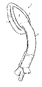
Referring now to the drawings, there is illustrated a laryngeal mask airway
device 1 for insertion into a patient to provide an airway passage to the
patient's glottic
opening, the device 1 comprising an airway tube 2, a mask 3 attached to the
airway tube
2, the mask 3 comprising a body 4 having a distal end 5 and a proximal end 6,
a
peripheral inflatable cuff 7, and defining an outlet 8 for gas, the mask 3
being connected
to the airway tube 2 for gaseous communication between the tube 2 and the
mask, the
distal end of the mask being ventrally displaced, relative to the proximal
end.
In this embodiment the device 1 further comprises an oesophageal drain 10, the
drain 10 comprising a conduit 11 extending from an opening 12 at the distal
end 5 to a
drain outlet 13 disposed to the outside of the patient when the device 1 is in
place,
wherein the conduit 11 is formed integrally in the material of the body 4.
As can be seen from the drawings, the device 1, in terms of overall appearance
is somewhat similar to prior art devices, in that it consists of the basic
parts which make
up most if not all laryngeal mask airway devices, i.e. an airway tube 2 and
mask 3
which includes a body part 4, and a cuff 7.
For the purposes of description it is appropriate to assign reference names to
areas of the device 1 and accordingly with reference to Figures 2 to 6, the
device 1 has a
dorsal side 14, a ventral side 15, a proximal end 16 (in a sense that this is
the end
nearest the user rather than the patient) a distal end 17 and right and left
sides 18 and 19.
Referring firstly to the airway tube 2, in the illustrated embodiments the
tube
comprises a relatively rigid PVC material such as a shore 90A Colorite PVC
moulded
into an appropriately anatomically curved shape. The tube 2 has some
flexibility such
that if it is bent it will return to its original shape. Although it is
resiliently deformable
in this way, it is sufficiently rigid to enable it to assist in insertion of
the device 1 into a
patient, acting as a handle and guide. In this embodiment the airway tube 2
does not
have a circular cross-section as in many prior devices, but instead is
compressed in the
dorsal/ventral direction which assists in correct insertion of the device 1,
helps prevent
kinking, and assists in comfortable positioning for the patient as the shape
generally
CA 02609468 2007-11-20
WO 2006/125986 PCT/GB2006/001908
8
mimics the shape of the natural airway. In this embodiment each side 18, 19 of
the
airway tube 2 includes a groove or channel 20 extending for most of the tube's
length
from the proximal to distal ends. These grooves 20 further assist in
preventing crushing
or kinking of the airway tube 2. Internally the grooves 20 form ridges along
the inner
surfaces of the sides 18 and 19.
Referring now to Figure 13, which shows an exploded view of the device 1, it
can be seen that the airway tube 2 includes a flared distal end 22 with
surfaces 22a
disposed to allow for attachment of the tube 2 to the mask 3, conveniently by
over
moulding of the mask 3 onto the airway tube 2. Thus, the airway tube 2 itself
can form
a pre-mould used in formation of the device 1, which substantially simplifies
manufacturing. Of particular note is the airway tube's dorsal mould surface 23
(Figure
13). This surface 23 is located at the flared distal end 22, and takes the
form of a flat
land extending between the outer dorsal surface 2a and the inner dorsal
surface 2b
(Figure 24) of the dorsal wall 2c. It includes optional through holes 2d to
allow the
over moulded back plate 4 to lock onto the tube 2, as will be described later
on. This
feature helps ensure a secure connection between the different materials
making up the
airway tube 2 and mask 3.
A further feature of the airway 2 is the oesophageal drain tube 41. This drain
tube 41 is located within airway tube 2, extending centrally through it from
one end to
the other, and in this embodiment it is disposed in contact with the inner
surface 2a of
the dorsal wall 2b of the airway tube 2, and bounded on each side by raised,
smooth
walls (not shown) which form a shallow channel through which it runs.
The proximal end of the airway tube 2 is provided with a connector 42, for
connection of the device 1 to a gas supply and drain (not shown) as shown for
example
in Figures 12 and 13 and in section in Figure 9. The connector 42 comprises a
connector body 43, an optional bite block 44 and a connector plug 45. The
connector
body 43 and bite block 44 correspond in shape and dimension with the internal
shape of
the proximal end of the airway tube 2 such that they fit inside it. The
connector body 43
has a perpendicularly extending peripheral flange 46 which extends at one
point on its
CA 02609468 2007-11-20
WO 2006/125986 PCT/GB2006/001908
9
circumference into a tab 47. Connector plug 45 attaches to connector body 43
by
adhesive or other suitable means applied to flange 46. The connector plug 45
comprises
major and minor bores 48, 49 which both lead into a common atrium 50 at the
distal end
of the connector plug 45 where it attaches to the connector body 43. Drain
tube 41
extends into and through minor bore 49, such that the bore of the airway tube
2 and the
bore of the drain tube 41 are separated from one another.
Turning now to the mask 3, the mask 3 consists of two parts, a body part 4
often referred to as a back plate, and a peripheral cuff 7.
The back plate 4 is formed in these embodiments by moulding from a shore
50A Vythene PVC + PU. This material is substantially softer and more
deformable
than the material of airway tube 2.
Referring now to Figure 23, the back plate 4 comprises a generally oval
moulding when viewed from the dorsal or ventral directions, having a smooth
dorsal
surface 24, a formed ventral surface 24a (Figure 17), a proximal joining
portion 24b,
and a distal tip 61.
The dorsal surface 24 has a convex curvature from one side to the other,
corresponding to the curvature of the dorsal surface of the airway tube 2, and
longitudinally, the dorsal surface 24 is also curved, having a curvature
beginning at the
joining portion 24b and extending with constant rate of curvature toward the
distal tip
61. As a result the tip 61 is ventrally biased relative to the distal end of
the airway tube,
in the assembled device 1, the extent of displacement of the distal tip 61
being
approximately 20mm or 10 degrees, in order to produce a curvature in the mask
that is
suited to the anatomy of the patient. This is shown schematically at X in
Figure 2. On
insertion, this displacement of the tip 61 assists the mask in "turning the
corner" in the
insertion path.
When viewed from the ventral side, the integrally moulded structures of the
back plate 4 can best be seen (Figures 4,7,12,17). The precise shape of the
ventral side
CA 02609468 2007-11-20
WO 2006/125986 PCT/GB2006/001908
24a of the back plate is illustrated particularly in the sectional views shown
in Figures
11A to 11K and in the enlarged perspective view in Figure 7. Referring to the
exploded
view shown in Figure 12, the convex curvature of the dorsal surface 24 of the
back plate
4 is mirrored in a corresponding concave curvature on the ventral side. Thus,
the ventral
surface 24a forms a shallow, elongate channel tapering towards the distal tip
61. The
channel is bounded by walls 26. The walls 26 have correspondingly shaped,
longitudinally extending convex outer surfaces 25. Each wall 26 extends
longitudinally
substantially the entire length of the back plate 4 from the proximal joining
portion 24b
towards the distal tip 61. Each wall 26 also has a convex inner surface 28,
but rather
than terminating at an angle normal to the channel floor, the curve of each
wall 26 is
continued, the walls curving back over the channel and terminating in inwardly
extending webs 27 (Figures 7 and 11). The inner surfaces 28 of the side walls
26 curve
down to form the floor of the channel but do not meet, because the base or
floor of the
channel is bisected by a longitudinally extending, integrally moulded conduit
which is
an oesophageal drain tube 11 extending along it for its entire length from
joining
portion 24b to distal tip 61. Thus, it can be seen that the channel has three
longitudinally extending conduits on its inner surface, the two open outer
conduits 28a
which are minor gas conduits in the assembled device 1, and the central drain
tube 11,
which forms a septum there between.
Referring now in greater detail to the drain tube 11, it will be seen that the
tube
11 has a sufficient diameter such that its upper wall section 11a, i.e. the
wall section
ftirthest from the floor of the channel, is on a similar level with the
inwardly extending
webs 27 of the side walls 26. Furthermore, the upper wall section lla itself
also has
outwardly extending webs 30, which taper toward, but do not meet, the
correspondingly
tapered edges of the webs 27. Thus, the upper surface l lb of the upper wall
section 1 la
of the drain tube 11, and the webs 27, 30, together define a surface llc shown
schematically by a dotted line in Figure 11), below the level of which run all
three
conduits 11, 28a.
Referring now particularly to Figure 7, it can be seen that although the drain
tube 11 extends the full length of the back plate 4 from its proximal joining
portion 24b
CA 02609468 2007-11-20
WO 2006/125986 PCT/GB2006/001908
11
to distal tip 61, the conduits 28a do not extend the fitll length of the back
plate 4, but
instead terminate about half way along its length. The floors 31 of the
conduits 28a
curve gently upwards as they extend towards the distal tip 61 of the back
plate 4 until
they terminate at a level approximately equal to the level of the webs 27 and
30. In the
embodiment shown in Figure 4a, these areas are hollowed out to form
depressions 31b.
As illustrated in Figure 12 and Figures 21 to 23 , drain tube 11 extends to
distal
tip 61, terminating in an opening 12. Thus, an end section l 1 e of the drain
tube 11
protrudes past the end of back plate 4. This end section 11 e is provided with
dorsal
webbing 11 a which extends to either side of it, and around it to form a hood
or pocket
36a which encloses the end section 11 e around its circumference. The hood or
pocket
36a is attached to the distal end of the drain tube 11 around the
circumference 12a of
opening 12 (Figure 22). This hood or pocket 36a is integrally formed in the
material of
the back plate 4 at distal tip 61. It completely surrounds and extends from
the
circumference of the drain tube opening 12 and the joint therebetween is
smooth. As
illustrated, the ventral extent of the hood is more limited than the dorsal
extent, the
dorsal extent being to about midway back towards the proximal end of the back
plate 4.
Referring to sectional views A-A and B-B in Figure 11, it can be seen that the
drain
tube I1 is supported on its right and left sides, and on its dorsal surface,
by
perpendicularly extending webs 62. These webs 62 are integrally formed, and
extend
back from the opening 12 to the point where the end section 11 e meets the
extent of the
back plate 4. In the illustrated embodiment the dorsal webs 62 extend
substantially
perpendicularly from the drain tube, but in a preferred embodiment, they may
extend to
one side or the other, at an angle of less than 90 degrees.
The second part of the mask 3 is the peripheral cuff 7. The cuff 7 is in this
embodiment blow moulded PVC and takes the form of a generally elliptical
inflatable
ring having a central aperture 7a, a relatively deeper proximal end 37 with an
inflation
port 38 and a relatively shallower distal end 7b tapering to a "wedge" profile
39. As
will be appreciated, particularly from the exploded views shown in Figure 12
and 13,
the cuff 7 is integrally formed in one piece. The wedge profile is provided
such that the
CA 02609468 2007-11-20
WO 2006/125986 PCT/GB2006/001908
12
ratio of dorsal to ventral side surface areas favours the dorsal side. Thus,
when deflated
the distal end 7b of the cuff 7 will curl with bias from dorsal to ventral
side.
In the assembled device 1, drain tube 41 is inserted into airway tube 2, such
that it protrudes from proximal end 16. The connector 42 is attached to the
airway tube
2 by inserting the connector body 43 and bite block 44 into proximal end 16.
The parts
are an interference fit and can be secured by adhesive. Plug 45 is attached to
connector
body 43 via flange 46, such that drain tube 41 passes into minor bore 49,
terminating at
or adjacent its mouth. Thus it will be seen that the minor bore 49 is solely
in fluid
communication with drain tube 41, and the major bore 48 is solely in fluid
communication with the interior of airway tube 2.
Airway tube 2 is attached to the back plate 4 conveniently by overmoulding the
back plate-4 onto the already formed tube 2. Thus, the joining portion 24b of
the back
plate 4 is moulded onto the dorsal arc of the airway tube 2 (Figure 13).
Secure
attachment is facilitated by the surfaces 22a, 23 which provide an increased
surface area
onto which the moulding occurs, and through-holes 2d, into which back plate
material
can flow. Drain tube 41 is connected in fluid tight manner to integrally
moulded drain
11, as demonstrated by arrow Z (Figure 13).
The cuff 7 is bonded to the back plate 4 as illustrated in Figures 12 and 13
by
inserting the wedge shaped distal end 7b of the cuff 7 into the hood or pocket
36a at the
distal tip 61 of the back plate 4 such that the wedge surface 39 mates with
the inner
surface 36b of the hood 36a, and sections of the inner periphery of the cuff 7
mate with
convex outer surfaces 25 of back plate walls 26. The cuff 7 is bonded into the
hood
such that the space between the hood and the cuff is airtight and in this
embodiment the
cuff is provided with a "pinch off' 40 (Figures 21 and 22) putting the cuff 7
and hood
36a into fluid communication so that the air space in the hood can also be
inflated, in
addition to the cuff 7 itself. However the cuff 7 pinch off does not extend
the entire
distance towards the distal tip of the cuff to prevent the pressure of
inflation occluding
the opening 12. The proximal dorsal surface of the cuff is bonded to the
ventral arc of
the distal end 22 of the airway tube 2. Thus, it will be appreciated that
unlike in
CA 02609468 2007-11-20
WO 2006/125986 PCT/GB2006/001908
13
previous devices incorporating oesophageal drains, in the invention the drain
11 does
not pierce the cuff 7, making manufacturing simpler. Furthermore, in prior
devices in
which the drain pierces the cuff, the cuff must be securely attached around
the
circumference of the drain tube at the distal tip. Such a secure attachment,
for example
with adhesive, can make the tip hard, and prevent the drain tube collapsing in
the
deflated, flattened device, which is highly desirable to enable the mask to
pass easily
around the curvature of the anatomy. In addition, the acute curvature of a
drain tube to
cuff joint would be highly susceptible to cracking. In the invention, these
problems are
avoided because the drain tube 11 is integrally moulded with the hood 36a,
which in
effect forms a second or minor cuff at the distal tip.
As will be appreciated, the airway of the device 1, which is the conduit
through
which gas is passed to the patient, is provided by the bore of airway tube 2,
which
terminates at flared distal end 22. Flared distal end 22 defines, along with
back plate 4
and cuff 7, outlet 8 for gas passing from tube 2 into mask 3. Outlet 8
includes three
routes by which gas may pass into the mask, namely a main gas conduit 8a
(Figure 6),
and two minor gas conduits 28a.
In use, the deflated device 1 is inserted into a patient in the usual manner
with
devices of this type. As noted above, the relative rigidity of the airway tube
2 allows a
user to grip it and use it to guide the device 1 into the patient, whilst the
relatively
softer, more compliant material of the back plate means that the mask will
more readily
deform to negotiate the insertion path without causing damage to the anatomy,
and will
return to its optimum shape to ensure that a good seal is achieved at the
furthest extent
of insertion. The ventral displacement of the distal tip 61 relative to the
join between the
back plate 4 and airway tube 2 further enhances ease of insertion, because the
distal tip
61 is thereby presented at the optimum angle to negotiate the "bend" in the
insertion
path. In devices formed from relatively rigid materials such as PVC, as
opposed to the
often used LSR these features are particularly important in easing insertion
and
providing for an enhanced seal.
CA 02609468 2007-11-20
WO 2006/125986 PCT/GB2006/001908
14
Referring now to the features of the moulded back plate 4, it will be seen
that
by providing drain tube 11 integrally moulded in the material of the back
plate 4,
problems of mask stiffness and difficulty of manufacture in prior designs
caused by the
presence of a separate drain tube bonded in place with adhesive can be
mitigated.
Moreover, with the back plate 4 of the invention, the combination of the
centrally located drain tube 11 and minor gas conduits 28a assist in solving
the problem
of occlusion of the airway by parts of the patient's anatomy. The minor gas
conduits
28a can be thought of as "nostrils" through which gas may continue to pass
into the
patient even if the main outlet 8a becomes occluded by, for example the
patient's
epiglottis, as the epiglottis will rest upon the septum provided by the drain
tube 11. As
illustrated particularly in Figures 11I and 11J the webs 27, 30 form a partial
closure
over the conduits 28a, to assist in preventing structures such as the
epiglottis from
falling into and blocking the conduits 28a, and also to make the back plate 4
more
resistant to lateral compression. It will be appreciated that in this
embodiment, the drain
11 fonns a convenient septum between the conduits 28a, however, in devices
with no
oesophageal drain, a solid septum could simply be formed in the material of
the back
plate by moulding. In addition, a larger number of conduits 28a could be
provided.
Thus, it can be seen that the above described embodiments address the
problems of prior art devices in novel and inventive ways.