Note: Descriptions are shown in the official language in which they were submitted.
CA 02610243 2013-09-25
METHOD OF FORMING DENDRITIC CELLS
FROM EMBRYONIC STEM CELLS
[0001] BACKGROUND OF THE INVENTION
[0002] Embryonic stem cells are pluripotent cells capable of both
proliferation in cell culture
as well as differentiation towards a variety of lineage restricted cell
populations that exhibit
multipotent properties (Odorico et al., (2001) Stem Cells 19:193-204). Human
embryonic stem
(ES) cells are thus capable of commitment and differentiation to a variety of
lineage-restricted
paths resulting in very specific cell types that perform unique functions.
[0003] Generally, ES cells are highly homogeneous, exhibit the capacity for
self-renewal,
and have the ability to differentiate into any functional cell in the body.
This self-renewal
property can lead under appropriate conditions to a long-term proliferating
capability with the
potential for unlimited expansion in cell culture. Furthermore, it is
understood, that if human ES
cells are allowed to differentiate in an undirected fashion, a heterogeneous
population of cells is
obtained expressing markers for a plurality of different tissue types (WO
01/51616; Shamblott et
al., (2001) Proc. Natl. Acad. Sci. U.S.A. 98:113). These features make these
cells a unique
homogeneous starting population for the production of cells having therapeutic
utility.
[0004] There have been efforts by researchers in the field to develop
methods to culture a
variety of progeny cell types from human ES cells. For example, U.S. Patent
6,280,718 describes
a method for culturing human ES cells into hematopoietic cells by culturing
the human ES cell
with stromal cells.
[0005] Some methods of creating progeny cell types from human ES cells
involve the
creation of embryoid bodies, which are three dimensional structures which can
be formed by ES
cells in culture and which foster the diverse differentiation of ES cells into
various differentiated
progeny lineages. Other methods for creating progeny lineages depend on the
culturing of human
ES cells with particular media, agents or types of cells to expose the ES
cells to factors which
encourage differentiation in a particular direction. All these methods have a
common objective,
which is to provide a source for particular cell types for scientific research
and experimentation
and, for some cell types, for ultimate transplantation into human bodies for
therapeutic purposes.
- 1 -
CA 02610243 2007-11-28
WO 2006/130651 PCT/US2006/021054
[9006] Dendritic cells are immune cells that perform a critical function in
the mammalian
immune system. Dendritic cells (sometimes here DCs) are powerful antigen-
presenting cells
which are present at low frequency in tissues of the body in contact with the
environment such as
skin, and linings of the nose, lungs, stomach and intestines. Dendritic cells
have the ability to
uptake antigens and induce primary T cell responses to initiate generalized
immune system
responses to pathogens. Dendritic cells are so named because of their long
processes or arms,
called dendrites, that are characteristic of dendritic cell morphology.
[0007] Dendritic cells are generated continuously in the bone marrow from
the
hematopoietic lineage and mature in the blood. The dendritic cells of an
individual have
heterogeneous phenotype and function. Dendritic cells develop in several ways,
and there may
be differences among the dendritic cells depending on their lineage of
derivation. Dendritic cells
that develop from CD34+ hematopoietic progenitors along two independent
pathways become
Langerhans cells and interstitial dendritic cells. Dendritic cells derived
from monocytes or from
plasmocytoid T cells are referred to as monocyte-derived DCs or plasmocytoid
DCs respectively.
On the basis of their cellular origin phenotype, dendritic cells are normally
classified broadly into
two major divisions, myeloid or lymphoid. It was believed that myeloid DCs
were developed
from a common myeloid precursor while lymphoid DCs developed from a common
lymphoid
precursors, although it has now also been proposed that a common myeloid DC
precursor gives
rise to all dendritic cell lineages.
[0008] The availability of human immature dendritic cells would be useful
for the study
' of antigen processing and presentation, as well as for understanding the
mechanisms of the
induction of immunity and tolerance. Functional analysis of human dendritic
cell subsets was
significantly facilitated by the development of in vitro systems for the
differentiation of dendritic
cells from CD34+ hematopoietic stem cells and monocytes. However, using these
existing
protocols, obtaining large numbers of human dendritic cell progenitors is a
laborious process and
is associated with potential risks for donors. Other aspects of dendritic cell
biology, such as
dendritic cell ontogeny, have not been studied in humans due to the
difficulties in obtaining
tissues during early development. The advent of human ES cells represents an
opportunity to
overcorne these limitations.
[0009] Functional dendritic cells have been generated from mouse ES cells
using
embryoid bodies and by co-culture with mouse macrophage colony-stimulating
factor deficient
bone-marrow stromal cell line, 0P9. We have previously demonstrated that 0P9
cells can be
used to induce hematopoietic cells from human ES cells. The full potency of
those
hematopoietic cells to produce progeny of the various lineages was unexplored
previously.
-2-
CA 02610243 2016-01-08
BRIEF SUMMARY OF THE INVENTION
In one embodiment, the present invention is a method of culturing human
embryonic
stem cells into dendritic cells, the method comprising the steps of co-
culturing human embryonic
stem cells with stromal cells that do not express macrophage colony-
stimulating factor, wherein
the stem cells are induced to differentiate into multipotent lympho-
hematopoietic progenitor cells
and wherein the culture is not in the presence of cytokines; culturing the
progenitor cells with
granulocyte/macrophage colony stimulating factor (GM-CSF) to cause the
expansion of myeloid
precursors cells; and recovering cells which have the phenotype of immature
dendritic cells.
Preferably the step of recovering cells with the phenotype of dendritic cells
includes culturing the
myeloid precursor cells with at least one cytokine selected from the group
consisting of IL-4,
TFN-a, IFN-a, and GM-CSF. Preferably, the stromal cells are 0P9 cells and the
culturing of step
(b) is under non-adherent conditions.
In another embodiment, the present invention includes the step of culturing
the
myeloid precursor cells with GM-CSF and TNFa or GM-CSF and INFa and recovering
regulatory accessory cells, wherein the regulatory accessory cells are
characterized by the
markers CD1a10W, CD9, CD801' and CD8610w
.
The present invention is also a culture of human dendritic cells, in which a
majority of
the cells in the culture have a phenotype of CD1a+, DC-SIGN+, CD4+, CD910w,
CD1 c+,
CD401', CD80+, CD86+, HLA-ABC+, HLA-DR+, and are negative for CD207 and CD208.
The present invention also relates to a culture of human dendritic cells,
wherein a
majority of the cells in the culture have a phenotype of CD1a+, DC-SIGN+,
CD4+, CD91', CD] 1c+,
CD4010w, CD80+, CD86+, HLA-ABC+, HLA-DR+, CD1410w, and are negative for CD207
and
CD208.
Preferably, at least 70% of the cells in the culture have the phenotype. In
another
embodiment, the invention is a culture of myeloid precursor cells in which a
majority of the cells
have a phenotype of myeloid precursors and in which an excess of 90% of the
cells are CD45+,
CD4+, CD1231' , negative for HLA-DR and include subpopulations of cells
expressing MPO, M-CSFR, CD1 lb, CD11c, CD15 and CD16.
In another embodiment, the present invention is a method of making of cellular
vaccine, comprising differentiating human embryonic stem cells into population
of dendritic
cells, characterized by the markers CD1a, CD80, CD86, DC-SIGN, HLA-DRhigh,
obtaining and
preparing single cell suspension of tumor cells from a patient, and fusing the
embryonic stem
cell-derived dendritic cells with the tumor cells so that a cellular vaccine
is created
- 3 -
CA 02610243 2014-11-25
In some embodiments, the present invention relates to a method of culturing
human
embryonic stem cells into immature dendritic cells, the method comprising: (a)
co-culturing human
embryonic stem cells with stromal cells that do not express macrophage colony-
stimulating factor,
wherein the stem cells are induced to differentiate into multipotent lympho-
hematopoietic progenitor
cells and wherein the culture is free of exogenously added cytokines; (b)
culturing the progenitor cells
with granulocyte/macrophage colony stimulating factor (GM-CSF) under non-
adherent conditions to
cause the expansion of myeloid precursor cells, wherein at least 90% of the
cells obtained are CD45+,
and at least 90% of the CD45+ cells also express myeloperoxidase (MPO) and
CD33, but not
deoxinucleotidyl transferase (TdT); (c) further differentiating the myeloid
precursor cells in the
presence of GM-CSF and IL-4 to form cells that have the phenotype of immature
dendritic cells; and
(d) recovering the cells which have the phenotype of immature dendritic cells,
wherein the recovered
immature dendritic cells are of myeloid lineage.
In another embodiment, the present invention relates to a method of culturing
human
embryonic stem cells into regulatory accessory cells, the method comprising:
(a) co-culturing human
embryonic stem cells with stromal cells that do not express macrophage colony-
stimulating factor,
wherein the stem cells are induced to differentiate into multipotent lympho-
hematopoietic progenitor
cells and wherein the culture is free of exogenously added cytokines; (b)
culturing the progenitor cells
with granulocyte/macrophage colony stimulating factor (GM-CSF) under non-
adherent conditions to
cause the expansion of myeloid precursors cells; and (c) culturing the myeloid
precursor cells with
GM-CSF and TNFct or GM-CSF and IFN-ct and recovering the regulatory accessory
cells, wherein the
regulatory accessory cells are characterized by the markers CD I alOW, CD9,
CD801' and CD8610W
.
In another embodiment, the present invention relates to a culture of human
dendritic cells,
wherein a majority of the cells in the culture have a phenotype of CD1a+, DC-
SIGN+, CD4+, CD910W
,
CD11c+, CD4010W, CD86+, CD80+, CD86+, HLA-ABC+, HLA-DR+, and are negative for
CD207
and CD208.
In another embodiment, the present invention relates to a culture of myeloid
precursor
cells, wherein a majority of the cells in the culture have a phenotype of
myeloid precursors and in
which at least 90% of the cells are CD45+, CD4+, CD12310W, negative for HLA-DR
and include
subpopulations of cells expressing myeloperoxidase (MPO), M-CSFR, CD11b, CD1 1
c, CD15 and
CD16.
In another embodiment, the present invention relates to a culture of human
regulatory
DCs, wherein a majority of the cells in the culture express CD1a10W, CD8010,
CD861" phenotype and
display diminished ability to induce the proliferation of naive T cells.
In another embodiment, the present invention relates to a method of making a
cellular
vaccine, the method comprising: (a) co-culturing human embryonic stem cells
with stromal cells that
do not express macrophage colony-stimulating factor, wherein the stem cells
are induced to
differentiate into multipotent lympho-hematopoietic progenitor cells and
wherein the culture is free of
- 3a -
CA 02610243 2014-11-25
exogenously added cytokines; (b) culturing the progenitor cells with
granulocyte/macrophage colony
stimulating factor (GM-CSF) under non-adherent conditions to cause the
expansion of myeloid
precursor cells, wherein at least 90% of the cells obtained are CD45+, and at
least 90% of the CD45+
cells also express myeloperoxidase (MPO) and CD33, but not deoxinucleotidyl
transferase (TdT); (c)
further differentiating the myeloid precursor cells in the presence of GM-CSF
and IL-4 to form
dendritic cells, wherein a majority of the dendritic cells is characterized by
the markers CD1a, CD80,
CD86, DC-SIGN, and HLA-DR; (d) preparing a single cell suspension of tumor
cells from a
patient; and (e) fusing the embryonic stem cell-derived dendritic cells with
the tumor cells so that a
cellular vaccine is created.
In another embodiment, the present invention relates to a method of making a
dendritic
cell vaccine for treating cancer, the method comprising: (a) co-culturing
human embryonic stem cells
with stromal cells that do not express macrophage colony-stimulating factor,
wherein the stem cells
are induced to differentiate into multipotent lympho-hematopoietic progenitor
cells and wherein the
culture is free of exogenously added cytokines; (b) culturing the progenitor
cells with
granulocyte/macrophage colony stimulating factor (GM-CSF) under non-adherent
conditions to cause
the expansion of myeloid precursor cells, wherein at least 90% of the cells
obtained are
CD45+CD4+CD1231' myeloid precursors, wherein the cell population includes
subpopulations of
CD11b+, CD1 1c+, CD16+ cells; (c) altering the myeloid precursor cells to
express immunogenic
tumor proteins or peptides; and (d) differentiating the genetically modified
myeloid precursors into
immunogenic dendritic cells.
In another embodiment, the present invention relates to a method of making
dendritic
cells with tolerogenic properties which can be used for treatment of rejection
of human embryonic
stem cell-derived tissues obtained from the same cell line, the method
comprising: (a) co-culturing
human embryonic stem cells with stromal cells that do not express macrophage
colony-stimulating
factor, wherein the stern cells are induced to differentiate into multipotent
lympho-hematopoietic
progenitor cells and wherein the culture is free of exogenously added
cytokines; (b) culturing the
progenitor cells with granulocyte/macrophage colony stimulating factor (GM-
CSF) under non-
adherent conditions to cause the expansion of myeloid precursor cells; (c)
culturing the myeloid
precursor cells with GM-CSF and TNFa or GM-CSF and IFN-a; and (d) obtaining
regulatory
dendritic cells, wherein the cells are characterized by CDlai'w, CD9-, CD8ew,
CD8610w
.
Other embodiments of the present invention will be apparent to one of skill in
the art
after review of the specification, claims and drawings.
BRIEF DESCRIPTION OF THE SEVERAL VIEWS OF THE DRAWINGS
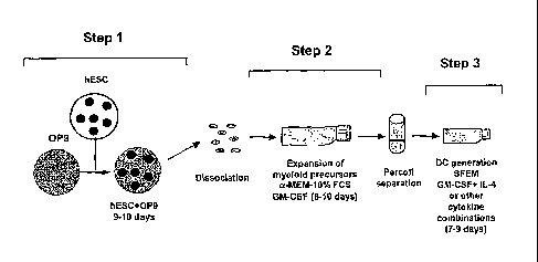
1000101
Figs. 1A and B are schematic illustrations of the overall method of the
present
invention.
- 3b -
CA 02610243 2013-09-25
[00011] Fig. 2 illustrates the morphology and phenotypical features of
myeloid precursor cells
generated in step 2 of Fig. 1. Fig 2A is a phase-contrast micrograph of
differentiated human ES
cells growing in the presence of GM-CSF. Fig 2B is a Wright-stained cytospin
of cells obtained
from that culture. Fig. 2C charts the colony forming cell (CFC) potential of
the expanded cells
(counts are mean of five experiments). Fig 2D are graphs of data from
representative experiments
demonstrating expression of surface and intracellular myeloid markers on GM-
CSF expanded
human ES cells.
[00012] Fig. 3 illustrates the morphology and light scatter properties of
hES cell-derived DSs.
(A) Phase contrast micrograph of culture and (C) Wright-stained smears of
differentiated H1 cells
demonstrate numerous thin cytoplasmic processes ("veils"); (A) bar is 15 pm
and (C) bar is 40
pm. (B) When cultured on flat-bottom ultralow attachment plates, cells form
long dendrites; bar
is 25 pm. (D) Light scatter properties and phenotype of cells obtained in step
3 after 9-day culture
of hES cell-derived myeloid progenitors with GM-CSF and IL-4. Phenotypic
analysis from
representative experiments using the H1 cell line shows that R1-gated cells
with a high scatter
profile express CD1a and weakly by CD14.
DETAILED DESCRIPTION OF THE INVENTION
[00013] We report here that dendritic cells can be created in large numbers
from human ES
cells. The co-culture system with a macrophage colony-stimulating factor (M-
CSF) deficient
stromal cell line, such as the murine line 0P9, fosters the differentiation of
human ES cells into
hematopoietic cells. These hematopoietic cells have the capacity to generate
dendritic cells, a
capacity which is exploited by GM-CSF culture of the hematopoietic cells. The
dendritic cells
derived from human ES cells are morphologically, phenotypically and
functionally comparable to
interstitial human dendritic cells naturally produced in vivo.
[00014] Slukvin et al., J. Immunology, 2006, 176: 2924-2932, is an academic
article by the
inventors describing the present invention.
[00015] The overall method is schematically illustrated in Figs. 1A and B,
in which the
process is broken down into three overall steps and is demonstrated in its
preferable form in the
Examples below and in Slukvin et al., J. Immunology, 2006, 176: 2924-2932. By
"multipotent
lymphohematopoietic progenitor cells," "myeloid dendritic cell (DC) precursor
cells," "immature
DC," "mature DC," and "regulatory DC," we mean the cell populations disclosed
in Fig. 1B.
[00016] In Step 1, human ES cells are co-cultured with stromal cells,
preferably M-CSF
deficient stromal cells, to induce differentiation of the cells into
multipotent
lymphohematopoietic progenitor cells. Preferably, the cells are 0P9 cells.
- 4 -
CA 02610243 2007-11-28
WO 2006/130651 PCT/US2006/021054
[00017] In Step 2, the disassociated ES-derived cells from that culture
are then cultured so
that myeloid cell expansion occurs. Preferably, this is done by culture of the
cells with
granulocyte/macrophage colony stimulating factor (GM-CSF) preferably as
described below in
the Examples. Also preferably, this step is performed in non-adherent
conditions. Preferable
non-adherent conditions require the tissue culture flask to be coated with
poly 2-hydroxyethyl
methacrylate(HEMA, Sigma) as described below. One could also prevent cell
adherence by
other means, such as cell shaking, using substances known to have non-adherent
properties to
cover the plastic container or using commercially available non-adherent
tissue flasks.
[00018] The result of this expansion step is a culture rich in myeloid
precursors, and that
culture is then used in Step 3 to make dendritic cells by culture in serum
free medium with GM-
CSF and IL-4, or other combination of cytokines (as described below) which
condition
development to dendritic cells.
[00019] An optional separation procedure, which can be done with Percoll
separation, is
shown between steps 2 and 3 and is used to remove both clumps of cells and
dead cells from the
culture prior to inducing dendritic cell formation.
[00020] In the examples described below, dendritic cells are generated
from human ES
cells by selective expansion of myeloid precursors obtained by the co-culture
of human ES cells
with M-CSF-deficient stromal cells without cytokine addition. The
hematopoietic cells resulting
from the co-culture step are competent to be induced to differentiate into
myeloid precursor cells
and then into immature dendritic cells.
[00021] A critical step in our protocol for generating DCs was the
efficiency of
hematopoietic differentiation in human ES cell/0P9 co-culture. Co-cultures
with a low number
of CD34+CD43+Lin- multipotent lymphohematopoietic progenitors (less than 3%)
failed to
expand myeloid precursors and, subsequently, differentiate to dendritic cells.
"Lin-" indicates
that these progenitors do not express CD1 lb, CD14, CD2, CD3, CD7, CD19, CD38,
CD45RA,
and HLA-DR markers present on more mature cells committed to specific
hemotopoietic lineage.
[00022] We used the entire cell suspension from the co-culture, rather
than isolated
lymphohematopoietic precursors for the next step in which
granulocyte/macrophage colony
stimulating factor (GM-CSF) is used to mediate expansion of myeloid precursors
which are
capable of differentiating into dendritic cells. In our hands, the most
effective factor to cause the
myeloid precursor cells to undergo this expansion was GM-CSF, in contrast to
other factors, such
as SCF and FLT3-L, which in our hands had little effect on the expansion of
myeloid dendritic
cell precursors.
[00023] The myeloid precursors derived from human ES cells and expanded
with GM-
CSF, contained myeloid colony-forming cells (CFCs), as well as small
populations of more
-5-
CA 02610243 2007-11-28
WO 2006/130651 PCT/US2006/021054
mature cells with the dendritic cell phenotype. However, the majority of the
cells
morphologically resembled blasts of the myelomonocytic lineage and expressed
CD4, CD45,
CD123I" and low levels of CD14. These cells were HLA-DR-negative. We found
that these
cells included subpopulations of cells expressing MPO, M-CSFR, CD1 lb, CD11c,
CD15, and
CD16.
[00024] The human ES cell-derived immature dendritic cells obtained by our
method had a
phenotype of CD la+, DC-SIGN+, CD4+, CD9I", CD1 1c+, CD40I0, CD80+, CD86+, HLA-
ABC+, HLA-DR+, CD207- and CD208-, a phenotype comparable with interstitial
dendritic cells
differentiated from the cord blood or bone marrow CD34+ hematopoietic stem
cells. Preferably,
at these immature dendritic cells comprise at least 70% of the cultured cells
at this point.
However, a distinct phenotypic feature of the human ES-derived dendritic cells
was co-
expression of CD14. The level of CD14 expression was the lowest in cells
differentiated using
IL-4, but was substantially higher on cells differentiated using TNF-a.
Dendritic cells that
develop from human CD34+ hematopoietic stem cells in the presence of GM-CSF
and TNF-a
differentiate into Langerhans cells and dermal and interstitial dendritic
cells through
intermediates that have phenotypes that are CD1a+CD14- and CD1a-CD14+
respectively. So
far, in our cultures, CD1a expression has always been associated with at least
low level
expression of CD14, and we have not seen distinct CD1a+CD14- or CD1a-CD14+
populations in
our cell cultures. Thus the culture conditions used for differentiation of the
human ES cells into
dendritic cells appears to use unique pathways that may not exact replicates
of the corresponding
pathways of differentiation from CD45+ hematopoietic stem cells in vivo.
[00025] The Examples below describe another embodiment of the present
invention, a
population of cells, wherein at least 70% are mature DCs.
[00026] Additionally, another embodiment of the invention is a population
of regulatory
DC cells. Myeloid DC precursors cultured with GM-CSF and TNF-a or GM-CSF and
IFN a
develop into CD laic", CD9-, CD8010, CD861" accessory cells with low
stimulatory activity.
These cells can represent regulatory DCs.
[00027] The co-culture system used here with the M-CSF deficient stromal
cells (0P9
cells) differs from the system based on 0P9 cells used with murine ES cells.
The method
described here does not use a second co-culture with the 0P9 cells, unlike the
mouse system.
We collected the human ES cell derivatives from the co-culture when the
maximal amount of
myeloid progenitors were generated and then expanded those progenitors with GM-
CSF in
feeder-free non-adherent conditions. This technique resulted in the discrete
population of
dendritic cell precursors which is useful for further studies of dendritic
cell development.
-6-
CA 02610243 2013-09-25
[00028] It has been shown recently that during embryoid body
differentiation, cells expressing
HLA-DR and capable of triggering proliferation of adult lymphocytes were
generated. Zhan et
al., 2004, Lancet Jul 10; 364(9429): 163-71. However, the antigen-presenting
properties and the
phenotype of the cells generated in this system were not demonstrated. It is
possible that cells
obtained as described in this report were macrophages. Our results provide,
for the first time,
evidence that human ES cells can be directly differentiated into cells with
morphology,
phenotype and functional properties of antigen-presenting dendritic cells.
Furthermore, this
process is already relatively efficient. We have been able to grow as many as
4 x 10⁷ dendritic
cells at a time from 107 initially plated human ES cells.
[00029] While dendritic cells have evident scientific interest and
application, they also have
potential use in human medicine. Several studies have demonstrated that
peptide-pulsed dendritic
cells transferred in vivo were able to induce efficiently anti-tumor immune
response in mice.
These studies have encouraged subsequent development of dendritic cell-based
vaccines for
cancer immunotherapy in humans. In these techniques, immature dendritic cell
precursors
isolated from peripheral blood or dendritic cells generated from peripheral
blood mononuclear
cells and CD34+ hematopoietic progenitors are used in clinical trials of
dendritic cell based
vaccines. However, these techniques are laborious, require repeated generation
of new dendritic
cells for each vaccination and are difficult to standardize. Embryonic stem
cells can be expanded
without limit and can differentiate into multiple types of cells, and
therefore can be universal and
scalable source of cells for dendritic cell vaccines. Potentially, dendritic
cells with major HLA
haplotype combinations can be obtained from human ES cells to match donor MHC
haplotype. In
the clinical setting, human ES cell-derived dendritic cells would have several
advantages over
dendritic cells from conventional sources. Large absolute numbers of dendritic
cells could be
generated from the same donor cell line, and the same line of dendritic cells
could be used for
multiple vaccinations. Derivation of dendritic cells from human ES cells can
be less laborious and
more amendable for standardization with implementation of bioreactor
technology. Low risk of
pathogen contamination and risk free donor collection are another important
advantages of
clinical use of human ES cell-derived dendritic cells.
[00030] In another embodiment, the present invention is a method of making
a cellular
vaccine, comprising differentiating the human embryonic stem cells into a
population of dendritic
cells, characterized in that they are CD 1 a+, CD80 , CD86+, DC-SIGN, HLA-
DRhIgh, obtaining
and preparing single cell suspension of tumor cells from a patient, and fusing
the embryonic stem
cell-derived dendritic cells with cancer cells. Gong et al., J. Immunology,
2000, 165: 1705-1711
and Parkhurst et al., 2003, J. Immunology, 170: 5317-5325 describe general
techniques for
cellular fusion.
- 7 -
CA 02610243 2007-11-28
WO 2006/130651 PCT/US2006/021054
100031] In another embodiment, the invention is a method of forming a
dendritic cell
vaccine for treating of cancer, comprising dendritic cells differentiating
from human embryonic
stem cells, where dendritic cells have been fused with allogeneic cancer
cells. One of skill in the
art would understand and appreciate the various methods of creating tumor
vaccines. For
example, U.S. Patent Application Publications US2002/0131962 Al and
US2006/0063255 Al
disclose several methods.
[00032] In another embodiment, the present invention is a method of making
a dendritic
cell vaccine for treating cancer, comprising differentiating human embryonic
stem cells into
CD45+CD4+CD1231' myeloid precursors which include subpopulations of cells
expressing
CD1 lb, CD11c, and CD16, genetically altering the myeloid precursors to
express immunogenic
tumor proteins/peptides, and differentiating the genetically modified myeloid
precursors into
immunogenic dendritic cells. For example, one may wish to transfect cells with
tumor genes that
will be the target of an immune response. For example, one may wish to
transfect cells with
melanoma-antigen-3 (MAGE-3), prostatic acid phosphatase (PAP) or prostate
specific membrane
antigen (PSMA).
[00033] In another embodiment, the present invention is a method of making
dendritic
cells with tolerogenic properties which can be used for treatment of rejection
of human
embryonic stem cell-derived tissues obtained from the same cell line. By
"tolerogenic
properties," we mean that the cell suppresses rejection of a transplant by the
host immune
system. The cells will down-regulate a detrimental immune response of the host
towards a
transplanted tissue. For this purpose hES cell-derived myeloid precursors will
be induced to
differentiate into regulatory DCs by culture with GM-CSF and TNF-a or GM-CSF
and IFN a.
[00034] EXAMPLES
[00035] EXPERIMENTAL PROTOCOL AND RESULTS
[00036] Expansion of human ES cell-derived myeloid progenitors with GM-CSF.
[00037] Recently we developed an in vitro culture system for hematopoietic
differentiation
from human ES cells, using cells of mouse M-CSF deficient bone marrow stromal
cell line 0P9
as feeder cells, a step used to start the protocols described here. Human ES
cells were co-
cultured with 0P9 cells so that they would differentiate into CD34+ cells
which are highly
enriched in colony-forming cells and contain erythroid, myeloid, as well as
lymphoid,
progenitors and include a population of CD34+CD43+Lin- multipotent
hemotopoietic
progenitors. This step does not require cytokine addition. The maximal
expansion of myeloid
colony-forming cells (CFCs) in the 0P9 co-culture system was observed on days
9 to 10 of
-8-
CA 02610243 2013-09-25
differentiation. To induce selective expansion of myeloid progenitors, we
harvested the resulting
cells front days 9 or 10 of human ES cell/0P9 co-culture and cultured the
cells in non-adherent
conditions in presence of GM-CSF. At the beginning of culture, aggregates of
large cells were
formed. Approximately 3 days after initiation of GM-CSF culture, individual
cells appeared and
rapidly expanded. After 9-10 clays of culture with GM-CSF, and following the
removal of
clumps and dead cells by Percoll separation, we obtained a population of cells
of which 90% of
the cells were CD45 positive. More than 90% of these CD45+ cells contained
intracellular MPO
(myeloperoxidase, a marker of myeloid cells) but not TdT (terminal
deoxynucleotidyl
transferase, a marker of lymphoid cells) and expressed a marker of myeloid
progenitors, CD33.
In addition, these human ES cell-derived myeloid cells were CD4 positive, and
weakly expressed
IL-3 receptor a-chain CD123. More than 50% of these cells expressed CD16,
CD15, CD1lb and
CD1 1 c (Table 2). Morphologically, the GM-CSF-expanded cells had multi-lobed
or round
nuclei and a moderate amount grayish, occasionally vacuolated, cytoplasm
without visible
granules (Fig.2B), resembling bone marrow myelomonocytic precursors. Some of
the GM-CSF-
expanded cells retained myeloid CFC potential, but no erythroid Of multi-
lineage CFC potential
was detected (Fig.2C). In addition, a relatively small population of cells at
advanced stages of
maturation that expressed a moderate level of CD14, low level of CD1a as well
as the HLA-DR,
and CD80 and CD86 co-stimulator molecules were present (Table 2).
[00038] Cutaneous lymphocyte-associated antigen (CLA) expression on
peripheral blood
CD34+ cells defines progenitors which further differentiate into Langerhan's
cells, while
CD34+CLA- cell give rise to interstitial DC-like cells. No significant CLA
expression was
detected in the total cell population obtained front 0P9 co-cultures or
isolated human ES cell-
derived CD34+ cells. However, CLA expression was found on a small subset of
myeloid
progenitors generated with GM-CSF.
[00039] GM-CSF appeared to be the most important factor in expansion of
myeloid
precursors. Separately, the addition of SCF, FLT3L, or SCF with FLT3L to GM-
CSF-
supplemented cultures had little effect on total cell output and myeloid CFCs
numbers during 10
days of culture (Table 1). These data demonstrate that culture of
differentiated human ES cells
generated in 0P9 system with GM-CSF predominantly expand into a unique
population of
CD45+CD4+CD12310' myeloid precursors which include subpopulation of cells
expressing
MPO, M-CSFR, CD1 lb, CD1 lc, CD15 and CD16,
[00040] Differentiation of human ES-cell derived myeloid precursors into
dendritic cells.
[00041] = To induce differentiation of myeloid precursors into dendritic
cells, we cultured
the culture of precursor cells with GM-CSF and various combinations of IL-4,
TNF-a, and IFN-
a. In typical experiment, after 7-10 days of culture with GM-CSF and IL-4,
most of the cells
-9-
CA 02610243 2007-11-28
WO 2006/130651 PCT/US2006/021054
appeared as clumps. In addition, individual floating cells with well-defined
dendrites appeared in
the cultures. Morphologically, these cells were large, had high nuclear
cytoplasmic ratio, and had
oval or kidney-shaped nuclei and nonvacuolated, occasionally granular
cytoplasm with very fine
cytoplasmic processes (Fig 3A and C). Based on flow cytometric analysis of
size and
granularity, two cell populations were observed (Fig 3D): R1, cells with high
scatter profile and
dendritic cell phenotype; and R2, cells with a low scatter profile, which
lacked dendritic cell
markers and which were more phenotypically similar to myeloid progenitors
generated in the
second step. Dendritic cells identified as R1 gated cells expressed CD1a, DC-
SIGN, CD4,
CD1 lc, HLA-ABC and HLA-DR, CD80, and CD86. Additionally, these cells
expressed a low
level of CD9, CD11b, CD123, and CD40. CD14 expression was very weak, but
detectable, and
most of the CD14-positive cells co-expressed CD1a. However all cells were
lacking CD83
expression.
[00042] In addition to IL-4, differentiation of myeloid precursors into
dendritic cells was
achieved by using other cytokines such as TNF-a and IFN-a or their
combinations. However,
most of the cells in cultures with 'TNF-a co-expressed low level of CD1a, high
levels of CD14
and were lacking expression of CD9. In addition, in cultures with TNF-a, cells
downregulated
expression of costimulatoiy molecules. As expected, addition of IFN-a to these
cell cultures
resulted in increased expression of MHC class I molecules. However, IFN-a
culture resulted in a
decreased number of CD1a+ cells, as well decreased CD14 expression. Similar to
the monocyte-
DC differentiation pathway, expression of DC-SIGN on human ES cell-derived
dendritic cells
was primarily dependent on IL-4. Based on cell yield, phenotypic, and
functional properties
(Table 1 and 2), we concluded that a combination of GM-CSF and IL-4 provides
the best
conditions for generation of functional dendritic cells from human ES cells.
[00043] By immunocytochemistry, human ES cell-derived dendritic cells were
positive for
CD68, but not strongly so, and expressed a very low level of intracytoplasmic
but not
membranous CD83. Fascin, an actin-binding protein that has been shown to be a
highly selective
marker of mature dendritic cells, was not detected. Prom this, we concluded
that the dendritic
cells generated by the process described so far were immature. To investigate
whether these
immature dendritic cells could be further matured, we treated cells generated
from the above
protocols with calcium ionophore A23187. This treatment resulted in the up-
regulation of CD83,
CD86 and HLA-DR expression. The intensity of intracytoplasmic CD68 staining
substantially
increased and perinuclear condensation of CD68 was evident in the cells so
produced. In
addition, some cells became fascin-positive. LPS, TNF-a, IL-lp, PGE2, and IL-6
were not
efficient in induction of maturation of liES cell-derived DCs. Taken together,
these data
-10-
CA 02610243 2013-09-25
demonstrate that cells with typical dendritic cell morphology and phenotype
can be generated
from human ES cells.
[00044] The dendritic cells induce allogeneic T cell response and are
capable of antigen
Processing and presentation.
[00045] We next investigated to determine whether our human ES cell-derived
dendritic
cells were fully functional as dendritic cells. As determined by DQ ovalbumin
assay, human ES
cell-derived dendritic cells were capable of taking up and processing antigen.
Cells obtained in
cultures treated with GM-CSF and IL-4 were the most efficient in antigen
processing, while the
dendritic cells differentiated with GM-CSF and TNF-ct were less efficient.
[00046] A hallmark of the functionality of dendritic cells is their ability
to stimulate naïve
cells. By our tests, human ES cell-derived dendritic cells were able to
trigger cord blood T cells,
which are en6rely naïve. Immature dendritic cells, generated in cultures with
GM-CSF and IL-4
added, were the most powerful stimulatory cells, while addition of TNF-a to
the cell culture
significantly diminished ability of the cells to stimulate naïve T
lymphocytes. In addition, the
dendritic cells were able to stimulate adult donor T-cells.
[00047] To evaluate the capacity of dendritic cells to present antigens
through the MHC
class I pathway, we pulsed HLA-A02 I-11 cell line-derived dendritic cells with
inactivated CMV
virus and evaluated the ability of the cells to stimulate HLA-A0201 restricted
CMV-specific T
cell clone HLA with specificity to CMV pp65 NLVPMVATV peptide. While the
addition of
dendritic cells to T-cells induced allogeneic response, a significant increase
in response by the T
cells was obtained when cells were stimulated with CMV pulsed Hl-derived
dendritic cells
(Table 3). Altogether, these data demonstrate that our culture system allows
generation of cells
with phenotype, morphology and unique antigen-presenting properties
characteristic of dendritic
cells.
[00048] METHODS AND MATERIALS
[00049] Cell lines, cytokines and monoclonal antibodies (mAbs).
[00050] Human ES cell lines H1 (passages 32-51) and H9 (passages 40-44)
were
maintained in an undifferentiated state by weekly passage on mouse embryonic
fibroblasts. A
mouse bone marrow stromal cell line 0P9 was obtained from Dr. Toru Nakano
(Research
Institute for Microbial Diseases, Osaka University, Japan). This cell line was
maintained on
gelatinized 10 cm dishes (BD Bioscience, Bedford, MA) in 0P9 growth medium
consisting of a-
MEM (Invitrogen, Carlsbad, CA), supplemented with 20% defined fetal bovine
serum (FBS;
HyClone Laboratories, Logan, UT). Sterile, recombinant, endotoxin and pyrogen-
free SCF,
FLT3-L, TNF-a, IL-4 were obtained from Peprotech (Rocky Hill, NJ), GM-CSF from
Berlex
-11-
CA 02610243 2013-09-25
Laboratories (Richmond, CA) and IFN-a from Schering Corporation (Kenilworth,
NJ). The
following mouse anti-human mAbs without detectable cross-reactivity with
murine cells have
been used for flow cytometric analysis: CD1a-PE, CD4-PE, CD11b-FITC, CD16-PE,
CD33FITC, CD8O-PE, CD86-PE, HLA-DR-PE, myeloperoxidase (MPO)-FITC, terminal
deoxinucleotidyl transferase (TdT)-FITC (Caltag, Burlingame, CA); CD9-PE, CD14-
FITC,
CD4O-PE, CD43-FITC, CD45-PE, CD209 (DC-SIGN)-FITC, CLA-FITC (BD Pharmingen);
CD1 lc-PE, CD34-PerCP-Cy5.5 (Becton Dickinson Immunocytometry Systems [BDIS],
San
Jose, CA); CD83-FITC, CD208 (DC-LAMP; Beckman Coulter, Miami, FL); CD123-FITC
(Miltenyi Biotech, Auburn, CA); HLA-ABC-FITC (Sigma, St. Louis, MO); CD207
(Vector
Laboratories).
[00051] Hematopoietic differentiation of human ES cells in co-culture with
0P9 cells.
[00052] The induction of human ES cells differentiation into hematopoietic
cells was done as
previously described, Vodyanik et al., 2005, Blood 105: 617. Briefly,
undifferentiated human ES
cells were harvested by treatment with 1 mg/ml collagenase IV (Invitrogen) and
added to 0P9
cultures at approximate density of 1.5x10⁶/20 ml per 10 cm dish in aMEM
supplemented with
10% FBS (HyClone) and 100 M Methyl P-D-thiogalactopyranoside (MTG) (Sigma,
St. Louis,
MO). Human ES cell/0P9 co-cultures were incubated for 9-10 days with a half
medium change
on days 4, 6, and 8 without added cytokines. The human ES cells then
differentiated into
hematopoietic cells.
[00053] Generation of human ES cell-derived dendritic cells.
[00054] A schematic diagram of the protocol used for generation of
dendritic cells from
human ES cells is depicted in Figure 1. On day 9-10 of human ES cell/0P9 co-
culture,
differentiated derivatives of human ES cells were harvested by treatment with
collagenase IV
(Invitrogen; 1 mg/ml in a-MEM) for 20 min at 37 C, followed by treatment with
0.05% Trypsin
0.5 mM EDTA (Invitrogen) for 15 min at 37 C. After trypsin inactivation by
FBS, these cells
were re-suspended in a-MEM supplemented with 10% FBS (HyClone) and 100 ng/ml
GM-CSF,
and transferred into tissue culture flasks (BD Bioscience) coated with poly 2-
hydroxyethyl
methacrylate (HEMA, Sigma) to prevent cell adherence. The cells were then
cultured for 8-10
days with a half medium change every fourth day to expand dendritic cell
precursors. To evaluate
the effect of SCF and FLT3-L on the expansion of these human ES cell-derived
dendritic cell
precursors, we cultured the cells in the presence of (1) 100 ng/ml GM-SCF + 20
ng/ml SCF; (2)
100 ng/ml GM-SCF + 50 ng/ml FLT3-L; or (3) 100 ng/ml GM-SCF + 20 ng/ml SCF +
50 ng/ml
FLT3-L. Subsequently, the cells were spun over 20% Percoll (Sigma) to remove
dead cells and
cell aggregates. As a third step, Percoll-isolated cells were cultured for 79
days in HEMA-coated
flasks in StemSpan serum-free expansion medium (SPEM; Stem Cell
- 12 -
CA 02610243 2007-11-28
WO 2006/130651 PCT/US2006/021054
Technologies, Vancouver, Canada) supplemented with lipid mixture 1 (Sigma) and
100 ng/ml
GM-CSF, with the addition of the following cytokines: (1) 100 ng/ml IL-4, (2)
20 ng/ml TNF-a,
(3) 104 U/m1IFN-a, and (4) 100 ng/ml IL-4 + 20 ng/ml TNF-a. Cells were
cultured for 7-9 days
with a half medium change every fourth day. To further maturate dendritic
cells, we cultured the
cells obtained in step 3 in SFEM medium with 400 ng/ml of A23187 calcium
ionophore (Sigma)
for 48 hours.
[00055] Flow cytometrv analysis
[00056] Cells were prepared in PBS-FBS (PBS containing 0.05% sodium azide,
1mM
EDTA, and 2% FBS), supplemented with 2% normal mouse serum (Sigma), and
labeled with a
combination of mAbs. Samples were analyzed using a FACSCalibur flow cytometer
(BDIS)
with CellQuest acquisition software (BDIS). List mode files were analyzed by
FlowJo software
(Tree Star, Inc., Ashland, OR). Control staining with appropriate isotype-
matched control mAbs
(BD Pharmingen) was included to establish thresholds for positive staining and
background
linear scaled mean fluorescence intensity (MFI) values. The percentage (%) of
positive cells was
calculated as % of positive cells stained with specific mAb ¨ % of background
staining with
corresponding isotype control. AMFI was calculated as MFI of cells stained
with specific mAb ¨
MFI of cells stained with corresponding isotype control. Linear scaled MFI was
used as an
indicator of relative antigen density on given cells.
[00057] Antigen processing assay
[00058] Ovalbumin (OA) processing assays were performed using self-
quenched
conjugate of ovalbumin (DQ-OVA; Molecular Probes, Eugene, OR) that exhibits
bright green
fluorescence upon proteolytic degradation. Dendritic cells obtained as in step
3 of Fig. 1 were
incubated with 100 i_ig/rril DQ-OVA for 30 min at 37 C in DMEM/F12
(Invitrogen) containing
2% FBS, and 1% of non-essential amino acids. Cells incubated at 4 C were used
as a control for
background fluorescence. OVA proteolysis was evaluated by flow cytometry.
[00059] Clonogenic progenitor cell assay
[00060] Hematopoietic clonogenic assays were performed in 35mm low
adherent plastic
dishes (Stein Cell Technologies) using a 1 ml/dish of MethoCult GF with H4435
semisolid
medium (Stem Cell Technologies) consisting of 1% methylcellulose, 30% FBS, 1%
BSA, 50
ng/ml SCF, 20 ng/ml granulocyte-macrophage colony stimulating factor (GM-CSF),
20 ng/ml
IL-3, 20 ng/ml IL-6, 20 ng/ml granulocyte colony stimulating factor (G-CSF),
and 3 units/ml
erythropoietin. All clonogenic progenitor assays were performed in duplicates
and CFCs were
scored after 14-21 days of incubation according to their colony morphology as
erythroid (E-
CFC), granulocyte, macrophage, megakaryocyte (GEMM-CFC), granulocyte-
macrophage (GM-
-13-
CA 02610243 2013-09-25
CFC), granulocyte (G-CFC) and macrophage (M-CFC). The frequency of CFC was
calculated
per 106 total cells.
[00061] Allogenic mixed lymphocyte reaction (MLR)
[00062] Adult mononuclear cells were isolated from peripheral blood samples
obtained from
healthy laboratory volunteers by density gradient centrifugation on Histopaque-
1077.
Mononuclear cord blood cells were also purchased from Cambrex Bio Science
(Walkersville,
MD). The mononuclear cells were depleted of monocytes by plastic adherence and
used as
responder cells. Graded numbers (1x10³ to 3x10⁴/wel1) of irradiated (35Gy)
stimulatory cells
were co-cultured with 1x10⁵ responder cells for 6 days in 96-well flat bottom
plates (Coming) in
RPPMI 1640 containing 5% human AB serum (Sigma). PH]thymidine (Sigma) was
added (1
Ci/well) during the last 16 hours of incubation. Cells were harvested onto
glass fiber filters and
incorporation of [3H]thymidine was measured by scintillation counting.
[00063] Although the foregoing invention has been described in some detail
by way of
illustration and example for purposes of clarity of understanding, the scope
of the claims should
not be limited by the preferred embodiments set forth in the examples, but
should be given the
broadest interpretation consistent with the description as a whole.
- 14 -
CA 02610243 2013-09-25
. .
table 1. Relative cell yield after each culture step*
Relative Cell Yield
Step 1 8.8 + 4.4
Step 2
GM-CSF 5.5 + 3.7
GM-CSF+SCF 5.6 + 5.7
GM-CSF+FLT3-L 4.6 + 4.2
GM-CSF+SCF+FLT3-L 5.1 -T 5.0
Step 3
GM-CSFAL-4 3.3 4.1
GM-CSF+TNF-a 2.3 + 1.7
GM-CSF+IFN-a, 2.3 + 1.6
GM-CSF+IL-4+TNF-a 1.9 1.3
*Relative cell yield at each step calculated as a number of cells obtained
from one
initially plated undifferentiated human ES cell (total number human ES cells
plated on
0P9/ total number of cells obtained after corresponding step); results
calculated as mean
+ SD of 4 to 10 experiments.
-15-
CA 02610243 2013-09-25
1 = ,
Table 2. Phenotypic analysis of DCs induced by different cytokine
combinations*
Cell Step 2 Step 3 Step 3 Step 3 Step
3
subset GM-CS17+ GM-CSF+ GM-CSF+ GM-CSFH-
1L-4 , TNF-a, IL-4+TNF- IFN-
cc
R1 gated % NA 58.8 + 12.3 45.5 + 12,1 46.7
+ 14.9 39.9 + 7,5
cells
CD1a % 3.3 + 2.1 82.9 + 12.4 66.9 + 24.0
78.2 + 7.7 30.3 + 27.1
AMFI 750.2 700.7 74.8 + 60.8 148.3
161.9 77.1 T 72.1
_
CD14 % 12.6 + 7.1 25.6 + 7.5 71.1 + 12.2
39.0 + 19.3 19.8 + 15.1
AMFI 14.7 4.2 27.6 + 15.5 55.3 + 38.1
31.5 + 29.0 60.7 T 50.8 .
_
DC- % <1 87.6 + 7.7 < 2 84.7 + 4.2
1'7.3 + 15.4
SIGN AMFI 460.3 352.0 213.8-+ 160.1
40.2 + 39.1
_
CD83 % <1 <1 <1 <1 <1
,
CD1 1 c % 60.0 + 14.2 94.1 + 5.3 98.0 + 1.6
93.7 33 . 91.0 + 8.5
AMFI 132.fil- 59.9 282.3 37.2 2021+ 19.8
237.6+ 17.8 97.4 + 41.8
CD11b % 59.4 + 13.1 67.4 + 29.0 48.8 + 24.9
56.0 + 5.4 59.6 + 8.4
AMFI 69.3 + 23.0 52.9 + 33.6 24.6 T 1 4 . 3
47.9 + 32.5 40.1 + 35.3
_
CD123 % 35.5 + 14.6 58.8 + 12.3 63.5 + 16.6
45.1 + 7.9 29.4 + 18.6
AMFI 27.8 + 15.2 35.9 + 14.6 28.3 + 12.6
33.9 20.1 18.9 + 15.3
HLA- % 79.6 + 8.8 90.3 + 8.4 91.8 + 4.1
84.8 + 9.3 99.2 + 0.9
ABC ANTI 125.7 61.6 92.4 10.6 130.3 62.1
111.2 55.4 258.0 47.9
HLA-DR % 14.9 + 12.0 90.1 + 6.3 90.1 + 4.1
82.1 + 8.0 89.4 + 7.7
AMFI 189.-+ 83.7 5971+ 204.3 267.3-+ 208.3-
+ 82.9 509.8-+
123.1 340.2
CD86 % 35.1 9.1 93.4 + 3.5 85.4 + 7.3
90.1 2.9 82.4 14.1
AMFI 60.2 + 24.3 1767.4+ 158.5-+ 94.6 439 +
131.0 125.3+
1122.3 107.2
CD80 % 7.9 + 7.8 81.2 + 21.8 84.8 10.7
81.8 + 11.6 81.6 + 19.3
AMFI 621,27+ 492.9 128.9+ 80.4 295.8 +
353.7 61.0 -1 13.2
CD40 % 4.6 + 4.4 46.4 + 16.9 43.3 + 23.7
57.0 + 1.6 53.9 + 26.8
AMFI 27.0 To 11.4 16.6 5.2
47.2 32.5 21.0 T 10.2 _
,
Cell Step 2 Step 3 Step 3 Step 3 Step
3
subset GM-CSF+ GM-CSF+ GM-CSF+ GM-CSF+
IL-4 INF-a. IL-4+TNF- IFN-
a.
*Results are mean + SD of 4 to 5 independent experiments; for step 3 cultures
% and AMFI of R1
gated cells calculated.
-16-
CA 02610243 2007-11-28
WO 2006/130651 PCT/US2006/021054
Table 3.
Antigen-presenting capacity of Hl-derived DCs*
T Cells DC CMV Proliferation (cpm) IFN-7 (pg/ml)
652 + 129 0
17225 + 579 224 + 26.7
20303 + 1279 326 + 11.8
* HLA-A02 Hl-derived dendritic cells (cells obtained in step 3 with GM-CSF+IL-
4)
incubated overnight with or without CMV virus and then added to the HLA-A0201
restricted allogeneic T cell clone with specificity to CMV pp65. Results
expressed as a
mean + SD of triplicate.
-17-