Note: Descriptions are shown in the official language in which they were submitted.
CA 02610827 2013-03-04
Device for Gastric Feeding and Drainage via an Artificial Stoma
BACKGROUND OF THE INVENTION
The present invention relates to a device for creating and maintaining an
artificial stoma
enabling access to a body cavity, such as used in the direct feeding of a
patient's stomach.
More particularly, the present invention relates to a device for
percutaneously placing various
gastric catheters, forming artificial stomas capable of accessing the
gastrointestinal tract, and
ultimately providing a gastric feeding capability. Beyond the initial
placement procedure, the
device meets the requirements for permanent placement in the patient, such
that when used
for enteral feeding, the device enables a low-force, dynamically self-
adjusting, directed seal
between the inside of the stomach or gastric wall and an external body
surface, i.e., the outside
of the abdominal wall.
In particular, the invention addresses the problem of the seal or permanent
fusion of the
tissues surrounding the stoma that needs to be established between the
abdominal cavity and
the inside of the stomach immediately after the surgical creation of the
fistula, i.e., during the
initial insertion of the catheter. It also is concerned with ways in which
subsequent to
placement, the catheter can be changed simply and atraurnatically, even by a
trained layperson,
without damaging the stoma site.
It is recognized that numerous medical conditions exist in which it becomes
necessary to gain
percutaneous access to viscera such as the stomach or small intestines.
Situations where a
patient has lost the ability to swallow and will require long term nutritional
support may dictate
feeding directly into the stomach or jejunum. Feeding in this manner may be
accomplished by
inserting a feeding tube into the patient's stomach such that one end remains
anchored in the
stomach, while the other end remains external to the patient's body for
connection to a
nutrient source. A variety of different feeding tubes or catheters intended
for enteral feeding
1
CA 02610827 2007-12-03
WO 2006/133927 PCT/EP2006/005733
have been developed over the years, including some having a "low profile"
relative to the
patient during use and those having the more traditional or non-low profile
configuration.
Such feeding tubes may be inserted into a patient's stomach in a number of
ways. Feeding
tubes may be endoscopically placed, surgically placed through an open
incision, laproscopically
placed, or percutaneously placed under endoscopic, fluoroscopic or ultrasonic
guidance.
Different types of feeding tubes may be placed using these procedures,
examples include
gastrostomy, jejunostomy or gastro-jejunostomy. These tubes may be retained in
the lumen
(stomach or intestine) with a variety of retention anchors. These anchoring
mechanisms
include: inflatable balloons, obturatable domes, fixed dome-type bumpers, or
suture wings.
It is known that many of the catheters on the market today are commonly
referred to as
"replacement" catheters because they are substituted for an enteral feeding
tube that is initially
placed in a patient for six to eight weeks until a fistula stoma tract is
established. Once the
stoma tract is established, the initial placement device is generally removed,
and the
"replacement" enteral feeding device is inserted into the stoma tract.
Historically, prior to
placing the actual enteral feeding device, it has been preferred to perform a
gastropexy
procedure during placement. This procedure enables the physician to attach the
visceral wall
to the abdomen and to create the stoma tract through the two. This attachment
is critical to
prevent inadvertent separation and exposure of the peritoneal cavity to
contamination and
possible peritonitis.
Initial placement devices are often not readily removable without additional
invasive surgical
procedures. That is, many initially placed enteral catheters contain rigid
retention members
which cannot readily be passed through the stoma of the patient when it is
desired to remove
the initially placed device. Typically the t-shaped fastener or t-bar is not
removable and is left
in the body cavity where it is allowed to pass naturally in the patient's
stool. In many cases the
t-bar is not passed and remains within the body cavity. Moreover, during the
six to eight weeks
it takes for the fistula's stoma tract to be established, the anchoring
mechanism of the prior art
gastropexy device which typically consists of a small metal t-shaped fastener
may embed itself
into the gastric or intestinal wall and ultimately lead to infection.
Furthermore, the t-bar itself
may have sharp edges which can be uncomfortable for the patient.
In many of these procedures, in order to achieve the desired seal between the
stomach and the
abdominal wall, a traction force must be applied to the anchoring mechanism.
The force is
2
CA 02610827 2007-12-03
WO 2006/133927 PCT/EP2006/005733
applied in such a way as to pull the stomach cavity to the abdominal wall so
that the
penetration through both may heal together thereby creating the passage or
stoma leading
from the patient's stomach, through the abdominal wall, to an external
environment. It is
necessary to apply this traction force for a period of a couple of days
through a couple of
weeks until the stoma site adequately heals. During this period the patient
has reduced
mobility which may lead to additional post-operative complications.
There is a need and desire for a device which may be used during initial
placement or creation
of a stoma site and which also may serve as the "replacement" enteral feeding
device itself.
Such a device would foster the permanent fusion of the stomach wall to the
abdomen; it
would replace standard catheter placement technology and thus substitute a
single step
procedure for the standard multi-step procedure. This would serve to reduce
the invasiveness
of the procedure, greatly enhance wound healing, and enable immediate, post-
placement
gastric access for feeding and drainage, and ultimately allow atraumatic
exchange of the low
profile device.
SUMMARY OF THE INVENTION
In response to the foregoing problems and difficulties encountered by those of
skill in the art,
the present invention is directed toward a device for the creation of an
artificial stoma into and
subsequent fluid transfer to or from a living body. Such a device may provide
the following
advantages: it would foster approximation of the gastric wall and abdominal
wall in a
sufficiently large area to enable tissue fusion; it would reduce the number of
punctures to only
one transcutaneous gastric puncture or incision; it would create a self-
adjusting seal with
respect to the puncture site and do so while taking into account body movement
and resulting
sheer forces; it would provide secure anchoring even under high pull-forces;
it would reduce
or prevent initial leakage or bleeding from the puncture channel thus reducing
the likelihood
of peritoneal infections; it would allow immediate access to the stomach and
maintain the
initial and continuous dilation of the puncture channel; it would reduce the
risk of ulceration
within the gastric lumen due to low-pressure filling of the balloon; it would
enable enhanced
patient mobility and comfort; and it would provide one device, meeting the
requirements of
both initial and long-term placement.
3
CA 02610827 2007-12-03
WO 2006/133927 PCT/EP2006/005733
The above-mentioned problems and difficulties can be solved by a device
according to
independent claim 1. Further advantageous features, aspects and details of the
invention are
evident from the dependent claims, the description, and the drawings.
The device in one embodiment would include a thin foil having a first and a
second end with a
length disposed therebetween. The thin foil would be arranged in a manner such
that one of
said ends is backfolded or introverted into the other of said ends. A cap
having at least one
port therethrough would also be provided. The cap would securely capture each
end of the
foil. The port would terminate between the first and second foil ends within a
space created by
introversion of the foil. By application of an inflation source to the port,
the length of the foil
would inflate and form a generally torus shaped balloon having both exterior
and interior
externally facing concentric surfaces. The cap is situated at one end of the
balloon.
In many embodiments, the device would contain a bore through the cap so that
communication with a passage formed by the interior externally facing surface
may be had.
Such communication would pass through the interior of the device. In many
embodiments,
the torus shaped balloon may be adapted to exert an increasingly greater force
upon increasing
inflation, the force being exerted axially along the foil and directed toward
the cap.
Some embodiments may be provided with an insertion device used for placing the
foil in a
deflated state within the living body and situating the cap adjacent a body
surface at the stoma.
The insertion device may contain a user manipulable introducer and a capturing
element. The
introducer may be configured as a tapered probe having a cavity therein within
which the foil
is temporarily captured. The capturing element would be sized to fit
frictionally within the
bore and in conjunction with the foil would retain the introducer in position
proximate to the
cap. An extension rod may be affixed to the introducer. The extension rod
would enable the
tapered probe to be inserted deeper into the living body until the foil is
disengaged from
within the cavity. This could be accomplished without affecting the position
of the cap or the
capturing element. To remove the introducer, the balloon is inflated and the
introducer is
extracted from the living body by passing it out via the passage and bore,
again, subsequent to
inflation of the balloon and removal of the capturing element.
The device in any of its embodiments may be adapted to be placed in the living
body by
engaging a guide wire previously situated in the living body, a process known
and understood
by those of skill in the art.
4
CA 02610827 2007-12-03
WO 2006/133927 PCT/EP2006/005733
According to one specific embodiment, the present invention relates to a
device for supplying
patients by means of a transcutaneous fistula (stoma) for direct feeding into
the stomach,
comprising a balloon which is backfolded in itself, whereby the inner end of
the balloon serves
as an open lumen for inserting a feeding catheter therethrough, whereby
moreover an axially
oriented tractive force acts between the balloon and the ends of the balloon,
which causes a
force component pressing the inner abdominal wall onto the stomach, and
whereby on the
body surface a disc element and/or a diameter of the outer one of the two
concentric ends of
the balloon, enlarged at least about 25 A compared to the ratio of the
diameter of the fistula,
serves as a bearing for an axially oriented back rolling of a torus onto the
inner stomach wall.
Specifically, the diameter of the outer of the two concentric ends of the
balloon may be larger
than about 50 to about 75 % of the fistula.
Other objects, advantages and applications of the present invention will be
made clear by the
following detailed description of a preferred embodiment of the invention and
the
accompanying drawings wherein reference numerals refer to like or equivalent
structures.
BRIEF DESCRIPTION OF THE DRAWINGS
FIG. 1 depicts an illustrative view of one embodiment of the present
invention.
FIGs. 2 and 3 depict intermediate steps in the creation of the backfolding
principle described
in many of the embodiments of the present invention.
FIG. 4 depicts an alternative embodiment of the FIG. 1 device.
FIG. 5 depicts an alternative embodiment of the FIG. 1 device having a second
embodiment
of a useful retention mechanism.
FIGs. 6 and 7 depict further embodiments of the device incorporating features
from the FIG.
1 device as well as the second embodiment of retention mechanism.
FIG. 8 depicts yet another embodiment of the device incorporating a third
embodiment of the
retention mechanism.
FIG. 9 depicts an insertion device adapted to be used with the FIG. 8
embodiment.
5
CA 02610827 2013-03-04
FIG. 10 depicts a channel enabling venting of the device; such a channel may
be used on any
embodiment of the device described herein.
DETAILED DESCRIPTION OF THE PRESENT INVENTION
In response to the foregoing challenges that have been experienced by those of
skill in the art,
the present invention is directed toward making initial feeding catheter
placement less invasive
and more comfortable for the patient. The invention is intended to reduce
complications
associated with enteral feeding and the initial placement of the enteral
feeding device such as
bleeding or leakage of gastric fluids. It provides a single device capable of
accommodating
both the procedure of placement and long-term wear and use which addresses the
needs
associated with initial as well as prolonged placement of the catheter.
To solve these problems, the invention envisions an introverted or backfolded
balloon
arrangement similar to that described, in a basic form, in WO 2004/069057 to
Dr. Lothar
Gobel. It should be understood that the introversion of one end of a balloon
element through
the opposite end produces a "torus" shape. In the inflated state, this torus
structure has the
tendency to move the balloon ends into a median plane of the torus ring. A
tractive force
operates axially between the balloon and the balloon ends, thus serving, in
the present
invention, as an important basic functional element.
If the balloon ends are passed through the fistula channel and secured outside
the body on the
outside wall of the abdomen within a fixing cap element, the resulting axial
tractive force
would generate a force component capable of pressing the stomach against the
inside wall of
the abdomen. This would permit the permanent fusion of the two tissue layers
which prior to
this device would have necessitated a separate treatment step prior to the
actual placement of
the enteral feeding device and intra-gastric access channel through the fused
tissue. This torus
shaped closing element would also allow immediate access to the stomach
through a free
central lumen of the element. This lumen may be used for immediate feeding,
drainage or
insertion of a catheter there through.
An initial version as well as alternative embodiments of such a device has
been described in
DE 10 2005 028 428.0 entitled "Votrichtung zur gastrischen Ernahrung und
Drainage iiber
6
CA 02610827 2013-03-04
eine transkutan angelegte Fistel" and was filed on June 17, 2005 by Dr. Fred
Gabel.
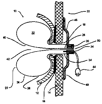
Looking now to FIG. 1, a device 10 in accordance with the present invention is
depicted. FIG.
1 depicts the spatial relationships between the device 10 and the adjacent
organs to it: the
gastric wall 12, the abdominal wall 14 and the anatomical fusion site 16
between the two wall
structures 12 and 14, where the perforation forming the fistula or stoma 18,
in a patient's
living body 20, is situated. At this point, it should be understood that for
convenience the
description of the device will generally be directed toward accessing the
stomach, for example,
to enterally feed a patient. However, the devices as shown and described
herein may also be
used to create a communication between two cavity located organs, spaces or
structures, or
one cavity located organ, space or structure and an external environment. As
such, no
limitations specifically requiring the invention to be associated with enteral
feeding or gastric
access should be read into the specification.
In many of the contemplated embodiments, the device 10 is formed from a thin
foil 22 having
first and second ends 24 and 26 respectively. As shown in FIG. 2, the foil 22
may be
cylindrical in shape and have a length 28 disposed between the ends 24 and 26,
which along
with the ends may define a three-dimensional volumetric space bounded by a
surface or
surfaces formed by the foil itself. Among the simplest of volumetric spaces is
the cylinder, as
shown. However, other contemplated volumetric shapes may include the sphere,
cube, cone,
cylinder, and more generally, the polyhedra.
In common embodiments, and referring to the cylindrical embodiment of FIG. 2
in particular,
FIG. 3 depicts one of the ends of the foil 22 being backfolded or introverted
into the other,
for example, end 26 may be backfolded into end 24 such that end 26 is situated
within end 24.
Of course, this configuration may be switched with end 24 situated within end
26 and still
perform in accordance with the invention.
In any event turning back to FIG. 1, it may be seen that by capturing the ends
24 and 26
within a cap 30, an interior space 32 is created. This space 32 is created by
the introversion of
the foil 22 itself and may be seen to be defined by its length 28 and bounded
by its ends 24
and 26. Providing the cap 30 with a port 34 that terminates within this space
32 between the
first foil end 24 and the second foil end 26 creates a torus shaped structure
or balloon 36
7
CA 02610827 2007-12-03
WO 2006/133927 PCT/EP2006/005733
having exterior and interior externally facing surfaces 38 and 40
respectively. The space 32
forms the interior of the balloon 36 and is adapted to inflate and deflate
upon application or
removal of a fluid source such as air, water, or saline. Other fluids may be
used and would be
understood by those of skill in the art.
The surface 38 may be seen to be an externally facing, exterior surface of the
balloon 36. The
surface 40 is also an exterior surface, however, it is considered an
internally facing, exterior
surface of the balloon in that it forms a passage 42 through the center of the
torus shaped
balloon yet does not enter the space 32. The diameter of this passage 42
formed by the surface
40 in many embodiments is smaller than the puncture channel through the
gastric 12 and
abdominal wall 14. The diameter of the passage 42 determines the flow-
characteristics through
the device. Further, secondary catheter elements, described below, may be
inserted into the
passage 42 if desired.
In order to inflate the balloon 36, an inflation source or mechanism 44 of
some kind should be
capable of connection to the port 34. As stated above, the fluid used to
inflate the balloon 36
may be a suitable gas or liquid, such as air or saline. A retention mechanism
46 may also be
provided in order to hold the device 10 properly within the living body 20.
Such a retention
mechanism 46 is envisioned to have numerous possible configurations each of
which will be
discussed at greater length in this specification. In a first embodiment, the
retention
mechanism 46 may be configured as a simple disc, button, or retaining ring 48.
The retaining
ring 48 may be secured to the cap 30 or to the balloon 36 itself via a
friction fit and may
simply be adapted to slidably attach to the exterior of the device in some
manner so that it
may be slid against the skin of the abdominal wall of the patient when in
place.
A bore 50 through the cap 30 connects the passage 42 to an external
environment. The bore
50 forms an opening through which fluids may pass into or out of the living
body 20. In many
embodiments, the bore 50 enables the injection of enteral feeding solutions.
It may also be
used to vent gases or other fluids from within the cavity as described in more
detail below.
However, in any of the embodiments described, dedicated pathways may be
provided, one for
feeding and one for venting. This concept would be readily understood by those
of skill in the
art and may be accommodated by numerous configurations including but not
limited to the
insertion of a dedicated catheter 52 through the bore 50, through the passage
42, and into the
stomach or other organ within which the device is in communication. Such a
catheter may be
8
CA 02610827 2007-12-03
WO 2006/133927 PCT/EP2006/005733
seen in FIGs. 5 through 7 which will be further described below. The catheter
52 may have
dual lumens, one for feeding and one for venting. Alternatively, the catheter
52 may be used
for one of the functions whereas the other function is performed by ensuring
that there is
ample room between the exterior of the catheter and the passage 42 and/or bore
50 diameters.
Referring generally back to this principle of an introverted foil 22 forming a
torus shaped
balloon 36, it may be seen that this is an improvement over the prior art
devices currently in
existence. For example, it should be noted that the present device 10 forms a
shaftless catheter
structure which effectively eliminates the need for the prior art rigid shaft
elements. It should
also be understood by those of skill in the art that unlike a balloon bearing
a rigid shaft, the
present invention may be reduced to a collapsed tape-like structure when in
the deflated and
evacuated state. With such a device 10, it would be possible to insert the
balloon portion 36
through the stoma 18 and into the living body 20 via a low-invasive, small
bore access
penetration.
Furthermore, due to the controllable collapsibility of the device 10 it is
more amenable to
atraumatic removal from the stoma than are prior art devices. This is because
the present
invention does not require the significant trans-abdominal exertion typically
associated with
those prior art devices containing a rigid shaft element for carrying the
balloon component. In
the prior art devices, the mechanics of the balloon member are typically
altered negatively over
time, for example, balloon members associated with the prior art are known to
stiffen and lose
their ability to retract fully into the shaft completely. This results in the
creation of
traumatizing folds that may exacerbate healing of the stoma site upon removal
or subsequent
manipulation of the catheter.
Turning now to FIG. 4, it may be seen that by giving the trans-abdominal
section, i.e., that
portion of the balloon 36 that is in contact with the stoma site 18,
appropriate dimensions, the
device 10 when inflated may be enabled to produce a certain radial force onto
the trans-
abdominal structures of the surgically perforated fistula channel or stoma 18.
This force would
serve to keep the penetration channel under permanent expansion and would
therefore
provide an efficient seal against gastric material leaving the stomach and
entering the
peritoneal cavity between the stomach and abdominal wall 14. Moreover this
feature could
also slow or stop bleeding at the site and foster a permanent and continuous
dilation of the
stoma itself.
9
CA 02610827 2007-12-03
WO 2006/133927 PCT/EP2006/005733
Continuing to look at FIG. 4, it may be seen that the outer of the two
concentric foil ends, in
this case end 24 is dimensioned in such a way, that the exterior surface 38
exceeds the
diameter of the stoma perforation. This may be seen by viewing that portion of
the exterior
surface 38 in contact with the gastric wall 12. In many embodiments, the
surface 38 may be
made to exceed the stoma diameter by a significant amount. A significant
amount may be
thought of in general terms as an amount that exceeds the widest section of
the perforation by
at least about 10%, but may range significantly higher including ranges from
about 25 to about
50% greater than the stoma diameter, and in some embodiments may range up to
about 75%
greater than the stoma diameter.
Due to the material selection of the balloon, the mechanical properties of the
material, and the
balloon's wall thickness, the device 10 may be designed to function at
inflation pressures that
inhibit bleeding in the stoma 18 without subjecting the foil to a tensive or
extensive force.
That is, the rest of the sheath, introverted in the transmural passage area,
forms a tight closure
in this section of the device by virtue of the proposed material wall
thickness, discussed in
greater detail below. This serves to prevent the escape of gastric secretions
in spite of
longitudinal folding of the exterior surface 38. Hemostatic inflation of the
device 10 precisely
tailored to the particular blood flow situation can thus be achieved in the
area of the stoma
perforation. When thin-walled balloon membranes with a residual dimension are
used, the
transrnural force which the balloon exerts on the puncture channel corresponds
largely to the
inflation pressure measurable in the case in question.
In order to promote this radial expansion effect, the wall thickness of the
balloon 36 would
likely be no greater than about 100 micrometers, especially in those regions
where radial
expansion is desirable, such as at the trans-abdominal section. Even so, in
many embodiments,
the balloon may be made of a soft membrane having a wall thickness of from
about 30 to
about 60 micrometers. While devices having wall thicknesses in this particular
range are well-
suited for use as initial placement devices, where higher seal forces are
desired, a structurally
identical device which is anticipated to remain in place for long-term
treatment could be made
of even thinner walled, less pressure resistant materials. In such devices, it
is envisioned that
the outer or exterior surface 38 especially at the trans-abdominal region
would not even exceed
about 50 micrometers, and may actually be thinner, in the range of from about
10 to about 30
micrometers.
CA 02610827 2007-12-03
WO 2006/133927 PCT/EP2006/005733
A material capable of functioning in the prescribed manner and capable of
functioning with
these wall thicknesses may be manufactured of Pellethane 2363 from DOW
Chemical, a
thermoplastic polyurethane. However, other materials having similar mechanical
characteristics
should work equally as well. Suitable materials would be mechanically low-
compliant and
therefore stable in shape under elevated balloon filling pressures. They would
exhibit little
volume expansibility, and as such, as in the example stated above, a
polyurethane is particularly
well-suited in this application. Such materials, even under heavy traction,
would not permit any
considerable shape deformation of the torus balloon and thus would minimize
the possibility
that the balloon could inadvertently slip through the gastric wall. This
capability is of some
importance so as to ensure the continued reliability of the device under
conditions associated
with daily use.
FIG. 5 depicts an alternative embodiment of the device 10 having a bulge 54,
in this case a
disc-shaped bulge, formed into the exterior surface 38 of the balloon 36. This
bulge 54, if
present, would desirably be situated proximate to the end 24 such that it
would be external to
the stoma 18 and the living body 20 itself The bulge 54 would serve as a
second embodiment
of the retention mechanism 46 and it may be seen that such a feature would
counteract the
force associated with the torus section of the balloon 36 internal to the
living body 20. This
embodiment may further facilitate homeostasis in the superficial wound area
immediately after
the perforation of the stoma 18. Moreover, the bulge 54 may also be desirable
when a more
rigid retention mechanism 46 can not be used due to the development of ulcers
or other
irritations of the skin.
In any of the aforementioned embodiments as well as in further embodiments
described
below, the foil 22 may be designed so that in the freely deployed state, that
is, when the
pressure within the space 32 is equal to the ambient environmental pressure,
the exterior
surface 38 of the balloon 36 at the trans-abdominal region may have a residual
diameter which
allows for the infolding of that surface 38 and thus provides for the best
possible equali7ation
of acting force and measured inflation pressure.
Turning now to FIG. 6, it may be seen that a version of the device 10 may be
manufactured so
as to place a secondary bulge 56 between the gastric wall 12 and the abdominal
wall 14. Such a
device 10 would serve as a bolster against the intra-gastric balloon 36, and
thus would enable a
fluid tight seal and/or a haemostatic compression against gastric perforation
when the balloon
11
CA 02610827 2007-12-03
WO 2006/133927 PCT/EP2006/005733
36 and secondary bulge 56 were inflated. This may be of use, for example, in
situations in
which a patient has suffered a severe perforation and its associated bleeding.
The device of
FIG. 6 would enable the clinician to perform efficient compression of the
puncture site. That
portion of the device which would serve as the anchor or retention mechanism
external to the
living body 20 may consist of the bulge 54 or of a retention mechanism 46
similar to that
shown in FIGs. 1 or 4.
FIG. 7 shows yet a further alternative version of the device 10. The FIG. 7
embodiment
depicts the torus shaped balloon 36 portion as being established inside the
gastric wall 12 yet
has no direct anchoring capability outside the abdominal wall 14. Such an
intra-abdominal
device grants free abdominal movement of the perforated organ and would also
enable gastric
access, yet the gastric wall 12 and the abdominal wall 14 would not be brought
into direct
contact and would thus not fuse to one another. This may be desirable for some
medical or
anatomical reasons. In this example, the device would be connected to the body
outside
through a hose connection.
As shown in FIGs. 5 through 7 the catheter may be used in any of the
embodiments, including
the others described herein. In particular, having the ability to slide a
catheter 52 through the
bore 50 and passage 42 into the stomach or other site internal to the living
body 20 would be
beneficial in cases of long-term use, as the catheter 52 could be changed
simply and
atraiimatically, i.e. without damaging the puncture channel, and it could be
accomplished even
by a trained layperson.
Referring to FIG. 8 there is shown a cross-sectional view of still another
embodiment of the
present invention. This embodiment is similar to the previous embodiments in
that the
balloon 36 may be configured similarly to any of the balloon embodiments
described above.
Like the prior embodiments, the FIG. 8 embodiment includes the thin foil 22
having a length
28 terminating in the first and second ends 24 and 26. Additionally, one end
is backfolded or
introverted with respect to the other so as to create the space 32 that forms
the balloon 36
between the two ends 24 and 26 respectively. However, FIG. 8 depicts yet a
third embodiment
of the retention mechanism 46. In that FIG. 8 forms a more detailed cross-
sectional view of
one possible embodiment, a number of items are described as pertaining to FIG.
8. It should
be noted that these items may also be found on other embodiments, including
those described
12
CA 02610827 2007-12-03
WO 2006/133927 PCT/EP2006/005733
above. Those items not capable of being utilized on previous embodiments will
be specifically
noted.
In more general terms, this embodiment integrates the retention mechanism 46
into the cap 30
itself. As such, the two components may be thought of as forming a head 58.
The head 58
serves at least in part to capture the ends 24 and 26 but also serves to
contain a valve or valves
which are used to regulate the flow of fluids through the entire device 10. As
such, the head 58
may be made of a medical grade silicone but should be sufficiently designed to
capture the foil
ends 24 and 26 without undue failure. As is the case with each embodiment of
the retention
mechanism 46, the head 58 also serves to prevent the device 10 from completely
advancing
through the stoma 18 and into the stomach or intestine of the living body 20.
A first of said valves would serve as the port 34 and as such would be adapted
to couple the
space 32 with the inflation source or mechanism 44. As in the previous
embodiments, the port
34 would serve as a means to inject fluids into or remove fluids from the
space 32 forming the
interior of the balloon 36. A lumen 60 may be provided that leads from the
port 34 to the
space 32. Such a lumen 60 though not shown may be desirable on each of the
other
embodiments. As would be apparent, control of the inflation mechanism 44
through the port
34 enables the user or a physician, etc., to selectively control inflation and
deflation of the
balloon 36. To assist in this, a releasable one-way valve 62 may be disposed
between- the space
32 and the port 34, for example in the lumen 60. Appropriate valves capable of
serving in this
function are known and would be understood by those having skill in the art
and may be
actuated by means of a syringe.
A second of said valves, if provided, may be situated in the bore 50 located
in the head 58 and
would enable the injection of enteral feeding solution, etc., through the
device 10 and into the
user. The valve may comprise an anti-reflux valve 64 which is configured to
allow nutrient
solutions, etc., to pass into the user, but to prevent the flow of fluids out
of the user unless
properly engaged by a syringe or other sampling device having a nipple which
corresponds to
the anti-reflux valve. The anti-reflux valve 64 would be disposed such that it
is in
communication with the passage 42.
Looking now to FIG. 9, the FIG. 8 embodiment is depicted in conjunction with
an insertion
device 66. The insertion device 66 may be configured as a user manipulable
introducer 68
having a hollow probe 70 at one end. The probe 70 may be tapered at a distal
end 72 to allow
13
CA 02610827 2007-12-03
WO 2006/133927 PCT/EP2006/005733
ease of passage through the stoma 18 so as to minimize aggravation of the
tissue. A proximal
end 74 of the probe 70 may also be tapered to allow subsequent withdrawal of
the probe 70
from the stoma 18 with minimal tissue damage as well. Protruding from the
proximal end 74 is
an extension rod 76 adapted to be grasped by the user, physician, or
clinician. The rod 76 may
be configured as a hollow cannula so as to be deployable over a guide wire
(not shown)
previously placed within the living body 20.
Prior to installation in the living body 20, the foil 22 is captured within a
cavity 78 formed in
the hollow probe 70. The extension rod 76 is situated so as to extend from the
cavity 78,
through the passage 42 and the bore 50, and ultimately extend outward through
the head 58. A
capturing element 80 is designed to be slid over the extension rod 76 and
seated within the
bore 50 in the head 58. By ensuring that the capturing element 80 is held in
contact with the
foil 22, which in turn is pressed against proximal end 74 of the probe 70, the
insertion device
66 may be placed in situ within the cavity.
Once the foil 22 is in place, the user would continue to advance the probe 70
deeper into the
living body 20 until the foil 22 is adequately deployed from the cavity 78.
Prior to or during
this step, the capturing element 80 may be removed from the extension rod 76
or at least
backed away from the probe 70. This may all be accomplished by manipulation of
the
extension rod 76. Subsequent inflation of the balloon 36 would ensure that the
foil 22 is
completely free of the cavity 78. After the balloon 36 is inflated, the probe
70 may be
withdrawn from the living body by backing it out through the passage 42 and
the bore 50, and
ultimately completely removing it from the device 10.
Finally, FIG. 10 has been included to address ventilation of the living body
20. In FIG. 10, it
may be seen that the channel 42 contained within the balloon 36 may be shaped
in order to
prevent its total collapse into the tape-like structure. That is, by
manufacturing the end 26 as
well as a portion of the interior surface 40 to a given foil wall thickness,
one can prevent a total
collapse of the channel 42, and instead it is possible to form one or more
laterally positioned-
tubulnr paths 82. These tubular paths 82 would serve to grant a permanent,
noncollapsible
passageway for gases. The diameter of the resulting tubular paths 82 may be
configured by
choosing an appropriate wall thickness of the foil 22. Alternatively a tubular
reinforcement
exhibiting an appropriate stiffness may be inserted and permanently affixed
within the channel
42. This may be desirable in instances as described above where a patient
having an existing
14
CA 02610827 2007-12-03
WO 2006/133927 PCT/EP2006/005733
anatomical or functionally insufficient communication between the stomach and
the ambient
surrounding of the patient requires the release of accumulating stomach gases
As used herein and in the claims, the term "comprising" is inclusive or open-
ended and does
not exclude additional unrecited elements, compositional components, or method
steps.
While various patents have been incorporated herein by reference, to the
extent there is any
inconsistency between incorporated material and that of the written
specification, the written
specification shall control. In addition, while the invention has been
described in detail with
respect to specific embodiments thereof, it will be apparent to those skilled
in the art that
various alterations, modifications and other changes may be made to the
invention without
departing from the spirit and scope of the present invention. It is therefore
intended that the
claims cover all such modifications, alterations and other changes encompassed
by the
appended claims.